ventricular excitation in experimental left bundle branch block
TRANSCRIPT

Ventricular Excitation in Experimental Left Bundle Branch Block
R. A. Becker, M.D., A. M. Scher, Ph.D., and R. 1’. Erickson, M.D., Seattle, Wash.
Experimental bundle branch block (BBB) has been a means of studying the role of conduction tissue in ventricular excitation, particularly as applied to clinical conditions. When Lewis’ first established electrocardiographic cri- teria for right and left BBB, he concluded that the increased duration of the QRS complex in BBB resulted mainly from the increased time required for septal activation. He also believed that spread through the affected ventricle was altered. Wilson2 and Barker,3 in studying the excitatory pathways of the heart and their relationship to the QRS complex, noted that Lewis had confused right and left BBB. They corrected this error, and established the presently ac- cepted electrocardiographic criteria for presence of these conduction defects.
More recently, several groups have studied the mechanism of ventricular activity in BBB using intracavity leads, esophageal leads, and records of ven- tricular mechanical activity. Wolferth,4 Braun-Menendez,5 and Smiths noted delay in mechanical contraction of the blocked side. Wener and his co-workers7 agreed with Sodi-Pallares,8~s Rosenbaum,ro and Smith” that the initial intra- cavity potential was reversed from negative to positive on the homolateral side in experimental BBB.
Although agreeing with Lewis’ that altered septal excitation was the major cause of the prolongation of the QRS during BBB, Smith and his co-worker@ proposed that the conduction system was also altered so that mural activation proceeded by less direct Purkinje pathways than those normally traversed. Rodriguez12 and Sodi-Pallares,13J4 however, believed that the speed, direction, and sequence of activation in the free ventricular wall was unchanged in BBB, and ascribed the changes in the QRS complex to “delay” of conduction and decreased speed of activation in the interventricular septum.
Using multichannel recording techniques, Erickson and his co-worker+ studied septal and mural activation in right BBB, and dealt with many of these
From the Department of Physiology and Biophysics, University of Washington School of Medicine, Seattle, Wash.
This investigation was supported by a research grant (H1315) from the National Heart Institute of the National Institutes of Health, Department of Health, Education, and Welfare, and by a grant from the State of Washington Research Fund for Biology and Medicine.
Received for publication Nov. 21, 1957.
54

548 IXCKER. S(‘HER, A\?;11 ERICKSON Am. Heart J. April, 1958
controversial points. With similar recording techniques, a more detailed study- of ventricular excitation in left BBB was undertaken in the hope of further resolving some of the points in question and of elucidating the mechanisms of BBB.
MATERIALS AND METHODS
Nine dogs weighing 6 to 10 Kg. were placed under barbiturate anesthesia, and maintained by artificial respiration. A sternum-splitting procedure gave a wide exposure of the heart, which was cradled in the pericardium to make the left ventricle accessible. Warm physiologic saline was applied to the epicardium throughout an experiment.
The recording apparatus, described in detail elsewhere,‘6 consisted of multipolar electrodes, multichannel oscilloscope, and Grass camera. The electrodes consisted of 15 fine insulated wires, staggered along a central shaft, with their bare tips exposed at l-millimeter intervals. A bank of 16 oscilloscopes displayed the activity at the electrode tips. Time pips at S-millisecond inter- vals were fed into all channels simultaneously from a master oscillator. A switch permitted the taking of unipolar records (potential between each terminal and an indifferent body surface point) or bipolar records (difference between adjacent terminals).
The fifteenth oscilloscope was used to record a fixed time-reference potential, obtained from an electrode inserted in the wall of the right ventricle. The sixteenth oscilloscope recorded a Lead II electrocardiogram.
Recording electrodes were inserted into the myocardium to cover all areas of the septum and walls. Most often, planes parallel to the base were used, but some mid-coronal, anterior, and posterior left ventricular placements were also utilized. India ink or colored threads identified electrode paths for anatomic-physiologic correlation.
After normal excitation had been recorded, a specially designed knife was inserted through the left ventricular myocardium, and directed toward the aortic valve. The knife was advanced until its tip was felt by a finger placed posterior to the ascending aorta. The tip was then with- drawn a few millimeters, and the blade was rotated to incise the septal endocardium near the junction of the posterior and right anterior cusps. Potentials recorded on an electrode in the interventricular septum and a Lead II electrocardiogram were monitored to determine when a complete block had been produced. Normally the septum is excited by double envelopment from both endocardial surfaces. After block it is excited entirely from the right. This change in direction of excitation can be easily detected by an electrode across the septum. When the electrocardiogram is concurrently changed, we can be sure that left bundle branch block (LBBB) is present. When block was obtained, the pattern of excitation was again plotted.
All potentials recorded were first related to the time-reference potential and then, by a fixed time correction, to the beginning of the QRS. Although there were variations between the various experiments, the usual experiment included about 20 electrode insertions, i.e., records from about 300 ventricular points.
RESULTS
Figs. 1 and 2 show potentials recorded on one insertion of the multipolar electrode across the mid-central interventricular septum before and after LBBB. A negative potential gave a downward deflection in a unipolar lead.
In the unipolar control records (Fig. l,A), left cavity fiotentials (Channels 1 and 2) show only negativity, indicating predominance of activity receding from that cavity through the septum and left wall. The right cavity potential (Channel 14) shows a slight positive deflection before the dominant negative deflection. This early positivity reflects the initial activity on the left. From Channel 3 through Channel 13 there is a rapid negative component indicative of local depolarization. While the initial component in Channels 4 and 5 is

~:‘%z,“r “d VENTRICULAR EXCITATION IN EXPERIMENTAL LBBB 549
negative, the remaining channels show a short, low-voltage, positive wave pre- ceding the rapid negative deflection. The slow, prominent negative component has a duration of 25 to 35 msec.
Following LBBB, an initial positive potential lasting some 40 msec. was recorded on Channels 1 through 4 (Fig. l,B). This positive potential resulted from spread of activation from right to left across the septum. The positive wave, although it decreased in magnitude, persisted up to Channel 13. Channel 14 showed only a negative deflection, in contrast to the small initial positivity recorded before block. The shape of the potential on all channels also changed. Plateauing of the slow component increased the duration of the negative wave to 55 msec., 20 msec. longer than in the normal record. This negative potential probably represents late activity, receding through the left wall.
In the bipolar control records (Fig. 2,A), Channels 3 through 8 showed a downward deflection, indicating activation from electrode tip to base, i.e., from left to right across the septum; while Channels 9 through 13 showed upright deflections, indicating movement of activity from right to left across the septum. This excitation pattern is characteristic of the normal double envelopment of the septum. The records on Channels 2 and 14 are from electrodes near the endocardium, in the left and right cavities, respectively, and show some evidence of nearby muscular activity.
After LBBB, septal activation was entirely from the right as shown by the bipolar records in Fig. 2,B. Ch annels 3 through 8 now displayed upright de- flections indicating excitation from the right, but the shape of the potentials on Channels 9 through 13 and their relationship to the time-reference potential were unaltered (Fig. 2,/l). The earliest point of depolarization was on the right endocardium (Channel 3). These records demonstrate that there is no delay at .the junction of the septal tissues previously activated from the right and from the left (between Channels 8 and 9).
Fig. 3, top, shows the sequence of depolarization in a heart cut by 4 planes parallel to the base. The advance of the wave front (darkened area) through the ventricles is shown sequentially. Normally, a central region of the left septal endocardium is excited first. A few milliseconds later the right endo- cardium is excited, followed by depolarization of tissues surrounding the cavities. The wave front then moves apically and in a circular fashion around each cavity, toward the epicardial surfaces. Ventricular invasion is virtually complete 20 to 30 msec. after the onset of QRS; the basal septum and the posterior wall are the only portions which remain to be excited. A more detailed description of this process can ‘be found elsewhere.17 Noteworthy points in this picture of normal excitation are (1) double envelopment of the septum, starting slightly earlier on the left, (2) simultaneous depolarization of much of the right and left ventricular walls, (3) late activation of the basal septum and the posterior wall and the tips of the papillary muscles. In the normal dog heart the entire process takes about 35 msec.
In LBBB both the pattern of spread and the total time of activation are markedly altered, as seen in Fig. 4,/l, bottom. As in the control, the heart is cut into 4 planes paralleling the base. The drawing represents a single experi-

ment utilizing more than 40 elec.trode insertions and over 500 separate recording points. The Lillclings in S other cqeriments, some of which fvere equnll~ Ed- tensive, agree completeI\- with those presented. The earliest ;Ictivity, occurring a few milliseconds before the onset of the QRS, is seen in Plane II I at the anterior border of the right cavit\-. During the first 15 msec. of the QRS, depolarization
Fig. I.--Unipolar septal pot,entials before and after LBBB. Negative potentials are downward. Time pips are 5 msec. apart. Potentials from left cavity on Channels 1 and 2, from septum On Channels 3 through 13, and from right cavity on Channel 14. Channels 15 and 16 record fixed time-reference potential and Lead II ECG, respectively. A, Potentials during normal excitation. B, After LBBB, same electrode. This electrode was in the position of the septal arrow in Fig. 4,C.
occurred in tissues normally activated by the right bundle, i.e., the right septal and ventricular myocardium. Depolarized muscle formed a conelike structure on the right side of the heart, extending into all 4 planes. During the first 1.5
msec. there was no depolarization of septal tissue normally activated from the left side.
For the next 30 msec. the wave front progressed in an orderly manner across the septum from right to left, roughly paralleling the curvature of the septum. The earliest activation on the left septal endocardium is seen posteriorly in Plane IV. This area became excited early in the interval between 30 and 45 msec.

x”lmre~ ;4” u VENTKICULAR EXCITATNIO IN EXPERIMENTAL LBBB 5.51
after the onset of QRS. Within the 30-to-45msec. period small areas on the anterior left septal endocardium, at the level of Planes II and IV became excited, followed early in the 45to-60-msec. period by similar areas in Planes I and III.
While parts of the left septal endocardium were not yet activated during the period from 45 to 60 msec. after the beginning of the QRS, ventricular exci- tation extended to the anterior and posterior epicardial surfaces in all planes, reaching posterolaterally in Plane I. One of the earliest points on the left to be excited appears on the septal portion of the anterior epicardium at about the level of Plane I. During this 45to-60-msec. period, depolarization of the an- terior and posterior walls was nearly completed, with a ring of activated tissue appearing in Plane I. This activated muscle extended into Planes II, III, and IV as a continuous sheet. During a few milliseconds of this period a wide area of the anterior endocardium in Plane IV became activated.
Fig. 2.-Bipolar septal potentials (dilference between adjacent tips) before and after LBBB. Rec- ords from the same electrode insertion as Fig. 1. Upper potentials (A) during normal excitation. Lower records (B) after LBBB. Channels 3 through 8 show negative deflection in the normals, indi- cating activation from electrode tip to base (i.e., left to right). After block the potentials become positive on these channels. indicating activation from the right. Potentials on Channels 9 through 13 are positive normally. After block these channels show no change compared to the normal. Channels 2 and 14 are near the endocardium in the right and left cavities, respectively. Discussion in text. Channel6 are numbered as in Fig. 1.
Within the 60-to-75msec. period activation in Plane I was completed. In the other 3 planes the remaining left endocardium became completely activated, suggesting spread by the Purkinje network. Most of the left lateral myocardium was excited during this period. The papillary muscles and extreme lateral myocardium remained to be excited. In the subsequent 15 msec. most of these

552 BE~‘KlSK, SCHEK, AND EKI(‘IiSON Am. Heart J. .\lvil, 1958
areas became activated, depolarization being completed in Plane 11. ‘The re- maining tissues (a small segment on the left lateral wall in Planes III and I\!, and a tip of the anterior papillary muscle in Plane III) were activated in the next 10 msec., to complete the process approximately 100 msec. after the onset of the QRS.
Fig. 3.-Summary of ventricular depolarization patterns before and after LBBB. Shaded area represents volume of myocardium depolarized up to the particular instant indicated by the figures in the columns. These figures represent time in milliseconds after the beginning of QRS. Both the normal and LBBB patterns are referred to the respective Lead II electrocardiograms shown on the lower left of each series of drawings. Normally, depolarization is almost entirely complete by 25 msec. After LBBB the altered mechanism of excitation accounts for the prolonged depolarization. Discussion in text.
Several significant points are not clearly apparent in Fig. 4,A. First, since the wave front is concave as viewed from the left, activity occurs at some left epicardial points before it reaches the underlying endocardium. As previously noted, the earliest activity on the left is intraseptal and epicardial after LBBB. Second, a transition occurs following septal activation. Fig. 4,C shows this transition, the arrows indicating the direction of spread and amount of tissue activated by the advancing wave front. With the early activation of the left epicardium, outside-in spread occurs. In time, the left septal endocardium is excited and the wave front spreads endocardially, and depolarization commences
~%~~ P VENTRICULAR EXCITATION IN EXPERIMENTAL LBBB 553
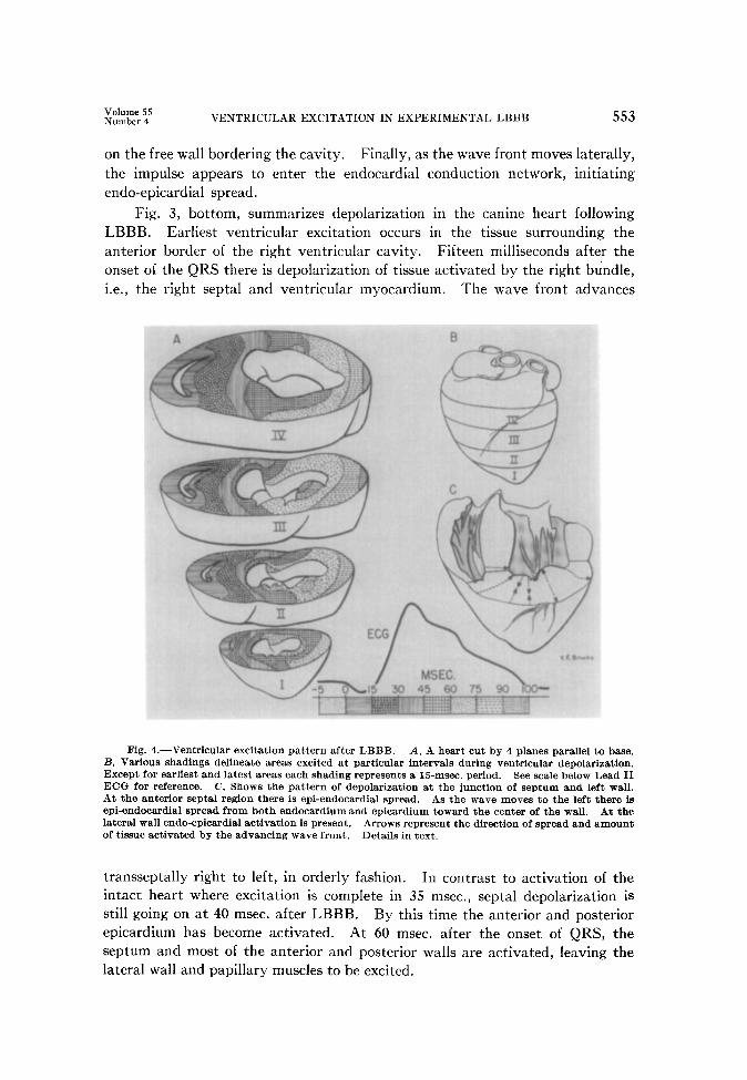
on the free wall bordering the cavity. Finally, as the wave front moves laterally, the impulse appears to enter the endocardial conduction network, initiating endo-epicardial spread.
Fig. 3, bottom, summarizes depolarization in the canine heart following LBBB. Earliest ventricular excitation occurs in the tissue surrounding the anterior border of the right ventricular cavity. Fifteen milliseconds after the onset of the QRS there is depolarization of tissue activated by the right bundle, i.e., the right septal and ventricular myocardium. The wave front advances
Fig. 4.-Ventricular excitation pattern after LBBB. A, A heart cut by 4 planes parallel to base. B, Various shadings delineate areas excited at particular intervals during ventricular depolarization. Except for earliest and latest areas each shading represents a 15-msec. period. See scale below Lead II ECG for reference. C, Shows the pattern of depolarization at the junction of septum and left wall. At the anterior septal region there is epi-endocardial spread. As the wave moves to the left there is epi-endocardial spread from both endocardium and epicardium toward the center of the wall. At the lateral wall endo-epicardial activation is present. Arrows represent the direction of spread and amount of tissue activated by the advancing wave front. Details in text.
transseptally right to left, in orderly fashion. In contrast to activation of the intact heart where excitation is complete in 35 msec., septal depolarization is still going on at 40 msec. after LBBB. By this time the anterior and posterior epicardium has become activated. At 60 msec. after the onset of QRS, the septum and most of the anterior and posterior walls are activated, leaving the lateral wall and papillary muscles to be excited.

554 BECKEH, SCHHK, AND ERICKSON ,\m. Heart J. April, 1958
DISCUSSION
Sepal Activation in LBBB:- ~-Lewis’ described the left ventricular conduction tissue as fanning out from the main left bundle and running from the septum to the bases of the papillary muscles. In LBBB this main bundle is interrupted and the septum is activated entirely from the right-single envelopment as opposed to normal double envelopment. Initial activation of the left ventricular myocardium is entirely from the right, and is completely independent of any branches of the main left bundle.
In nearly 50 septal insertions, the wave of excitation advanced across the septum at a uniform rate with no evidence of intraseptal delay as has been postu- lated.12-1a These findings concur with previous reports” that alteration of septal activation in LBBB accounts for the major portion of the QRS prolongation.
The QRS complex in a normal Lead II canine electrocardiogram lasts 30 to 40 msec., but after LBBB this complex lasts as long as 100 msec. Over half of this time, i.e., 50 to 60 msec., is needed for septal activation by single envelop- ment.
Activation of the Free Wall.-While some workers 12-14 believe that ventricular mural excitation proceeds in normal fashion following LBBB, others” have found changes in the spread through the left ventricle. Our experiments reveal an abnormal pattern of depolarization in the free left wall after LBBB.
The mural phase of ventricular depolarization is lengthened as a result of these conduction and excitation changes in the blocked ventricle. Normal myocardial activation requires 30 to 40 msec. When this period is added to the time required for septal activation in LBBB, 10 to 20 msec. remain unac- counted for. Alterations in (1) rate of conduction, (2) site of early activation, (3) direction of spread, and (4) sequence of depolarization account for this time difference. These changes can be seen by comparing the patterns of acti- vation before and after LBBB (Fig. 3).
Normally, the left endocardium is excited early and almost simultaneously. After LBBB, recordings from both endocardium and epicardium reveal that a small area on the anterior left epicardium is excited before any of the left endo- cardium. The wave of excitation advances by muscle conduction through the medial half of the left ventricle. The interval between the earliest and latest points is increased after LBBB. The altered pattern of spread after block is shown by arrows in Fig. 4. The anatomic arrangement (spiral) of the apical musculature may account for the epi-endocardial spread on the left.
Canine studies’* reveal that an anterior myocardial infarction is difficult to determine in the presence of LBBB. Recently, GrantI reported that some anterior myocardial infarctions produce QRS prolongation resembling that caused by LBBB. He offers limited criteria for differentiating the two entities, but does not explain why they result in similar electrocardiographic patterns. The epi-endocardial spread of activation noted at the junction of the septum and the left free wall (Fig. 4) is one probable reason for the difficulty in differenti- ating and diagnosing myocardial infarction and LBBB.
Cavity Potentials.-Several workers have noted that following LBBB the initial intracavity potential on the homolateral side is positive instead of nega-

“N::g: “4” VENTRICULAR EXCITATION IN EXPERIMENTAL LBBB 55.5
tive. Normally, the left cavity potentials show only negativity, but after LBBB an initial positive potential, lasting 40 msec., precedes the negative deflection. The positive deflection indicates right-to-left septa1 activation, and the following negative deflection reflects later activity receding through the left wall. Right cavity potentials show the converse change. While a positive deflection normally precedes a large negative potential, only negativity is seen after LBBB. Septal potential changes, from negative to positive, strengthen the conclusion reached from cavity records’-lo that septal activation spreads from right to left after LBBB.
QRS Complex.-The QRS complex in a Lead II electrocardiogram is pro- longed after LBBB. The new sequence of depolarization, caused by alterations of the septal and mural phases of activation, accounts for the changes in this complex. Depolarization of the septal tissue normally activated from the right end of the right free wall produces the initial (negative) deflection, lasting about 15 msec. After a rapid positive deflection due to activation of most of the septum, a plateau appears in the complex. This plateau persists as the remaining septal tissue, much of the apical region, and the anterior and posterior myo- cardium are activated. In the last 40 msec., the remaining free wall is excited, as the QRS complex returns to the base line.
Wilson and his co-workers20 compared direct (epicardial) and precordial leads in dogs with conduction defects, and found marked similarity in the QRS complexes. Also, the precordial electrocardiograms obtained in canine bundle branch block were not essentially different from those obtained in human bundle branch block. Comparison of bipolar limb leads from Wilson’s work21 with those taken during these experiments revealed no significant difference. This finding further supports the concept that canine and human electrocardiograms are similar, and justifies the interpretation of experimental work on dogs in terms of human electrocardiography.
SUMMARY
The pattern and mechanism of ventricular excitation following experimental left bundle branch block was studied in detail by means of a multichannel re- cording system.
After interruption of the main left bundle, the septum is activated from right to left. This depolarization proceeds in orderly fashion with no evidence of intraseptal delay. Single rather than double envelopment of the septum accounts for the septal phases of the prolonged QRS complex. This type of septal activation accounts for more than half the duration of the QRS after LBBB.
Mural activation on the blocked side occurs first on the anterior epicardium. The excitation wave apparently reaches this region by muscle conduction across the septum. Initially there is epi-endocardial spread in this region of the mural myocardium. Such a pattern of spread may be one reason why differentiating LBBB and anterior myocardial infarction is sometimes difficult. As the exci- tation wave proceeds laterally, its direction of spread becomes endo-epicardial.

Changes in (1) the earliest ac.tiv;tted site, (2) the rate of conduction, (3) the direction of spread, and (4) the sequence of depolarization in the left ventricular wall also contribute to the prolongation of the QRS.
The inscription of the QRS complex in a Lead II electrocardiogram is corre- lated with the order of depolarization. Records from bipolar limb leads further substantiate the similarity between human and canine electrocardiograms.
REFERENCES
a: 3. 4.
i: 7.
t :
10.
:1: 13.
14.
15. 16. 17. 18.
19. 20.
21.
Lewis, T.: Philos. Trans. Roy. Sot. B 207:247, 1916. Wilson, F. N., MacLeod, A. G., and Barker, P. S.: AM. HEART J. 6:637, 1931. Barker, P. S., MacLeod, A. G., and Alexander, J.: AM. HEART J. 5:720, 1930. Wolferth, C. C., and Margolies, A.: AM. HEART J. 10:425, 1935. Braun-Menendez, E., and Solari, L. A.: Arch. Int. Med. 63:830, 1939. Smith, L. A., Fields, J., Kennamer, R., and Prinzmetal, M.: AM. HEART J. 44:231, 1952. Wener, J., Scherlis, L., and Sandberg, A. A.: AM. HEART J. 41:864, 1951. Sodi-Pallares, D., Vizcaino, M., Soberbn, J., and Cabrera, E.: AM. HEART J. 33:819, 1947. Sodi-Pallares, D., Estandia, A., Soberhn, J., and Rodriguez, M. I.: AM. HEART J. 40:655,
1950. Rosenbaum, F. F., Erlanger, H., Cotrim, iVI., Johnston, F. D., and Wilson, F. N.: AM.
HEART J. 27:783. 1944. Smith, L. A., Kennamer, R., and Prinzmetal, M.: Circulation Res. 2:221, 1954. Rodriguez, M. I., and Sodi-Pallares, D.: AM. HEART J. 44:715, 1952. Sodi-Pallares, D., Rodriguez, M. I., Chait, L. O., and Zuckerman, R.: AM. HEART J.
41:569, 1951. Sodi-P;ik-es. D., Bisteni, A., Medrano, G. A., and Cisneros, F.: AM. HEART J. 49:587,
Efickson, i. V., Scher, A. M., and Becker, R. A.: Circulation Res. 5:5, 1957. Scher, A. M., Young, A. C., and Malmgren, A. L.: Rev. Sci. Instr. 26:603, 1955. Scher, A. M., and Young, A. C.: Circulation Res. 4:461, 1956. Wilson, F. N., Johnston, F. D., Rosenbaum, F. F., Erlanger, H., Kossman, C. E., Hecht, HI.,
Cotrim, N., Menezes de Oliveira, R., Scarsi, R., and Barker, P. S.: AM. HEART J. 27:19, 1944.
Grant, R. P., and Dodge, H. T.: Am. J. Med. 20:834, 1956. Wilson, F. N., Johnston, F. D., Hill, I. G. W., MacLeod, A. G., and Barker, P. S.: AM.
HEART J. 9:459. 1934. Wilson, F. N.: J. Mt. Sinai Hosp. 8:1110, 1942.