vestibular function and anatomy
TRANSCRIPT

Vestibular Function and Anatomy
Gordon Shields, MD
Faculty Advisor: Arun Gadre, MD
The University of Texas Medical Branch
Department of Otolaryngology
Grand Rounds Presentation
April 14, 2004

System of balance
Membranous and bony labyrinth embedded in petrous bone
5 distinct end organs
– 3 semicircular canals: superior, lateral, posterior
– 2 otolith organs: utricle and saccule

Semicircular canals sense angular acceleration
Otolithic organs (utricle and saccule) sense linear acceleration

Embryology
3rd week of embryonic development
Otic placode formed from neuroectoderm and ectoderm
Otocyst or otic vesicle 4th week
Embryology
Endolymphatic duct forms
Utricular chamber becomes utricle/semicircular canals
Saccular chamber becomes saccule/cochlea
Separation of saccule and cochlea-ductus reuniens


Embryology
Week 3 sensory epithelia develop from ectoderm
3 cristae, 2 maculae
Vestibulocochlear ganglion starts as one then spits into superior and inferior divisions
– Superior division: Superior/lateral canals, utricle
– Inferior division: saccule, posterior canal (via singular nerve)
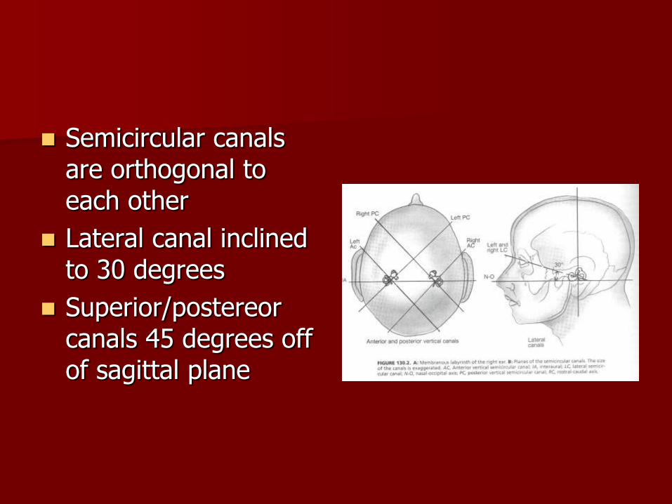
Semicircular canals are orthogonal to each other
Lateral canal inclined to 30 degrees
Superior/postereor canals 45 degrees off of sagittal plane

Utricle is in horizontal plane
Saccule is in vertical plane

Anatomy
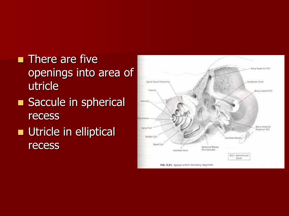
There are five openings into area of utricle
Saccule in spherical recess
Utricle in elliptical recess
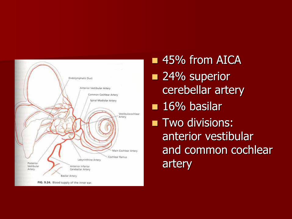
45% from AICA
24% superior cerebellar artery
16% basilar
Two divisions: anterior vestibular and common cochlear artery

Superior vestibular nerve: superior canal, lateral canal, utricle
Inferior vestibular nerve: posterior canal and saccule

Membranous labyrinth is surrounded by perilymph
Endolymph fills the vestibular end organs along with the cochlea

Perilymph
– Similar to extracellular fluid
– K+=10mEQ, Na+=140mEq/L
– Unclear whether this is ultrafiltrate of CSF or blood
– Drains via venules and middle ear mucosa

Endolymph
– Similar to intracellular fluid
– K+=144mEq/L, Na+=5mEq/L
– Produced by marginal cells in stria vascularis from perilymph at the cochlea and from dark cells in the cristae and maculae
– Absorbed in endolymphatic sac which connected by endolymphatic, utricular and saccular ducts

Sensory structures
Ampulla of the semicircular canals
Dilated end of canal
Contains sensory neuroepithelium, cupula, supporting cells

Cupula is gelatinous mass extending across at right angle
Extends completely across, not responsive to gravity
Crista ampullaris is made up of sensory hair cells and supporting cells

Sensory cells are either Type I or Type II
Type I cells are flask shaped and have chalice shaped calyx ending
One chalice may synapse with 2-4 Type I cells
Type II cells – cylinder shaped, multiple efferent and afferent boutons
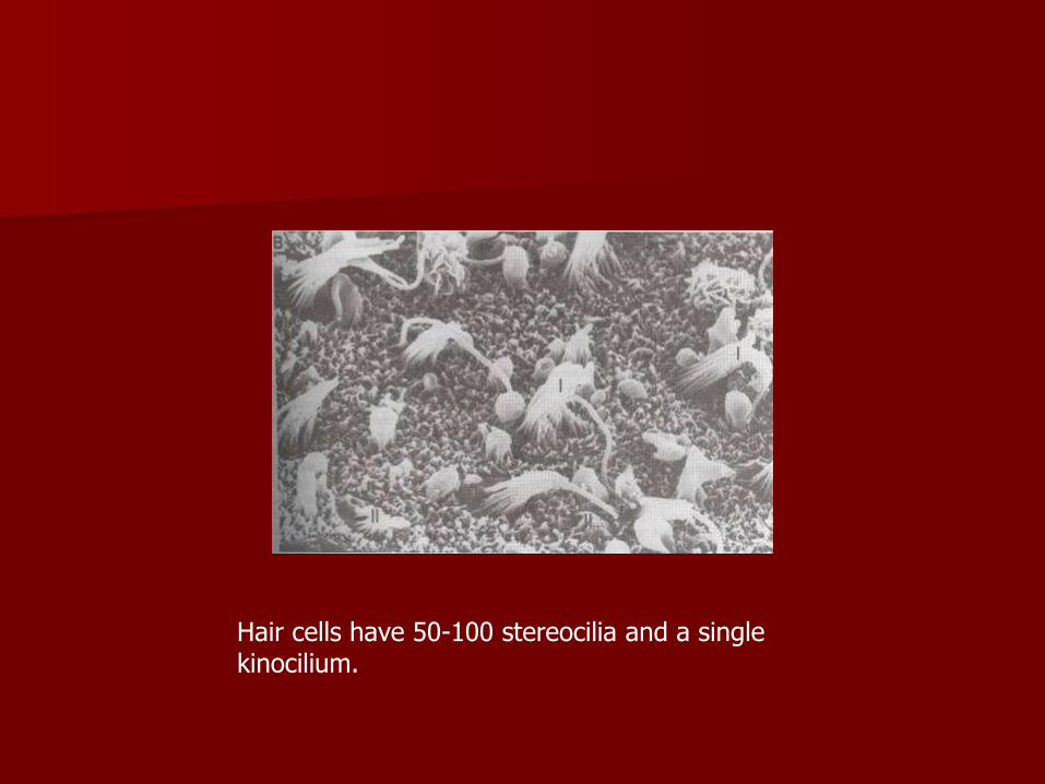
Hair cells have 50-100 stereocilia and a single kinocilium.
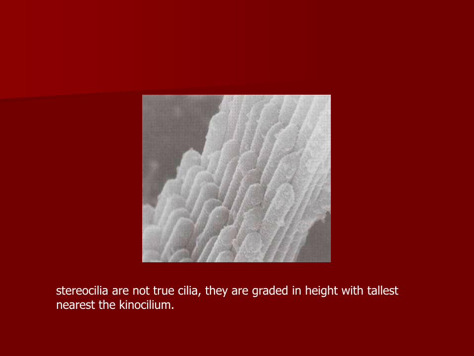
stereocilia are not true cilia, they are graded in height with tallest nearest the kinocilium.
Kinocilium is located on one end of cell giving each cell a polarity
Has 9+2 arrangement of microtubule doublets
Lacks inner dynein arms, and central portion of microtubules not present near ends – may mean they are immobile or weakly mobile

Each afferent neuron has a baseline firing rate
Deflection of stereocilia toward kinocilium results in an increase in the firing rate of the afferent neuron
Deflection away causes a decrease in the firing rate


kinocilia are located closest to utricle in lateral canals and are on canalicular side in other canals
Ampullopetal flow (toward the ampulla) excitatory in lateral canals, inhibitory in superior/posterior canals
Ampullofugal flow (away from the ampulla) has opposite effect

Semicircular canals are paired
– Horizontal canals
– Right superior/left posterior
– Left superior/right posterior
– Allow redundant reception of movement
– Explains compensation after unilateral vestibular loss
Otolithic organs
Utricle and saccule sense linear acceleration
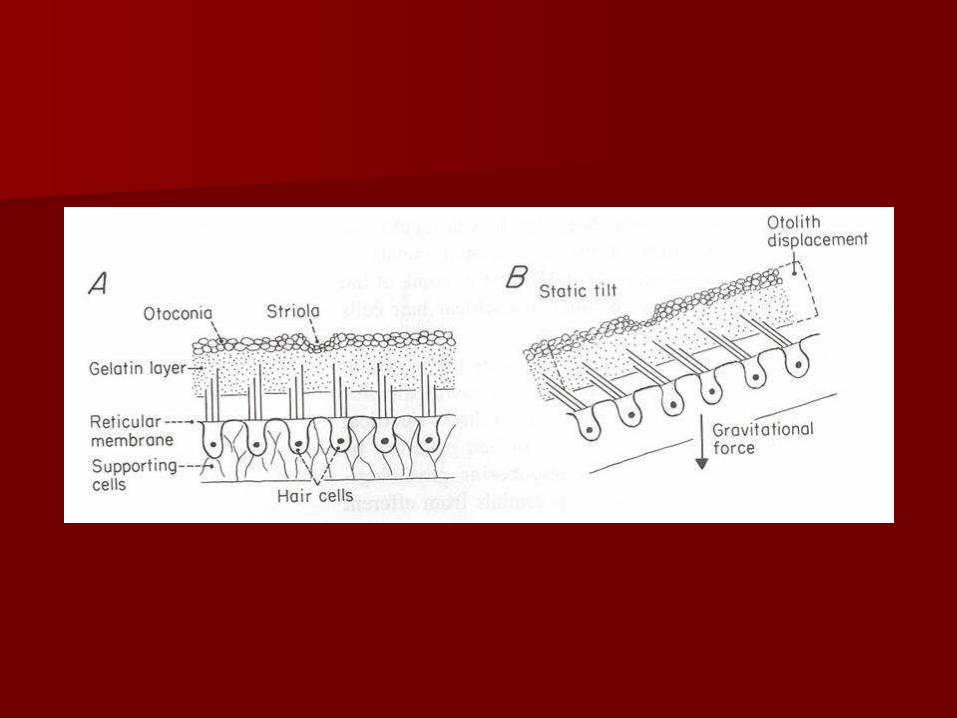
Cilia from hair cells are embedded in gelatinous layer
Otoliths or otoconia are on upper surface

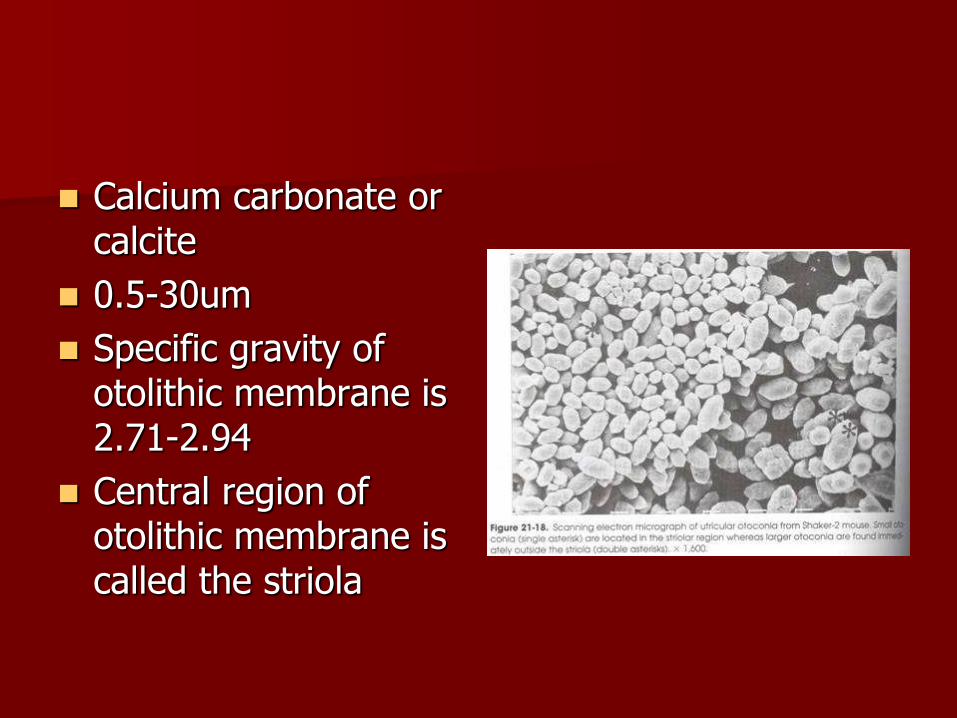
Calcium carbonate or calcite
0.5-30um
Specific gravity of otolithic membrane is 2.71-2.94
Central region of otolithic membrane is called the striola

Saccule has hair cells oriented away from the striola
Utricle has hair cells oriented towards the striola
Striola is curved so otolithic organs are sensitive to linear motion in multiple trajectories
Central connections
Scarpa’s ganglion is in the internal auditory canal
Contains bipolar ganglion cells of first order neurons
Superior and inferior divisions form common bundle which enters brainstem
No primary vestibular afferents cross the midline

Afferent fibers terminate in the vestibular nuclei in floor of fourth ventricle
– Superior vestibular nucleus
– Lateral vestibular nucleus
– Medial vestibular nucleus
– Descending vestibular nucleus
Vestibular nuclei project to
– Cerebellum
– Extraocular nuclei
– Spinal cord
– Contralateral vestibular nuclei

Senses and controls motion
Information is combined with that from visual system and proprioceptive system
Maintains balance and compensates for effects of head motion

Vestibulo-ocular reflex – Membranous labyrinth
moves with head motion – Endolymph does not
causing relative motion – Cupula on right canal
deflected towards utricle causing increase in firing rate, left deflects away causing a decrease in firing rate.
– Reflex causes movement of eyes to the left with saccades to right
– Stabilizes visual image

If acceleration stops, and spin to right is at constant velocity, sensation of motion stops after 14-20 seconds as does nystagmus
Cupula only takes 8-10 seconds to return to equilibrium position
Vestibular integrator is the term for the prolongation and is mediated by the vestibular nuclei and cerebellum

Vestibulospinal Reflex
Senses head movement and head relative to gravity
Projects to antigravity muscles via 3 major pathways:
– Lateral vestibulospinal tract
– Medial vestibulospinal tract
– Reticulospinal tract

How do calorics work?
Patient is lying down with horizontal canals oriented vertically (ampulla up)
Cold water irrigation causes endolymph in lateral portion to become dense and fall causing deflection of cupula away from utricle with a decrease in the firing rate
This causes nystagmus with fast phase (beat) away from the stimulus

With warm water irrigation column of endolymph becomes less dense, rises and causes deflection of cupula toward the utricle
Results in increase firing rate and nystagmus which beats towards the stimulation
COWS (cold opposite, warm same)

Sources Shepard NT, Solomon D. Functional Operation of the Balance System in Daily
Activities. Otolaryngologic Clinics of North America 2000;33(3):455-468. Minor LB. Physiological principles of vestibular function on earth and in
space. Otolaryngology-Head and Neck Surgery 1998;118(3 part 2):S5-S15.
Abdel Razek OA. Anatomy of the Vestibular System. www.emedicine.com Hoffman R, Strunk C. Vestibular Anatomy and Physiology. Department of
Otolaryngology Grand Rounds University of Texas Medical Branch December 9, 1992.
Baloh RW. Dizziness, Hearing Loss, and Tinnitus. Philadephia, F.A. Davis Company, 1998.
Jahn AF, Santos-Sacchi J. Physiology of the Ear. Second edition. San Diego, Singular, 2001.
Friedman I, Ballantyne J. Ultrastructural Atlas of the Inner Ear. London, Butterworth & Co., 1984.
Janfaza P, Nadol JB. Temporal Bone and Ear. In: Janfaza P ed. Surgical Anatomy of the Head and Neck. Philadelphia, Lippincott Williams & Wilkins, 2001:419-479.
Wall C, Vrabec JT. Vestibular Function and Anatomy. In: Head & Neck Surgery-Otolaryngolog. Philadelphia, Lippincott Williams & Wilkins, 2001:1641-1650.

Vestibular Function and Anatomy
Gordon Shields, MD
Faculty Advisor: Arun Gadre, MD
The University of Texas Medical Branch
Department of Otolaryngology
Grand Rounds Presentation
April 14, 2004