vigo, daniele; torre, marina l.; faustini, massimo; … roberta; villani, simona; asti, annalia;...
TRANSCRIPT
Vigo, Daniele; Torre, Marina L.; Faustini, Massimo; Russo, Vincenzo; Norberti, Roberta; Villani, Simona; Asti, Annalia; Bini, Pier Paolo; Conte, Ubaldo (2004) Barium alginate capsules for 3D immobilisation of living cells: morphology, membrane properties and permeability. Journal of Drug Delivery Science and Technology, Vol. 14 (3), p. 167-172. ISSN 1157-1489.
http://eprints.uniss.it/7296/
Documento digitalizzato dallo Staff di UnissResearch

z V) V)
Jou alof
(Formerly STP Pharma Sciences)
MAY-JUNE 2004
J. DRUG DEL. SCI. TECH., 14 (3) 167-1722004
Barium alginate capsules for 3D immobilisation of living cells: morphology, membrane properties and permeability
O. Vigo1, M.L. Torre2*, M. FaustinP, E. MunarP, v. Russo1, R. Norberti2, s. VillanP, A. Asti4 , P.P. Bini5, u. Conte2
1 Dipartimento di Scienze e Tecnologie Veterinarie per la Sicurezza Alimentare, via Celoria 10, 20133 Milano, Italia 2Dipartimento di Chimica Farmaceutica, via Taramelli 12, 27100 Pavia, Italia
3Dipartimento di Scienze Sanitarie Applicate e Psicocomportamentali, via Bassi 21, 27100 Pavia, Italia 4Centro Grandi Strumenti, via Bassi 21, 27100 Pavia, Italia
5Dipartimento di Biologia Animale, via Vienna 2, 07100 Sassari, Italia *Correspondence: [email protected]
Encapsulation in a bariwn alginate membrane is a promising strategy to obtain a three dimensionai culture ojliving cells: membrane properties are crucialjor a realistic clinical application. A olle-step ellcapsulatioll tecll1liqlle, recemly developedjor C01lfrolled release oj boar semen, was employed to prepare bariwll alginate and protamine-algillate membranes: per11leability to two modelmolecules (haemoglobin and glllcose) was evaluated. Capsllies were evaluatedjor technological properties and scanning electron microscopy was IIsed to examine tlze externalmorpllOlogy oj the capsllies and the 3D distriblltioll oj the cells witlzill tlze core. The reslllts indicate that 3D arrange11le1lf and cell shape are mai1lfained, capsule di11lensions and mechanical properties call be 11l0dlllated, as well as tlzeir permeability to model molecllies slIcll as Izaemoglobin and glllcose.
Key words: Celi encapsulatioll- Alginate - Membrane permeability - SEM - 3D celi arrangement.
CelI encapsulation is a strategy in which three-dimensional (3D) celI cultures are obtained by entrapping a pool oflive celIs within a semi-permeable membrane [1,2]. The opti mal degree of selectivity and permeability of the membrane to allow both diffusion of metabolites, oxygen and nutrients and cell life is the primary challenge in capsule development [3]. Other challenges include obtaining the desired mechanical properties ofthe membrane in terms of resistance to rupture, elasticity, particle size distribution, surface properties and morphology. All these characteristics combine to determine the ease of handling and the possible applications of the capsules [4].
In vitro cultures of mammalian cells isolated from tissues have a broad range of applications, including reproductive strategies [4]; nevertheless, standard monolayer in vitro celI cultures cannot adequately simulate the in vivo development of tissues which require both a 3D arrangement of the cells and a physiological extracellular matrix (ECM). There have been several successful attempts to use polymeric matrices, or scaffolds to culture isolated cells in 3D space [5,6]. Hydrogels, and in particular alginate, mimic ECM well, as reviewed by Gombotz and Fong Wee [7].
A one-step encapsulation technique in barium alginate, described by Klein et al. [8], was recently developed for controlled release of semen for the artificial insemination of swine [9-14] and for 3D culture of ovarian follicular cells [15]. The barium alginate capsules are formed of a core containing the live cells surrounded by a gel membrane which may, if desired, be coated by an extemallayer of protamine. The advantage of this method is its versatility: core properties can be modified, and depending on celI type, different suitable polymers can be employed to mimic artificial ECM. Altematively, cells in the core can be maintained into their physiological matrix. Another advantage of this method is the minimal effect of concentration of gellifying cation on cell functions: ions diffuse very
167
quickly from the core leading to the alginate membrane growth. Membrane properties can also be modulated in terms of gel thickness, mechanical properties [12] and release kinetics [Il] by changing the ion type, its concentration in the core, and by cross-linking of the extemal surface with polyamines, as for example, protamine. Aiso capsules dimensions can be adapted to the use, changing the extrusor needle diameter.
The aim of this work was to evaluate the permeability of the membrane to two mode l molecules (haemoglobin (Hb) and glucose), as well as to characterize the physical and mechanical properties of the capsules (weight, total diameter, core diameter, gel thickness and capsule strength). Bovine erythrocytes were encapsulated as a source of haemoglobin and as model cells for encapsulation process: in fact, these kinds of cells have fragile membranes and are easily damaged in non-physiological environment.
Capsule membranes were modified by superficial treatment with protamine sulfate (PS) at different concentrations and for different time intervals in order to modulate morphological and functional properties. Furthermore, the capsules were examined by scanning electron microscopy (SEM) to investigate their extemal morphology and the 3D distribution of the cells within the core.
lo MATERIALS ANO METHOOS In order to study the permeability of the capsules to the
chosen molecules, a protein (haemoglobin, MW 64,000) and a small non-ionic carbohydrate (glucose, MW 180), and to examine the 3D arrangement of encapsulated live cells (bovine erythrocytes), two sets of capsules were produced. One set contained bovine red blood cells (RBC) in the form of an erythrocyte-rich fraction, the other set contained physiological saline in order to evaluate the reduction in glucose concentration in the supematant after incubation.
J. DRUG DEL. SCI. TECH., 14 (3) 167-1722004
1. Preparation of the erythrocyte-rich fraction Blood samples were colIected into evacuated heparin-con
taining glass tubes (Vacutainer) from six adult Friesian Holstein cows 30 min after milking; at least 30 mI of blood/sample was centrifuged (1500 x g, lO min) to produce an erythrocyte-rich fraction (ERF). The ERF was separated from the plasma and buffy coat, resuspended, washed with NaCI 0.9% at 4°C and centrifuged three times. The haematocrit of the final ERF ranged between 80 to 85% (microhaematocrit, Sanyo micro Centaur).
2. Encapsulation The encapsulation was performed using a method previously
described for swine spermatozoa [lO]. For the haemoglobin diffusion study, a solution of xanthan
gum 0.25% w/v (Satiaxane CX2, SKW Biosystems, I) in NaCI 0.9% w/v was added to the ERF (ERF/xanthan solution volume ratio 1: 1) and then a saturated BaCl
2 solution was added to
obtain a final Ba++ ion concentration of 20 mM. The resulting suspension was dropped (through a 26Gx1/2" needle) into a 0.5 % w/v sodium alginate solution (sodium alginate medium viscosity, Sigma-Aldrich, Germany) where barium alginate capsules were obtained. These capsules, calI ed gel capsules, were collected by filtration, rinsed twice in NaCI 0.9% solution and resuspended in the same medium.A second kind of capsule was prepared by treating the external surface ofthe gel capsules with PS to modulate the membrane properties. Different PS concentrations and cross-linking times were assessed: 0.5% w/v PS solution for 3 min (0.5% 3'); 0.5% w/v PS solution for 30 min (0.5% 30'); 1 % w/v PS solution for 3 min (1 % 3') and 1 % w/v PS solution for 30 min (1 % 30').
For the glucose permeability study, the capsules were prepared as above, but with physiological solution (NaCI 0.9%) instead of ERF.
3. Scanning electron microscopy (SEM) Membrane and core structure were investigated by scanning
electron microscopy (SEM): the capsules were dehydrated with serial ethanol solutions from 50 to 100%, criticaI point dried, go Id sputtered (purity degree 99.9%) with an Edwards S l50A sputter coater (Boc Edwards Italia SpA, Milan, Italy) and then observed by a scanning electron microscope (Cambridge Stereoscan 250, Cambridge Instruments Ltd., Cambridge, United Kingdom), operating at 20 kY.
4. Technological characterization of capsules Capsules were macroscopicalIy photographed by a digitaI
video camera connected to an image anal yser (CV 9000 Vero 4.0 Image Anal yzer, FKV Srl, Soriso le, BG , Ital y) . The who le capsule diameter, the core diameter and gel capsule thickness were measured and the weight of the capsules was determined.
The mechanical properties of the capsule membranes were evaluated by means of a texture analyser (Stable MicroSystems, mode l TA-XT2, UK) equipped with a 3 cm diameter plexiglass probe. During the test, the probe descends at the rate ofO.l mm/ s until a force resistance of 50 g is detected, and then maintains this position for 30 s. The force versus time plot characterizes the consistency of the capsule and the force value (in grams)
168
Barium alginate capsules for 3D immobilisation of Iiving cells: morphology, membrane properties and permeability
D. Vigo, M.L. Torre, M. Faustini, E. Munari, V. Russo, R. Norberti, S. Villani, A. Asti, P.P. Bini, U. Conte
detected after 30 s can be considered as the strength index (F30) of the capsule. All measurements were repeated on five randomised capsules per batch.
5. Haemoglobin diffusion measurements Three sample capsules were incubated at 2SOC in a hypo
osmotic sodium chloride solution (500 ILI, 0.45% w/v, pH 7.1, equivalent to the isoelectric point of Hb, 25°C) in an Eppendorf tube. The osmotic shock caused the red blood cells to rupture and the haemoglobin to leak out. Diffusion of Hb through membrane was assessed by measuring light absorbance in the supernatant (100 ILI of extracapsular medium + 100 ILI ofDrabkin reagent to convert Hb into cyanometaHb). Optical density (OD) readings were performed by a microplate reader (Labsystems Multiskan EX), with filter at A = 540 nm. The percentage of Hb diffused as a function of time was estimated as the increase in Hb concentration in the supernatant, with respect to the Hb concentration afterthe mechanical membrane disruption of a standard sample, taking into account the mean volume of capsules for each batch.
The folIowing formula was applied:
Hb % increase = [500/(500 + nV)]*100 (ODt/ODd)
where n is the number of capsules added in each tube, Ve the mean capsule volume for each batch, ODt the optical density at each sampling time, and ODd the optical density after membrane disruption.
6. Glucose permeability measurements Three sample capsules were incubated at 25°C in 500 ILI
of an aqueous glucose solution (180 mg glucose/lOO mI) in an Eppendorf tube. The concentration gradient leads to the diffusion of glucose into the capsule, thus decreasing the concentration of glucose in the external solution. At fixed times, the reduction in glucose concentrations was determined by an automated haematochemistry analyser (Kone modo Specificselecti ve, Kone Instrument Corp., Evry, France) with a specific glucose kit (hexokinase method), based on end-point reduction of NAD to NADH at a A = 340 nm, linearity 600 mglIOO mI, and incubation temperature 37°C (Dasit Italia, Cornaredo, Milan, Italy).
Results were calculated as the decrease in glucose concentration in the supernatant, with respect to the initial concentration, taking into account the mean volume of capsules for each batch, using the following formula:
glucose % decrease = [500/(500 + nV )]*100 (C/C) e t O
where n is the number of capsules added in each tube, V the mean capsule volume for each batch, C
t the glucose conce~tra
tion at each sampling time, and Co the glucose concentration at time zero.
The complement to 100 is reported in the results.
7. Statistical analysis Results concerning the technological properties of the
capsules (weight, total diameter, core diameter, gel thickness
Barium alginate capsules for 3D immobilisation of living cells: morphology, membrane properties and permeability D. Vigo, M.L. Torre, M. Faustini, E. Munari, V Russo, R. Norberti, S. Villani, A. Asti, P. P. Bini, U. Conte
and capsule strength) for each formulation were analysed by a one-way ANOVA procedure, followed by a least significant difference (LSD) test. Results were reported as mean value, standard deviation (SD) and sample size (n). For each molecule model, concentration reduction profiles were analysed using a two-way ANOVA, with capsule formulation and ti me of sampling as factors , as described by O'Hara et al. [16] and Yuksel et al. [17]. Differences between formulations were assessed by the LSD test.
The level of statistical significance was set at an alpha of 0 .05. Differences between groups for each variable are represented with different superscripts .
II. RESULTS ANO OISCUSSION With the aid of a digitaI camera connected to an image
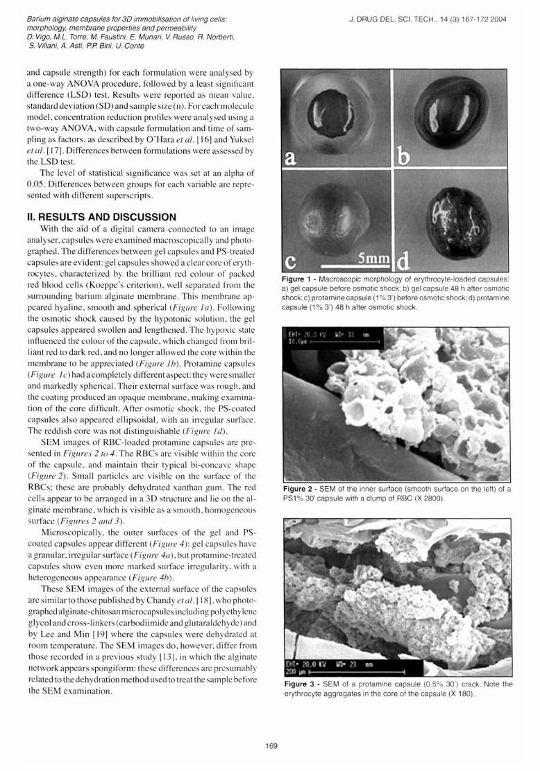
analyser, capsules were examined macroscopically and photographed. The differences between gel capsules and PS-treated capsules are evident: gel capsules showed a clear core of erythrocytes, characterized by the brilliant red colour of packed red blood cells (Koeppe's criterion), well separated from the surrounding barium alginate membrane. This membrane appeared hyaline, smooth and spherical (Figure la) . Following the osmotic shock caused by the hypotonic solution, the gel capsules appeared swollen and lengthened. The hypoxic state influenced the colour of the capsule, which changed from brilliant red to dark red, and no longer allowed the core within the membrane to be appreciated (Figure lb) . Protamine capsules (Figure J c) hadacompletely differentaspect: they were smaller and markedly spherical. Their extemal surface was rough, and the coating produced an opaque membrane, making examination of the core difficult . After osmotic shock, the PS-coated capsules also appeared ellipsoidal , with an irregular surface. The reddish core was not distinguishable (Figure l d).
SEM images of RBC-Ioaded protamine capsules are presented in Figures 2 fa 4. The RBCs are visible within the core of the capsule, and maintain their typical bi-concave shape (Figure 2). Small particles are visible on the surface of the RBCs; these are probably dehydrated xanthan gum. The red cells appear to be arranged in a 3D structure and lie on the alginate membrane , which is visible as a smooth , homogeneous surface (Figures 2 and 3).
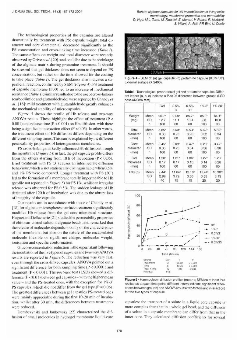
Microscopically, the outer surfaces of the gel and PScoated capsules appear different (Figure 4): gel capsules have a granular, irregular surface (Figure 4a), but protamine-treated capsules show even more marked surface irregularity, with a heterogeneous appearance (Figure 4b).
These SEM images of the extemal surface of the capsules are similar to those published by Chandy et al. [18], who photographed alginate-chitosan microcapsules including polyethylene gJycol and cross-linkers (carbodiimide and glutaraldehyde) and by Lee and Min [19] where the capsules were dehydrated at room temperature . The SEM images do, however, differ from those recorded in a previous study [13], in which the alginate network appears spongiform: these differences are presumably related to the dehydration method used to treat the sample before the SEM examination.
169
J. DRUG DEL. SCI. TECH., 14 (3) 167-1722004
Figure 1 - Macroscopic morphology of erythrocyte-Ioaded capsules: a) gel capsule before osmotic shock; b) gel capsule 48 h after osmotic shock; c) protamine capsule (1 % 3') before osmotic shock; d) protamine capsule (1 % 3') 48 h after osmotic shock.
Figure 2 - SEM of the inner surface (smooth surface on the left) of a PS1 % 30' capsule with a clump of RBC (X 2800).
Figure 3 - SEM of a protamine capsule (0.5% 30') crack. Note the erythrocyte aggregates in the core cf the capsule (X 180).
J . ORUG DEL. SCI. TECH., 14 (3) 167-1722004
The technological properties of the capsules are altered dramatically by treatment with PS: capsule weight, total diameter and core diameter all decreased significantly as the PS concentration and cross-linking time increased (Table I). The same effects on weight and totai diameter were recently observed by Orive et al . [20], and could be due to the shrinkage of the alginate matrix during protamine treatment. It should be stressed that gel thickness does not seem to depend on PS concentration, but rather on the time allowed for the coating to take pIace (Table I). The gel thickness aiso indicates a superficial reaction, confirmed by SEM (Figure 4). PS treatment of capsule membrane (F30) led to an increase of mechanical resistance (Table I); similarresultsdue to the useofcross-linkers (carbodiimide and glutaraldehyde) were reported by Chandy et al., [18]: mild treatment with glutaraIdehyde greatly enhances the mechanical stability of microcapsules .
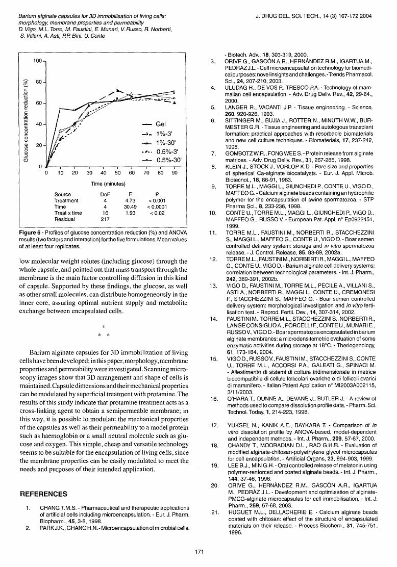
Figure 5 shows the profile of Hb release and two-way ANOVA results . These highlight the effect of treatment (P < 0.001) and release time (P < 0.001 ) on Hb diffusion , with there being a significant interaction effect (P < 0.05). In other words, the treatment effect on Hb diffusion differs depending on the different ampling times. This can be explained by the different permeability properties of heterogeneous membranes.
PS cross-linking markedIy inftuenced Hb diffusion through the membrane (Figure 5): in fact, the gel capsule profile differs from the others starting from 18 h of incubation (P < 0.05). Brief treatment with PS (3') causes an intermediate diffusion behaviour, which is not stati stically distinguishable when 0.5% and 1% PS were compared. Longer treatment with PS (30') led to the forrnation of a membrane totally impermeable to Hb (profile not reported in Figure 5) for PS 1%, whiIst an irregular release was observed for PS 0 .5%. The sudden leakage of Hb detected after 120 h of incubation was due to the abrupt loss of integrity of the capsule.
Our results are in accordance with those of Chandy et al. [18] for alginate microspheres: surface treatment significantly modifies Hb release from the gel core microbead structure. Huguet and Dellacherie [21] studied the permeability properties of chitosan-coated calcium alginate beads, and remarked that the release of molecules depends not only on the characteristics of the membrane, but also on the nature of the encapsulated molecule (ftexible or rigid), net charge, molecular weight, ionisation and specific conforrnation.
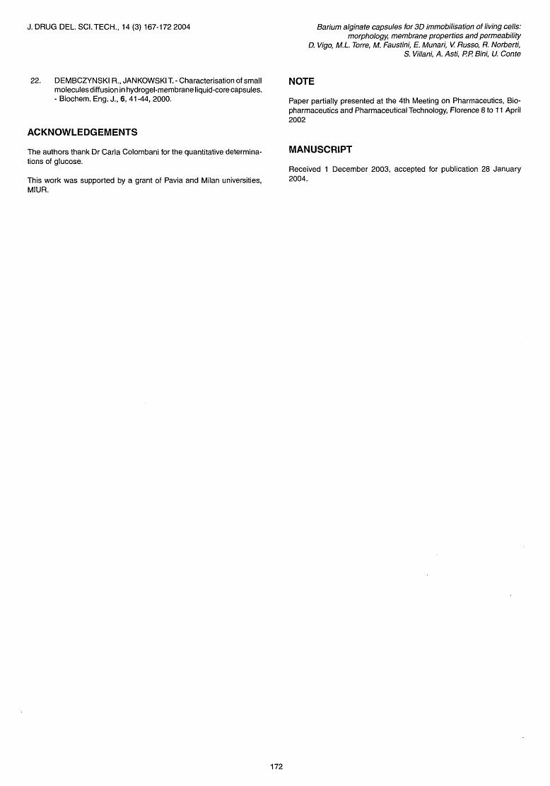
Glucose concentration reduction in the supematant following the incubation of the fi ve types of capsules and two-way ANOVA resu]ts are reported in Figure 6. The reduction was very fast, even through the cross-linked capsules. ANOVA pointed out a significant difference for both sampling time (P < 0.0001) and treatment (P < 0.001). The post-hoc test (LSD) showed a difference (P < 0.0] ) between gel capsules - with the higher mean value - and the PS-treated ones, with the exception for 1%-3' PS capsules, which did not differ from the gel type (P = 0.06). The greatest differences between gel capsules PS-treated ones were mainly appreciable during the first 10-20 min of incubation, whilst after 30 min , the differences between treatments were reduced.
Dembczynski and Jankowski [22] characterized the diffusion of small molecules in hydrogel membrane liquid-core
170
Barium alginate capsules for 3D immobilisation of living cells: morphology, membrane properties and permeability
D. Vigo, M.L. Torre, M. Faustini, E. Munari, V Russo, R. Norberti, S. Villani, A. Asti, P P Bini, U. Conte
Figure 4 - SEM of: (a) gel capsule; (b) protamine capsule (0.5% 30'). External surface (X 2400).
Table 1- Technological properties of gel and protamine capsules. Oifferent letters (a, b, c) indicate a P<0.05 difference between groups (LSD post-ANOVA test) .
Weight Mean (mg) SO
n
Total Mean diameter SO
(mm) n
Core Mean diameter SO
(m m) n
Gel Mean thickness SO
(mm) n
F30 (g) Mean SO n
100
~ 80
"O al CI)
60 :::J :t= '6 c :o 40 o Cl o E al
20 co I
O , O 24
Source Trealmenl Time Treal x lime Residual
Gel 0.5% 0.5% 3' 30'
90.7a 91.8a 85.7b
12.7 11.1 13.4 160 60 60
5.85a 5.60b 5.53b
0.33 0.23 0.26 160 60 60
3.45a 3.09b 3 .47a
0.35 0.23 0.34 160 60 60
1.20a 1.21 a 1.06b
0.17 0.17 0.18 160 60 60
9.44a 11.64b 12.19b
2.89 3.72 3.35 40 15 15
1% 3' 1% 30'
85.0b 84.1b
9.8 10.8 100 80
5.62b 5.62b
0.32 0.34 100 80
3.26c 3.47a
0.36 0.38 100 80
1.22a 1.28c
0.14 0.28 100 80
11.44b 10.30ab
3.55 3.13 25 20
gel
-~_ . 1%3'
~ 0.5%3'
.......... 1%30'
0.5%30'
48 72 96 120 144 168
Time (hours)
DoF F P 3 35.62 < 0 .001 5 19.76 < 0.001 10 1.96 < 0.05
143
Figure 5 - Haemoglobin diffusion profiles (mean ± SEM on at least four replicates at each time point; different letters indicate significant differences between groups) and ANOVA results (twofactors and interaction) for the five types of capsule .
capsules: the transport of a solute in a liquid core capsule is more complex than that in a whole gel bead , and the diffusion of a solute in a capsule membrane can differ from that in the inner core . They calculated diffusion coefficients for several
Barium alginate capsules for 3D immobilisation of living cells: morphology, membrane properties and permeability D. Vigo, M.L. Torre, M. Faustini, E. Munari, V. Russo, R. Norberti, S. Villani, A. Asti, P.P. Bini, U. Conte
100
~ 80
c .Q t3 :::J 60 "O ~ C o ~ 40 c - Gel ID U C ~- 1%-3' o U ID 20 ~ 1%-30' CI)
o U .~. I 0.5%-3' :::J
a ----- 0.5%-30'
10 20 30 40 50 60 70 80 90
Time (minutes)
Source DoF F P Treatment 4 4.73 < 0.001 Time 4 30.49 < 0.0001 Treat x time 16 1.93 < 0.02 Residual 217
Figure 6 - Profiles of glucose concentration reduction (%) and ANOVA results (twofactors and interaction) forthe five formulations. Mean values of at least four replicates.
low molecular weight solutes (including glucose) through the whole capsule, and pointed out that mass transport through the membrane is the main factor controlling diffusion in this kind of capsule. Supported by these findings, the glucose, as well as other small molecules, can distribute homogeneously in the inner core, assuring optimal nutrient supply and metabolite exchange between encapsulated cells.
* * *
Barium alginate capsules for 3D immobilization of living cells have been developed; in this paper, morphology, membrane properties and permeability were investigated. Scanning microscopy images show that 3D arrangement and shape of cells is maintained. Capsule dimensions and their mechanical properties can be modulated by superficial treatment with protamine. The resu1ts of this study indicate that protamine treatment acts as a cross-linking agent to obtain a semipermeable membrane; in this way, it is possible to modulate the mechanical properties of the capsules as well as their permeability to a model protein such as haemoglobin or a small neutral molecule such as glucose and oxygen. This simple, cheap and versatile technology seems to be suitable for the encapsulation of living cells, since the membrane properties can be easily modulated to meet the needs and purposes of their intended applicatiorl.
REFERENCES
1. CHANG T.M.S. - Pharmaceutical and therapeutic applications of artificial cells including microencapsulation. - Eur. J. Pharm. Biopharm., 45,3-8, 1998.
2. PARKJ.K., CHANG H.N.- Microencapsulation of microbial cells.
171
J. DRUG DEL. SCI. TECH., 14 (3) 167-1722004
- Biotech. Adv., 18,303-319,2000. 3. ORIVE G., GASCON A.R., HERNANDEZ R.M., IGARTUA M.,
PEDRAZ J.L. - Celi microencapsulation technology for biomedical purposes: novel insights and challenges. -Trends Pharmacol. Sci., 24, 207-210, 2003.
4. ULUDAG H., DE VOS P., TRESCO P.A. - Technology of mammalian celi encapsulation. - Adv. Drug Deliv. Rev., 42, 29-64., 2000.
5. LANGER R., VACANTI J.P. - Tissue engineering. - Science, 260, 920-926, 1993.
6. SIITINGER M., BUJIA J., ROTTER N., MINUTH W.W., BURMESTER G.R. - Tissue engineering and autologous transplant formation: practical approaches with resorbable biomaterials and new celi culture techniques. - Biomaterials, 17, 237-242, 1996.
7. GOMBOTZ W. R., FONG WEE S. - Protein release from alginate matrices. - Adv. Drug Deliv. Rev., 31, 267-285,1998.
8. KLEIN J., STOCK J., VORLOP K.D. - Pore size and properties of spherical Ca-alginate biocatalysts. - Eur. J. Appl. Microb. Biotecnol., 18, 86-91, 1983.
9. TORRE M.L., MAGGI L., GIUNCHEDI P., CONTE U., VIGO D., MAFFEO G. - Calcium alginate beads containing an hydrophilic polymer for the encapsulation of swine spermatozoa. - STP Pharma Sci., 8, 233-236, 1998.
10. CONTE u., TORRE M.L., MAGGI L., GIUNCHEDI P., VIGO D., MAFFEO G., RUSSO V. - European Pat. Appl. n° Ep0922451, 1999.
11. TORRE M.L., FAUSTINI M., NORBERTI R., STACCHEZZINI S., MAGGI L., MAFFEO G., CONTE U., VIGO D. - Boar semen controlled delivery system: storage and in vitro spermatozoa release. - J. Control. Release, 85, 83-89, 2002a.
12. TORRE M.L., FAUSTINI M., NORBERTI R., MAGGI L., MAFFEO G., CONTE U., VIGO D. - Barium alginate celi delivery systems: correlation between technological parameters. - Int. J. Pharm., 242,389-391,2002b.
13. VIGO D., FAUSTINI M., TORRE M.L., PECILE A., VILLANI S., ASTI A, NORBERTI R., MAGGI L., CONTE U., CREMONESI E, STACCHEZZINI S., MAFFEO G. - Boar semen controlled delivery system: morphological investigation and in vitro fertiIisation test. - Reprod. Fertil. Dev., 14,307-314,2002.
14. FAUSTINI M., TORRE M.L., STACCHEZZINI S., NORBERTI R., LANGE CONSIGLIO A, PORCELLI E, CONTE U., MUNARI E., RUSSO V., VIGO D. - Boar spermatozoa encapsulated in barium alginate membranes: a microdensitometric evaluation of some enzymatic activities during storage at 18°C. - Theriogenology, 61, 173-184, 2004.
15. VIGO D., RUSSOV., FAUSTINI M., STACCHEZZINI S., CONTE U., TORRE M.L., ACCORSI P.A., GALEATI G., SPINACI M. - Allestimento di sistemi di coltura tridimensionale in matrice biocompatibile di cellule follicolari ovariche e di follicoli ovarici di mammifero. - Italian Patent Application n° M12003A002115, 3/11/2003.
16. O'HARA T., DUNNE A., DEVANE J., BUTLER J. - A review of methods used to compare dissolution profile data. - Pharm. Sci. Technol. Today, 1, 214-223, 1998.
17. YUKSEL N., KANIK AE., BAYKARA T. - Comparison of in vitro dissolution profile by ANOVA-based, model-dependent and independent methods. - Int. J. Pharm., 209, 57-67, 2000.
18. CHANDY T., MOORADIAN D.L., RAO G.H.R. - Evaluation of modified alginate-chitosan-polyethylene glycol microcapsules for celi encapsulation. - Artificial Organs, 23, 894-903, 1999.
19. LEE B.J., MIN G.H. - Oral controlled release of melatonin using polymer-renforced and coated alginate beads. - Int. J. Pharm., 144, 37-46, 1996.
20. ORIVE G., HERNÀNDEZ R.M., GASCÒN AR., IGARTUA M., PEDRÀZ J.L. - Development and optimisation of alginatePMCG-alginate microcapsules for celi immobilisation. - Int. J. Pharm., 259, 57-68, 2003.
21. HUGUET M.L., DELLACHERIE E. - Calcium alginate beads coated with chitosan: effect of the structure of encapsulated materials on their release. - Process Biochem., 31, 745-751, 1996.
J. DRUG DEL. SCI. TECH., 14 (3) 167-1722004
22. DEMBCZYNSKI R., JANKOWSKI T. - Characterisation of small molecules diffusion in hydrogel-membrane liquid-core capsules. - Biochem. Eng. J., 6, 41-44, 2000.
ACKNOWLEDGEMENTS
The authors thank Dr Carla Colombani for the quantitative determinations of glucose.
This work was supported by a grant of Pavia and Milan universities, MIUR.
172
Barium alginate capsules for 3D immobilisation of living cells: morphology, membrane properties and permeability
D. Vigo, ML. Torre, M Faustini, E. Munari, V. Russo, R. Norberti, S. Villani, A. Asti, P.P. Bini, U. Conte
NOTE
Paper partially presented at the 4th Meeting on Pharmaceutics, Biopharmaceutics and Pharmaceutical Technology, Florence 8 to 11 Aprii 2002
MANUSCRIPT
Received 1 December 2003, accepted for publication 28 January 2004.