viii. pectoral girdle & upper extremity - articulation between upper extremities and axial...
TRANSCRIPT
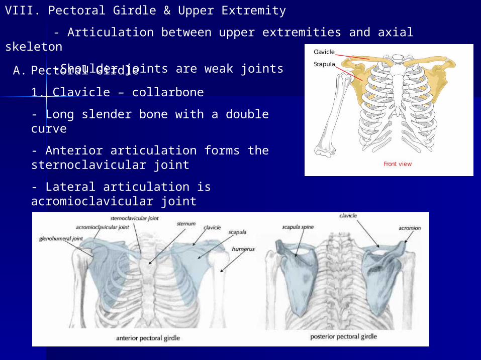
VIII. Pectoral Girdle & Upper Extremity
- Articulation between upper extremities and axial skeleton
-Shoulder joints are weak jointsA. Pectoral Girdle
1. Clavicle – collarbone
- Long slender bone with a double curve
- Anterior articulation forms the sternoclavicular joint
- Lateral articulation is acromioclavicular joint
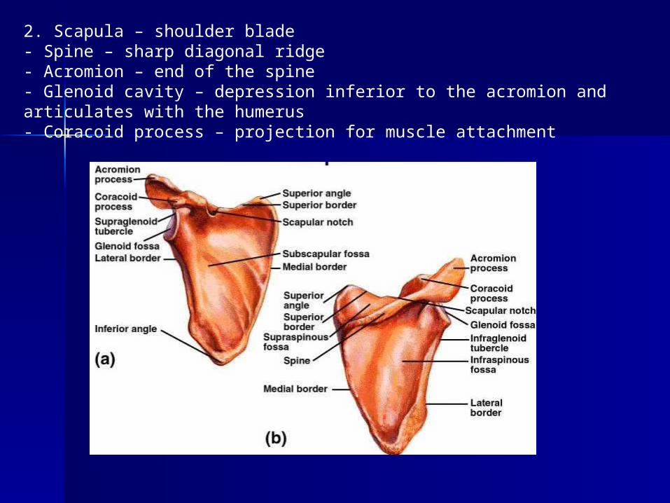
2. Scapula – shoulder blade- Spine – sharp diagonal ridge- Acromion – end of the spine- Glenoid cavity – depression inferior to the acromion and articulates with the humerus- Coracoid process – projection for muscle attachment
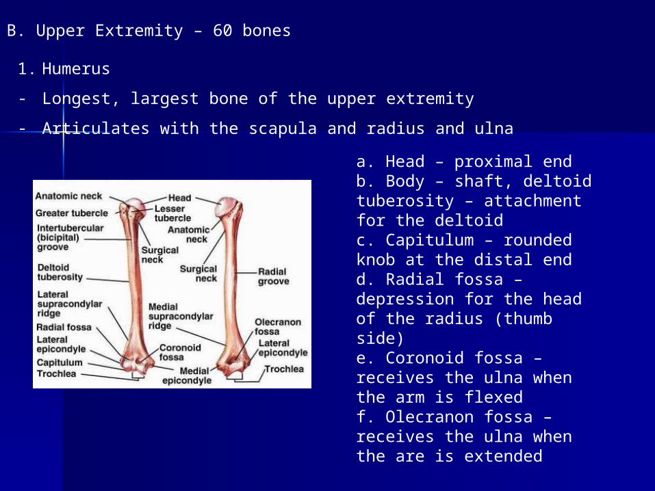
B. Upper Extremity – 60 bones
1. Humerus
- Longest, largest bone of the upper extremity
- Articulates with the scapula and radius and ulna
a. Head – proximal endb. Body – shaft, deltoid tuberosity – attachment for the deltoidc. Capitulum – rounded knob at the distal endd. Radial fossa – depression for the head of the radius (thumb side)e. Coronoid fossa – receives the ulna when the arm is flexedf. Olecranon fossa – receives the ulna when the are is extended
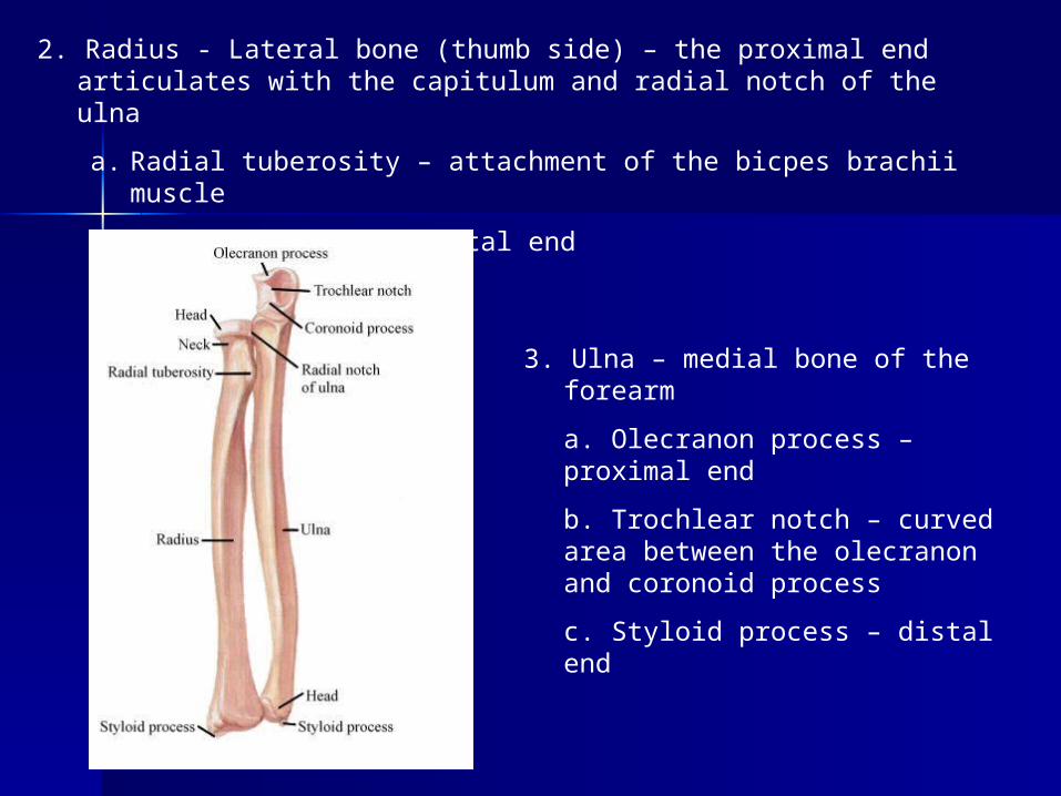
2. Radius - Lateral bone (thumb side) – the proximal end articulates with the capitulum and radial notch of the ulna
a. Radial tuberosity – attachment of the bicpes brachii muscle
b. Styloid process – distal end
3. Ulna – medial bone of the forearm
a. Olecranon process – proximal end
b. Trochlear notch – curved area between the olecranon and coronoid process
c. Styloid process – distal end
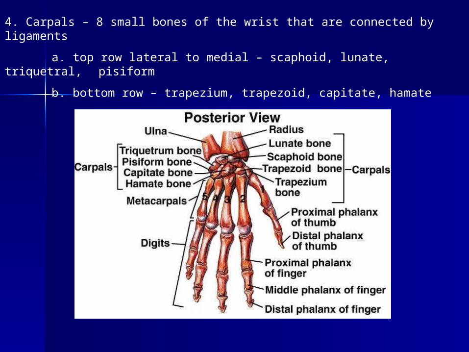
4. Carpals – 8 small bones of the wrist that are connected by ligaments
a. top row lateral to medial – scaphoid, lunate, triquetral, pisiform
b. bottom row – trapezium, trapezoid, capitate, hamate
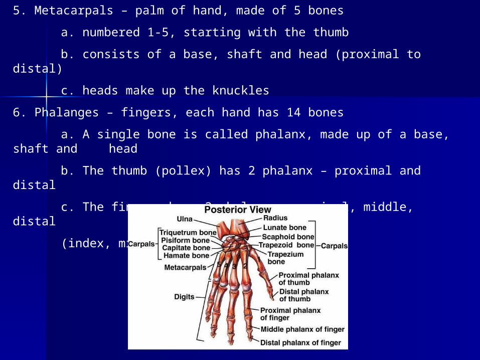
5. Metacarpals – palm of hand, made of 5 bones
a. numbered 1-5, starting with the thumb
b. consists of a base, shaft and head (proximal to distal)
c. heads make up the knuckles
6. Phalanges – fingers, each hand has 14 bones
a. A single bone is called phalanx, made up of a base, shaft and head
b. The thumb (pollex) has 2 phalanx – proximal and distal
c. The fingers have 3 phalanx – proximal, middle, distal
(index, middle, ring, little)
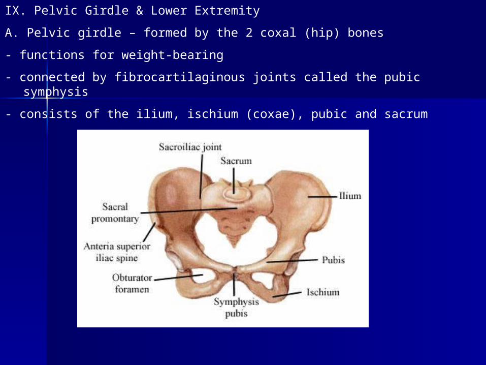
IX. Pelvic Girdle & Lower Extremity
A. Pelvic girdle – formed by the 2 coxal (hip) bones
- functions for weight-bearing
- connected by fibrocartilaginous joints called the pubic symphysis
- consists of the ilium, ischium (coxae), pubic and sacrum
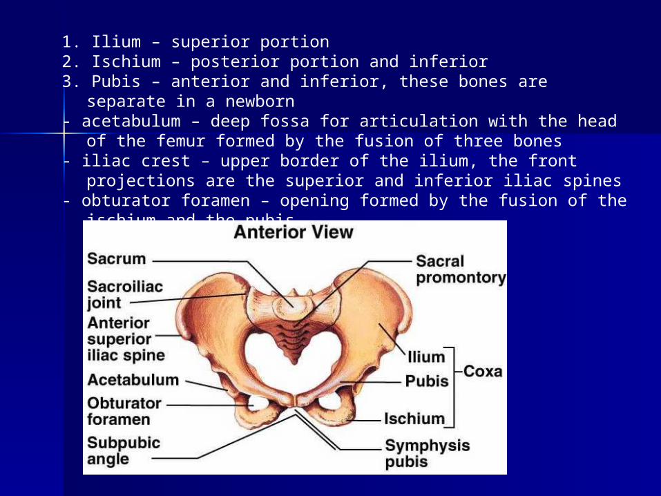
1. Ilium – superior portion2. Ischium – posterior portion and inferior 3. Pubis – anterior and inferior, these bones are separate in a
newborn- acetabulum – deep fossa for articulation with the head of the
femur formed by the fusion of three bones- iliac crest – upper border of the ilium, the front projections are the
superior and inferior iliac spines- obturator foramen – opening formed by the fusion of the ischium
and the pubis
B. Lower extremity – Leg
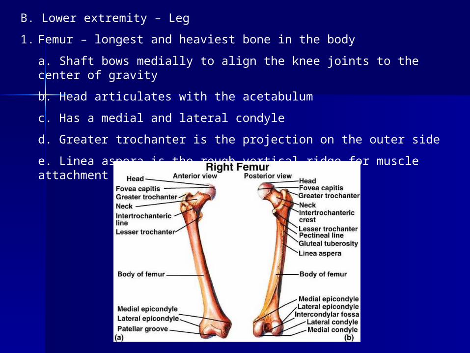
1. Femur – longest and heaviest bone in the body
a. Shaft bows medially to align the knee joints to the center of gravity
b. Head articulates with the acetabulum
c. Has a medial and lateral condyle
d. Greater trochanter is the projection on the outer side
e. Linea aspera is the rough vertical ridge for muscle attachment
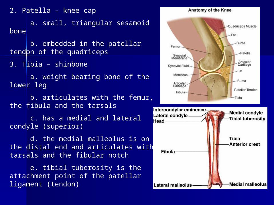
2. Patella – knee cap
a. small, triangular sesamoid bone
b. embedded in the patellar tendon of the quadriceps
3. Tibia – shinbone
a. weight bearing bone of the lower leg
b. articulates with the femur, the fibula and the tarsals
c. has a medial and lateral condyle (superior)
d. the medial malleolus is on the distal end and articulates with tarsals and the fibular notch
e. tibial tuberosity is the attachment point of the patellar ligament (tendon)
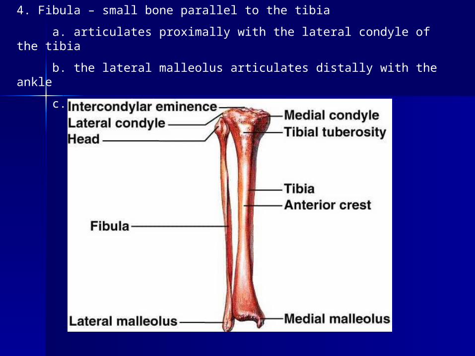
4. Fibula – small bone parallel to the tibia
a. articulates proximally with the lateral condyle of the tibia
b. the lateral malleolus articulates distally with the ankle
c. non weight-bearing
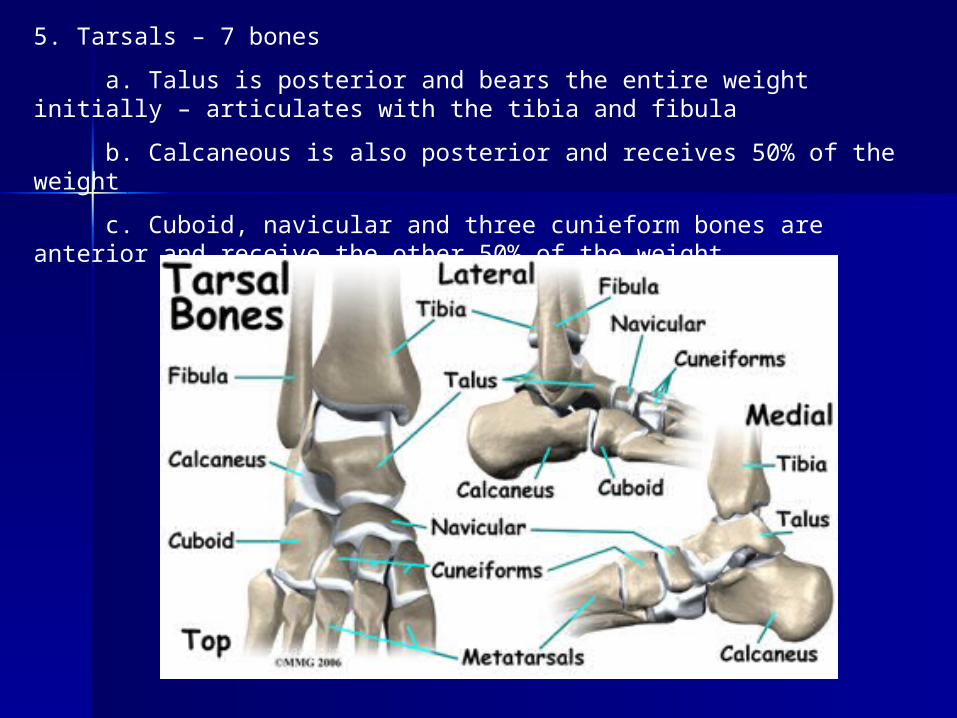
5. Tarsals – 7 bones
a. Talus is posterior and bears the entire weight initially – articulates with the tibia and fibula
b. Calcaneous is also posterior and receives 50% of the weight
c. Cuboid, navicular and three cunieform bones are anterior and receive the other 50% of the weight
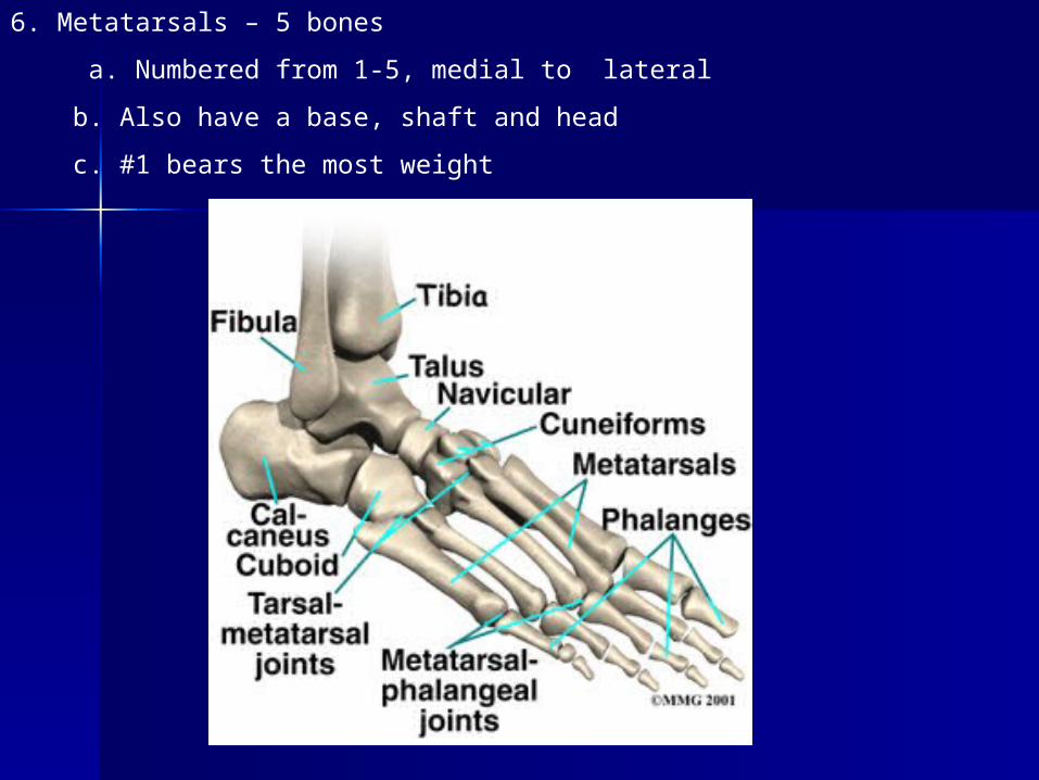
6. Metatarsals – 5 bones
a. Numbered from 1-5, medial to lateral
b. Also have a base, shaft and head
c. #1 bears the most weight