virtual rounds presentation a case of hypercalcemia heather taylor november 9, 2010
TRANSCRIPT

Virtual RoundsPresentation
A Case of Hypercalcemia
Heather TaylorNovember 9, 2010
Hypercalcemia
57 year old
Caucasian female
Generally healthy

Hypercalcemia
Chief Complaint: review lab results (June 30)
History of Illness: seen by GP in April c/o bilateral shoulder pain. Recently started exercise program O/E full ROM but weakness rotator cuff muscles & tender supraspinatus.
Xrays -> bilateral calcification in rotator cuff Ca+ 2.88 May 5th PTH normal. Repeat calcium normal. Dx rotator cuff tendonitis. Plan: repeat calcium. Exercise. NSAIDS.
May 20th calcium 3.01 PTH normal

Hypercalcemia
June 30th Presented at clinic for lab results. c/o “normal aches and pains”, itchy eyes, drippy nose
Sent for TSH, PTHrp, ionized calcium, Vit D 1,25 & Vit D 25
July 8th : c/o hx crampy abdominal pain, constipation, nausea, fatigue, skin “feels dry”, headache, irritability, leg weakness & trouble concentrating; sx for 2-3 weeks, thought was dehydrated so took Pedialyte...sx improved.
similar episode 4 months ago, lasted only 2 days.
Hypercalcemia
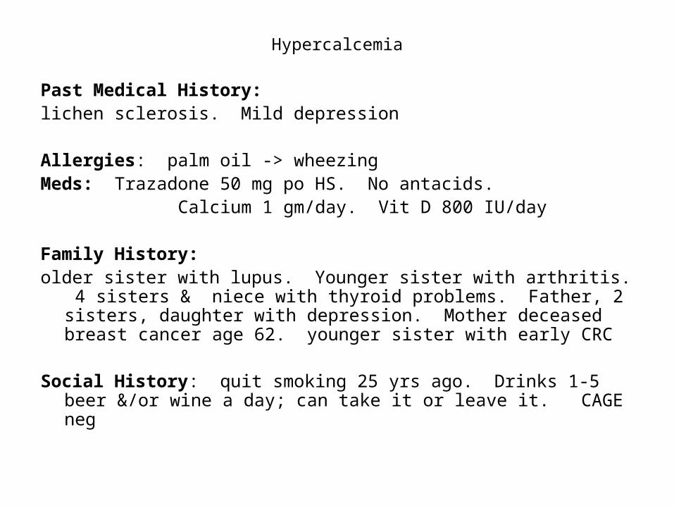
Past Medical History: lichen sclerosis. Mild depression
Allergies: palm oil -> wheezingMeds: Trazadone 50 mg po HS. No antacids. Calcium 1 gm/day. Vit D 800 IU/day Family History: older sister with lupus. Younger sister with arthritis. 4 sisters & niece
with thyroid problems. Father, 2 sisters, daughter with depression. Mother deceased breast cancer age 62. younger sister with early CRC
Social History: quit smoking 25 yrs ago. Drinks 1-5 beer &/or wine a day; can take it or leave it. CAGE neg
Hypercalcemia
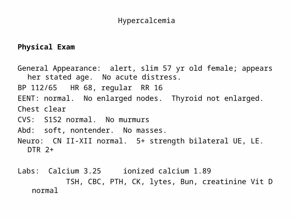
Physical Exam
General Appearance: alert, slim 57 yr old female; appears her stated age. No acute distress.
BP 112/65 HR 68, regular RR 16
EENT: normal. No enlarged nodes. Thyroid not enlarged.
Chest clear
CVS: S1S2 normal. No murmurs
Abd: soft, nontender. No masses.
Neuro: CN II-XII normal. 5+ strength bilateral UE, LE. DTR 2+
Labs: Calcium 3.25 ionized calcium 1.89
TSH, CBC, PTH, CK, lytes, Bun, creatinine Vit D normal
Hypercalcemia
Differential Dx: ?
Plan: ?
Refer to Internal Medicine
Pt went to Prince George for 3 wks.

Hypercalcemia
• Results when calcium entering circulation exceeds calcium excreted in urine or deposited in bone
• Due to: increased bone resorption, increased GI absorption, decreased renal excretion.
• Hyperparathyroidism & malignancy make up 90% of cases
Hypercalcemia
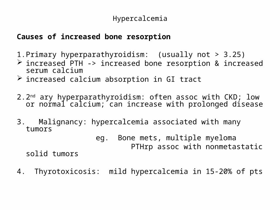
Causes of increased bone resorption
1. Primary hyperparathyroidism: (usually not > 3.25) increased PTH -> increased bone resorption & increased
serum calcium increased calcium absorption in GI tract
2. 2nd ary hyperparathyroidism: often assoc with CKD; low or normal calcium; can increase with prolonged disease
3. Malignancy: hypercalcemia associated with many tumors eg. Bone mets, multiple myeloma PTHrp assoc with nonmetastatic solid tumors
4. Thyrotoxicosis: mild hypercalcemia in 15-20% of pts
Hypercalcemia
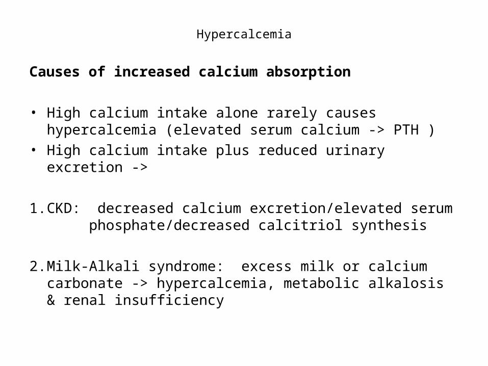
Causes of increased calcium absorption
• High calcium intake alone rarely causes hypercalcemia (elevated serum calcium -> PTH )
• High calcium intake plus reduced urinary excretion ->
1. CKD: decreased calcium excretion/elevated serum phosphate/decreased calcitriol synthesis
2. Milk-Alkali syndrome: excess milk or calcium carbonate -> hypercalcemia, metabolic alkalosis & renal insufficiency

Hypercalcemia
3. Excessive Vit D: increases calcium absorption & bone resorption
1,25 -> results from:
a) excess intake calcitriol
eg. tx for hypoparathyroidism, or hypocalcemia/2nd’ary hyperparathyroidism of renal failure.
Lasts 1-2 days; tx with salt & fluids, stopping calcitriol.
b) Increased endogenous production
eg pts with lymphoma, chronic granulomatous disorders (sarcoidosis, Wegeners)
high 25OHD from excess Vit D or calcidiol intake
lasts longer; more aggressive tx needed

Hypercalcemia
Other less common causes of hypercalcemia Lithium -> increased PTH secretion Thiazide diuretics -> decreases urinary calcium excretion Pheochromocytoma -> rare; ?due to concurrent hyperparathyroidism
Adrenal insufficiency Rhabdomyolysis & diuretic phase of ARF Theophylline toxicity Familial hypocalciuric hypercalcemia Metaphyseal chondrodysplasia: rare form of dwarfism

Hypercalcemia
Diagnostic Approach Clinical & lab evaluation to distinguish between hyperparathyroidism
& malignancy
PTH-mediated: 1’ary hyperparathyroidism, familial hyperparathyroid syndromes
Non PTH-mediated: malignancy, Vit D excess, granulomatous dx
1. Repeat sample to confirm hypercalcemia2. Clinical evaluation, duration of high calcium, presence of sx, FH, &
meds may give clues.3. PTH: frankly elevated or in upper half of normal -> primary
hyperparathyroidism4. PTH < 20 pg/ml: look for other causes. 5. PTHrp & Vit D metabolites
Hypercalcemia
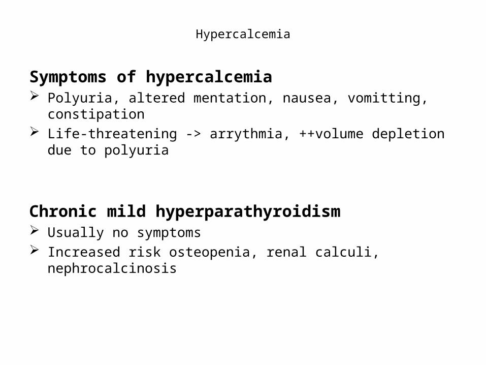
Symptoms of hypercalcemia Polyuria, altered mentation, nausea, vomitting, constipation Life-threatening -> arrythmia, ++volume depletion due to polyuria
Chronic mild hyperparathyroidism Usually no symptoms Increased risk osteopenia, renal calculi, nephrocalcinosis
Hypercalcemia
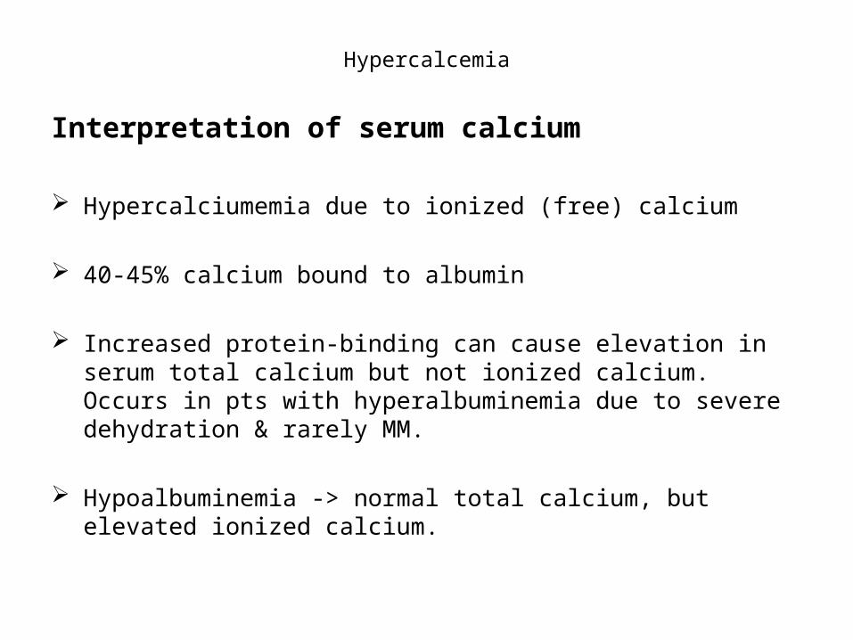
Interpretation of serum calcium
Hypercalciumemia due to ionized (free) calcium
40-45% calcium bound to albumin
Increased protein-binding can cause elevation in serum total calcium but not ionized calcium. Occurs in pts with hyperalbuminemia due to severe dehydration & rarely MM.
Hypoalbuminemia -> normal total calcium, but elevated ionized calcium.
Hypercalcemia
Degree of hypercalcemia useful
< 2.75 mmol/L more likely primary hyperparathyroidism
> 3.25 mmol/L more common with malignancy
10-20% of pts with 1’ary hyperparathyroidism have PTH in upper end of normal
Hypercalcemia
Key PointsKnow when to check calciumKnow what to look for if elevatedComprehensive hx can give clues
Hypercalcemia
Questions?
Thank you!

