visual associative agnosia: clinico-anatomical study of a ... · by presenting three figures from...
TRANSCRIPT

Journal of Neurology, Neurosurgery, and Psychiatry 1986;49:1233-1240
Visual associative agnosia: a clinico-anatomical studyof a single case
ROSALEEN A McCARTHY, ELIZABETH K WARRINGTON
From the National Hospitalfor Nervous Diseases, London, UK
SUMMARY A single case study of a patient with visual associative agnosia is described. The patienthad well preserved language, spatial, visual, and perceptual abilities but nevertheless was impairedin recognising visually presented common objects. It is argued that his deficit cannot be accountedfor in terms of a disconnection syndrome. Behavioural and anatomical (MRI scan) evidence forfocal unilateral dysfunction is presented. It is concluded that the left hemisphere plays a crucial rolein recognising the meaning of common objects.
Although impairment in the recognition of visuallypresented common objects with preserved perceptualskills may occur occasionally in the context of severeaphasia or dementia, it is very rarely observed as aselective deficit. Indeed convincing cases of visualassociative agnosia have been so rare that its existenceas a neurological syndrome has been disputed.' In aretrospective analysis of over 400 cases of unilateralcerebral lesion Hecaen and Angelergues2 detectedonly one patient in whom the deficit was relativelyselective and three further cases were noted in whomit occurred in the context of other severe cognitiveimpairments. De Renzi et al have reported a similarlow incidence.3
Several cases of visual associative agnosia havebeen described in the literature; however, the majorityhave complicated additional disorders4-7 or exten-sive bilateral brain lesions.8"- It is possible thatthere is only one case (although mild) of a unilaterallesion giving rise to this syndrome in its "pure" form.Hecaen et al2 investigated quantitatively a patientwho not only had some difficulty in naming commonobjects but was also impaired in describing their func-tion, in demonstrating their use and in allocatingthem to a category of similar objects. He was alsoprofoundly dyslexic. At the same time he was able toperform stringent perceptual tests (such as the Pop-pelreuter overlapping figures task) indicating that hisvisual processing capacity and perceptual analysis
Address for reprint requests: Dr RA McCarthy, Dept of Experi-mental Psychology, Downing St, Cambridge, UK.
Received 15 August 1985.Accepted 28 September 1985
(apperception) was intact. This patient had a rela-tively large tumour occupying the posterior half ofthe left hemisphere.We report our investigations of a patient with an
associative visual agnosia in whom the lesion local-isation was established precisely by MRI scanning.Our aim is to corroborate and extend the findings ofHecaen et al and to discuss the contibution of the lefthemisphere to object recognition.
Case report
FRA was a right-handed 77 year-old retired shopkeeper.Apart from one undiagnosed episode of unconsciousness in1975, he had been well until 1 March 1985 when he awokeand discovered that he was unable to read the newspaper.His dyslexia persisted and he was admitted to the NationalHospital, Queen Square, under the care of Professor Gilliattfor investigation of his difficulties.On examination his motor system was normal. However,
he was noted to have a right homonymous hemianopia anda profound dyslexia. Serology, urea and liver function testswere normal as was his ECG. CT scan revealed a medial leftoccipital infarct which possibly involved the posterior tem-poral lobe. His MRI scan (fig 1) revealed a lesion in thelower part of the occipital lobe involving the white and greymatter. The medial occipital cortex and the fusiform andlateral occipital gyri were particularly affected. Sagittalimages confirmed involvement of the inferior surfaces of theoccipital lobe and occipital pole. The corpus callosum(including the splenium) and the cerebellum appeared nor-mal. He was diagnosed as having sustained an infarction inthe territory of the left posterior cerebral artery. His condi-tion remained static and he was discharged home on 16April 1985.
Psychological Test FindingsFRA's test scores are summarised in tables 1 and 2. He was
1233
guest. Protected by copyright.
on 14 Septem
ber 2018 byhttp://jnnp.bm
j.com/
J Neurol N
eurosurg Psychiatry: first published as 10.1136/jnnp.49.11.1233 on 1 N
ovember 1986. D
ownloaded from

1234
Fig I MRIscan showing infarction in the territory oftheleft posterior cerebral artery. (a) Sagittal view, (b)horizontal view.
able to function at an average level on the WAIS and toscore satisfactorily on tests of memory. His expressivespeech was fluent with appropriate content, vocabulary, syn-tax and prosody. He completely failed to recognise or nameany items on the Graded Difficulty Naming test or theOldfield pictures test. In contrast to his very poor per-
McCarthy, Warringtonformance on these two object naming tests his ability toname "action" pictures16 (for example digging, peeling,swimming) was remarkably good (50/60 correct). However,there was evidence of only very mild word-finding difficultieson auditory and oral tests (naming from description, verbalfluency), His identification of objects by touch (for example,corkscrew, tape measure, mitten) was relatively satisfactory.
His literacy skills were impaired having the characteristicsof a total dyslexia with a mild dysgraphia. His naming ofsingle letters was weak, though significantly better with tac-tile than with visual presentation (table 2). He was unable torecognise that a visually presented "a" and an "A" were thesame letter. He attempted to read words by spelling themaloud but this strategy was completely vitiated by his letterrecognition difficulties (indeed our corpus does not contain a
single correct reading response). Furthermore, there was noevidence of his being able to comprehend even the common-est printed word. In contrast, his reading of arabic numeralswas intact (table 2). His ability to write was considerablybetter than his reading; thus short common words and sim-ple sentences were legibly and accurately produced to dicta-tion. On a Graded Difficulty Spelling test using words whichhave unusual sound-spelling correspondences (for example"two", "flood", "sword") his score was low average (Baxter,personal communication). In this context it is of interest tonote that his spelling of nonsense syllables was excellent(table 2).There was no evidence of motor apraxia or constructional
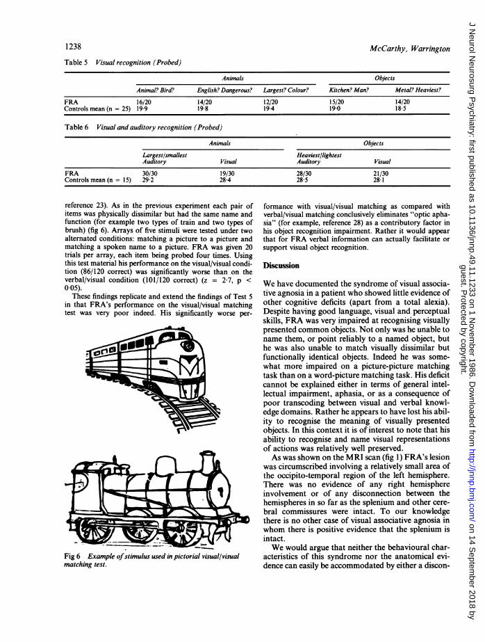
apraxia (fig 2). FRA had normal visual acuity (N6). His dis-crimination between a square and an oblong matched fortotal flux (Efron shapes), shape detection (presence orabsence of a degraded "X"), and position discrimination(identification of centrally positioned dot in a square) wereall intact (fig 3 and table 1). His ability to recognise photo-graphs of famous personalities (selected from an array ofthree visually similar faces) was fairly satisfactory (table 1).
Despite the excellence of his visual and spatial abilitiesFRA reported difficulties in everyday life that suggestedimpaired object recognition. For example he stated that hefrequently became muddled when attempting such simpletasks as setting a table. On word picture matching tests hisperformance was poor: thus he only scored at the 25th per-centile on the Peabody Picture Vocabulary23 and on arevised version of this test which uses closely related dis-tractor items (British Picture Vocabulary24), his score was
very defective falling below the first percentile. There wasalso evidence of difficulties in colour recognition in so far ason a simple task of pointing to one of four named primary
Table 1 Verbal and non-verbal test scores.
Verbal Non-verbal
Verbal IQ 111 Performance IQ 94Raw.Score Age-Scaled Raw Score Age-Scaled
Arithmetic 11 13 P Completion 6 10Similarities 9 11 Block Design 20 11Digit Span 10 12 P Arrangement 6 7Vocabulary 45 12Forced-choice words13 34/50 Efron shapes"9 19/20Picture naming14 15 0 Shape detection (adapted from ref 20) 30/30Naming from description"' 9/15 Position discrimination21 20/20Fluency animals (90,)17 13 Forced-choice famous facesTactile identification 32/40 (adapted from ref 22) 19/30
guest. Protected by copyright.
on 14 Septem
ber 2018 byhttp://jnnp.bm
j.com/
J Neurol N
eurosurg Psychiatry: first published as 10.1136/jnnp.49.11.1233 on 1 N
ovember 1986. D
ownloaded from
Visual associative agnosia: a clinico-anatomical study of a single case
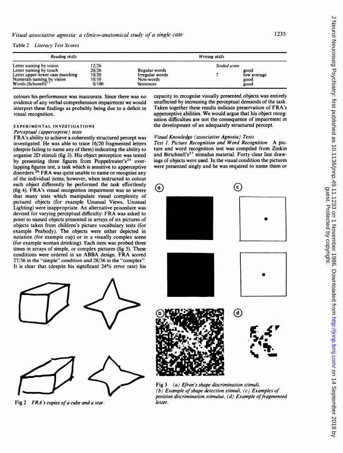
Table 2 Literacy Test Scores
Reading skills Writing skills
Letter naming by vision 12/26 Scaled scoreLetter naming by touch 20/26 Regular words goodLetter upper-lower case matching 10/20 Irregular words 7 low averageNumerals naming by vision 10/10 Non-words goodWords (Schonell)'' 0/100 Sentences good
colours his performance was inaccurate. Since there was noevidence of any verbal comprehension impairment we wouldinterpret these findings as probably being due to a deficit invisual recognition.
EXPERIMENTAL INVESTIGATIONSPerceptual (apperceptive) testsFRA's ability to achieve a coherently structured percept wasinvestigated. He was able to trace 16/20 fragmented letters(despite failing to name any of them) indicating the ability toorganise 2D stimuli (fig 3). His object perception was testedby presenting three figures from Poppelreuter's25 over-lapping figures test, a task which is sensitive to apperceptivedisorders.26 FRA was quite unable to name or recognise anyof the individual items; however, when instructed to coloureach object differently he performed the task effortlessly(fig 4). FRA's visual recognition impairment was so severethat many tests which manipulate visual complexity ofpictured objects (for example Unusual Views, UnusualLighting) were inappropriate. An alternative procedure wasdevised for varying perceptual difficulty: FRA was asked topoint to named objects presented in arrays of six pictures ofobjects taken from children's picture vocabulary tests (forexample Peabody). The objects were either depicted inisolation (for example cup) or in a visually complex scene(for example woman drinking). Each item was probed threetimes in arrays of simple, or complex pictures (fig 5). Theseconditions were ordered in an ABBA design. FRA scored27/36 in the "simple" condition and 28/36 in the "complex".It is clear that (despite his significant 24% error rate) his
capacity to recognise visually presented objects was entirelyunaffected by increasing the perceptual demands of the task.Taken together these results indicate preservation of FRA'sapperceptive abilities. We would argue that his object recog-nition difficulties are not the consequence of impairment inthe development of an adequately structured percept.
Visual Knowledge (associative Agnosia) TestsTest 1. Picture Recognition and Word Recognition A pic-ture and word recognition test was compiled from Zinkinand Birtchnell's" stimulus material. Forty clear line draw-ings of objects were used. In the visual condition the pictureswere presented singly and he was required to name them or
0
K>Fig 2 FRA's copies ofa cube and a star.
Fig 3 (a) Efron's shape discrimination stimuli.(b) Example ofshape detection stimuli, (c) Examples ofposition discrimination stimulus, (d) Example offragmentedletter.
1235
guest. Protected by copyright.
on 14 Septem
ber 2018 byhttp://jnnp.bm
j.com/
J Neurol N
eurosurg Psychiatry: first published as 10.1136/jnnp.49.11.1233 on 1 N
ovember 1986. D
ownloaded from

3McCarthy, Warrington
Fig 4 FRA's attempt to colour the individual objects in Poppelreuter's overlapping figures.
1 236
guest. Protected by copyright.
on 14 Septem
ber 2018 byhttp://jnnp.bm
j.com/
J Neurol N
eurosurg Psychiatry: first published as 10.1136/jnnp.49.11.1233 on 1 N
ovember 1986. D
ownloaded from

Visual associative agnosia: a clinico-anatomical study of a single case
Table 3 Picture recognition and word recognition
Picture Word
(1) 20/40 40/40(2) 22/40 39/40
identify them by description or function. In the auditorycondition he was asked to define the same 40 object names.The visual and auditory conditions were tested in an ABBAdesign. The items which were presented in the first half of thevisual condition were presented in the second half of theauditory condition and vice versa. FRA attempted this taskon two occasions. A lenient scoring criterion was used andresponses were accepted as correct if there was reasonableevidence that the core concept had been conveyed. Therewas a significant error rate on the visual condition. Unlikereported cases of "optic aphasia"28 there was no evidence ofperseverative responding. His errors consisted of semanticapproximations and of complete failures to recognise items.His error rate was similar on the two test occasions. He alsoshowed consistency on the items he failed to recogniseQ = 0-7 (X2 = 6 45, p < 0-02) suggesting that his impairmentwas not one of access to semantic representations. On theauditory condition his performance was virtually error-free(table 3). These findings establish that FRA has a visualrecognition deficit of a degradation type with intact know-ledge of the same information presented in the auditory ver-bal domain.
Test 2. Picture Word Matching The aim of this test was todocument FRA's recognition of a very high frequency visualvocabulary and at the same time to explore the possibilitiesof category specificity. The test stimuli consisted of arrays offive realistic coloured pictures of animals, foods or objectstaken from the Ladybird series of books for very youngchildren (see reference 29). The three stimulus categorieswere tested in a 3 x 3 Latin square. Recognition was testedby requiring FRA to point to a named stimulus. Twentytrials were given on each five-item array. The percentagecorrect for each category is shown in table 4. It is clear thathis performance is less than perfect even on this excep-tionally simple task. There was a weak category effect(chi-square = 6-9, p < 0 05).
Test 3. Probed Visual Knowledge More detailed knowledgeof visual representation of animals and of objects was
Fig 5 Example ofsimnple and complex stimulus.
Table 4 Picture word matching No. correct
Objects Foods Animals
45/60 52/60 55/60
assessed using a probe technique (described in full in refer-ence 7). Triplets of coloured drawings were presented andFRA was asked to point to a picture having a particularproperty. The distractor items were animals and objectswhich did not have the property in question. The numbercorrect for each type of probe, together with the mean scorefor control group, are given in table 5. FRA's scores werewell below those of the controls on all types of probe.
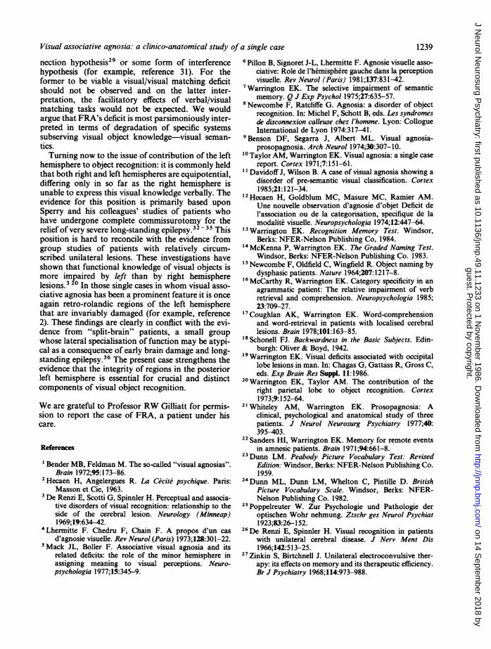
Test 4. Probed Visual Knowledge Our aim in this test wasto probe knowledge of attributes of animals and objectsusing both auditory and visual presentation. Triplets ofblack and white photographs of animals and of commonhousehold objects were assembled. There were 15 sets ofstimuli in both animal and object versions. In the animalversion FRA was asked to point first to the largest and sec-ondly to the smallest (for example elephant, monkey,camel). In the object version FRA was asked to point first tothe heaviest and then to the lightest item (for example drill,spectacles, jug). The auditory version was identical exceptthat the names of the stimuli were spoken. On both visualversions of this test FRA's performance was very weak andsignificantly below the level of the worst control subjects onthis test. His very satisfactory scores on the auditory versionprovide further evidence on the modality specificity of hisdeficit (table 6).
Test 5. Visuall Visual Matching of Objects This test wasdevised in order to establish whether FRA's comprehensiondeficit was entirely within the visual domain or the con-sequence of impairment in transcoding between visual andverbal knowledge systems ("optic aphasia"). A visual/visualobject matching test was devised. Forty pairs of commonobjects were selected so that the members of each pair werephysically dissimilar but had a common function and name(for example two types of razor, two types ofjug). FRA wastested with arrays of five objects in two conditions. First, hewas asked to match an object with its pair and secondly tomatch the spoken name to the object. The two conditionswere tested in an ABBA/BAAB design. He was given 10trials for each array, each stimuli being probed twice. Itemsthat were in the visual/visual condition for the first half ofthe experiment were tested in the verbal/visual condition inthe second and vice versa.He was as impaired on the visual/visual condition (46/80
correct) as in the verbal/visual condition (43/80 correct).This establishes that he has an object recognition deficitwithin the visual domain that is not exacerbated by trans-coding between visual and verbal domains, rather than beingworse on the verbal/visual condition as would be expected ifhe were an optic aphasic.28
Test 6. Visual/Visual Matching of Pictures Our aim in thistest was to replicate the findings of Test 5, that visual/visualmatching was impaired, using pictorial material. Thirtypairs of line drawings were selected, the majority being frompublished children's picture vocabulary tests (for example,
1237
guest. Protected by copyright.
on 14 Septem
ber 2018 byhttp://jnnp.bm
j.com/
J Neurol N
eurosurg Psychiatry: first published as 10.1136/jnnp.49.11.1233 on 1 N
ovember 1986. D
ownloaded from
1238
Table 5 Visual recognition (Probed)
McCarthy, Warrington
Animals Objects
Animal? Bird? English? Dangerous? Largest? Colour? Kitchen? Man? Metal? Heaviest?
FRA 16/20 14/20 12/20 15/20 14/20Controls mean (n = 25) 19 9 19 8 19-4 19 0 18 5
Table 6 Visual and auditory recognition (Probed)
Animals Objects
Largest/smallest Heaviest/lightestAuditory Visual Auditory Visual
FRA 30/30 19/30 28/30 21/30Controls mean (n = 15) 29-2 28-4 28-5 28 1
reference 23). As in the previous experiment each pair ofitems was physically dissimilar but had the same name andfunction (for example two types of train and two types ofbrush) (fig 6). Arrays of five stimuli were tested under twoalternated conditions: matching a picture to a picture andmatching a spoken name to a picture. FRA was given 20trials per array, each item being probed four times. Usingthis test material his performance on the visual/visual condi-tion (86/120 correct) was significantly worse than on theverbal/visual condition (101/120 correct) (z = 2-7, p <0 05).
These findings replicate and extend the findings of Test 5in that FRA's performance on the visual/visual matchingtest was very poor indeed. His significantly worse per-
Fig 6 Example ofstimulus used in pictorial visual/visualmatching test.
formance with visual/visual matching as compared withverbal/visual matching conclusively eliminates "optic apha-sia" (for example, reference 28) as a contributory factor inhis object recognition impairment. Rather it would appearthat for FRA verbal information can actually facilitate orsupport visual object recognition.
Discussion
We have documented the syndrome of visual associa-tive agnosia in a patient who showed little evidence ofother cognitive deficits (apart from a total alexia).Despite having good language, visual and perceptualskills, FRA was very impaired at recognising visuallypresented common objects. Not only was he unable toname them, or point reliably to a named object, buthe was also unable to match visually dissimilar butfunctionally identical objects. Indeed he was some-what more impaired on a picture-picture matchingtask than on a word-picture matching task. His deficitcannot be explained either in terms of general intel-lectual impairment, aphasia, or as a consequence ofpoor transcoding between visual and verbal knowl-edge domains. Rather he appears to have lost his abil-ity to recognise the meaning of visually presentedobjects. In this context it is of interest to note that hisability to recognise and name visual representationsof actions was relatively well preserved.As was shown on the MRI scan (fig 1) FRA's lesion
was circumscribed involving a relatively small area ofthe occipito-temporal region of the left hemisphere.There was no evidence of any right hemisphereinvolvement or of any disconnection between thehemispheres in so far as the splenium and other cere-bral commissures were intact. To our knowledgethere is no other case of visual associative agnosia inwhom there is positive evidence that the splenium isintact.We would argue that neither the behavioural char-
acteristics of this syndrome nor the anatomical evi-dence can easily be accommodated by either a discon-
guest. Protected by copyright.
on 14 Septem
ber 2018 byhttp://jnnp.bm
j.com/
J Neurol N
eurosurg Psychiatry: first published as 10.1136/jnnp.49.11.1233 on 1 N
ovember 1986. D
ownloaded from
Visual associative agnosia: a clinico-anatomical study of a single case
nection hypothesis29 or some form of interferencehypothesis (for example, reference 31). For theformer to be viable a visual/visual matching deficitshould not be observed and on the latter inter-pretation, the facilitatory effects of verbal/visualmatching tasks would not be expected. We wouldargue that FRA's deficit is most parsimoniously inter-preted in terms of degradation of specific systemssubserving visual object knowledge-visual seman-tics.Turning now to the issue of contribution of the left
hemisphere to object recognition: it is commonly heldthat both right and left hemispheres are equipotential,differing only in so far as the right hemisphere isunable to express this visual knowledge verbally. Theevidence for this position is primarily based uponSperry and his colleagues' studies of patients whohave undergone complete commissurotomy for therelief of very severe long-standing epilepsy.32 " Thisposition is hard to reconcile with the evidence fromgroup studies of patients with relatively circum-scribed unilateral lesions. These investigations haveshown that functional knowledge of visual objects ismore impaired by left than by right hemispherelesions.3 20 In those single cases in whom visual asso-ciative agnosia has been a prominent feature it is onceagain retro-rolandic regions of the left hemispherethat are invariably damaged (for example, reference2). These findings are clearly in conflict with the evi-dence from "split-brain" patients, a small groupwhose lateral specialisation of function may be atypi-cal as a consequence of early brain damage and long-standing epilepsy.36 The present case strengthens theevidence that the integrity of regions in the posteriorleft hemisphere is essential for crucial and distinctcomponents of visual object recognition.
We are grateful to Professor RW Gilliatt for permis-sion to report the case of FRA, a patient under hiscare.
References
'Bender MB, Feldman M. The so-called "visual agnosias".Brain 1972;95: 173-86.
2Hecaen H, Angelergues R. La Cecite psychique. Paris:Masson et Cie, 1963.
3De Renzi E, Scotti G, Spinnler H. Perceptual and associa-tive disorders of visual recognition: relationship to theside of the cerebral lesion. Neurology (Minneap)1969;19:634-42.
4Lhermitte F. Chedru F, Chain F. A propos d'un casd'agnosie visuelle. Rev Neurol (Paris) 1973;128:301-22.
'Mack JL, Boller F. Associative visual agnosia and itsrelated deficits: the role of the minor hemisphere inassigning meaning to visual perceptions. Neuro-psychologia 1977;15:345-9.
6 Pillon B, Signoret J-L, Lhermitte F. Agnosie visuelle asso-ciative: Role de l'hemisphere gauche dans la perceptionvisuelle. Rev Neurol (Paris) 1981;137:831-42.
'Warrington EK. The selective impairment of semanticmemory. Q J Exp Psychol 1975;27:635-57.
8Newcombe F, Ratcliffe G. Agnosia: a disorder of objectrecognition. In: Michel F, Schott B, eds. Les syndromesde disconnexion calleuse chez l'homme. Lyon: CollogueInternational de Lyon 1974:317-41.
9 Benson DF, Segarra J, Albert ML. Visual agnosia-prosopagnosia. Arch Neurol 1974;30:307-10.
Taylor AM, Warrington EK. Visual agnosia: a single casereport. Cortex 1971;7:151-61.
l Davidoff J, Wilson B. A case of visual agnosia showing adisorder of pre-semantic visual classification. Cortex1985;21:121-34.
'2Hecaen H, Goldblum MC, Masure MC, Ramier AM.Une nouvelle observation d'agnosie d'objet Deficit del'association ou de la categorisation, specifique de lamodalite visuelle. Neuropsychologia 1974;12:447-64.
13Warrington EK. Recognition Memory Test. Windsor,Berks: NFER-Nelson Publishing Co, 1984.
14McKenna P, Warrington EK. The Graded Naming Test.Windsor, Berks: NFER-Nelson Publishing Co. 1983.
s Newcombe F, Oldfield C, Wingfield R. Object naming bydysphasic patients. Nature 1964;207: 1217-8.
16 McCarthy R, Warrington EK. Category specificity in anagrammatic patient: The relative impairment of verbretrieval and comprehension. Neuropsychologia 1985;23:709-27.
17Coughlan AK, Warrington EK. Word-comprehensionand word-retrieval in patients with localised cerebrallesions. Brain 1978;101:163-85.
Schonell FJ. Backwardness in the Basic Subjects. Edin-burgh: Oliver & Boyd, 1942.
9Warrington EK. Visual deficits associated with occipitallobe lesions in man. In: Chagas G, Gattass R, Gross C,eds. Exp Brain Res Suppl. 11:1986.
20Warrington EK, Taylor AM. The contribution of theright parietal lobe to object recognition. Cortex1973;9:152-64.
21 Whiteley AM, Warrington EK. Prosopagnosia: Aclinical, psychological and anatomical study of threepatients. J Neurol Neurosurg Psychiatry 1977;40:395-403.
22 Sanders HI, Warrington EK. Memory for remote eventsin amnesic patients. Brain 1971;94:661-8.
23Dunn LM. Peabody Picture Vocabulary Test: RevisedEdition: Windsor, Berks: NFER-Nelson Publishing Co.1959.
24Dunn ML, Dunn LM, Whelton C, Pintille D. BritishPicture Vocabulary Scale. Windsor, Berks: NFER-Nelson Publishing Co. 1982.
25Poppelreuter W. Zur Psychologie und Pathologie deroptischen Wohr nehmung. Ztschr ges Neurol Psychiat1923;83:26-152.
26De Renzi E, Spinnler H. Visual recognition in patientswith unilateral cerebral disease. J Nerv Ment Dis1966;142:513-25.
27Zinkin S, Birtchnell J. Unilateral electroconvulsive ther-apy: its effects on memory and its therapeutic efficiency.Br J Psychiatry 1968;114:973-988.
1239
guest. Protected by copyright.
on 14 Septem
ber 2018 byhttp://jnnp.bm
j.com/
J Neurol N
eurosurg Psychiatry: first published as 10.1136/jnnp.49.11.1233 on 1 N
ovember 1986. D
ownloaded from
1240
28Beauvois MF. Optic Aphasia: a process of interactionbetween vision and language. Phil Trans R Soc LondB298 1982:35-47.
29Warrington EK, Shallice T. Category specific semanticimpairments. Brain 1984;107:829-53.
30Geschwind N. Disconnexion syndromes in animals andman. Brain 1965;88:585-644.
31Kinsboume M. Mechanisms of hemispheric interaction inman. In: Kinsbourne M, Smith WL, eds. Hemisphericdisconnection and cerebralfunction. Springfield, Illinois:Charles C Thomas. 1974.
Sperry RW. Cerebral dominance in perception. In: YoungFA, Lindsley DB. eds. Early experience in visual infor-mation processing in perceptual and reading disorders.Washington DC: National Academy of Science
McCarthy, Warrington
1970:167-78.33Sperry RW, Gazzaniga MS, Bogen JE. Interhemispheric
relationships: The neocortical commissures syndromesof hemisphere disconnection. In: Vinken PJ, BruynGW, eds. Handbook ofClinical Neurology, Amsterdam:North-Holland Publishing Co. 1969.
34Sperry RW, Zaidel E, Zaidel D. Self recognition andsocial awareness in the disconnected minor hemisphere.Neuropsychologia 1979;17: 153-6.
" Beaumont JG. The split-brain studies. In: Beaumont JG,ed. Divided Visual Field Studies of CerebralOrganisation. London: Academic Press 1982:113-28.
36Whitaker HA, Ojemann GA. Lateralisation of highercortical functioning: A critique. Ann NY Acad Sci1977;299:459-73.
guest. Protected by copyright.
on 14 Septem
ber 2018 byhttp://jnnp.bm
j.com/
J Neurol N
eurosurg Psychiatry: first published as 10.1136/jnnp.49.11.1233 on 1 N
ovember 1986. D
ownloaded from