visual impairment intracranial pressure (viip) [aka microgravity … · “visual impairment...
TRANSCRIPT
Page No. 1
Visual Impairment Intracranial Pressure (VIIP)
[aka Microgravity Ocular Syndrome (MOS)]
Bill Tarver, M.D. VIIP Lead Clinician; RAM ‘96 (USAF)
Tyson Brunstetter, O.D., Ph.D. Navy Aerospace/Research Optometrist
CAPT, MSC, USN
01 February 2017
https://ntrs.nasa.gov/search.jsp?R=20170001329 2020-03-22T21:45:49+00:00Z

Page No. 2
Why We Do What We Do…
Page No. 3
Recent VIIP/MOS Headlines:

Page No. 4
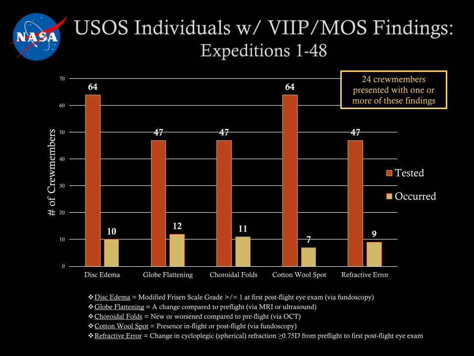
To date, 24 USOS ISS long-duration spaceflight astronauts have developed some or all of the following findings:
• Hyperopic shift
• Globe flattening
• Choroidal folds
• Cotton wool spots
• Optic disc edema
• Optic Nerve Sheath Distention
OcularALL are potential signsof elevated intracranial pressure (ICP)
• Mildly elevated post-flight intracranial pressure- 21 - 29 cm H2O range
• Upper limit of normal: ~20 cm H2O• Gray zone: 20.1 – 24.9 cm H2O
Page No. 5
Disc Edema = Modified Frisen Scale Grade >/= 1 at first post-flight eye exam (via fundoscopy)
Globe Flattening = A change compared to preflight (via MRI or ultrasound)
Choroidal Folds = New or worsened compared to pre-flight (via OCT)
Cotton Wool Spot = Presence in-flight or post-flight (via fundoscopy)
Refractive Error = Change in cycloplegic (spherical) refraction ≥0.75D from preflight to first post-flight eye exam
64
47 47
64
47
1012 11
79
0
10
20
30
40
50
60
70
Disc Edema Globe Flattening Choroidal Folds Cotton Wool Spot Refractive Error
# o
f C
rew
mem
ber
s
Tested
Occurred
24 crewmembers
presented with one or
more of these findings

Page No. 6
VIIP/MOS Clinical Findings

Page No. 7
Of the active astronaut population…
• 80% wear vision correction (32% contact lenses)
• Mean age = 47 yrs
• Majority are presbyopic (i.e., a normal, age-related, progressively
worsening inability to focus clearly on near objects)
From postflight questionnaires (1989 - 2011): 25% of short-duration
(Shuttle) & 50% of long- duration (ISS) mission astronauts report a
subjective degradation in vision, especially at near
• Provided “Space Anticipation Glasses”

Page No. 8
Subjective Degradation in Vision (cont):
• Associated w/ Hyperopic Shifts in refractive error due to Globe Flattening
A 1 mm decrease in axial length will produce a ~3 diopter hyperopic shift
Largest shift to date is +1.75 diopters
In presbyopes: Typically decreases near visual
acuity (VA), but leaves distant VA intact

Page No. 9
Case Example:
ssociated w/ papilledema(i.e., disc edema 2o to increased
intracranial pressure); typically
bilateral 1 year post-flightPre-flight6 days post-flight
MRI
Page No. 10
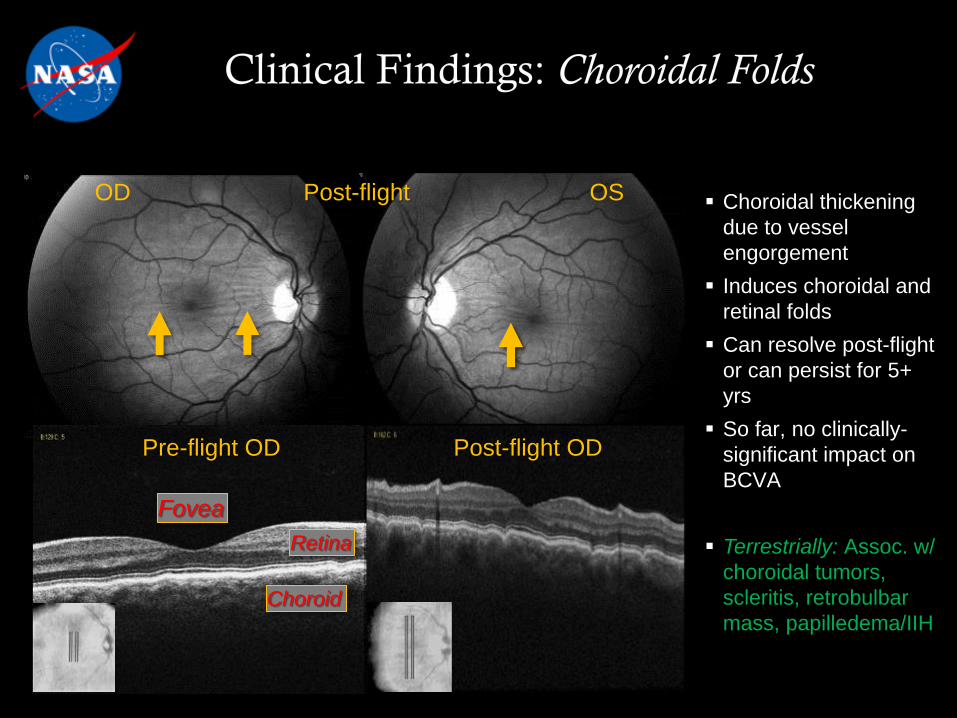
OD Post-flight OS
Pre-flight OD Post-flight OD
Choroid
Retina
Fovea
Choroidal thickening
due to vessel
engorgement
Induces choroidal and
retinal folds
Can resolve post-flight
or can persist for 5+
yrs
So far, no clinically-
significant impact on
BCVA
Terrestrially: Assoc. w/
choroidal tumors,
scleritis, retrobulbar
mass, papilledema/IIH

Page No. 11
Example 2Posterior pole fundoscopic images
OD & OS for two ISS crewmembers• Top arrows: Choroidal folds
• Bottom arrows: Cotton wool spots
Cotton wools spots• Abnormal retinal finding
• Accumulations of axoplasmic
material w/in retinal nerve fiber
layer
• Caused by ischemia
reduced axonal transport
swelling of axon damaged
nerve fibers
• Terrestrially: Associated w/
diabetes, HTN, central retinal
vein occlusion
Example 1
Pre-flightPost-flight

Page No. 12
Pre-flight fundoscopic
images of the right
(OD) & left (OS) optic
discs
Post-flight images of
optic discs, showing
Grade 3 edema OD &
Grade 1 edema OS
OD OS
OD OS

Page No. 13
Fundoscopic image of optic disc OD,
10 days after return to Earth• Arrows: “C” shaped halo of edema
Terrestrially: Optic disc edema is
associated with:
• Unilateral: Optic neuritis, optic
neuropathy, retinal artery/vein
occlusion
• Bilateral: Increase in ICP… IIH ( “papilledema”)
Intracranial mass
Cerebral edema
Increased CSF production
Decreased CSF absorption
Obstructive hydrocephalus
Venous outflow obstruction
• Typically reduces VA, enlarges
blind spot, causes relative
afferent pupillary defect & color
impairment

Page No. 14
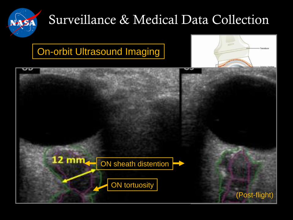
Post-flight ultrasound image of
globe, optic nerve (ON; purple), and
optic nerve sheath (green). Showing:• ON Sheath distention
• ON tortuosity
ON Sheath terrestrially:• Normal diameter (ONSD)
< 5.9 mm
• Enlargement typically
associated w/ increased ICP
12 mm
Post-Flight OD

Page No. 15
Common Characteristics of the Cases

Page No. 16
~6 month duration ISS mission
• [No clinically-significant signs
during short-duration flights]
• Dose response??
All had normal pre-flight eye
exams
Past medical history:
• Negative for systemic disease
• None had used medications before/during their mission that
could increase ICP (e.g., vitamin A, tetracycline, corticosteroids, or
nalidixic acid)
Common Characteristics of the Cases

Page No. 17
None complained of headaches, transient vision loss, double
vision, pulsatile tinnitus, or vision changes during eye mvmts(i.e., the classic symptoms of idiopathic intracranial hypertension)
None experienced loss
in best-corrected visual
acuity, color vision, or
stereopsis
Right eye affected more than
left in all cases
ISS cabin
• Normal pressure & oxygen
• Elevated CO2
~0.33-0.5% avg, w/ avg peak ~0.7%
10x terrestrially: ~0.03-0.04%
Common Characteristics of the Cases

Page No. 18
0 50 100 150 200 250 300 350
Average Days
Next Generation Missions
Mercury n = 6
Gemini *n = 20
Apollo *n = 33
Skylab n = 9
Apollo-Soyuz n = 3
Space Shuttle *n = 710
Mir n = 7
International
Space Station*n = 55 as of 6/14/16
* Person-flights; may include multiple-time flyers w/in program
We are just entering, relatively speaking, the
long-duration phase of space exposure…

Page No. 19
Why is this Happening?

Page No. 20
Microgravity Cephalad fluid shift Cerebral venous congestion
Why is this Happening?

Page No. 21
Current Risk Statement:
“Visual Impairment Intracranial Pressure” (VIIP)
"Given that the microgravity environment causes cephalad fluid
shift in astronauts, there is a probability that astronauts will have
intracranial hypertension (IHT) to some degree, which if left
untreated, could lead to deleterious health effects.”
Why is this Happening?

Page No. 22
Hypothesis #1: Increased intracranial pressure
• The original theory, hence the name “Visual Impairment Intracranial
Pressure”
Hypothesis #2: This is a local ocular eye problem
Hypothesis #3: Slight IOP reduction + slight ICP increase
Hypothesis #4: Folate-dependent 1-carbon metabolic pathway
altered
Hypothesis #5: Vessel congestion placing pressure locally
around optic nerve (“Circle of Zinn-Haller” theory)
• In µGravity, head venous pressure ≈15-20 mmHg
Standing terrestrially ≈ −20 mmHg
Why is this Happening?

Page No. 23
In-flight Exacerbating Factors??
Resistive Exercise
High Oral Sodium Intake
Prepackaged Foods…
Up to 5000+ mg/day
In-flight Pharmaceuticals
High CO2
~10x terrestrial levels

Page No. 24
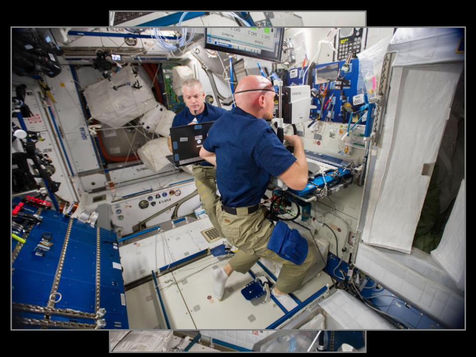
Medical Surveillance

Page No. 25
Surveillance & Medical Data Collection
49 ISS expedition missions have been completed (since 2000)
Sentinel case occurred in 2005
• Optic disc edema and cotton wool spot
Surveillance/medical data collection is ongoing and has
evolved
– Began some “VIIP” related testing in 2008 (w/ Exp 18)
– Inconsistent testing until Feb 2010 (Exp 23) when Eye MED B
came into effect

Page No. 26
Surveillance & Medical Data Collection
Terrestrially
3T MRI – Special “NASA Astronaut”
protocol 12-18 months prior to launch
Terrestrially & On-Orbit
Vision Exam
• Visual Acuity (near & far)
• Amsler grid
Ocular Ultrasound
Fundoscopy
Optical Coherence Tomography (OCT)
Tonometry (when clinically indicated)

Page No. 27
Surveillance & Medical Data Collection
Visual Acuity & Amsler Grid
Page No. 28
Surveillance & Medical Data Collection
On-orbit Ultrasound Imaging
Elevated optic disc
(Post-flight)
ON sheath distention
ON tortuosity
(Post-flight)

Page No. 29
Surveillance & Medical Data Collection
Fundoscope

Page No. 30
Surveillance & Medical Data Collection
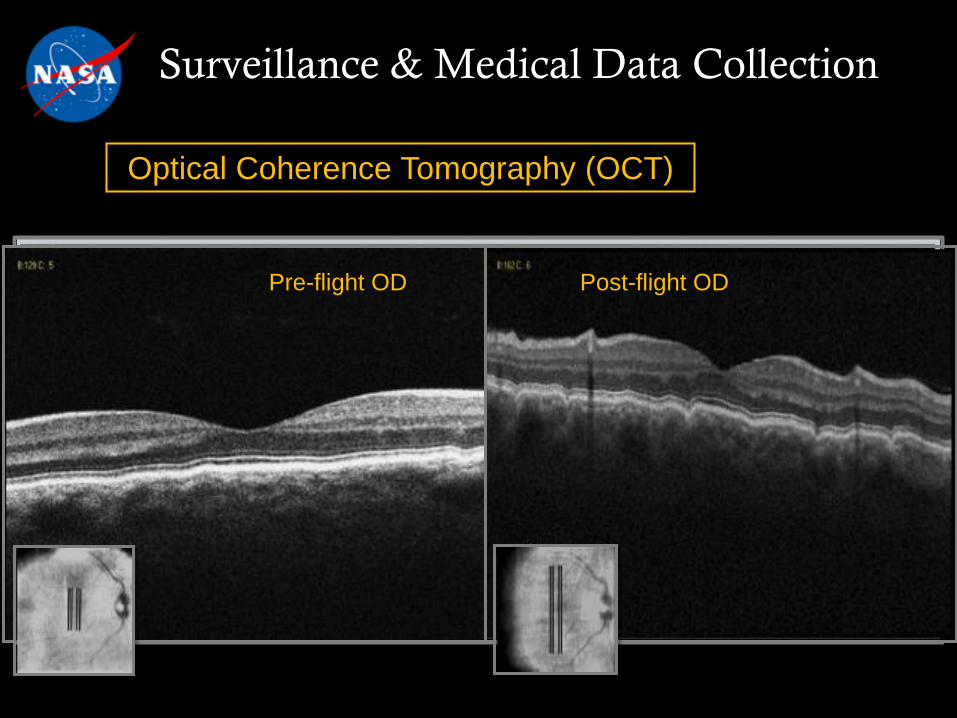
Optical Coherence Tomography (OCT)
Page No. 31
Surveillance & Medical Data Collection
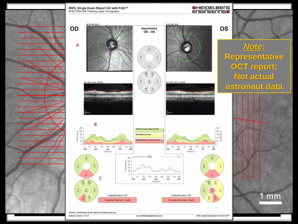
Page No. 32
Surveillance & Medical Data Collection
Optical Coherence Tomography (OCT)Note:
Representative
OCT report;
Not actual
astronaut data
Page No. 33
Surveillance & Medical Data Collection
Optical Coherence Tomography (OCT)
Pre-flight OD Post-flight OD

Page No. 34
Clinical & Research Update

Page No. 35
Clinical Update: Feb17
Ongoing clinical work
Correlation between ocular structural changes (OCT) and chronic effect on
visual function (visual fields testing)
Correlation of subcortical white matter hyperintensities (WMH) found on MRI
and VIIP/MOS signs – 2017
Refinement of cardiovascular parameters and their correlation with
VIIP/MOS signs – 2017
We are evaluating the next generation OCT, “OCT2” to determine if it will
enhance on orbit imaging/data acquisition

Page No. 36
What We Are Watching Coming From Our Research Colleagues
Ocular Health Study and the Fluid Shifts Study – both finish data
collection this summer
Clinical relevance of MRI-based findings
Implementation of direct ICP measures study pre and post
mission
Correlation between HDT with CO2 and VIIP/MOS (EnviHab)

Page No. 37
Questions?

Page No. 38
Back-Up

Page No. 39
The Lamina Cribosa & the Translaminar Pressure Gradient: A Mechanism for Papilledema
1G 0G
CSFp
IOP
Area of Interest:
Area of Magnification
Translaminar Pressure Gradients: