visualization of microbleeds with optical histology in...
TRANSCRIPT

Microvascular Research 105 (2016) 109–113
Contents lists available at ScienceDirect
Microvascular Research
j ourna l homepage: www.e lsev ie r .com/ locate /ymvre
Visualization of microbleeds with optical histology in mouse model ofcerebral amyloid angiopathy
Patrick Lo a,b, Christian Crouzet a,b, Vitaly Vasilevko c,1, Bernard Choi a,b,d,⁎,1a Beckman Laser Institute and Medical Clinic, University of California, Irvine, 1002 Health Sciences Road East, Irvine, CA 92612, USAb Department of Biomedical Engineering, University of California, Irvine, 3120 Natural Sciences II, Irvine, CA 92697, USAc Institute for Memory Impairments and Neurological Disorders, University of California, Irvine, 1207 Gillespie NRF, Irvine, CA 92697-4540, USAd Edwards Lifesciences Center for Advanced Cardiovascular Technology, University of California, Irvine, 2400 Engineering Hall, Irvine, CA 92697, USA
⁎ Corresponding author at: University of California, IrviMedical Clinic, 1002 Health Sciences Road East, Irvine, CA
E-mail addresses: [email protected] (P. Lo), [email protected](V. Vasilevko), [email protected] (B. Choi).
1 These authors contributed equally to the manuscript.
http://dx.doi.org/10.1016/j.mvr.2016.02.0020026-2862/© 2016 Elsevier Inc. All rights reserved.
a b s t r a c t
a r t i c l e i n f oArticle history:Received 14 May 2015Revised 3 February 2016Accepted 4 February 2016Available online 10 February 2016
Cerebral amyloid angiopathy (CAA) is a neurovascular disease that is strongly associated with an increase in thenumber and size of spontaneous microbleeds. Conventional methods of magnetic resonance imaging for detec-tion of microbleeds, and positron emission tomography with Pittsburgh Compound B imaging for amyloiddeposits, can separately demonstrate the presence of microbleeds and CAA in affected brains in vivo; however,there still is a critical need for strong evidence that shows involvement of CAA in microbleed formation. Here,we show in a Tg2576 mouse model of Alzheimer's disease, that the combination of histochemical staining andan optical clearing method called optical histology, enables simultaneous, co-registered three-dimensional visu-alization of cerebral microvasculature, microbleeds, and amyloid deposits. Our data suggest that microbleeds arelocalized within the brain regions affected by vascular amyloid deposits. All observed microhemorrhages (n =39) were in close proximity (0 to 144 μm) with vessels affected by CAA. Our data suggest that the predominanttype of CAA-related microbleed is associated with leaky or ruptured hemorrhagic microvasculature. The pro-posed methodological and instrumental approach will allow future study of the relationship between CAA andmicrobleeds during disease development and in response to treatment strategies.
© 2016 Elsevier Inc. All rights reserved.
Keywords:Intracerebral hemorrhageDiIThioflavin SPrussian blueAmyloid-βCerebral amyloid angiopathyMicrohemorrhagesStroke
Introduction
Intracerebral microbleeds result from rupture or leaking of cerebralblood vessels. They are routinely visualized with magnetic resonanceimaging (MRI) and are defined in humans as round foci b5 mm indiameter that appear hypointense and distinct from vascular flowvoids, leptomeningeal hemasiderosis, and nonhemorrhagic subcorticalmineralization (Fazekas et al., 1999). Hemosiderin, a hemoglobinbreakdown product, causes magnetic susceptibility-induced relaxation,leading to T2* signal loss. Initially, microbleeds tend to be clinicallyasymptomatic. In the long term, they are a contributing factor to age-related mental decline and dementia (Gregoire et al., 2012; Poelset al., 2012; Poels et al., 2010).
Two categories of vascular events can lead to the formation of amicrobleed. First, microscopic deposits of lysed red blood cell productsfound in postmortem human studies suggest that microvessels alsomay leak blood into the brain parenchyma, especially in vessels affectedby cerebral amyloid angiopathy (CAA) (Hartz et al., 2012). Second,
ne, Beckman Laser Institute and92612.u (C. Crouzet), [email protected]
occlusions of small vessels can prevent blood flow from reaching a re-gion of the brain, leading to ischemic damage, followed by hemorrhagicconversion of the infarction site (Fisher, 1986). Study of the pathophys-iology of these small lesions is limited due to the small size of the vesselsinvolved, resulting in problems with early detection of microbleedswith contemporary MRI techniques.
CAA is a neurovascular disease that is strongly associated with theincrease in the number and size of spontaneous microbleeds. CAA ischaracterized by the deposition of fibrillar forms of amyloid peptidesin the blood vessels of the cortex and leptomeninges. Amyloid depositson the cerebral vasculature promote degeneration of the tunica mediaand loss of smooth muscle and endothelial cells, inducing thickeningof the vessel wall and vascular dysfunction (Vinters, 1987). Recently,we reported the progressive accumulation of CAA in parallel withmicrobleeds in the Tg2576 transgenic mouse model of amyloidosis(Fisher et al., 2011). Older Tg2576 mice develop CAA in leptomeningealand pial vessels, which is associatedwith spontaneousmicrobleeds thatcan be further exacerbated by anti-amyloid immunotherapy (Fisheret al., 2011; Wilcock et al., 2004).
Combined in-vivo and ex-vivo analyses strongly imply co-localization of CAA affected regions and microbleeds (Ni et al., 2015).Several imaging methods exist to visualize both amyloid deposits andmicrohemorrhages, including 1) in-vivo MRI imaging for microbleeds(Chan andDesmond, 1999; Kwa et al., 1998), 2) in-vivo 11C-PIB Positron

110 P. Lo et al. / Microvascular Research 105 (2016) 109–113
Emission Tomography (PET) for cerebral amyloid burden (Yates et al.,2011), 3) postmortem H&E and hemosiderin staining for microbleeds(Craelius et al., 1982; Fisher et al., 2011) and 4) postmortem CongoRed, Thioflavin S or immunostaining with anti-amyloid beta antibodiesfor amyloid deposits (Vinters et al., 1988). Microscopic analysestraditionally include two-dimensional histochemistry with Prussianblue for hemosiderin and Congo Red or Thioflavin S for amyloid deposits(Fisher et al., 2011;Wilcock et al., 2004).Multiphoton in-vivomicroscopyenables co-registered visualization of amyloid deposits, blood vessels andcerebral cellular elements (Dong et al., 2010); however, there is a restric-tion in the analyzed size and volume. The entire brain vasculature can bevisualized ex vivo and in three dimensions using the corrosion castmethod (Meyer et al., 2008), although this method limits application ofother immunohistochemical approaches to visualize other cerebral struc-tures. However, none of the current methods combines microscopic res-olution and three-dimensional structural analysis of the affected areas.
Here, for the first time, we show that the combination of histochem-ical staining and a new optical clearingmethodwe call optical histology(Moy et al., 2013a, 2013b), enables simultaneous, co-registered three-dimensional visualization of cerebral microvasculature, microbleeds,and amyloid deposits. With this combined approach, we present datathat supports the hypothesis that microbleeds occur in the brain areasmost affected by CAA in the Tg2576 model of amyloidosis.
Materials and methods
Animal model
We used naive 21–22-month-old Tg2576 mice (n = 4) andnontransgenic (nTg) (n = 2) littermates (Fisher et al., 2012; Hsiaoet al., 1996). All animal procedures followed the “Principles of LaboratoryAnimal Care” from NIH publication No. 85-23 and were approved inaccordance with the Institutional Animal Care and Use Committee atUniversity of California, Irvine.
Vessel painting
To achieve high-resolution, three-dimensional imaging of micro-scopic features in the brain,we used a technique called optical histology,described extensively in previous publications (Moy et al., 2013a,2013b). The first step of optical histology involves fluorescent labelingof themicrovasculature. Briefly, to label blood vessels, animals receiveda cardiac perfusion of DiI (Life Technologies, Grand Island, NY), acarbocyanine fluorescent dye, at a rate of 5 mL/min (Li et al., 2008;Moy et al., 2013b) followed by perfusion with 4% paraformaldehyde.After perfusion, brains were extracted and immersed in 4% paraformal-dehyde for 72 h.
Tissue sectioning
Each brain was embedded in 5% agarose gel and placed into a16 × 38 mm hollow cylinder. Using a micrometer (ThorLabs, NewJersey, USA) for precise positioning and a microtome blade (LeicaMicrosystems, Wetzlar, Germany), 500 μm-thick brain sections were
Fig. 1. Brain sections preparation stages for optical histology. (A) 500 μm-thick wild-type mousThioflavin S staining. (C) Section after optical clearing in FocusClear for three hours. (D) Brain
generated. After sectioning, excess agarose was removed from eachsection.
Tissue staining and clearing
Each section was stained for hemosiderin and fibrillar beta-amyloiddeposits using Prussian blue and Thioflavin S staining, respectively(Liu et al., 2014; Passos et al., 2013). Briefly, Prussian blue by Mallorymodifications for hemosiderin was performed using freshly prepared 5%potassium hexacyanoferratetrihydrate and 5% hydrochloric acid (bothSigma-Aldrich, St. Louis, MO) in water for 60 min, followed by 3 washesinwater, then brain sectionswere preincubatedwith 50% ethanol, follow-ed by one-hour immersion in 0.5% Thioflavin S with mild agitation, twowashes with 50% ethanol, two washes in PBS, and storage in PBS buffer.To render the samples transparent, brain sections were incubated in0.3 mL of FocusClear (CelExplorer Labs, Hsinchu, Taiwan) for threehours. Cleared sections were placed in a custom-built tissue holder(Moy et al., 2013b). After tissue staining and clearing, a visual change inthe appearance of the sections was observed from the combination ofPrussian blue and Thioflavin S staining with the clearing of the tissue(Fig. 1).
Microscopy
A confocal microscope (Meta 150, Carl Zeiss, Germany) was used tovisualize DiI-stained microvasculature and Thioflavin S-stained amyloiddeposits. The fluorescence of DiI was collected using a 543 nm HeNelaser for excitation and a 565–615 nm bandpass filter for fluorescenceemission. Thioflavin S fluorescence was collected using a 458 nm Argonlaser for excitation and a 500-530 nm bandpass filter for emission.Since hemosiderin stained with Prussian blue is not fluorescent, trans-missionmicroscopy at 543 nmwas used to visualize the absorption con-trast of regions stained by Prussian blue. Z-stacks of the brain werecollected using a 10× objective (Plan-Neofluar 10×/0.30 Ph1), allowingfor 0.7 μm lateral resolution and 9 μm longitudinal resolution. A 20× ob-jective (Plan-Neufluar 20×/0.5 Ph2)was employed to examine individu-al microbleed sites. In addition, brightfield microscopic images of thetransilluminated brain section were collected with an inverted micro-scope (Diaphot TMD, Nikon, Melville, NY) equipped with a color CCDcamera (Grasshopper, Point Grey, Richmond, BC, Canada).
Image processing
Algorithms in ImageJ (http://imagej.nih.gov/ij/index.html) and Fiji,an open-source plugin (Preibisch et al., 2009), were used to create awide-field mosaic image comprised of adjacent microscope fields ofview, and to generate fly-through videos and maximum intensity pro-jection (MIP) images of fluorescence emission and absorption contrast.The ImageJ “Analyze Particles” function with a threshold diameter of5 μm was used to determine the area and number of amyloid depositsin the MIP image of each brain section. Hemosiderin size was assessedusing the ImageJ “Scale Bar” feature.
e brain section stained with DiI before optical clearing. (B) Section after Prussian blue andsection mounted between two glass cover slides.
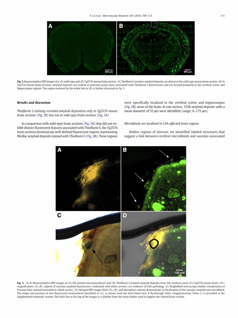
Fig. 2.RepresentativeMIP images of a (A)wild-type and (B) Tg2576mouse brain section. (A) Thioflavin S positive amyloid deposits are absent in thewild-typemouse brain section. (B) InTg2576 mouse brain sections, amyloid deposits are evident as punctate green areas associated with Thioflavin S fluorescence and are located primarily in the cerebral cortex andhippocampus regions. The region enclosed by the white line in (B) is further discussed in Fig. 3.
111P. Lo et al. / Microvascular Research 105 (2016) 109–113
Results and discussion
Thioflavin S staining revealed amyloid deposition only in Tg2576 mousebrain sections (Fig. 2B) but not in wild-type brain sections (Fig. 2A)
In comparisonwith wild-type brain sections (Fig. 2A) that did not ex-hibit distinctfluorescent features associatedwith Thioflavin S, the Tg2576brain sections demonstratewell-definedfluorescent regions representingfibrillar amyloid deposits stainedwith Thioflavin S (Fig. 2B). These regions
Fig. 3. (A, B) Representative MIP images of (A) DiI-stained microvasculature and (B) Thioflamagnification). In (B), regions of vascular amyloid fluorescence (indicated with white arrowPrussian blue-stained hemosiderin (black arrow). (D) Merged MIP images from (A), (B), andThe shape and position of non-fluorescent hemosiderin identified in (C), is shown with tSupplemental materials' section. The thick line at the top of the images is a shadow from the t
were specifically localized in the cerebral cortex and hippocampus(Fig. 2B) areas of the brain. In one section, 1528 amyloid deposits with amean diameter of 33 μmwere identified (range: 6–175 μm).
Microbleeds are localized in CAA-affected brain regions
Within regions of interest, we identified labeled structures thatsuggest a link between cerebral microbleeds and vascular-associated
vin S-stained amyloid deposits from the cerebral cortex of a Tg2576 mouse brain (10×s) are evidence of CAA pathology. (C) Brightfield microscopy enables visualization of
absorption contrast demonstrate co-localization of the vascular amyloid and microbleed.he thick black line. A fly-through video (Supplementary Video 1) is provided in theissue holder used to support the cleared brain section.

Fig. 4. Hippocampus region of Tg2576 mouse brain section stained with Thioflavin S (A, D) and Prussian blue (B, E), imaged with the 10× (A, B, C) and 20× (D, E, F) objectives. (A, D)Vascular amyloid and amyloid plaques stained with Thioflavin S are visualized with green fluorescence emission. (B, E) The absorption contrast of hemosiderin stained with Prussianblue is indicative of a microbleed and characterizes a leaky/ruptured hemorrhagic vessel. (C, F) Following merging of images from Fig. 2 (A, B, D, E) with those of DiI fluorescence, weidentified that the microbleed was associated with CAA-affected vasculature. A fly-through video (Supplementary Video 2) of Figs. (D–F) is provided in the Supplemental materials'section.
112 P. Lo et al. / Microvascular Research 105 (2016) 109–113
fibrillar amyloid, a hallmark of CAA pathology (Fig. 3, SupplementalFig. 1). As a representative example, in the subregion of interestidentified in Fig. 2B, we observed DiI-labeled vasculature (Fig. 3A,Supplemental Figs. 1A, 3A) and vascular amyloid (Fig. 3B, whitearrows, Supplemental Figs. 1B, 3B). With transmission (Supple-mental Fig. 1C) and brightfield microscopy (Fig. 3C, SupplementalFig. 1D), we observed non-fluorescent Prussian blue positivehemosiderin (Fig. 3C, black arrow) that was adjacent to vascularamyloid (Fig. 3D). With overlays of the co-registered microscopyimages, the spatial overlap of vascular amyloid, hemosiderin, andPrussian blue is more readily visualized (Fig. 3D, SupplementalFig. 1E).
Fig. 5. Hippocampus region of Tg2576mouse brain sectionwith an occluded ischemic vessel anabruptly (white arrow), suggesting either a blood clot or narrowing of the vessel. (B) With meevident (black arrow). Scale bar= 100 μm. (C) Themicrobleed is confirmedwith brightfieldmiPrussian blue-stained hemosiderin (black arrows), suggesting transformation of ischemia intothis section is provided in the Supplemental materials' section.
Three-dimensional visualization with optical histology enables identificationof leaky vessels closely associated with CAA pathology (Fig. 4, SupplementalFigs. 2 and 3)
After merging the images of DiI, Thioflavin S, and transmissionmicroscopy channels, clusters of hemosiderin along the vessel weredetected. The hemosiderin is observed by absorption contrast andappears as black (Fig. 4A, Supplemental Figs. 2B, 3C) and confirmedwith brightfield microscopy (Supplemental Figs. 2C, 3D). CombiningPrussian blue staining of hemosiderin with tracing of the CAA-affectedcerebral vessel, we identified the presence of a leaky/ruptured vesselin the Tg2576 mouse brain section (Fig. 4B, Supplemental Figs. 2D, 3E).
d hemosiderin deposition (A) DiI vessel painting reveals vascular structure that terminatesrged fluorescence and transmission microscopy channels, a cerebral microbleed is clearlycroscopy, which shows both extravasated red blood cells (diffuse red region in center) anda hemorrhagic event. Scale bar = 50 μm. A fly-through video (Supplementary Video 3) of

113P. Lo et al. / Microvascular Research 105 (2016) 109–113
Optical histology enabled identification of a single occurrence of an appar-ently occluded vessel in Tg2576 mice with the CAA pathology (Fig. 5)
DiI staining enabled identification of a microvessel that appeared tobe occluded (Fig. 5A, white arrow). Merging the images from threechannels in a manner similar to Fig. 4F, we identified a microischemiclesion that apparently transformed into a ~130 μm-wide hemorrhagicmicrobleed (Fig. 5B,C). Co-localization of red blood cells and hemosider-in inside the brain parenchyma further supports the transformation ofischemia into a hemorrhagic event. Such ischemic events were notpresent in brain sections of wild-type animals.
Collectively, our data from nine Tg2576 mouse brain sections suggest thatcerebral microbleeds are heterogeneous in terms of size and location
From these sections, we identified 39 microbleeds with characteris-tic diameters ranging from 5 to 240 μm (median diameter= 39 μm). Ofthe 39 microbleeds, 30 occurred within the cerebral cortex and ninewithin the hippocampus; all were closely localizedwith the distributionof fibrillar forms of amyloid deposits, with a median distance of 0 μmbetween the vessel and amyloid (range: 0–144 μm). Twelve (31%)microbleeds originated from capillaries (≤10 μm diameter) and 27(69%) from venules and arterioles (10 to 70 μm). These preclinicalfindings are consistent with clinical MRI data (Poels et al., 2010) show-ing a median diameter of the CAA-affected vessels of 17 μm (range:3–50 μm), suggesting that microbleeds arise primarily from smallarterioles. Of the 39 microbleeds, all but one (Fig. 5) demonstratedcharacteristics of leaky/ruptured hemorrhagic blood vessels (Fig. 4).Additional studies are warranted to assess the potential relationshipbetween CAA and microbleed formation; optical histology is expectedto facilitate these studies.
In the transgenic mouse data sets, the CAA fluorescence is strongoverall interrogated depths, suggesting that the optical clearing processwas effective. In the wild-type mice, the DiI fluorescent signal also wasstrong; however, in the transgenic mice, the DiI fluorescent signal wasless consistent. This may be due to compromised vascular staining dueto vascular amyloid deposition and compromised flow in smallervessels in Tg animals (Meyer et al., 2008). Additional work is plannedto study this discrepancy and ascertain the reasons underlying thisdifference.
Optical histology enables high-resolution and wide field-of-viewimaging of bulk tissue sections across multiple body planes. With thecombination of thick-tissue sectioning, chemical-based optical clearing,and fluorescence microscopy, we can study tissue depths that wouldotherwise be inaccessible. Future studies that stem from optical histolo-gy can include visualizing multiple fluorescently-labeled structures(Moy et al., 2013a) in brain sections that allow comprehensive charac-terization of cerebral pathology. Optical histology can also expand intopharmacological studies by evaluating treatment efficacy and responsethrough visualization of biological markers at a high resolution andwide field-of-view.
Supplementary data to this article can be found online at http://dx.doi.org/10.1016/j.mvr.2016.02.002.
Acknowledgments
We would like to thank Kelley Kilday, Sneha Shivkumar andDr. Tatiana Krasieva (University of California, Irvine) for their intellec-tual contributions to this project. The work was supported in part bythe Alzheimer's Association (NIRG-12-242781), Arnold and MabelBeckman Foundation, theNational Institutes of Health LaserMicrobeamand Medical Program (LAMMP, a P41 Technology Research Resource,grant number EB015890), and the Undergraduate Research Opportuni-ties Program at University of California, Irvine.
References
Chan, S., Desmond, D.W., 1999. Silent intracerebral microhemorrhages in stroke patients.Ann. Neurol. 45, 412–413.
Craelius, W., et al., 1982. Iron deposits surrounding multiple sclerosis plaques. Arch.Pathol. Lab. Med. 106, 397–399.
Dong, J., et al., 2010. Multiphoton in vivo imaging of amyloid in animal models ofAlzheimer's disease. Neuropharmacology 59, 268–275.
Fazeka, F., et al., 1999. Histopathologic analysis of foci of signal loss on gradient-echo T2*-weighted MR images in patients with spontaneous intracerebral hemorrhage: evi-dence of microangiopathy-related microbleeds. AJNR Am. J. Neuroradiol. 20,637–642.
Fisher,M., 1986. Aspirin, anticoagulants, and hemorrhagic conversion of ischemic infarction:hypothesis and implications. Bull. Clin. Neurosci. 51, 68–72.
Fisher, M., et al., 2011. Therapeutic modulation of cerebral microhemorrhage in a mousemodel of cerebral amyloid angiopathy. Stroke 42, 3300–3303.
Fisher, M., et al., 2012. Mixed cerebrovascular disease and the future of stroke prevention.Transl. Stroke Res. 3, 39–51.
Gregoire, S.M., et al., 2012. Cerebral microbleeds and long-term cognitive outcome: longi-tudinal cohort study of stroke clinic patients. Cerebrovasc. Dis. 33, 430–435.
Hartz, A.M., et al., 2012. Amyloid-beta contributes to blood–brain barrier leakage in trans-genic human amyloid precursor protein mice and in humans with cerebral amyloidangiopathy. Stroke 43, 514–523.
Hsiao, K., et al., 1996. Correlative memory deficits, Abeta elevation, and amyloid plaquesin transgenic mice. Science 274, 99–102.
Kwa, V.I., et al., 1998. Silent intracerebral microhemorrhages in patients with ischemicstroke. Amsterdam Vascular Medicine Group. Ann. Neurol. 44, 372–377.
Li, Y., et al., 2008. Direct labeling and visualization of blood vessels with lipophiliccarbocyanine dye DiI. Nat. Protoc. 3, 1703–1708.
Liu, S., et al., 2014. Comparative analysis of H&E and Prussian blue staining in a mousemodel of cerebral microbleeds. J. Histochem. Cytochem.
Meyer, E.P., et al., 2008. Altered morphology and 3D architecture of brain vasculature in amouse model for Alzheimer's disease. Proc. Natl. Acad. Sci. U. S. A. 105, 3587–3592.
Moy, A.J., et al., 2013a. High-resolution visualization of mouse cardiac microvasculatureusing optical histology. Biomed Opt. Express. 5, 69–77.
Moy, A.J., et al., 2013b. Optical histology: a method to visualize microvasculature in thicktissue sections of mouse brain. PLoS ONE 8, e53753.
Ni, J., et al., 2015. Cortical localization of microbleeds in cerebral amyloid angiopathy: anultra high-field 7T MRI study. J. Alzheimers Dis. 43, 1325–1330.
Passos, G.F., et al., 2013. The bradykinin B1 receptor regulates Abeta deposition andneuroinflammation in Tg-SwDI mice. Am. J. Pathol. 182, 1740–1749.
Poels, M.M., et al., 2010. Prevalence and risk factors of cerebral microbleeds: an update ofthe Rotterdam scan study. Stroke 41, S103–S106.
Poels, M.M., et al., 2012. Cerebral microbleeds are associated with worse cognitivefunction: the Rotterdam scan study. Neurology 78, 326–333.
Preibisch, S., et al., 2009. Globally optimal stitching of tiled 3Dmicroscopic image acquisitions.Bioinformatics 25, 1463–1465.
Vinters, H.V., 1987. Cerebral amyloid angiopathy. A critical review. Stroke 18, 311–324.Vinters, H.V., et al., 1988. Immunohistochemical study of cerebral amyloid angiopathy. II.
Enhancement of immunostaining using formic acid pretreatment of tissue sections.Am. J. Pathol. 133, 150–162.
Wilcock, D.M., et al., 2004. Passive immunotherapy against Abeta in aged APP-transgenicmice reverses cognitive deficits and depletes parenchymal amyloid deposits in spiteof increased vascular amyloid and microhemorrhage. J. Neuroinflammation 1, 24.
Yates, P.A., et al., 2011. Cerebral microhemorrhage and brain beta-amyloid in aging andAlzheimer disease. Neurology 77, 48–54.