visualizing molecular diffusion through passive permeability...
TRANSCRIPT
COCHBI-1072; NO. OF PAGES 9
Visualizing molecular diffusion through passive permeabilitybarriers in cells: conventional and novel approachesYu-Chun Lin1, Siew Cheng Phua1, Benjamin Lin1,2 and Takanari Inoue1,3
Available online at www.sciencedirect.com
Diffusion barriers are universal solutions for cells to achieve
distinct organizations, compositions, and activities within a
limited space. The influence of diffusion barriers on the
spatiotemporal dynamics of signaling molecules often
determines cellular physiology and functions. Over the years,
the passive permeability barriers in various subcellular locales
have been characterized using elaborate analytical techniques.
In this review, we will summarize the current state of knowledge
on the various passive permeability barriers present in
mammalian cells. We will conclude with a description of several
conventional techniques and one new approach based on
chemically inducible diffusion trap (CIDT) for probing
permeable barriers.
Addresses1 Department of Cell Biology, Center for Cell Dynamics, School of
Medicine, Johns Hopkins University, United States2 Department of Biomedical Engineering, Johns Hopkins University,
United States3 PRESTO Investigator, JST, 4-1-8 Honcho, Kawaguchi, Saitama 332-
0012, Japan
Corresponding authors: Lin, Yu-Chun ([email protected]) and Inoue,
Takanari ([email protected])
Current Opinion in Chemical Biology 2013, 17:xx–yy
This review comes from a themed issue on Molecular Imaging
Edited by James Chen and Kazuya Kikuchi
1367-5931/$ – see front matter, # 2013 Elsevier Ltd. All rights reserved.
http://dx.doi.org/10.1016/j.cbpa.2013.04.027
IntroductionDiffusion is the random motion of molecules driven by
thermal energy, resulting in molecular movement from
areas of high concentration to areas of low concentration.
As an energetically favorable process, diffusion occurs
without energy expenditure and thus is a ubiquitous
strategy used by cells for molecular transport. However,
uncontrolled diffusion can be disadvantageous to achiev-
ing localized signaling, which often requires the spatial
enrichment of signaling species and is critical to funda-
mental cellular functions such as cell polarity, growth,
proliferation, and death [1�,2–4,5��]. To address this
issue, cells have evolved diffusion barriers, which serve
as gatekeepers in filtering molecules based on size, shape,
charge, and other intrinsic properties. Diffusion barriers
thus enable cellular compartmentalization and spatiotem-
poral control of signaling. Intracellular diffusion barriers
Please cite this article in press as: Lin Y-C, et al.: Visualizing molecular diffusion through passive p
http://dx.doi.org/10.1016/j.cbpa.2013.04.027
www.sciencedirect.com
exist at a variety of cellular structures, including the
nuclear envelope, the annulus of spermatozoa, the lead-
ing edge of migrating cells, the cleavage furrow of divid-
ing cells, and the budding neck of yeast, as well as in
cellular extensions such as primary cilia, dendritic spines,
and the initial segment of the neuronal axon [1�,6�,7].
While some diffusion barriers exist constitutively in cells,
others are highly dynamic. The importance of diffusion
barriers is further underscored by the various human
diseases which result from their dysfunction [3,6�,8,9].
Diffusion barriers can be categorized into two major
classes based on the substrates they affect: lateral diffu-
sion barriers and permeability barriers. Lateral diffusion
barriers localize in membranes and restrict the movement
of molecules within the membrane plane such as trans-
membrane proteins and membrane lipids [1�]. Conver-
sely, permeability barriers embedded within membranes
act as conduits regulating the movement of solutes
through the membrane. Permeability barriers also localize
within aqueous cellular compartments to hinder solute
diffusion [2,10��]. Generally speaking, lateral diffusion
barriers are well characterized [1�], primarily because
membrane molecules move relatively slowly and are in
a two-dimension environment, allowing easy observation
of their dynamics. In contrast, it has been challenging to
measure the dynamics of solutes in cells, owing to their
generally fast diffusion as well as technical limitations in
precisely determining the axial position of solute mol-
ecules inside living cells [10��]. However, recent
advances in microscopy techniques have enabled the
refinement of our understanding of passive permeability
barriers. In this review, we will provide an overview of the
current knowledge of permeable diffusion barriers in
various subcellular regions (summarized in Figure 1
and Table 1). Subsequently, we will describe six methods
used to measure the dynamics of solutes in cellular
aqueous compartments and their application to probing
permeable diffusion barriers, with a particular focus on
the strengths and weaknesses of these approaches (sum-
marized in Figure 2 and Table 2).
Passive permeability barriers in cellsNuclear pore complex
The eukaryotic nucleus is surrounded by the nuclear
envelope, a double layered membrane structure that func-
tionally separates the nucleus from the cytosol. Communi-
cation between the cytosol and nucleus is regulated by
specialized conduits, known as nuclear pore complexes
(NPCs), which are anchored in the nuclear envelope at
ermeability barriers in cells: conventional and novel approaches, Curr Opin Chem Biol (2013),
Current Opinion in Chemical Biology 2013, 17:1–9
2 Molecular Imaging
COCHBI-1072; NO. OF PAGES 9
Please cite this article in press as: Lin Y-C, et al.: Visualizing molecular diffusion through passive permeability barriers in cells: conventional and novel approaches, Curr Opin Chem Biol (2013),
http://dx.doi.org/10.1016/j.cbpa.2013.04.027
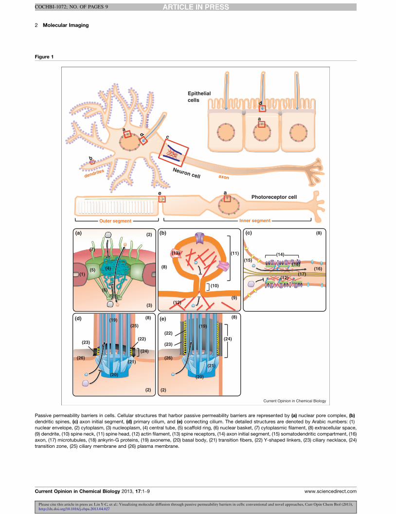
Figure 1
Photoreceptor cell
Epithelial cells
b
a
Neuron cell
d
Outer segment Inner segment
axondendrites
(1)
(a) (b) (c)
(d)
(2)
(3)
(4)(5)
(6)
(7)
(9)
(8)
(10)
(11)
(12)
(8)
(15)
(16)(17)
(18)
(12)
(8)
(2)
(19)
(20)
(21)
(22)
(14)
(24)(26) (26)
(25)
(23)
(e) (8)
(2)
(20)
(21)
(19)
(23)
(22)(24)
(13)
a
d
c
da
b
ee
d
dddddd
Current Opinion in Chemical Biology
Passive permeability barriers in cells. Cellular structures that harbor passive permeability barriers are represented by (a) nuclear pore complex, (b)
dendritic spines, (c) axon initial segment, (d) primary cilium, and (e) connecting cilium. The detailed structures are denoted by Arabic numbers: (1)
nuclear envelope, (2) cytoplasm, (3) nucleoplasm, (4) central tube, (5) scaffold ring, (6) nuclear basket, (7) cytoplasmic filament, (8) extracellular space,
(9) dendrite, (10) spine neck, (11) spine head, (12) actin filament, (13) spine receptors, (14) axon initial segment, (15) somatodendritic compartment, (16)
axon, (17) microtubules, (18) ankyrin-G proteins, (19) axoneme, (20) basal body, (21) transition fibers, (22) Y-shaped linkers, (23) ciliary necklace, (24)
transition zone, (25) ciliary membrane and (26) plasma membrane.
Current Opinion in Chemical Biology 2013, 17:1–9 www.sciencedirect.com

Probing passive permeability barriers in cells Lin et al. 3
COCHBI-1072; NO. OF PAGES 9
Table 1
Summary of different permeable diffusion barriers in cells
Permeable diffusion
barrier
Key components
of diffusion barrier
Functional pore size
for passive diffusion
Diffusion tracers Methods used to study References
Nuclear pore complex FG-rich nucleoporins 9–10 nm Dextran, gold particles,
recombinant proteins
SPT, PAFP, FCS, FRAP,
microinjection, CID
[12–15,
40,44��]
Dendritic spines neck Unknown <2.4 nm GFP, dextran, Ca2+ dye, PAFP [9]
Axon initial segment Actin, ankyrin-G >10 kDa, <70 kDa Dextran, GFP Microinjection, FRAP [21��]
Ciliary pore complex Nucleoporin 8 nm (�650 kDa) [31��]
or <67 kDa [30��]
Dextran, recombinant
proteins, YFP-FKBP-POIs
CIDT, microinjection,
FRAP
[30��,31��]
Connecting cilium Unknown >81 kDa GFP, PAFP, arrestin,
transducin, recoverin
PAFP [26,28,29��]
Permeable diffusion
barrier in cytoplasm
Actin 20–40 nm (average
pore size)
Dextran, ficoll, DNA,
recombinant proteins, IgG
Microinjection, FRAP [10��,32,33]
junctions between the inner and outer membrane
(Figure 1a). Because of their importance in regulating
nuclear function, NPCs are one of the most well-charac-
terized diffusion barriers in cells [7]. Each NPC consists of
several major domains: a central channel, a core scaffold
that supports the central channel, a nuclear basket, and
cytoplasmic filaments. Nuclear trafficking is regulated
through the central channel created by the core scaffold,
which is assembled by approximately 30 different nucleo-
porin proteins arranged in rotational symmetry to form a
conduit-like structure. Two-thirds of these nucleoporins
constitute the core scaffold, while the remaining one-third
of nucleoporins extend out phenylalanine–glycine (FG)-
rich repeats to fill the central channel of the NPC [7].
Importantly, these FG-rich proteins have been proposed to
assemble into a molecular sieve that regulates nucleocy-
toplasmic shuttling [11]. This molecular sieve-like diffu-
sion barrier, with a calculated diameter of �9–10 nm,
allows passive diffusion of molecules such as ions, metab-
olites, and cargo smaller than �40 kDa [12]. Larger cargo,
however, require active transport-mediated and signal-de-
pendent mechanisms to pass through the NPC [2]. The
largest cargo that can be actively transported across NPCs is
�39 nm, which approximates the narrowest region of the
central channel (�50 nm) [7,13]. It is still unknown
whether these passive and active transport systems occupy
Please cite this article in press as: Lin Y-C, et al.: Visualizing molecular diffusion through passive p
http://dx.doi.org/10.1016/j.cbpa.2013.04.027
Table 2
Summary of the approaches used to study the permeable diffusion b
Technique Protein
tracers
Non-protein
tracers
Very fast
tracers (ms)
V
trac
Single-particle tracking (SPT) U U UU UU
Photoactivatable fluorescence
protein (PAFP)
U UU
Fluorescence correlation
spectroscopy (FCS)
U U UUU U
Fluorescence recovery after
photobleaching (FRAP)
U U U
Microinjection U U UU
Chemically inducible diffusion
trapping (CIDT)
U U
www.sciencedirect.com
distinct or similar channels of the NPC [14,15]. In sum-
mary, NPCs enable cells to properly regulate the localiz-
ation and mobility of molecules across the nuclear
envelope.
Dendritic spine neck
Dendritic spines are small membranous protrusions
originating from neuronal dendrites, which serve as post-
synaptic terminals of excitatory synapses. As postsynaptic
compartments of chemical neurotransmission, dendritic
spine structure and functional regulation can determine
the strength with which a receiving neuron is stimulated
by a given connection. Spines are dynamic structures, as
they retract from the dendritic shaft when inactive and
reform in response to stimulation [16]. Despite their
morphological and temporal variability along dendrites,
they typically have a relatively large head attached to the
dendritic shaft by a narrow neck (Figure 1b) [17].
Postsynaptic induction requires NMDAR (NMDA-sen-
sitive glutamate receptor)-mediated calcium influx and
calcium-dependent signaling in the dendritic spine.
During synaptic transmission, NMDAR-mediated
calcium entry is tightly restricted to the spine head, with
minimal calcium diffusion into the connected dendrite
[18�]. As a result, the spine neck has been suggested to
ermeability barriers in cells: conventional and novel approaches, Curr Opin Chem Biol (2013),
arriers
ery slow
ers (hours)
Tracking single
molecule
Tracking
population
Intact
cells
In vitro
system
Photo-
damage
U U U UU
U U U U UUU
U U U U
U U U UUUU
U U U U
U U U
Current Opinion in Chemical Biology 2013, 17:1–9

4 Molecular Imaging
COCHBI-1072; NO. OF PAGES 9
restrict calcium diffusion and also appears to limit that
of soluble GFP, indicating the existence of a passive
permeability barrier at the neck of dendritic spines
[9,16,17,18�]. The strength of the cytosolic compartmen-
talization varies according to spine morphology and cor-
relates inversely with the width of the neck, providing
further evidence for a permeability barrier [18�]. The
neck of the dendritic spine thus effectively enables
compartmentalized signaling in neurons.
Axon initial segment
During development, axon and dendrite specification in
neurons depends on the orchestration of positive and
negative signals that regulate protein trafficking and
cytoskeletal dynamics. Functional differentiation be-
tween axons and dendrites depends on the presence of
different sets of proteins in their respective membranes.
A lateral membrane diffusion barrier has been found at
the initial segment of the axon, called the axon initial
segment (AIS), which contributes to maintaining the
asymmetric distribution of proteins in this compartment
(Figure 1c) [19]. Actin, ankyrin-G, voltage-dependent
sodium channels, neurofascin, and NrCAM have all been
shown to concentrate within the AIS and directly con-
tribute to its lateral diffusion barrier function [4,19,20]. A
size-selective permeable diffusion barrier also exists in
the cytoplasm of the AIS, with actin and ankyrin-G
serving as key players in maintaining its functionality
[21��]. Taken together, the AIS barrier serves to maintain
the polarity of axonal extensions from the neuronal soma
[4,20].
Primary cilium
The primary cilium is a hair-like membrane projection on
the apical surface of many vertebrate cells [22]. The
primary cilium acts as a sensory and signaling organelle
which regulates several signaling systems such as photo-
transduction, olfaction, and developmental pathways
[22]. Although the ciliary membrane is contiguous with
the plasma membrane and the ciliary lumen is open to the
cytoplasm (Figure 1d,e), the primary cilium is highly
enriched with specific proteins that support ciliary func-
tions. Disruption of canonical proteins that normally
localize to and function within primary cilia leads to
ciliary dysfunction and a suite of congenital conditions
known as ciliopathies [23].
Each primary cilium contains several structurally distinct
domains: the axoneme, basal body, transition fibers,
Y-shaped linkers, ciliary necklace and transition zone
(Figure 1d,e) [24]. The axoneme is composed of a radial
array of nine doublet microtubules with no central pair of
singlet microtubules (the 9+0 arrangement), while the
basal body is a cytosolic microtubule-organizing center,
that is derived from the mother centriole. Transition
fibers form a pinwheel-like structure radiating from
the basal body and anchor to the most proximal region
Please cite this article in press as: Lin Y-C, et al.: Visualizing molecular diffusion through passive p
http://dx.doi.org/10.1016/j.cbpa.2013.04.027
Current Opinion in Chemical Biology 2013, 17:1–9
of the ciliary membrane. Y-shaped linkers connect the
axoneme to a specialized membrane domain, known as
the ciliary necklace. The transition zone describes a
region between the axoneme and transition fibers where
the Y-shaped linkers and the necklace can be observed
(Figure 1d,e). The transition fibers cover the entrance to
the ciliary lumen and the space between two consecutive
transition fibers accommodates particles smaller than
60 nm in diameter [25]. It has been widely believed that
transition fibers act as a diffusion barrier to contribute to
the specialization of the privileged ciliary environment.
The diffusion of soluble molecules between the cytosol
and the ciliary lumen has been well characterized in rod
photoreceptor cells, which possess a connecting cilium
(CC) that serves as a conduit between the cell body and
the outer segment (OS) (Figure 1) [26]. The CC shares a
similar structure with primary cilia but possesses an
elongated transition zone (Figure 1e) [27]. Diffusion
across the CC occurs during phototransduction when
photons expose arrestin-binding sites on rhodopsin in
the OS. The initial pool of arrestin in the OS is removed
from the soluble environment by binding to rhodopsin
and induces soluble arrestin from the cell body to diffuse
across the CC into the OS due to the newly induced
concentration gradient [28]. Investigation of the barrier
properties of the CC using fluorescent proteins has
demonstrated that monomers, dimers and trimers of
green fluorescent protein (GFP) freely diffuse across
the CC, suggesting that there is no fixed pore that limits
the diffusion of soluble proteins up to 81 kDa [29��].
The diffusion of soluble proteins into primary cilia of
epithelial cells is beginning to be evaluated. Kee et al.found that molecules above 67 kDa are excluded from the
primary cilia lumen by a fixed size NPC-like pore at the
base of the primary cilia [30��]. Particular nucleoporins
were found to localize at the base of primary cilia, pre-
sumably as part of a permeable diffusion barrier in
primary cilia [30��]. In distinct contrast, we recently
indicated that the interior of the cilium was accessible
to proteins as large as 79 A (650 kDa), and the kinetics of
protein diffusion was exponentially limited by molecular
size. Together with computational modeling, we con-
cluded that the permeable diffusion barrier of primary
cilia behaves like a molecular sieve rather than a fixed
pore [31��]. The discrepancy between these two studies
may have resulted from the use of different techniques
with various sensitivity and resolution (see below for
thorough discussions on the techniques).
Cytoplasm
The cytoplasm is a fairly crowded soluble environment
filled with small solutes, soluble macromolecules, cytos-
keleton, and membrane structures such as vesicles and
organelles. Therefore solute diffusion is slower in cyto-
plasm than in saline, especially with increasing molecular
ermeability barriers in cells: conventional and novel approaches, Curr Opin Chem Biol (2013),
www.sciencedirect.com
Probing passive permeability barriers in cells Lin et al. 5
COCHBI-1072; NO. OF PAGES 9
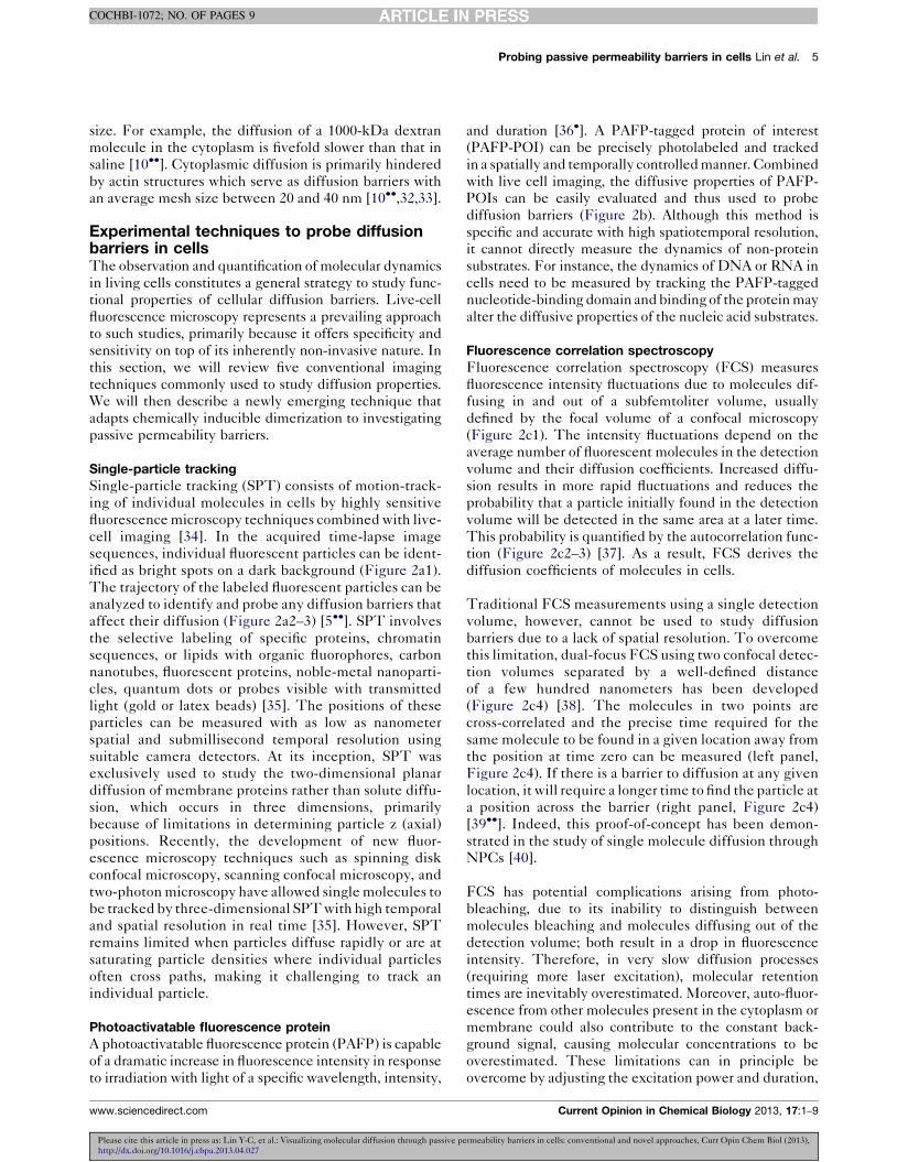
size. For example, the diffusion of a 1000-kDa dextran
molecule in the cytoplasm is fivefold slower than that in
saline [10��]. Cytoplasmic diffusion is primarily hindered
by actin structures which serve as diffusion barriers with
an average mesh size between 20 and 40 nm [10��,32,33].
Experimental techniques to probe diffusionbarriers in cellsThe observation and quantification of molecular dynamics
in living cells constitutes a general strategy to study func-
tional properties of cellular diffusion barriers. Live-cell
fluorescence microscopy represents a prevailing approach
to such studies, primarily because it offers specificity and
sensitivity on top of its inherently non-invasive nature. In
this section, we will review five conventional imaging
techniques commonly used to study diffusion properties.
We will then describe a newly emerging technique that
adapts chemically inducible dimerization to investigating
passive permeability barriers.
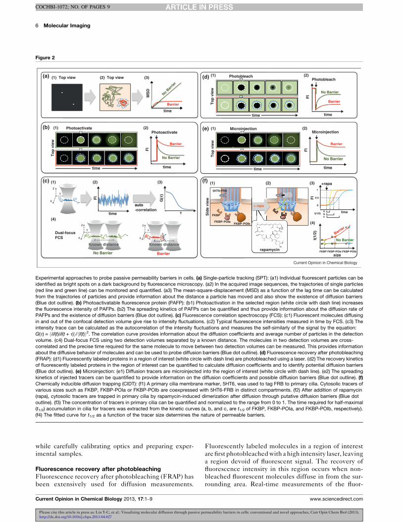
Single-particle tracking
Single-particle tracking (SPT) consists of motion-track-
ing of individual molecules in cells by highly sensitive
fluorescence microscopy techniques combined with live-
cell imaging [34]. In the acquired time-lapse image
sequences, individual fluorescent particles can be ident-
ified as bright spots on a dark background (Figure 2a1).
The trajectory of the labeled fluorescent particles can be
analyzed to identify and probe any diffusion barriers that
affect their diffusion (Figure 2a2–3) [5��]. SPT involves
the selective labeling of specific proteins, chromatin
sequences, or lipids with organic fluorophores, carbon
nanotubes, fluorescent proteins, noble-metal nanoparti-
cles, quantum dots or probes visible with transmitted
light (gold or latex beads) [35]. The positions of these
particles can be measured with as low as nanometer
spatial and submillisecond temporal resolution using
suitable camera detectors. At its inception, SPT was
exclusively used to study the two-dimensional planar
diffusion of membrane proteins rather than solute diffu-
sion, which occurs in three dimensions, primarily
because of limitations in determining particle z (axial)
positions. Recently, the development of new fluor-
escence microscopy techniques such as spinning disk
confocal microscopy, scanning confocal microscopy, and
two-photon microscopy have allowed single molecules to
be tracked by three-dimensional SPT with high temporal
and spatial resolution in real time [35]. However, SPT
remains limited when particles diffuse rapidly or are at
saturating particle densities where individual particles
often cross paths, making it challenging to track an
individual particle.
Photoactivatable fluorescence protein
A photoactivatable fluorescence protein (PAFP) is capable
of a dramatic increase in fluorescence intensity in response
to irradiation with light of a specific wavelength, intensity,
Please cite this article in press as: Lin Y-C, et al.: Visualizing molecular diffusion through passive p
http://dx.doi.org/10.1016/j.cbpa.2013.04.027
www.sciencedirect.com
and duration [36�]. A PAFP-tagged protein of interest
(PAFP-POI) can be precisely photolabeled and tracked
in a spatially and temporally controlled manner. Combined
with live cell imaging, the diffusive properties of PAFP-
POIs can be easily evaluated and thus used to probe
diffusion barriers (Figure 2b). Although this method is
specific and accurate with high spatiotemporal resolution,
it cannot directly measure the dynamics of non-protein
substrates. For instance, the dynamics of DNA or RNA in
cells need to be measured by tracking the PAFP-tagged
nucleotide-binding domain and binding of the protein may
alter the diffusive properties of the nucleic acid substrates.
Fluorescence correlation spectroscopy
Fluorescence correlation spectroscopy (FCS) measures
fluorescence intensity fluctuations due to molecules dif-
fusing in and out of a subfemtoliter volume, usually
defined by the focal volume of a confocal microscopy
(Figure 2c1). The intensity fluctuations depend on the
average number of fluorescent molecules in the detection
volume and their diffusion coefficients. Increased diffu-
sion results in more rapid fluctuations and reduces the
probability that a particle initially found in the detection
volume will be detected in the same area at a later time.
This probability is quantified by the autocorrelation func-
tion (Figure 2c2–3) [37]. As a result, FCS derives the
diffusion coefficients of molecules in cells.
Traditional FCS measurements using a single detection
volume, however, cannot be used to study diffusion
barriers due to a lack of spatial resolution. To overcome
this limitation, dual-focus FCS using two confocal detec-
tion volumes separated by a well-defined distance
of a few hundred nanometers has been developed
(Figure 2c4) [38]. The molecules in two points are
cross-correlated and the precise time required for the
same molecule to be found in a given location away from
the position at time zero can be measured (left panel,
Figure 2c4). If there is a barrier to diffusion at any given
location, it will require a longer time to find the particle at
a position across the barrier (right panel, Figure 2c4)
[39��]. Indeed, this proof-of-concept has been demon-
strated in the study of single molecule diffusion through
NPCs [40].
FCS has potential complications arising from photo-
bleaching, due to its inability to distinguish between
molecules bleaching and molecules diffusing out of the
detection volume; both result in a drop in fluorescence
intensity. Therefore, in very slow diffusion processes
(requiring more laser excitation), molecular retention
times are inevitably overestimated. Moreover, auto-fluor-
escence from other molecules present in the cytoplasm or
membrane could also contribute to the constant back-
ground signal, causing molecular concentrations to be
overestimated. These limitations can in principle be
overcome by adjusting the excitation power and duration,
ermeability barriers in cells: conventional and novel approaches, Curr Opin Chem Biol (2013),
Current Opinion in Chemical Biology 2013, 17:1–9
6 Molecular Imaging
COCHBI-1072; NO. OF PAGES 9
Figure 2
(a) (1) Top view (2) Top view
time
MS
D
No Bar
rier
Barrier
(3)
time
Photoactivate
To
p v
iew
(b)
ti
p
(1)
time
Barrier
No Barrier
FI
ti
Barrier
Photoactivate(2)
Z
X
Y
G(
)
(c) (1)
FI
time
(2) (3)
Z
X
Y
Z
X
Y
Known distance
Dual-focus FCS
(4)
Known distance
auto -correlation
No Barrier Barrier
time
Barrier
No Barrier
FI
PhotobleachPhotobleach
time
time
(d) (1) (2)
FI
time
Barrier
No Barrier
ti
Barrier
MicroinjectionMicroinjectionj
To
p v
iew
(1) (2)
5HT6-FRB
FKBP
FKBP-POIaFKBP-POIb
(f)
1
0.5
0
FI
(1) (2) (3) +rapa
abc
time
(4)
t(1/
2)
size
Barrier
t(1/2)
No Barrier
FKBP FKBP-POIa FKBP-POIb
aa’
b
b’
c
c’
(e)
To
p v
iew
Sid
e vi
ewauto + rapa
rapamycin
Current Opinion in Chemical Biology
Experimental approaches to probe passive permeability barriers in cells. (a) Single-particle tracking (SPT): (a1) Individual fluorescent particles can be
identified as bright spots on a dark background by fluorescence microscopy. (a2) In the acquired image sequences, the trajectories of single particles
(red line and green line) can be monitored and quantified. (a3) The mean-square-displacement (MSD) as a function of the lag time can be calculated
from the trajectories of particles and provide information about the distance a particle has moved and also show the existence of diffusion barriers
(Blue dot outline). (b) Photoactivatable fluorescence protein (PAFP): (b1) Photoactivation in the selected region (white circle with dash line) increases
the fluorescence intensity of PAFPs. (b2) The spreading kinetics of PAFPs can be quantified and thus provide information about the diffusion rate of
PAFPs and the existence of diffusion barriers (Blue dot outline). (c) Fluorescence correlation spectroscopy (FCS): (c1) Fluorescent molecules diffusing
in and out of the confocal detection volume give rise to intensity fluctuations. (c2) Typical fluorescence intensities measured in time by FCS. (c3) The
intensity trace can be calculated as the autocorrelation of the intensity fluctuations and measures the self-similarly of the signal by the equation:
G(t) = hdI(t)dI(t + t)i/hI(t)i2. The correlation curve provides information about the diffusion coefficients and average number of particles in the detection
volume. (c4) Dual-focus FCS using two detection volumes separated by a known distance. The molecules in two detection volumes are cross-
correlated and the precise time required for the same molecule to move between two detection volumes can be measured. This provides information
about the diffusive behavior of molecules and can be used to probe diffusion barriers (Blue dot outline). (d) Fluorescence recovery after photobleaching
(FRAP): (d1) Fluorescently labeled proteins in a region of interest (white circle with dash line) are photobleached using a laser. (d2) The recovery kinetics
of fluorescently labeled proteins in the region of interest can be quantified to calculate diffusion coefficients and to identify potential diffusion barriers
(Blue dot outline). (e) Microinjection: (e1) Diffusion tracers are microinjected into the region of interest (white circle with dash line). (e2) The spreading
kinetics of injected tracers can be quantified to provide information on the diffusion coefficients and possible diffusion barriers (Blue dot outline). (f)
Chemically inducible diffusion trapping (CIDT): (f1) A primary cilia membrane marker, 5HT6, was used to tag FRB to primary cilia. Cytosolic tracers of
various sizes such as FKBP, FKBP-POIa or FKBP-POIb are coexpressed with 5HT6-FRB in distinct compartments. (f2) After addition of rapamycin
(rapa), cytosolic tracers are trapped in primary cilia by rapamycin-induced dimerization after diffusion through putative diffusion barriers (Blue dot
outline). (f3) The concentration of tracers in primary cilia can be quantified and normalized to the range from 0 to 1. The time required for half-maximal
(t1/2) accumulation in cilia for tracers was extracted from the kinetic curves (a, b, and c, are t1/2 of FKBP, FKBP-POIa, and FKBP-POIb, respectively).
(f4) The fitted curve for t1/2 as a function of the tracer size determines the nature of permeable barriers.
while carefully calibrating optics and preparing exper-
imental samples.
Fluorescence recovery after photobleaching
Fluorescence recovery after photobleaching (FRAP) has
been extensively used for diffusion measurements.
Please cite this article in press as: Lin Y-C, et al.: Visualizing molecular diffusion through passive p
http://dx.doi.org/10.1016/j.cbpa.2013.04.027
Current Opinion in Chemical Biology 2013, 17:1–9
Fluorescently labeled molecules in a region of interest
are first photobleached with a high intensity laser, leaving
a region devoid of fluorescent signal. The recovery of
fluorescence intensity in this region occurs when non-
bleached fluorescent molecules diffuse in from the sur-
rounding area. Real-time measurements of the fluor-
ermeability barriers in cells: conventional and novel approaches, Curr Opin Chem Biol (2013),
www.sciencedirect.com

Probing passive permeability barriers in cells Lin et al. 7
COCHBI-1072; NO. OF PAGES 9
escence recovery kinetics allow estimates of diffusion
coefficients (Figure 2d).
FRAP is generally limited in measurement duration, such
that long-tail recovery curves expected in anomalous
diffusion are easily overestimated. Furthermore, diffu-
sion rates of fast diffusing molecules, such as small soluble
molecules, are always underestimated due to the rela-
tively long photobleaching time required to overcome the
rapid diffusion of surrounding non-photobleached mol-
ecules into the region of interest.
Microinjection of diffusion tracers
Microinjection has been widely applied to studies of
diffusion. Injection can be applied transiently and locally
to increase the concentration of diffusion tracer mol-
ecules. In combination with live cell imaging, the
dynamics of injected solutes can be monitored and the
diffusion coefficient can be extracted from the spreading
kinetics (Figure 2e). Microinjection has been used to
study NPCs [15], primary cilia [30��], AIS [21��], and
diffusion barriers in cytoplasm [32,33].
To determine the functional size of permeable diffusion
barriers, the ideal injected diffusion tracers should meet
the following conditions: first, inert (unable to affect
cellular physiology and does not have any affinity with
unrelated endogenous proteins and diffusion barriers);
second, spherical shape; third, stable; fourth, appropriate
fluorescence signal; fifth, soluble in aqueous buffer; sixth,
can form polymers with a continuous series of sizes; and
seventh, neutral in electric charge. Dextran, ficoll, poly-
ethylene glycol (PEG), gold particles and globular
proteins have been frequently used as diffusion tracers
[13,21,30��,41]. Dextran, ficoll, and PEG can form poly-
mers of various sizes and are essentially non-interacting
macromolecules [41]. Ficoll and PEG, but not dextran,
are relatively spherical macromolecules [41]. Gold
particles are also spherical molecules with well-defined
sizes and very low affinity with other endogenous com-
ponents in cells. Globular proteins such as insulin, lyso-
zyme, myoglobin and hemoglobin, although they are not
neutral molecules and may have affinity to endogenous
cellular components, are still frequently used to study
permeable diffusion barriers more physiologically.
The delivery of microinjected diffusion tracers can be
precisely controlled by automated equipment. Several
current models of air-pressure-driven microinjectors
allow fine-tuning of injection pressure, compensation
pressure as well as injection duration, making the trans-
ferred dosage highly controlled in a reproducible manner.
There is also a diverse array of diffusion tracers ranging
from synthetic molecules such as pharmacological drugs
to physiological molecules including nucleotides, pep-
tides, recombinant proteins and macromolecules. The
disadvantages of microinjection include potential damage
Please cite this article in press as: Lin Y-C, et al.: Visualizing molecular diffusion through passive p
http://dx.doi.org/10.1016/j.cbpa.2013.04.027
www.sciencedirect.com
to cells due to its invasive application and technical
difficulties in injecting small structures such as dendritic
spines.
Chemically inducible diffusion trap
Chemically inducible dimerization (CID) techniques
have been widely used in cell biology for over 20 years
since their inception [42�]. The most common CID
system relies on the rapamycin-inducible interaction of
FK506-rapamycin-binding domain (FRB) and immuno-
philin FK506-binding protein-12 (FKBP) [42�,43�]. In a
typical CID system, two chimeric proteins are coex-
pressed in cells [42�,43�]. One protein contains FRB
and a plasma membrane anchor sequence that localizes
it to the cytoplasmic leaflet of the plasma membrane,
while the other FKBP-tagged cytosolic protein of interest
(FKBP-POI) freely diffuses throughout the cytoplasm.
Addition of rapamycin rapidly traps FKBP-POIs at the
membrane, where the FRB-tagged protein is localized
(Figure 2f1–2) [42�]. The kinetics of FKBP-POI motility
from cytoplasm to the membrane can be quantified to
provide information about its diffusion rate (Figure 2f3).
On the basis of this methodology, two groups recently
have improvised CID systems to probe the dynamics of
soluble proteins across diffusion barriers in living cells.
Raschbichler et al. generated a bipartite assay called
NEX-TRAP (Nuclear EXport Trapped by RAPmycin)
for the analysis of protein nuclear export in cells [44��].This method used the CID system to trap NLS-tagged
proteins that have shuttled out of the nucleus to specific
membrane compartments in the cytoplasm. In this man-
ner, the kinetics of protein nuclear export can be con-
veniently represented by the relative amounts of NLS-
tagged proteins trapped on the cytoplasmic membrane
compartment, which in turn allows probing of the diffu-
sion barrier properties of the NPC.
In contrast to the large-volume nucleus, the primary
cilium is a tiny organelle, whose volume only comprises
�0.01% of the total cell volume. This has posed a great
challenge to fluorescently visualizing the translocation of
soluble proteins into this tiny organelle. However, Lin
et al. recently overcame this technical difficulty with a
newly developed CID-based methodology termed the
chemically inducible diffusion trap (CIDT) at cilia to
visualize diffusion of cytosolic proteins into primary cilia
in real time in intact living cells [31��]. In this system, a
specific cilia-enriched membrane protein, the 5-hydroxy-
tryptamine receptor 6 (5HT6), was tagged with FRB on
its cytoplasmic tail, while cytosolic proteins to be tested
for ciliary influx are fused with FKBP (Figure 2f1). Upon
addition of a chemical dimerizer, cytosolic diffusion probe
proteins that are able to diffuse between the cytosol and
ciliary lumen are trapped inside primary cilia (Figure 2f2).
The diffusion kinetics of FKBP-tagged cytosolic proteins
into primary cilia can be visualized via wide-field fluor-
escence microscopy from the accumulating fluorescence
ermeability barriers in cells: conventional and novel approaches, Curr Opin Chem Biol (2013),
Current Opinion in Chemical Biology 2013, 17:1–9

8 Molecular Imaging
COCHBI-1072; NO. OF PAGES 9
signal (Figure 2f3). The advantages of CIDT lie in the
ability to rapidly trigger diffusion measurements without
damaging the plasma membrane by microinjection
or detergent-mediated permeabilization. Furthermore,
analysis of the diffusion kinetics similarly provides esti-
mates of diffusion coefficients by using Fick’s first law:
J ¼ �D½Fcy� � ½Fci�
x
where [Fcy] and [Fci] are concentrations of the FKBP
tagged proteins in the cytoplasm and cilium, respectively,
D is the diffusion coefficient of the FKBP-tagged proteins,
and x is the proposed length of a diffusion barrier. Esti-
mates of the flux, J, can be obtained by quantifying the
change in fluorescence intensity (proportional to FKBP
tagged proteins) in cilia over time through an estimated
cross sectional area provided by structural studies. Thus,
diffusion coefficients of different sized FKBP-tagged
proteins can be derived from CIDT measurements and
comparisons can be made between theoretical and exper-
imentally determined diffusion coefficients to shed light
on the regulation of diffusion into cilia (Figure 2f4).
Despite its utility, the CIDT technique presents a few
limitations. First, the CID diffusion trap system is not
suitable to study the diffusion of non-protein substrates.
Second, to enable visualization and inducible dimeriza-
tion of cytosolic proteins, each cytosolic diffusion probe
has to be tagged with either FRB or FKBP fused with a
fluorescent protein, placing a restriction on their minimal
molecular size (e.g. YFP-FKBP, molecular weight,
�40 kDa; stokes radius, �32 A). Therefore, CIDT cannot
readily probe diffusion barriers with a pore size smaller
than 40 kDa. The derivation of diffusion coefficients is
also limited to early time points to minimize the effects of
the lateral diffusion of FKBP–FRB pairs in the ciliary
membrane.
ConclusionsDysfunction of subcellular diffusion barriers severely
impacts cellular functions, leading to various disease
conditions such as cancers, brain disorders, triple A syn-
drome, and ciliopathies. Because of their importance in
regulating cellular functions, several approaches have
been established to evaluate the motion of solutes across
diffusion barriers in cells. In this review, we have high-
lighted each approach while emphasizing their pros and
cons. Measurements of solute motions in a given cellular
aqueous compartment with present techniques require
specialized microscopy techniques and careful analysis.
In contrast, a newly emerging approach such as CIDT
complements nicely with existing techniques, as it uses
an ordinary wide-field fluorescence microscope. CIDT is
well suited to probing submicron compartments that have
been otherwise challenging. Armed with these tech-
niques, we continue our endeavor of elucidating elegant
passive permeability architectures in living cells.
Please cite this article in press as: Lin Y-C, et al.: Visualizing molecular diffusion through passive p
http://dx.doi.org/10.1016/j.cbpa.2013.04.027
Current Opinion in Chemical Biology 2013, 17:1–9
References and recommended readingPapers of particular interest, published within the period of review,have been highlighted as:
� of special interest
�� of outstanding interest
1.�
Caudron F, Barral Y: Septins and the lateralcompartmentalization of eukaryotic membranes. Dev Cell2009, 16:493-506.
A comprehensive review that covers the different lateral diffusion barrierin cells.
2. Strambio-De-Castillia C, Niepel M, Rout MP: The nuclear porecomplex: bridging nuclear transport and gene regulation. NatRev Mol Cell Biol 2010, 11:490-501.
3. Buffington SA, Rasband MN: The axon initial segment innervous system disease and injury. Eur J Neurosci 2011,34:1609-1619.
4. Winckler B, Forscher P, Mellman I: A diffusion barrier maintainsdistribution of membrane proteins in polarized neurons.Nature 1999, 397:698-701.
5.��
Ruthardt N, Lamb DC, Brauchle C: Single-particle tracking as aquantitative microscopy-based approach to unravel cell entrymechanisms of viruses and pharmaceutical nanoparticles.Mol Ther 2011, 19:1199-1211.
A thorough review that covers the introduction of SPT and recentadvances of SPT in three dimensions.
6.�
Nachury MV, Seeley ES, Jin H: Trafficking to the ciliarymembrane: how to get across the periciliary diffusion barrier?Annu Rev Cell Dev Biol 2010, 26:59-87.
A thorough review that covers the different mechanisms of ciliary influx ofprotein into cilia.
7. Hoelz A, Debler EW, Blobel G: The structure of the nuclear porecomplex. Annu Rev Biochem 2011, 80:613-643.
8. Cronshaw JM, Matunis MJ: The nuclear pore complex: diseaseassociations and functional correlations. Trends EndocrinolMetab 2004, 15:34-39.
9. Bloodgood BL, Sabatini BL: Neuronal activity regulatesdiffusion across the neck of dendritic spines. Science 2005,310:866-869.
10.��
Dix JA, Verkman AS: Crowding effects on diffusion in solutionsand cells. Annu Rev Biophys 2008, 37:247-263.
A thorough review that describes the molecular crowding of solutes incellular aqueous compartments. It also introduces several experimentalmeasurements of solute diffusion in cellular aqueous compartments.
11. Grunwald D, Singer RH, Rout M: Nuclear export dynamics ofRNA–protein complexes. Nature 2011, 475:333-341.
12. Paine PL, Moore LC, Horowitz SB: Nuclear envelopepermeability. Nature 1975, 254:109-114.
13. Pante N, Kann M: Nuclear pore complex is able to transportmacromolecules with diameters of about 39 nm. Mol Biol Cell2002, 13:425-434.
14. Mohr D, Frey S, Fischer T, Guttler T, Gorlich D: Characterisationof the passive permeability barrier of nuclear pore complexes.EMBO J 2009, 28:2541-2553.
15. Naim B, Brumfeld V, Kapon R, Kiss V, Nevo R, Reich Z: Passiveand facilitated transport in nuclear pore complexes is largelyuncoupled. J Biol Chem 2007, 282:3881-3888.
16. Alvarez VA, Sabatini BL: Anatomical and physiologicalplasticity of dendritic spines. Annu Rev Neurosci 2007,30:79-97.
17. Lee KF, Soares C, Beique JC: Examining form and function ofdendritic spines. Neural Plast 2012, 2012:704103.
18.�
Noguchi J, Matsuzaki M, Ellis-Davies GC, Kasai H: Spine-neckgeometry determines NMDA receptor-dependent Ca2+
signaling in dendrites. Neuron 2005, 46:609-622.This work demonstrates that the geometry of the dendritic spine neckregulates the permeability of the diffusion barrier for Ca2+.
ermeability barriers in cells: conventional and novel approaches, Curr Opin Chem Biol (2013),
www.sciencedirect.com

Probing passive permeability barriers in cells Lin et al. 9
COCHBI-1072; NO. OF PAGES 9
19. Bender KJ, Trussell LO: The physiology of the axon initialsegment. Annu Rev Neurosci 2012, 35:249-265.
20. Hedstrom KL, Ogawa Y, Rasband MN: AnkyrinG is required formaintenance of the axon initial segment and neuronal polarity.J Cell Biol 2008, 183:635-640.
21.��
Song AH, Wang D, Chen G, Li Y, Luo J, Duan S, Poo MM: Aselective filter for cytoplasmic transport at the axon initialsegment. Cell 2009, 136:1148-1160.
This work focuses on the diffusion barrier for solutes in the axon initialsegment. The authors found that a size-selective permeable diffusionbarrier exists in the cytoplasm of AIS.
22. Singla V, Reiter JF: The primary cilium as the cell’s antenna:signaling at a sensory organelle. Science 2006, 313:629-633.
23. Hildebrandt F, Benzing T, Katsanis N: Ciliopathies. N Engl J Med2011, 364:1533-1543.
24. Satir P, Christensen ST: Overview of structure and function ofmammalian cilia. Annu Rev Physiol 2007, 69:377-400.
25. Anderson RG: The three-dimensional structure of thebasal body from the rhesus monkey oviduct. J Cell Biol 1972,54:246-265.
26. Calvert PD, Strissel KJ, Schiesser WE, Pugh EN Jr, Arshavsky VY:Light-driven translocation of signaling proteins in vertebratephotoreceptors. Trends Cell Biol 2006, 16:560-568.
27. Horst CJ, Forestner DM, Besharse JC: Cytoskeletal-membraneinteractions: a stable interaction between cell surfaceglycoconjugates and doublet microtubules of thephotoreceptor connecting cilium. J Cell Biol 1987,105:2973-2987.
28. Nair KS, Hanson SM, Mendez A, Gurevich EV, Kennedy MJ,Shestopalov VI, Vishnivetskiy SA, Chen J, Hurley JB, Gurevich VVet al.: Light-dependent redistribution of arrestin in vertebraterods is an energy-independent process governed byprotein–protein interactions. Neuron 2005, 46:555-567.
29.��
Najafi M, Maza NA, Calvert PD: Steric volume exclusion setssoluble protein concentrations in photoreceptor sensory cilia.Proc Natl Acad Sci USA 2012, 109:203-208.
This work focuses on the diffusion barrier for soluble proteins in theconnecting cilium of photoreceptor cells. The authors found that mono-mers, dimers and trimers of green fluorescent protein (GFP) freely dif-fused across the CC, suggesting that there is no fixed pore that limits thediffusion of soluble proteins at least up to 81 kDa.
30.��
Kee HL, Dishinger JF, Lynne Blasius T, Liu CJ, Margolis B,Verhey KJ: A size-exclusion permeability barrier andnucleoporins characterize a ciliary pore complex thatregulates transport into cilia. Nat Cell Biol 2012, 14:431-437.
This work focuses on the diffusion barrier for soluble proteins into primarycilia. The authors found that molecules above 67 kDa are excluded fromthe primary cilia lumen by a fixed size NPC-like pore at the base of theprimary cilia.
31.��
Lin YC, Niewiadomski P, Lin B, Nakamura H, Phua SC, Jiao J,Levchenko A, Inoue T, Rohatgi R, Inoue T: Chemically induciblediffusion trap at cilia reveals molecular sieve-like barrier. NatChem Biol 2013 http://dx.doi.org/10.1038/nchembio.1252.
Please cite this article in press as: Lin Y-C, et al.: Visualizing molecular diffusion through passive p
http://dx.doi.org/10.1016/j.cbpa.2013.04.027
www.sciencedirect.com
The first study to probe the diffusion barrier in primary cilia using chemicalchemically inducible dimerization technique. The authors found that amolecular-sieve like permeable barrier exists at the base of primary ciliawhich is accessible to proteins as large as 650 kDa.
32. Luby-Phelps K, Castle PE, Taylor DL, Lanni F: Hindered diffusionof inert tracer particles in the cytoplasm of mouse 3T3 cells.Proc Natl Acad Sci USA 1987, 84:4910-4913.
33. Luby-Phelps K, Taylor DL, Lanni F: Probing the structure ofcytoplasm. J Cell Biol 1986, 102:2015-2022.
34. Michalet X, Berglund AJ: Optimal diffusion coefficientestimation in single-particle tracking. Phys Rev E Stat NonlinSoft Matter Phys 2012, 85:061916.
35. Hellriegel C, Gratton E: Real-time multi-parameterspectroscopy and localization in three-dimensional single-particle tracking. J R Soc Interface 2009, 6(Suppl. 1):S3-S14.
36.�
Lukyanov KA, Chudakov DM, Lukyanov S, Verkhusha VV:Innovation: photoactivatable fluorescent proteins. Nat Rev MolCell Biol 2005, 6:885-891.
A comprehensive review that covers the instructional use of photoactiva-table proteins and the potential applications of photoactivatable proteins.
37. Ries J, Schwille P: Fluorescence correlation spectroscopy.Bioessays 2012, 34:361-368.
38. Dertinger T, Pacheco V, von der Hocht I, Hartmann R, Gregor I,Enderlein J: Two-focus fluorescence correlationspectroscopy: a new tool for accurate and absolute diffusionmeasurements. Chemphyschem 2007, 8:433-443.
39.��
Digman MA, Gratton E: Imaging barriers to diffusion by paircorrelation functions. Biophys J 2009, 97:665-673.
This work develops a dual-FCS system to map diffusion barriers in cells.
40. Cardarelli F, Gratton E: In vivo imaging of single-moleculetranslocation through nuclear pore complexes by paircorrelation functions. PLoS ONE 2010, 5:e10475.
41. Venturoli D, Rippe B: Ficoll and dextran vs. globular proteins asprobes for testing glomerular permselectivity: effects ofmolecular size, shape, charge, and deformability. Am J PhysiolRenal Physiol 2005, 288:F605-F613.
42.�
DeRose R, Miyamoto T, Inoue T: Manipulating signaling at will:chemically-inducible dimerization (CID) techniques resolveproblems in cell biology. Pflugers Arch 2013, 465:409-417.
A comprehensive review that introduces the principle of CID systems andreviews the different applications of CID-based techniques.
43.�
Putyrski M, Schultz C: Protein translocation as a tool: thecurrent rapamycin story. FEBS Lett 2012, 586:2097-2105.
A comprehensive review that covers the different CID-based techniquesused to modulate molecular activities by translocation of specific effec-tors to their functional sites.
44.��
Raschbichler V, Lieber D, Bailer SM: NEX-TRAP, a novel methodfor in vivo analysis of nuclear export of proteins. Traffic 2012,13:1326-1334.
This work uses chemically inducible dimerization system to evaluate thenuclear export activity of protein. The method can also be utilized tomeasure the kinetic of protein passing through the NPCs.
ermeability barriers in cells: conventional and novel approaches, Curr Opin Chem Biol (2013),
Current Opinion in Chemical Biology 2013, 17:1–9