vital pulp therapy with new materials: new directions and treatment perspectives—permanent teeth
DESCRIPTION
Aritculo sobre la terapia en la denticion permanente joven ,un caso clinicoTRANSCRIPT
Conference Paper
220 VITAL PULP THERAPY
PEDIATRIC DENTISTRY V 30 / NO 3 MAY / JUN 08
Vital Pulp Therapy with New Materials: New Directions and Treatment Perspectives—Permanent Teeth David E. Witherspoon, BDS, MS
Dental caries is one of the greatest challenges to the integrity of the developing tooth. It can result in irreversible pulpal damage, eventually causing necrosis of the pulpal tissues and associated arrested development of the tooth root. Ultimately, abnormal root development will impact the long-term prog- nosis for tooth retention.1–4 Thus, the primary goal when treating immature permanent teeth should be to maintain pulp vitality so that apexogenesis can occur.5–8 Direct pulp caps and pulpotomies in teeth with incomplete root formation promote normal development of the root complex. There are long-term prognostic advantages of this treatment outcome over apexification treatment. The tooth structure formed is of a great quantity, and its composition appears to have greater structural integrity.3 The result is that the fully developed tooth is more resistant to vertical root fractures.4
The classic study by Kakehashi et al9,10 eloquently establi-shed the role of bacteria in pulpal health and necrosis. In a germ-free environment, the pulp demonstrated the ability to heal and deposit additional dentin material. In the presence of bacteria, pulpal demise was inevitable. This fundamental premise is integral to the success of all vital pulp procedures. Thus, the basic principle of vital pulpal treatment can be broken down into 2 broad phases. The initial phase involves removing the diseased and bacterially contaminated tissue. The second phase involves establishing an environment that
will prevent any further and future bacterial contamination. The principal procedures aimed at maintaining pulpal vitality include direct pulp caps and pulpotomies. By removing the affected coronal pulp tissue and leaving the unaffected radicular pulp tissue in situ, sealed from the oral environment, normal root development can take place.
Historically, a number of materials11–13 have been advocated to induce normal root development. To date, the material of choice has been calcium hydroxide (Ca(OH)2).
14–20 Most recent- ly, an alternative material, mineral trioxide aggregate (MTA), has become available for use in pulpal procedures. Several properties are necessary when choosing a material to be used in vital pulp treatment. These include the ability of the material to kill bacteria, induce mineralization, and establish a tight bacterial seal. The ideal material for vital pulp treatment should be able to resist long-term bacterial leakage and stimulate the remaining pulp tissue to return to a healthy state, promoting the formation of dentin. The early data for MTA suggest that it is the optimum material for fulfilling these goals when vital pulp therapy is the treatment of choice. MTA’s Physical and Chemical Properties MTA is composed of tricalcium silicate, bismuth oxide, dicalcium silicate, tricalcium aluminate, and calcium sulfate dihydrate. MTA might also contain up to 0.6% insoluble residue, including free crystalline silica. Other trace constituents might include calcium oxide, free magnesium oxide, potas-sium, and sodium sulfate compounds.21 Hydration of the powder results in the formation of a finely crystalline gel of the
Dr. Witherspoon is an endodontist in private practice in Plano, Texas.Correspond with Dr. Witherspoon at [email protected]
Abstract: Pulp necrosis in immature teeth subsequent to caries has a major impact on long-term tooth retention. The aim of vital pulp therapy is to maintain pulp viability by eliminating bacteria from the dentin-pulp complex and to establish an environment in which apexogenesis can occur. A compli-cating factor in treating immature teeth is the difficulty predicting the degree of pulpal damage. The ability of the clinician to manage the health of the remaining pulpal tissue during the procedure is paramount. Currently, the best method appears to be the ability to control pulpal hemorrhage by using sodium hypochlorite. Mineral trioxide aggregate (MTA) currently is the optimum material for use in vital pulp therapy. Compared with the traditional material of calcium hydroxide, it has superior long-term sealing ability and stimulates a higher quality and greater amount of reparative dentin. In the medium-term clinical assessment, it has demonstrated a high success rate. Thus, MTA is a good substitute for calcium hydroxide in vital pulp procedures. (Pediatr Dent 2008;30:220-4)
KEYWORDS: APEXOGENESIS, DIRECT PULP CAP, MINERAL TRIOXIDE AGGREGATE, PULPOTOMY

PEDIATRIC DENTISTRY V 30 / NO 3 MAY / JUN 08
VITAL PULP THERAPY 221
hydrated forms of the components, with some Ca(OH)2 also being formed. This solidifies to a hard structure in less than 3 hours.22 It has a compressive strength equal to intermediate restorative material (IRM) and Super-EBA IRM but less than that of amalgam. MTA has been shown to have an antibacterial effect on some of the facultative bacteria and no effect on strict anaerobic bacteria.23 This limited antibacterial effect is less than that demonstrated by Ca(OH)2 pastes. MTA’s ability to resist the future penetration of microorganisms appears to be high. In in vitro leakage studies,24,25 MTA has resisted leakage predictably and repeatedly. MTA frequently performs better than amalgam IRM or Super EBA.26–30 Compared with composite resins placed under ideal conditions, MTA’s leakage patterns are similar.31,32 Furthermore, the presence of blood has little impact on the degree of leakage.29,33 It is commercially available as ProRoot MTA (Dentsply Tulsa Dental, Tulsa, OK) and has been advocated for use in vital pulp therapy.34–38 Mineralization MTA has demonstrated the ability to induce hard tissue formation in pulpal tissues when used as either a direct pulp capping or pulpotomy material.34, 36, 39–45 MTA promotes rapid cell growth in vitro.46 Compared with Ca(OH)2, in animal studies, MTA consistently induces the formation of dentin at a greater rate with a superior structural integrity. It develops more complete dentin bridges and demonstrates an improved ability to maintain pulp tissue integrity.34,39 Histologic evaluation in animal and human studies has shown that MTA stimulates reparative dentin formation, with thick dentinal bridging, minimal inflammation, and nominal hyperemia. The net result is that vital pulp therapy with MTA produces negligible pulpal necrosis.34,41–43,47
MTA also appears to induce the formation of a dentin bridge at a faster rate than Ca(OH)2.
48 In 1 case report52 partial pulpotomies were conducted on 2 cases of dens evaginatus. After 6 months, the teeth were removed as part of planned orthodontic treatment. Histologic examination of these teeth showed an apparent continuous dentin bridge formation in both teeth, and the pulps were free of inflammation. The process by which MTA acts to induce dentin bridge formation is not known. It has been theorized49 that the tricalcium oxide in MTA reacts with tissue fluids to form Ca(OH)2, resulting in hard tissue formation in a similar manner to Ca(OH)2. Clinical Outcomes The initial response reported in case reports has been very positive.50–52 Several human clinical studies with MTA for direct pulp capping and pulpotomies have recently been published. Direct Pulp Capping The clinical data available on MTA pulp capping of cariously exposed permanent teeth are limited to 2 studies. Both of these studies have reported a high rate of success, which ranges from 93%–98%.53,54 In 1 study,54 53 teeth with carious exposures
that had been diagnosed with reversible pulpitis and normal periradicular tissue were treated with MTA pulp caps. A total of 40 patients between 7 and 45 years old were treated. Briefly, the treatment consisted of caries removal with the aid of an operating microscope and extensive use of caries detector dye. Pulpal bleeding was controlled with 5.25%–6.00% NaOCl applied for up to 10 minutes. After hemostasis was achieved, the pulps were capped with MTA, and the teeth were temporized with unbonded composite Photocore (Kuraray Co Ltd, Osaka, Japan) and a moistened cotton pellet placed directly over the MTA. The teeth were restored with a bonded composite 5–10 days later. Forty-nine of the 53 teeth were available for recall at the 1-year time frame, with an average recall period of 3.94 years.
The maximum period of observation was 9 years. During that time, 98% of the cases presented a favorable outcome, with a normal radiographic appearance, no symptoms, and a normal response to cold testing. In addition, of the 15 teeth in younger patients that were not fully formed at the time of treatment, all subsequently demonstrated continued normal apexogenesis to complete root formation.54 In the second study, a 93% clinical and radiographic success rate was reported at the 24-month recall period. The study used a similar protocol to direct pulp cap 30 young, permanent, cariously exposed asymptomatic teeth with MTA. All the teeth showed signs of vitality and absence of periapical radiolucency, with evidence of continued root growth and no reported symptoms.53 Pulpotomy The human clinical data available on MTA pulpotomies in cariously exposed permanent teeth have reported high success rates, which ranged from 93%–100%. In a prospective clini-cal trial comparing success rates of partial pulpotomies with treated cariously exposed permanent molars by using Ca(OH)2 or MTA, there was no statistically significant difference in the success rate between each group (Ca(OH)2=91%, MTA=93%). Fifty-one teeth in 34 patients were available for recall. The patients ranged from 6.8–13.3 years old, and the follow-up period was from 25.4–45.6 months, with an average of 34.8 months.55
When comparing MTA and Ca(OH)2 pulpotomies within the same patient at the 12-month recall time frame, 2 of the 15 teeth in the Ca(OH)2 group were considered failures, whereas none of the teeth treated with MTA failed (0 of 15 teeth). Calcific metamorphosis was evident radiographically in 2 teeth treated with Ca(OH)2 and in 4 teeth treated with MTA.56
The use of gray MTA for partial pulpotomy in cariously exposed young permanent first molars diagnosed with reversible pulpitis and normal periradicular tissue has resulted in a very high success rate. Exposed pulp tissue was removed with a diamond bur, and after hemostasis, 2–4 mm of gray MTA was placed against the fresh wound and then covered with a layer of glass ionomer cement. The teeth were restored with amalgam or stainless steel crowns. At the 24-month recall period, 22 of 28 teeth showed no signs of clinical or radiographic failure
222 VITAL PULP THERAPY
PEDIATRIC DENTISTRY V 30 / NO 3 MAY / JUN 08
and responded within normal limits to pulp vitality tests. Although 6 of 28 teeth did not respond to vitality testing at the final follow-up, the radiographic appearance was within normal limits, and the teeth were asymptomatic. The patients’ ages ranged from 7.2–13.1 years, with an average age of 10 years.57
In a case series outcomes report that used MTA pulpotomies to treat cariously exposed immature permanent teeth (first or second molars) that had been diagnosed with irreversible pulpitis, a success rate of 92% was reported.58 The key factor in deci-ding whether to complete a pulpotomy as opposed to a pulpectomy was the ability to control pulpal hemor-rhage. In cases in which pulpal hemostasis could be achieved with NaOCl, a pulpotomy was completed. The patients’ ages ranged from 7–15 years, with an average age of 10 years. The recall period ranged from 6–53 months, with an average recall period of 21 months. The single tooth that required nonsurgical root canal treatment had completed normal root development before requiring nonsurgical root canal treatment.
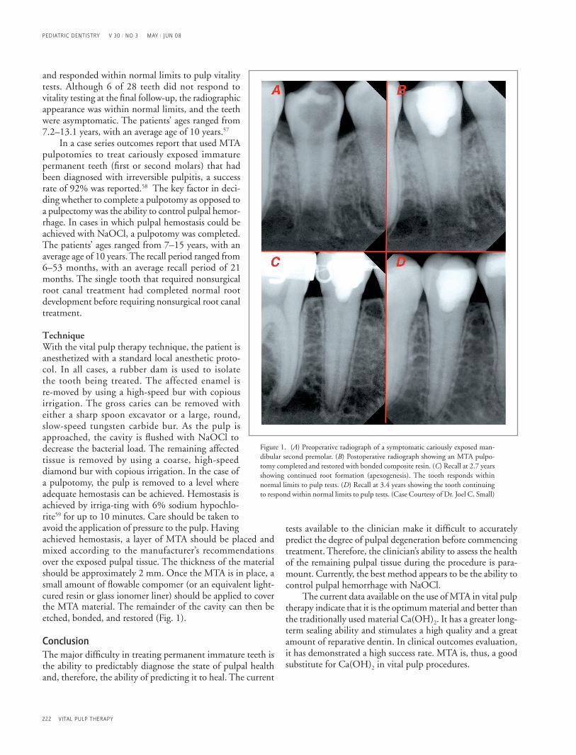
Technique With the vital pulp therapy technique, the patient is anesthetized with a standard local anesthetic proto-col. In all cases, a rubber dam is used to isolate the tooth being treated. The affected enamel is re-moved by using a high-speed bur with copious irrigation. The gross caries can be removed with either a sharp spoon excavator or a large, round, slow-speed tungsten carbide bur. As the pulp is approached, the cavity is flushed with NaOCl to decrease the bacterial load. The remaining affected tissue is removed by using a coarse, high-speed diamond bur with copious irrigation. In the case of a pulpotomy, the pulp is removed to a level where adequate hemostasis can be achieved. Hemostasis is achieved by irriga-ting with 6% sodium hypochlo-rite59 for up to 10 minutes. Care should be taken to avoid the application of pressure to the pulp. Having achieved hemostasis, a layer of MTA should be placed and mixed according to the manufacturer’s recommendations over the exposed pulpal tissue. The thickness of the material should be approximately 2 mm. Once the MTA is in place, a small amount of flowable compomer (or an equivalent light- cured resin or glass ionomer liner) should be applied to cover the MTA material. The remainder of the cavity can then be etched, bonded, and restored (Fig. 1). Conclusion The major difficulty in treating permanent immature teeth is the ability to predictably diagnose the state of pulpal health and, therefore, the ability of predicting it to heal. The current
tests available to the clinician make it difficult to accurately predict the degree of pulpal degeneration before commencing treatment. Therefore, the clinician’s ability to assess the health of the remaining pulpal tissue during the procedure is para-mount. Currently, the best method appears to be the ability to control pulpal hemorrhage with NaOCl.
The current data available on the use of MTA in vital pulp therapy indicate that it is the optimum material and better than the traditionally used material Ca(OH)2. It has a greater long-term sealing ability and stimulates a high quality and a great amount of reparative dentin. In clinical outcomes evaluation, it has demonstrated a high success rate. MTA is, thus, a good substitute for Ca(OH)2 in vital pulp procedures.
Figure 1. (A) Preoperative radiograph of a symptomatic cariously exposed man-dibular second premolar. (B) Postoperative radiograph showing an MTA pulpo-tomy completed and restored with bonded composite resin. (C) Recall at 2.7 years showing continued root formation (apexogenesis). The tooth responds within normal limits to pulp tests. (D) Recall at 3.4 years showing the tooth continuing to respond within normal limits to pulp tests. (Case Courtesy of Dr. Joel C. Small)
PEDIATRIC DENTISTRY V 30 / NO 3 MAY / JUN 08
VITAL PULP THERAPY 223
References 1. Robertson A, Andreasen FM, Andreasen JO, Noren
JG. Long-term prognosis of crown-fractured permanent incisors: the effect of stage of root development and associ-ated luxation injury. Int J Paediatr Dent 2000;10:191–9.
2. Rabie G, Trope M, Tronstad L. Strengthening of immature teeth during long-term endodontic therapy. Endod Dent Traumatol 1986;2:43–7.
3. Katebzadeh N, Dalton BC, Trope M. Strengthening im- mature teeth during and after apexification. J Endod 1998;24:256–9.
4. Cvek M. Prognosis of luxated nonvital maxillary incisors treated with calcium hydroxide and filled with guttaper-cha: a retrospective clinical study. Endod Dent Traumatol 1992;8:45–55.
5. Love RM. Effects of dental trauma on the pulp. Pract Periodontics Aesthet Dent 1997;9:427–36, 38 (quiz)
6. Shabahang S, Torabinejad M. Treatment of teeth with open apices using mineral trioxide aggregate. Pract Periodontics Aesthet Dent 2000;12:315–20, 22 (quiz)
7. Webber RT. Apexogenesis versus apexification. Dent Clin North Am 1984;28:669–97.
8. Massler M. Preventive endodontics: vital pulp therapy. Dent Clin North Am 1967;Nov:663–73.
9. Kakehashi S, Stanley HR, Fitzgerald RJ. The effects of surgical exposures of dental pulps in germ-free and conventional laboratory rats. Oral Surg Oral Med Oral Pathol 1965;20:340–9.
10. Kakehashi S, Stanley HR, Fitzgerald RJ. The effects of surgical exposures of dental pulps in germ-free and conventional laboratory rats. J South Calif Dent Assoc 1966;34:449–51.
11. Horsted-Bindslev P, Vilkinis V, Sidlauskas A. Direct cap- ping of human pulps with a dentin bonding system or with calcium hydroxide cement. Oral Surg Oral Med Oral Pathol Oral Radiol Endod 2003;96:591–600.
12. Trope M, McDougal R, Levin L, May KN Jr, Swift EJ Jr. Capping the inflamed pulp under different clinical conditions. J Esthet Restor Dent 2002;14:349–57.
13. Kiba H, Hayakawa T, Nakanuma K, Yamazaki M, Yamamoto H. Pulpal reactions to two experimental bond- ing systems for pulp capping procedures. J Oral Sci 2000; 42:69–74.
14. Ulmansky M, Sela J, Langer M, Yaari A. Response of pulpotomy wounds in normal human teeth to successi-vely applied Ledermix and Calxyl. Arch Oral Biol 1971; 16:1393–8.
15. Schroder U, Granath LE. Scanning electron microscopy of hard tissue barrier following experimental pulpotomy of intact human teeth and capping with calcium hydro-xide. Odontol Revy 1972;23:211–20.
16. Schroder U. Evaluation of healing following experimental pulpotomy of intact human teeth and capping with calcium hydroxide. Odontol Revy 1972;23:329–40.
17. Holland R, de Souza V, de Mello W, Nery MJ, Bernabe PF, Otoboni Filho JA. Healing process after pulpotomy and covering with calcium hydroxide, Dycal, or MPC: histological study in dog teeth. Rev Fac Odontol Araca-tuba 1978;7:185–91.
18. Holland R, de Souza V, de Mello W, Nery MJ, Bernabe PF, Otoboni Filho JA. Permeability of the hard tissue bridge formed after pulpotomy with calcium hydroxide: a histologic study. J Am Dent Assoc 1979;99:472–5.
19. Stanley HR. Criteria for standardizing and increasing credibility of direct pulp capping studies. Am J Dent 1998; 11(special issue):S17–34.
20. Haskell EW, Stanley HR, Chellemi J, Stringfellow H. Direct pulp capping treatment: a long-term follow-up. J Am Dent Assoc 1978;97:607–12.
21. Dentsply, Tulsa-Dental. Material safety data sheet: ProRoot MTA root canal repair material. Tulsa, OK: Dentsply, 2002:1–2.
22. Torabinejad M, Hong CU, McDonald F, Pitt Ford TR. Physical and chemical properties of a new root-end filling material. J Endod 1995;21:349–53.
23. Torabinejad M, Hong CU, Pitt Ford TR, Kettering JD. Antibacterial effects of some root end filling materials. J Endod 1995;21:403–6.
24. Wu MK, Kontakiotis EG, Wesselink PR. Long-term seal provided by some root-end filling materials. J Endod 1998;24:557–60.
25. Roy CO, Jeansonne BG, Gerrets TF. Effect of an acid environment on leakage of root-end filling materials. J Endod 2001;27:7–8.
26. Torabinejad M, Rastegar AF, Kettering JD, Pitt Ford TR. Bacterial leakage of mineral trioxide aggregate as a root-end filling material. J Endod 1995;21:109–12.
27. Fischer EJ, Arens DE, Miller CH. Bacterial leakage of mineral trioxide aggregate as compared with zinc-free amalgam, intermediate restorative material, and Super-EBA as a root-end filling material. J Endod 1998;24:176–9.
28. Lee SJ, Monsef M, Torabinejad M. Sealing ability of a mineral trioxide aggregate for repair of lateral root perfo-rations. J Endod 1993;19:541–4.
29. Martell B, Chandler NP. Electrical and dye leakage com- parison of three root-end restorative materials. Quintes-sence Int 2002;33:30–4.
30. Tang HM, Torabinejad M, Kettering JD. Leakage evalua-tion of root end filling materials using endotoxin. J Endod 2002;28:5–7.
31. Adamo HL, Buruiana R, Schertzer L, Boylan RJ. A com-parison of MTA, Super-EBA, composite, and amalgam as root-end filling materials using a bacterial microleakage model. Int Endod J 1999;32:197–203.
32. Fogel HM, Peikoff MD. Microleakage of root-end filling materials. J Endod 2001;27:456–8.
224 VITAL PULP THERAPY
PEDIATRIC DENTISTRY V 30 / NO 3 MAY / JUN 08
33. Torabinejad M, Higa RK, McKendry DJ, Pitt Ford TR. Dye leakage of four root end filling materials: effects of blood contamination. J Endod 1994;20:159–63.
34. Pitt Ford TR, Torabinejad M, Abedi HR, Bakland LK, Kariyawasam SP. Using mineral trioxide aggregate as a pulp-capping material. J Am Dent Assoc 1996;127:1491 1491–4.
35. Torabinejad M, Chivian N. Clinical applications of mineral trioxide aggregate. J Endod 1999;25:197–205.
36. Andelin WE, Shabahang S, Wright K, Torabinejad M. Identification of hard tissue after experimental pulp cap- ping using dentin sialoprotein (DSP) as a marker. J Endod 2003;29:646–50.
37. Bakland LK. Management of traumatically injured pulps in immature teeth using MTA. J Calif Dent Assoc 2000;28:855–8.
38. Schmitt D, Lee J, Bogen G. Multifaceted use of Pro-Root MTA root canal repair material. Pediatr Dent 2001;23: 326–30.
39. Faraco IM Jr, Holland R. Response of the pulp of dogs to capping with mineral trioxide aggregate or a calcium hydroxide cement. Dent Traumatol 2001;17:163–6.
40. Holland R, de Souza V, Murata SS, et al. Healing process of dog dental pulp after pulpotomy and pulp covering with mineral trioxide aggregate or Portland cement. Braz Dent J 2001;12:109–13.
41. Dominguez MS, Witherspoon DE, Gutmann JL, Opper-man LA. Histological and scanning electron microscopy assessment of various vital pulp-therapy materials. J Endod 2003;29:324–33.
42. Aeinehchi M, Eslami B, Ghanbariha M, Saffar AS. Mineral trioxide aggregate (MTA) and calcium hydroxide as pulp-capping agents in human teeth: a preliminary report. Int Endod J 2003;36:225–31.
43. Tziafas D, Pantelidou O, Alvanou A, Belibasakis G, Papadimitriou S. The dentinogenic effect of mineral trio-xide aggregate (MTA) in short-term capping experiments. Int Endod J 2002;35:245–54.
44. Parirokh M, Asgary S, Eghbal MJ, et al. A comparative study of white and grey mineral trioxide aggregate as pulp capping agents in dog’s teeth. Dent Traumatol 2005; 21:150–4.
45. Iwamoto CE, Adachi E, Pameijer CH, Barnes D, Romberg EE, Jefferies S. Clinical and histological evaluation of white ProRoot MTA in direct pulp capping. Am J Dent 2006;19:85–90.
46. Mitchell PJ, Pitt Ford TR, Torabinejad M, McDonald F. Osteoblast biocompatibility of mineral trioxide aggregate. Biomaterials 1999;20:167–73.
47. Chacko V, Kurikose S. Human pulpal response to mineral trioxide aggregate (MTA): a histologic study. J Clin Pediatr Dent 2006;30:203–9.
48. Junn DJ, McMillan P, Bakland L, Torabainejad M. Quantitative assessment of dentin bridge formation following pulp-capping with mineral trioxide aggregate (MTA) (abstract). J Endod 1998;24:278.
49. Holland R, de Souza V, Nery MJ, Otoboni Filho JA, Bernabe PF, Dezan Junior E. Reaction of rat connective tissue to implanted dentin tubes filled with mineral trioxide aggregate or calcium hydroxide. J Endod 1999; 25:161–6.
50. Karabucak B, Li D, Lim J, Iqbal M. Vital pulp therapy with mineral trioxide aggregate. Dent Traumatol 2005; 21:240–3.
51. Patel R, Cohenca N. Maturogenesis of a cariously exposed immature permanent tooth using MTA for direct pulp capping: a case report. Dent Traumatol 2006;22:328–33.
52. Koh ET, Ford TR, Kariyawasam SP, Chen NN, Torabi-nejad M. Prophylactic treatment of dens evaginatus using mineral trioxide aggregate. J Endod 2001;27:540–2.
53. Farsi N, Alamoudi N, Balto K, Al Mushayt A. Clinical assessment of mineral trioxide aggregate (MTA) as direct pulp capping in young permanent teeth. J Clin Pediatr Dent 2006;31:72–6.
54. Bogen G. Direct pulp capping with mineral trioxide aggregate: an observational study. J Am Dent Assoc 2008;139:305–15.
55. Qudeimat MA, Barrieshi-Nusair KM, Owais AI. Calcium hydroxide vs mineral trioxide aggregates for partial pulpotomy of permanent molars with deep caries. Eur Arch Paediatr Dent 2007;8:99–104.
56. El-Meligy OA, Avery DR. Comparison of mineral trioxide aggregate and calcium hydroxide as pulpotomy agents in young permanent teeth (apexogenesis). Pediatr Dent 2006; 28:399–404.
57. Barrieshi-Nusair KM, Qudeimat MA. A prospective cli- nical study of mineral trioxide aggregate for partial pulp-otomy in cariously exposed permanent teeth. J Endod 2006; 32:731–5.
58. Witherspoon DE, Small JC, Harris GZ. Mineral trioxide aggregate pulpotomies: a case series outcomes assessment. J Am Dent Assoc 2006;137:610–8.
59. Hafez AA, Cox CF, Tarim B, Otsuki M, Akimoto N. An in vivo evaluation of hemorrhage control using sodium hypochlorite and direct capping with a one- or two-com-ponent adhesive system in exposed nonhuman primate pulps. Quintessence Int 2002;33:261–72.
Conflict of Interest: David E. Witherspoon, BDS, MS, has acted as a Workshop Coordinator for the annual session of the American Association of Endodontists.
Copyright © 2008 American Academy of Pediatric Dentistry and American Association of Endodontists.
This article is being published concurrently in Journal of Endodontics July 2008;34:7S. The articles are identical. Either citation can be used when citing this article.