vitamin d analogs enhance the anticancer activity of 5-fluorouracil in an in vivo mouse colon cancer...
DESCRIPTION
Active vitamin D analogs that are less toxic than calcitriol can be useful in the combined treatment of patients suffering from colon cancer. In the present study we demonstrate, for the first time in an in vivo model system, the biological effect of combined therapy using 5-fluorouracil (5-FU) along with vitamin D analog PRI-2191 (tacalcitol, 1,24-dihydroxyvitamin D3) or PRI-2205 (5,6-trans-isomer of calcipotriol) on colon cancer.TRANSCRIPT

Milczarek et al. BMC Cancer 2013, 13:294http://www.biomedcentral.com/1471-2407/13/294
RESEARCH ARTICLE Open Access
Vitamin D analogs enhance the anticanceractivity of 5-fluorouracil in an in vivo mouse coloncancer modelMagdalena Milczarek1, Mateusz Psurski1,2, Andrzej Kutner3 and Joanna Wietrzyk1*
Abstract
Background: Active vitamin D analogs that are less toxic than calcitriol can be useful in the combined treatmentof patients suffering from colon cancer. In the present study we demonstrate, for the first time in an in vivo modelsystem, the biological effect of combined therapy using 5-fluorouracil (5-FU) along with vitamin D analog PRI-2191(tacalcitol, 1,24-dihydroxyvitamin D3) or PRI-2205 (5,6-trans-isomer of calcipotriol) on colon cancer.
Methods: We investigated the influence of vitamin D analogs on the anticancer activity of 5-FU or capecitabine inthe treatment of mice bearing MC38 mouse colon tumors implanted subcutaneously or orthotopically. The cellcycle distribution, E-cadherin expression and caspase 3/7 activity in vitro were also evaluated.
Results: We observed that both PRI-2191 and PRI-2205 significantly enhanced the antitumor activity of 5-FU; butthese results depend on the treatment regimen. Applying the optimal schedule of combined therapy we observeda significant decrease in tumor growth, metastasis and also a prolongation of the survival time of mice, incomparison with the administrations of 5-FU given alone. Both combinations indicated a synergistic effect and didnot cause toxicity. Moreover, analogs applied after completed course of administration of 5-FU, prolonged theantitumor effect of the drug. Furthermore, when the prodrug of 5-FU, capecitabine, was used, potentiation of itsactivity was also observed.
Conclusions: Our data suggest that vitamin D analogs (especially PRI-2191) might be potentially applied to clinicaluse in order to enhance the anticancer effect of 5-FU and also prolong its activity against colon cancer. The activityof PRI-2191 is realized through stopping the cells in the G0/G1 cell cycle phase and increasing the expression ofE-cadherin.
Keywords: Vitamin D analogs, Combined treatment, 5-Fluorouracil, Capecitabine, Anticancer activity, Colon cancer
BackgroundAccording to the World Health Organization’s Inter-national Agency for Research on Cancer, colorectal can-cer is the third most frequent malignancy and the fourthleading cause of deaths from cancer worldwide [1]. Des-pite significant progress in the treatment of patients suf-fering from colorectal cancer in the last decade, there isa constant need for new therapies. One of the directionsis the development of novel combined treatment
* Correspondence: [email protected] of Experimental Oncology, Ludwik Hirszfeld Institute ofImmunology and Experimental Therapy, Polish Academy of Sciences, R. WeiglaSt. 12, Wroclaw 53-114, PolandFull list of author information is available at the end of the article
© 2013 Milczarek et al.; licensee BioMed CentrCommons Attribution License (http://creativecreproduction in any medium, provided the or
strategies. The benefit of such an approach is the possi-bility of enhancing the therapeutic effect of a drug,which is the basis of a standard therapy. Promising can-didates for this strategy are vitamin D analogs.Epidemiological and clinical data and also research on
animals suggest a protective role for the active form ofvitamin D (calcitriol, 1,25-dihydroxyvitamin D3) in thedevelopment of colon cancer [2-4]. Data from thein vivo studies have shown that a diet supplementedwith vitamin D significantly delayed MC-26 colon cancertumor growth compared to a diet deficient in this
al Ltd. This is an Open Access article distributed under the terms of the Creativeommons.org/licenses/by/2.0), which permits unrestricted use, distribution, andiginal work is properly cited.

Milczarek et al. BMC Cancer 2013, 13:294 Page 2 of 19http://www.biomedcentral.com/1471-2407/13/294
vitamin [5]. Calcitriol affects proliferation, differentiationand apoptosis of human colon cancer cells. It exertsa biological effect mainly through the vitamin D receptor(VDR) [6]. It has been shown that the expression ofVDR increases from normal colon epithelial cells throughprecancerous lesions to well-differentiated tumors andthen decreases in advanced stages of cancer [6,7].The antitumor activity of calcitriol is observed only
when it is applied in hyper-physiological doses, whichcan cause the side effect of hypercalcemia and hypercal-ciuria [8-10]. For this reason, the synthesis of analogshas been initiated in order to dissociate the calcemiceffect from the anticancer activity of calcitriol. In ourprevious studies, we have examined the biological activ-ity of a series of side-chain modified analogs of vitaminD and a series of diastereometric and geometric onesagainst various cancer and normal cell lines [11,12]. Wealso evaluated the influence of vitamin D analogs on theactivity of a range of anticancer drugs in vitro andin vivo against the human and murine cancer cells[13-18]. We observed that vitamin D analogs increasedthe antitumor effect of cyclophosphamide and cisplatincompared to the cytostatic drug applied alone. Based onour results, we selected two analogs for further research:PRI-2191 (tacalcitol, 1,24-dihydroxyvitamin D3) andPRI-2205 (5, 6-trans calcipotriol), which reveal higherantitumor activity and lower calcemic activity, as well aslower toxicity than calcitriol [12,19]. These two analogs,used in combined HT-29 colon cancer treatment withirinotecan or oxaliplatin showed, in selected schedules oftreatment, improvement in mice survival and tumorgrowth delay [20].5-Fluorouracil (5-FU) is one of the oldest anticancer
drugs and is still used in the treatment of colorectal can-cer [21,22]. Two recent reports of in vitro studies dem-onstrate that calcitriol and calcipotriol promote thesensitivity of human colon carcinoma cells to 5-FU andenhance the cytotoxicity of the FOLFIRI anticancer regi-men. These results also indicate that the mechanism ofcalcitriol and calcipotriol action is dependent on the cal-cium sensing receptor (CaSR). Protein expression and thegene transcriptional activity of survivin and thymidylatesynthase are suppressed by inducing the expression andactivation of CaSR by calcitriol or calcipotriol. This leadsto an increase in the sensitivity of colon carcinoma cells to5-FU [23,24].Therefore, the aim of our present studies was to examine
the biological effect (antitumor activity, influence on the lifespan of mice, toxicity and antimetastatic activity) of com-bined therapy with the use of 5-FU along with PRI-2191 orPRI-2205 against MC38 mouse colon cancer in vivo. Inaddition, we examined whether vitamin D analogs wouldprolong the antitumor activity of 5-FU (application of ana-logs was initiated after administration of 5-FU ended).
MethodsCompoundsCalcitriol and its analogs: PRI-2191, PRI-2201 andPRI-2205 are certified synthetic materials obtained fromthe Pharmaceutical Research Institute, Warsaw, Poland.Samples of the compounds were stored, under argon, inamber ampoules at −20°C. Prior to usage, in the case ofin vitro studies, compounds were dissolved in 99.8%ethanol to the concentration of 10-4 M and subsequentlydiluted in culture medium in order to reach the concen-tration of 100 nM. For animal experiments, compoundswere dissolved in 99.8% ethanol, then diluted in 80%propylene glycol in order to reach the required concen-trations and administered subcutaneously (s.c.) or orally(p.o.) to mice in a volume of 5 μl per 1 g of body weight.5-Fluorouracil (5-FU) (Ebewe Pharma, Unterach,
Austria) solution in the concentration of 50 mg/ml wasdiluted prior to usage in in vitro studies in culturemedium in order to reach the required concentrationsand for in vivo experiments in saline in order to reachthe required concentrations and administered eitherintravenously (i.v.) or intraperitoneally (i.p.) to mice ata volume of 10 μl per 1 g of body weight.Capecitabine (CPC) (Pharmaceutical Research Institute,
Warsaw, Poland) was dissolved in 40% ethanol, thendiluted in water for injection in order to reach the requireconcentration and administered orally (p.o.) to mice ata volume of 10 μl per 1 g of body weight.
Cell linesThe mouse colon adenocarcinoma cell line MC38, cul-tured in vivo, was obtained from the Tumor Bank of theTNO Radiobiology Institute, Rijswijk, Holland. This cellline was adapted to growth in vitro as MC38/0 [25]. TheMC38/EGFP mouse colon cancer cells, transduced withgreen fluorescent protein gene and cultured in vitro,were obtained from the Institute of Immunology andExperimental Therapy, Wroclaw, Poland [25]. Humancolon adenocarcinoma cell lines HT-29 and LoVo werereceived from the Deutsches Krebsforschungszentrum,Heidelberg, Germany. All cell lines were stored inliquid nitrogen at the Cell Culture Collection of theInstitute of Immunology and Experimental Therapy,Wroclaw, Poland.The cell lines were cultured in vitro as follows: MC38/0
and MC38/EGFP in RPMI 1640 medium (IIET, Wroclaw,Poland), HT-29 and LoVo in RPMI 1640 + Opti-MEMI (1:1) (from IIET, Wroclaw, Poland and Gibco,Scotland, UK, respectively) all culture media weresupplemented with 2 mM L-glutamine, 1 mM sodiumpyruvate (both from Sigma-Aldrich Chemie GmbH,Steinheim, Germany), 5% fetal bovine serum (PAALaboratories GmbH, Pasching, Austria (MC38/0 andMC38/EGFP) or Thermo Fisher Scientific Inc., UK

Milczarek et al. BMC Cancer 2013, 13:294 Page 3 of 19http://www.biomedcentral.com/1471-2407/13/294
(HT-29 and LoVo) and 100 U/ml penicillin, 100 μg/mlstreptomycin (both from Polfa Tarchomin S.A.Warsaw, Poland). The cells were cultured at 37°C ina humid atmosphere saturated with 5% CO2.
MiceC57BL/6 female, 12-16-week-old mice, weighing 20–25 gwere obtained from the Maria Sklodowska-Curie Institute –Oncology Center (Warsaw, Poland) and maintainedunder specific pathogen-free (SPF) conditions. All experi-ments were performed according to EU Directive 2010/63/EU for animal experiments and were approved by the1st Local Committee for Experiments with the Use of La-boratory Animals, Wroclaw, Poland.
Details of the treatment schedulesThe MC38 colon cancer cells were passaged in vivo.Subcutaneous transplantation: mice were subcutane-ously (s.c.) inoculated in the right flank region witha 33% suspension of homogenized MC38 tumor tissuecoming from s.c. tumors from another mouse, 0.25 mlper mouse. Orthotropic transplantation: the anesthetizedmouse was placed on a wooden board in the right lateralposition and the incision was made through the leftupper abdominal pararectal line and peritoneum. Thececal wall was carefully exposed, placed and fixed be-tween layers of sterile gauze. 1 – 2 mm piece of speci-men, derived from primary tumor grown s.c. in anothermouse, was fixed to the serosal part of cecal wall with5–0 surgical sutures. After implantation, the peritoneumand abdominal wall was sutured with 4–0 surgical sutures(Dexon-“S”, Polfa, Poznań, Poland). Tumor cell transplanta-tions were performed under general anesthesia withthe mixture of ketamine hydrochloride (100 mg/kg,Ketamina 10%, Biowet, Puławy, Poland) and xylazine hydro-chloride (20 mg/kg, XylaRiem, Riemser Arzneimittel AG,Germany).The MC38/EGFP colon cancer cells derived from
in vitro culture were inoculated s.c. in the right flankregion with 1 × 10⁶ cells suspended in 0.2 ml saline permouse. After tumor inoculation, mice were randomly di-vided into different groups (day 0).
The evaluation, both the most effective dose and treatmentschedule of vitamin D analogs in combination with 5-FUVitamin D analog was administered s.c. three times aweek. In these experiments, different doses of vitamin Danalogs and varied doses and treatment schedules of 5-FUwere studied (Table 1 – 5-FU and PRI-2191, Table 2 –5-FU and PRI-2205). In each experiment, mice bearing s.c.MC38 colon cancer cells were randomly divided intodifferent groups (day 0). Then, the mice were injectedwith 5-FU and/or vitamin D analogs.
Vitamin D analog was administered s.c. five times aweek. Mice bearing s.c. MC38 colon cancer cells werei.v. injected with 5-FU at a dose of 100 mg/kg/day ondays: 2, 17 and/or vitamin D analogs: PRI-2191 at a doseof 0.2 μg/kg/day or PRI-2205 at a dose of 20 μg/kg/day.Both analogs were administrated s.c., five times a week ondays: 9, 10, 11, 14, 15, 16, 17, 18, 21, 22, 23, 24, 25.Vitamin D analog was administered p.o. three times a
week. Mice bearing s.c. MC38 colon cancer cells were i.v.injected with 5-FU at a dose of 100 mg/kg/day on days:9, 16, 23 and/or vitamin D analogs: PRI-2191 at a dose of1 μg/kg/day or PRI-2205 at a dose of 10 μg/kg/day. Bothanalogs were administrated p.o. by gavage, directly intothe lower esophagus using a feeding needle, three timesa week on days: 12, 14, 16, 19, 21, 23, 26, 28.
The effect of combined therapy with 5-FU and PRI-2191or PRI-2205 on MC38 colon tumor growth implantedorthotopicallyMice bearing MC38 tumors implanted i.i. were i.v.injected with 5-FU at a dose of 100 mg/kg/day on days:8, 15 and/or vitamin D analogs: PRI-2191 at a dose of1 μg/kg/day or PRI-2205 at a dose of 10 μg/kg/day. Bothanalogs were administrated s.c., three times a week ondays: 10, 13, 15, 17, 20. Mice were sacrificed on day 22after i.i. transplantation, tumors were weighed and bloodwas collected.
The prolongation of 5-FU antitumor activity by PRI-2191or PRI-2205Mice bearing s.c. MC38 colon cancer cells were i.v.injected with 5-FU at a dose of 100 mg/kg/day on days:20, 24, 28, 34. When 5-FU reduced tumor volume byabout 90% in comparison to the control group, the ad-ministration of analogs was initiated (after administra-tion of the cytostatic drug ended). Both analogs wereadministrated s.c., three times a week on days: 35, 38,40, 42, 45, 47, 49, 52, 54, 56, PRI-2191 at a doseof 1 μg/kg/day and PRI-2205 at a dose of 10 μg/kg/day.
The antimetastatic effect of combined therapy with 5-FUand PRI-2191 or PRI-2205Mice bearing s.c. MC38/EGFP colon cancer cells were i.p.treated with 5-FU at a dose of 75 mg/kg/day on days:19, 24, 29 and/or vitamin D analog PRI-2191 at a dose of1 μg/kg/day or PRI-2205 at a dose of 10 μg/kg/day. Bothanalogs were administrated s.c., three times a week ondays: 19, 21, 24, 26, 28, 31, 33, 35, 38, 40, 42, 45, 47, 49,52, 54, 56. On the 59th day of the experiment the lymphnodes were isolated and then observed undera NightOWL II LB 983 device at Wroclaw University ofEnvironmental and Life Sciences, Poland, in order tovisualize and calculate the metastasis of s.c. tumor cells toregional lymph nodes, liver, spleen and lung. Only the

Table 1 The effect of vitamin D analog PRI-2191 (in varied doses) alone or in combination with 5-FU on tumor growth, life span and body weight of micebearing subcutaneous colon cancer MC38
No. 5-FU PRI-2191
TGI [%] ILS [%] Maximal decrease
in body weight [%]
5-FU PRI-2191 5-FU+ %H TGI Effect 5-FU PRI-2191 5-FU+ %H ILS Effect 5-FU PRI-2191 5-FU+
PRI-2191 PRI-2191 PRI-2191
1 75 mg/kg; 2 μg/kg 48 6 70* 51 Synergism 21 53 41 62 Antagonism 7 18 19
2 150 mg/kg; 2 μg/kg 87 44 96* 92 Synergism nt. nt. nt. nt. nt. 9 11 21
3 100 mg/kg; 1 μg/kg 38 6 51 42 Synergism 26 −6 118*a 22 Synergism 4 6 12
4 100 mg/kg; 0.5 μg/kg 38 1 36 39 Antagonism 26 −6 43 22 Synergism 4 3 9
5 100 mg/kg; 0.25 μg/kg 38 −35 36 16 Antagonism 26 3 65 29 Synergism 4 9 9
Vitamin D analog was administered subcutaneously, three times a week.No. – number of experiment; TGI – tumor growth inhibition; %H TGI: hypothetical tumor growth inhibition; ILS: increase in life span; %H ILS: hypothetical increase in life span; nt.: not tested; No. 1 – TGI calculated onthe 27th day of the experiment, No. 2, 3, 4, 5 – TGI on the 32nd day of the experiment.No. 1: 5-FU was injected i.p. on the first day; PRI-2191 was injected on days: 1, 3, 6, 8, 10, 13, 15, 17, 20, 22; Number of mice: control - 10; in the each treatment group - 8.No. 2: 5-FU was injected i.p. on days: 9, 16, 23; PRI-2191 was injected on days: 11, 14, 16, 18, 21, 23; Number of mice in each group - 8.No. 3, 4 and 5: 5-FU was injected i.v. on days: 8, 15, 30, 38; PRI-2191 was injected on days: 9, 11, 14, 16, 18, 21, 23, 25, 28, 30, 32, 35, 37, 39, 42, 44; Number of mice: No. 3, 4 and 5: control – 7; 5-FU – 7, PRI-2191 – 5;5-FU + PRI-2191 – 6; *P < 0.05 as compared to control; *aP < 0.05 as compared to 5-FU.
Milczarek
etal.BM
CCancer
2013,13:294Page
4of
19http://w
ww.biom
edcentral.com/1471-2407/13/294

Table 2 The effect of vitamin D analog PRI-2205 (in varied doses) alone or in combination with 5-FU on tumor growth, life span and body weight of micebearing subcutaneous colon cancer MC38
No. 5-FU PRI-2205
TGI [%] ILS [%] Maximal decrease
in body weight [%]
5-FU PRI-2205 5-FU+ %H TGI Effect 5-FU PRI-2205 5-FU+ %H ILS Effect 5-FU PRI-2205 5-FU+
PRI-2205 PRI-2205 PRI-2205
1 75 mg/kg; 10 μg/kg 48 7 76* 44 Synergism 21 2 77*a 23 Synergism 7 10 8
2 50 mg/kg; 10 μg/kg 30 −16 70 19 Synergism 12 0 38* 12 Synergism 0 0 0
3 150 mg/kg; 10 μg/kg 87 25 96* 90 Synergism nt. nt. nt. nt. nt. 9 1 19
4 100 mg/kg; 5 μg/kg 31 −12 26 23 Antagonism 26 −6 12 22 Antagonism 4 1 6
5 100 mg/kg; 2.5 μg/kg 31 −41 19 2 Antagonism 26 −15 32 16 Synergism 4 7 4
Vitamin D analog was administered subcutaneously, three times a week.No. – number of experiment; TGI – tumor growth inhibition; %H TGI: hypothetical tumor growth inhibition; ILS: increase in life span; %H ILS: hypothetical increase in life span; nt.: not tested; No. 1 – TGI calculated onthe 27th day of the experiment, No. 2, 4, 5 - TGI on the 30th day of the experiment; No. 3 – TGI on the 32nd day of the experiment.No. 1: 5-FU was injected i.p. on the first day; PRI-2205 was injected on days: 1, 3, 6, 8, 10, 13, 15, 17, 20, 22; Number of mice: control - 10; in the each treatment group - 8.No. 2: 5-FU was injected i.p. on days: 2, 7, 11, 16; PRI-2205 was injected on days: 2, 4, 7, 9, 11, 14, 16, 18, 21; Number of mice: control – 9, 5-FU – 8, 5-FU + PRI-2205 – 8, PRI-2205 – 7.No. 3: 5-FU was injected i.p. on days: 9, 16, 23; PRI-2205 was injected on days: 11, 14, 16, 18, 21, 23, 25, 28; Number of mice in each group - 8.No. 4 and 5: 5-FU was injected i.v. on days: 8, 15, 30, 38; PRI-2205 on days: 9, 11, 14, 16, 18, 21, 23, 25, 28, 30, 32, 35, 37, 39, 42, 44; Number of mice: No. 4 and 5: control – 7, 5-FU – 7, PRI-2205 – 5, 5-FU + PRI-2205 – 6;*P < 0.05 as compared to control; *aP < 0.05 as compared to 5-FU.
Milczarek
etal.BM
CCancer
2013,13:294Page
5of
19http://w
ww.biom
edcentral.com/1471-2407/13/294

Milczarek et al. BMC Cancer 2013, 13:294 Page 6 of 19http://www.biomedcentral.com/1471-2407/13/294
metastases of MC38/EGFP colon cancer cells to thelymph nodes were observed.
The effect of combined therapy with capecitabine andPRI-2191 or PRI-2205 on MC38 colon tumor growthimplanted subcutaneouslyMice bearing MC38 tumors implanted s.c. were p.o.(by gavage, directly into the lower esophagus using afeeding needle) administered with capecitabine at a doseof 450 mg/kg/day on days: 3, 4, 5, 6, 7, 10, 11, 12, 13, 14and/or vitamin D analogs: PRI-2191 at a dose of 1 μg/kg/dayor PRI-2205 at a dose of 10 μg/kg/day. Both analogs wereadministrated s.c., three times a week on days: 3, 5, 7, 10,12, 14, 17, 19, 21, 24, 26, 28, 31, 33, 35, 38, 40, 42, 45,47, 49, 52, 54.
Evaluation of the therapeutic effectTumor volume was calculated using the formula (a2 × b)/2,where a = shorter tumor diameter in mm and b = longertumor diameter in mm. Inhibition of tumor growth wascalculated from the following formula: tumor growth inhib-ition (TGI) [%] = 100 – [(WT/WC) × 100], where WT is themedian tumor weight of treated mice and WC – that ofuntreated control mice. Mice obviously sick were sacrificedand the increase in life span (ILS) of treated mice overthe control was evaluated. ILS was calculated from thefollowing formula: 100 – [(MSTT/MSTC) × 100], whereMSTT is the median survival time of treated animals, andMSTC is the median survival time of untreated controlmice.
Evaluation of combination effectsThe TGI and ILS values were then compared with hypothe-tical tumor growth inhibition: %H TGI or %H ILS = 100 -[(100 - E for cytostatic) × (100 - E for vitamin D analog) /100][26], where E was TGI or ILS.As the result of the comparison of TGI to TGI hypothe-
tical value (%H TGI) or ILS to ILS hypothetical value(%H ILS) the type of interaction between two compoundsin combined treatment was designated, which can be:
� synergy – when the experimental value of TGI/ILSis greater than hypothetical TGI/ILS;
� additive effect – when those two values arecomparable;
� subadditive effect – if the experimental TGI/ILSvalue is smaller than the hypothetical, but largerthan TGI/ILS for cytostatic given alone;
� antagonism – if the experimental TGI/ILS is smallerthan the experimental TGI/ILS for cytostatic.
Tumor growth delayTumor growth delay (TGD) is a parameter describingthe time needed for tumors to reach the volume of
1 cm3. It can be also expressed as the direct difference indays: ΔTGDcontrol = TGD treated group–TGDcontrol, whereTGDtreated group are the days needed to reach the volume of1 cm3 for tumors in the treated group and TGDcontrol thedays needed for tumors to reach the volume of 1 cm3 incontrol group. In the case of combined treatment:ΔTGDcytostatic = TGDtreated group–TGDcytostatic, whereTGDcytostatic are the days needed for tumors to reach thevolume of 1 cm3 in the treated group with cytostatic drugs.
Body weight changesThe average body weight change (BWC) in all groups wascalculated using the formula: BWC= (ABWn/ABW1) ×100 – 100%, where ABWn is the average body weight onthe nth-day of experiment (during treatment) and ABW1
is the average body weight on the first day of treatment.
Blood leukocytes and plateletsThe level of blood leukocytes and platelets was measuredin each individual blood sample (Sysmex K4500SL, serialnumber F2872, Japan).
Serum calcium levelFemale BALB/c mice were treated i.p. with 5-FU ata dose of 50 and 75 mg/kg on days 1 and 8, respectivelyand/or with vitamin D analogs: PRI-2191 at a dose of2 μg/kg/day or PRI-2205 at a dose of 10 μg/kg/day. Bothanalogs were administrated s.c., three times a week ondays: 1, 3, 6, 8, 10, 13. Mice were sacrificed 16 days afterthe first injection of the compounds and blood sera werecollected. The calcium level was measured in each indi-vidual serum sample with the photometric Arsezano 3method (Olympus AU400; Olympus America Inc., Melville,NY, USA).
An anti-proliferative assay in vitro24 hours before the addition of the tested compounds,cells were plated in 96-well plates (Sarstedt, Newton,NC, USA) at a density of 2 × 10⁴ cells per well. The cellswere exposed for 120 h to 100 nM of calcitriol,PRI-2191, PRI-2205 or PRI-2201 and simultaneously to 5-FU: 0.1 μg/ml (HT-29 and LoVo) or 0.01 μg/ml (MC38/0); and the SRB assay for evaluating the cytostatic effectwas performed as described previously [12]. The resultswere calculated as the proliferation inhibition of the can-cer cell population. Each compound was tested four-foldeither alone or in combined treatment in a single experi-ment, which was repeated 3–5 times.The percentage of proliferation inhibition was calculated
according to the formula: % of proliferation inhibition =[(At-Am)/(Ac-Am)*100]-100; where: At – absorbanceof treated cells; Am – absorbance of culture medium;Ac – absorbance of control cells. The value of the percent-age of proliferation inhibition was then compared with the

Milczarek et al. BMC Cancer 2013, 13:294 Page 7 of 19http://www.biomedcentral.com/1471-2407/13/294
hypothetical percentage of proliferation inhibition:%H= 100-[(100-E for cytostatic) x (100-E for calcitriolanalog) /100], where E was the mean value of the percent-age of proliferation inhibition in order to determine theinteraction between two compounds in combined treat-ment [26]. The type of interaction was described above.
Cell cycle analysisCultured HT-29 cells were seeded at a density of7.5 × 103 cells/ml of culture medium on 6-well plates(Corning, NY, USA) to a final volume of 4 ml. The cellswere exposed to compounds at set concentrations for120 h: PRI-2191 and PRI-2205 100 nM; 5-FU 0.1 μg/ml.Ethanol, used as a solvent for all compounds, diluted corre-sponding to its highest concentration, produced no toxicitywhen applied to the compounds. After 120 h of incubation,the cells were collected with the use of trypsin/EDTA, pH 8(IIET, Wroclaw, Poland), washed in phosphate-buffered sa-line (PBS) supplemented with 2% of fetal bovine serum andcounted in a hemacytometer. Cells (1 × 10⁶ per sample)were then washed twice in cold phosphate buffered saline(PBS) and fixed for 24 h in 70% ethanol at −20°C. The cellswere then washed twice in PBS and incubated with RNAse(8 μg/ml, Fermentas GmbH, St. Leon-Rot, Germany) at37°C for 1 h. The cells were stained for 30 min withpropidium iodide (0.5 mg/ml; Sigma-Aldrich ChemieGmbH, Steinheim, Germany) at 4°C and the cellular DNAcontent was determined using a BD FACSCalibur instru-ment (Becton Dickinson, San Jose, CA, USA) and a ModFitLT 3.0 program (Verity Software House, Topsham, ME,USA). The experiment was repeated 4 times.
E-cadherin expressionCultured HT-29 cells were seeded at a density of1 × 105 cells/ml of culture medium on 6-well plates(Corning, NY, USA) to a final volume of 4 ml. The cellswere exposed to compounds at set concentrations for48 h: PRI-2191 and PRI-2205 100 nM; 5-FU 0.1 μg/ml.After 48 h of incubation, the cells were collected using anon-enzymatic cell dissociation solution (Sigma-AldrichChemie GmbH, Steinheim, Germany), washed inphosphate-buffered saline supplemented with 1% of fetalbovine serum (1% FBS) and counted in a hemacytome-ter. The cells (2 × 10⁵ per sample) were washed oncewith 1% FBS and then suspended in 0.1 ml of 5% bo-vine serum albumin solution in PBS (Sigma-AldrichChemie GmbH, Steinheim, Germany) and incubatedfor 20 min at room temperature. After incubation, thecells were washed with 1% FBS and then were stainedwith anti-E-cadherin conjugated with phycoerythrin(PE) or anti-IgG1-PE (both from Abcam, Cambridge,UK) for 30 min in the dark at room temperature. Afterincubation, the cells were washed with 1% FBS andthen suspended in 0.3 ml in the same solution. Data
analysis was performed by flow cytometry usinga BD LSRFortessa instrument (Becton Dickinson, SanJose, CA, USA). Next, data were analysed in a BDFACSDiva 6.2 program. The experiment was repeated 3times.
Active caspase-3/7 and cell death (subG1) analysisCultured MC38/0 cells were seeded at a density of1 × 105 cells/ml of culture medium on 24-well plates forcaspase-3/7 activity assay or 6-well plates for subG1
stage analysis (both from Corning, NY, USA) to a finalvolume of 2 ml or 4 ml, respectively. The cells wereexposed to compounds at set concentrations for 48 h:PRI-2191 and PRI-2205 100 nM; 5-FU 50 and 200 μg/ml. Ethanol, used as a solvent for all compounds anddiluted corresponding to its highest concentration, pro-duced no toxicity when applied to the compounds.
Enzymatic caspase-3/7 activity assayThe experiment was based on a previously describedmethod [27] adapted to cell-based assay. After treat-ment, MC-38/0 cell were lysed using ice-cold lysis buffer(50 mM HEPES, 10% (w/v) sucrose, 150 mM NaCl,2 mM EDTA, 1% (v/v) Triton X-100, pH 7.3) (IIET,Wroclaw, Poland). After 10 min., 40 μl of each samplewas transferred to a white, 96-well plate (Corning, NY,USA) containing 160 μl of reaction buffer (20 mMHEPES, 10% (w/v) sucrose, 100 mM NaCl, 1 mM EDTA,10 mM DTT, 0.02% (v/v) Trition X-100, pH 7.3) (IIET,Wroclaw, Poland) with 9 μM Ac-DEVD-ACC(λex = 360 nm, λem = 460 nm) as a fluorogenic substrate.Fluorescence increase correlated with caspase-3/7 levelwas continuously recorded at 37°C for 90 min using aBiotek Synergy H4 (Biokom, Warsaw, Poland). Resultswere normalized to the protein content determinedusing the Lowry method (BioRad, Warsaw, Poland) andare reported as relative caspase-3/7 activity in compari-son to the untreated control.
Death cell analysis with propidium iodide staining(subG1 stage)After 48 h of incubation the cells were prepared in thesame manner as cells for the assay of cell cycle distribu-tion described above. Next, data analysis was performedby flow cytometry using a BD LSRFortessa instrument(Becton Dickinson, San Jose, CA, USA). Next, data wereanalysed in a BD FACSDiva 6.2 program. The experi-ment was repeated 4 times.
Statistical evaluationStatistical analysis of in vivo results was performed byemploying STATISTICA version 7.1 (StatSoft, Inc.,USA). For tumor growth inhibition analysis and forantimetastatic effect analysis the Kruskal-Wallis ANOVA

Milczarek et al. BMC Cancer 2013, 13:294 Page 8 of 19http://www.biomedcentral.com/1471-2407/13/294
Multiple Comparison P value (2-tailed) test was used.For survival analysis, the Peto & Peto modification ofthe Gehan-Wilcoxon test was used. The results of in vitrostudies were analysed by employing STATISTICA version10 (StatSoft, Inc., USA) using the Mann–Whitney U testfor cell cycle, E-cadherin expression and death cell(subG1) analysis as well as the Tukey HSD test forcaspase-3/7 activity assay. P values less than 0.05 wereconsidered significant.
Figure 1 The influence of PRI-2191 on the antitumor activity of5-FU in MC38 colon cancer. PRI-2191 was administered at a doseof 1 μg/kg/day, s.c., three times a week, from days 9 to 44, 5-FUat a dose of 100 mg/kg/day, i.v., from days 8 to 38. Black arrowsindicate the days of 5-FU administration, gray arrows – days of PRI-2191 administration. A) Kinetics of tumor growth (mean of tumorvolume). P < 0.05: 5-FU combined with PRI-2191 in comparison tothe control from days 18 to 25 (Kruskal-Wallis ANOVA MultipleComparisons P values (2-tailed) test). B) Tumor growth inhibition(TGI) and hypothetical TGI values were calculated from days 8 to 32of the experiment (median of tumor volume). C) Survival analysis oftreated and untreated mice. Number of mice: control – 7; 5-FU – 7,PRI-2191 – 5; 5-FU + PRI-2191 – 6.
ResultsThe evaluation of both the most effective dose andtreatment schedule of vitamin D analogs in combinationwith 5-FUThe analogs were injected to mice subcutaneously (s.c.)or orally (p.o.) three or five times a week. We also testedvaried doses of 5-FU, which were administrated once orrepeatedly, intraperitoneally (i.p.) or intravenously (i.v.)to establish the best scheme of the combined treatmentof mice with MC38 colon cancer.Analog PRI-2191 or PRI-2205 administered s.c., three
times a week. The analog PRI-2191 was used in the fol-lowing doses: 2, 1, 0.5 and 0.25 μg/kg/day. Combinedtreatment with 5-FU and PRI-2191 (2 μg/kg/day) signifi-cantly retarded the tumor growth when compared to theuntreated mice. However, the analysis of ILS indicatedan antagonism between both agents. A significant de-crease was observed in the body weight of mice whichreceived PRI-2191 alone or in combination with 5-FU(Table 1, No. 1, 2).The application of lower doses of PRI-2191 (0.5 or
0.25 μg/kg/day) did not increase the tumor growth in-hibition caused by 5-FU (in 38% in relation to untreatedmice) (No. 4, 5). On the other hand, treatment with5-FU and PRI-2191 prolonged the survival of mice by43% (0.5 μg/kg/day) and 65% (0.25 μg/kg/day), while for5-FU alone by 26%, but not in a statistically significantmanner (Table 1, No. 4, 5).Our results indicate that the best effective dose for
analog PRI-2191 is 1 μg/kg/day in combined treatmentwith 5-FU (Table 1, No. 3, Figure 1). In this case, theanalysis of TGI (Figure 1B) as well as ILS (Table 1, No. 3)indicated synergy between 5-FU and PRI-2191. A statis-tically significant prolongation of the survival of micetreated with both agents (118%) was observed in com-parison with 5-FU alone (26%), as well as with un-treated mice (Table 1, No. 3; Figure 1C). The combinedtreatment retarded tumor growth from the 8th day tothe end of the experiment as compared to 5-FU treat-ment alone (in a statistically significant manner fromthe 18th to 25th day compared to untreated mice)(Figure 1A). The maximal decrease in body weight didnot exceed 12%.

Figure 2 The influence of PRI-2205 on the antitumor activity of5-FU in MC38 colon cancer. PRI-2205 was administered at a dose of10 μg/kg/day, s.c., three times a week, from day 1 to 22, 5-FU at a doseof 75 mg/kg/day, i.p., on the first day. Black arrow indicate day of 5-FUadministration, gray arrows – days of PRI-2205 administration.A) Kinetics of tumor growth (mean of tumor volume). P < 0.05: 5-FUcombined with PRI-2205 in comparison to the control from days 10 to27 (Kruskal-Wallis ANOVA Multiple Comparisons P values (2-tailed) test).B) Tumor growth inhibition (TGI) and hypothetical TGI values werecalculated from days 13 to 27 of the experiment (median of tumorvolume). C) Survival analysis of treated and untreated mice. Number ofmice: control - 10; in the each treatment group – 8. Statistical analysiswas performed for tumor growth inhibition.
Milczarek et al. BMC Cancer 2013, 13:294 Page 9 of 19http://www.biomedcentral.com/1471-2407/13/294
The application of PRI-2205 at a dose of 5 μg/kg/daydid not improve the therapeutic effect of 5-FU (Table 2,No. 4). In the case of the application of a lower dose(2.5 μg/kg/day), prolonged survival of mice was observedwhen treated with both agents, but not in a significantmanner. Moreover, the interaction between both agentsin the case of TGI analysis indicated antagonism(Table 2, No. 5). The results indicate that the most ef-fective dose for PRI-2205 is 10 μg/kg/day. We performedthree experiments, in which the same dose of PRI-2205(10 μg/kg/day) and also varied doses and treatmentschedules of 5-FU (50, 75 or 150 mg/kg/day) were ap-plied. A synergistic effect was observed in the case ofTGI as well as ILS, independent of the dose of 5-FU(Table 2, No. 1, 2, 3; Figure 2A, B, C). However, thecombined treatment with the highest dose of 5-FU(150 mg/kg/day) caused high toxicity (Table 2, No. 3).Analog PRI-2191 or PRI-2205 was administered s.c.,
five times a week. Both vitamin analogs slightly sloweddown MC38 tumor growth in mice treated with 5-FUand caused a subadditive or synergistic effect at the endof the experiment on the 25th day, respectively (Table 2).Furthermore, only analog PRI-2205 prolonged the sur-vival of mice treated with 5-FU. This combined treat-ment was not toxic (Table 3).Analog PRI-2191 or PRI-2205 administered p.o., three
times a week. Combined treatment with PRI-2191 de-creased tumor growth by 81%, while 5-FU alone by 72%(P > 0.05). The interaction between both agents indicatedsynergy. The maximal decrease in body weight did notexceed 15% (Table 4). However, analog PRI-2205 admin-istered orally did not improve the therapeutic effect of5-FU (Table 4).
The effect of combined therapy with 5-FU and PRI-2191or PRI-2205 on MC38 colon tumor growth - implantedorthopicallyAs described above, the most effective treatment sched-ule and dose of both vitamin D analogs is s.c. injection,three times a week and at a dose of 1 μg/kg/day forPRI-2191 and 10 μg/kg/day for PRI-2205. In this experi-ment, we evaluated the influence of vitamin D analogs(in the most effective dose and treatment schedule) onthe antitumor activity of 5-FU in the treatment of micebearing MC38 colon tumors transplanted orthopically.On the 22nd day of the experiment, intestinal tumorswere isolated and weighed. In mice treated with 5-FUalone, the reduction of tumor weight was 79% whencompared to untreated mice (P > 0.05). Combined treat-ment with 5-FU and PRI-2191 significantly (94%) re-duced the intestinal tumor growth. The interactionbetween these two compounds indicated synergy(Figure 3A, Table 4). The kinetics of body weightchanges in treated and untreated mice is illustrated in
Figure 3B. The combined treatment with 5-FU andPRI-2191 did not cause toxicity. The maximal decrease inbody weight did not exceed 5% (Figure 3B). The analog
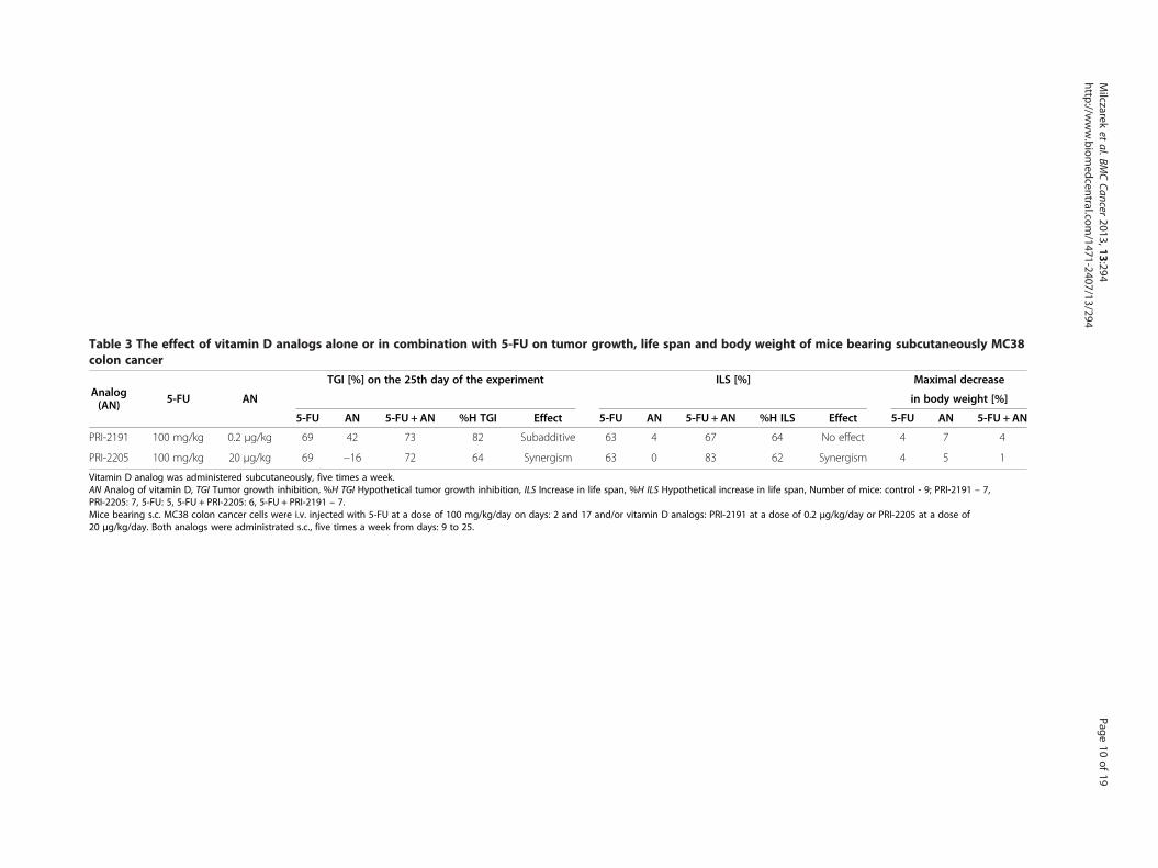
Table 3 The effect of vitamin D analogs alone or in combination with 5-FU on tumor growth, life span and body weight of mice bearing subcutaneously MC38colon cancer
Analog(AN) 5-FU AN
TGI [%] on the 25th day of the experiment ILS [%] Maximal decrease
in body weight [%]
5-FU AN 5-FU + AN %H TGI Effect 5-FU AN 5-FU + AN %H ILS Effect 5-FU AN 5-FU + AN
PRI-2191 100 mg/kg 0.2 μg/kg 69 42 73 82 Subadditive 63 4 67 64 No effect 4 7 4
PRI-2205 100 mg/kg 20 μg/kg 69 −16 72 64 Synergism 63 0 83 62 Synergism 4 5 1
Vitamin D analog was administered subcutaneously, five times a week.AN Analog of vitamin D, TGI Tumor growth inhibition, %H TGI Hypothetical tumor growth inhibition, ILS Increase in life span, %H ILS Hypothetical increase in life span, Number of mice: control - 9; PRI-2191 – 7,PRI-2205: 7, 5-FU: 5, 5-FU + PRI-2205: 6, 5-FU + PRI-2191 – 7.Mice bearing s.c. MC38 colon cancer cells were i.v. injected with 5-FU at a dose of 100 mg/kg/day on days: 2 and 17 and/or vitamin D analogs: PRI-2191 at a dose of 0.2 μg/kg/day or PRI-2205 at a dose of20 μg/kg/day. Both analogs were administrated s.c., five times a week from days: 9 to 25.
Milczarek
etal.BM
CCancer
2013,13:294Page
10of
19http://w
ww.biom
edcentral.com/1471-2407/13/294

Table 4 The effect of vitamin D analog (in the most effective dose, administered orally) alone or in combination with5-FU on tumor growth and body weight of mice bearing subcutaneously MC38 colon cancer
Analog(AN) 5-FU AN
TGI [%] on the 21st day of the experiment Maximal decrease
in body weight [%]
5-FU AN 5-FU + AN %H TGI Effect 5-FU AN 5-FU + AN
PRI-2191 100 mg/kg 1 μg/kg 72 12 81 75 Synergism 2 5 15
PRI-2205 100 mg/kg 10 μg/kg 72 6 73 73 No effect 2 5 5
Vitamin D analog was administered orally (p.o.), three times a week.AN Analog of vitamin D, TGI Tumor growth inhibition; %H TGI, Hypothetical tumor growth inhibition; Number of mice in each group - 8.Mice bearing s.c. MC38 colon cancer cells were i.v. injected with 5-FU at a dose of 100 mg/kg/day from days 9 to 23 and/or vitamin D analogs: PRI-2191 at a doseof 1 μg/kg/day or PRI-2205 at a dose of 10 μg/kg/day. Both analogs were administrated p.o., three times a week from days 12 to 28.
Milczarek et al. BMC Cancer 2013, 13:294 Page 11 of 19http://www.biomedcentral.com/1471-2407/13/294
PRI-2205 did not improve the antitumor effect of 5-FU(Figure 3A).On the other hand, 5-FU applied alone increased the
number of blood leukocytes and platelets in relation tothe control mice. In mice treated with 5-FU andPRI-2191 or PRI-2205, the tendency for a decrease inthe number of blood leukocytes was noted in compari-son with 5-FU alone, but there was no change comparedto control. In terms of the number of platelets, no differ-ence between 5-FU alone and in combined treatmentwas observed (Table 5).
The prolongation of 5-FU antitumor activity by PRI-2191or PRI-2205The application of analogs was started from the 35thday of the experiment after the end of 5-FU administra-tion (i.e. when cytostatic reduced tumor volume by 90%in relation to control untreated mice). As shown inFigure 4, after treatment with 5-FU alone ended a rapidincrease in tumor growth was noted. However, in thecase of combined treatment, we observed that the appli-cation of vitamin D analogs prolonged the antitumor ac-tivity of 5-FU. Both analogs delayed tumor growth aswell as prolonged the survival of mice in comparison
Figure 3 The effect of PRI-2191 or PRI-2205 alone or in combinationtumor weight. B) Body weight of mice during treatment. Mice bearing MC100 mg/kg/day on days 8 and 15 and vitamin D analogs: PRI-2191 at a doswere administrated s.c., three times a week from days 10 to 20. Mice wereweighed and blood was collected. Black arrows indicate the days of 5-FU aNumber of mice: control - 7; PRI-2191 – 4, PRI-2205: 6, 5-FU: 6, 5-FU + PRI-2control; *P < 0.05 – 5-FU + PRI-2191 compared to PRI-2191.
with 5-FU given alone (Figure 4, Table 6). Better resultswere observed in the case of PRI-2191 than PRI-2205.To compare the time needed to reach 1 cm3 tumor ofvolume in the treated groups in relation to the controlgroup, tumor growth delay (TGD) was calculated. Asshown in Table 6, administration of 5-FU alone delayedtumor growth by 23 days compared to untreated mice,in the case of combined treatment by 27 days usingPRI-2205 and 32 days using PRI-2191. Moreover, the105% prolongation of the survival of mice treated withPRI-2191 after 5-FU was noted, while prolongationamounted by 76% with 5-FU alone (Table 6). In addition,no toxicity for 5-FU and combined treatment was ob-served (data not shown).
The antimetastatic effect of combined therapy with 5-FUand PRI-2191 or PRI-2205Next, we investigated the influence of vitamin D analogson regional lymph node metastasis formation in thetreatment of mice bearing MC38/EGFP colon cancercells. The combined treatment using 5-FU and PRI-2191significantly decreased colon cancer metastasis to lymphnodes compared to 5-FU applied alone (Figure 5). Theapplication of 5-FU and PRI-2191 decreased metastasis
with 5-FU on MC38 tumors transplanted ortotopically. A) Intestinal38 tumors implanted i.i. were i.v. injected with 5-FU at a dose ofe of 1 μg/kg/day or PRI-2205 at a dose of 10 μg/kg/day. Both analogssacrificed on the 22nd day after i.i. transplantation, tumors weredministration, gray – days of vitamin D analogs administration.191 – 7; 5-FU + PRI-2205: 6; **P < 0.01 – 5-FU + PRI-2191 compared to

Table 5 The influence of PRI-2191 or PRI-2205 alone or in combination with 5-FU on intestinal tumor growth,leukocytes and platelets in the blood samples from mice with MC38 colon cancer
GroupTumor weight Leukocytes Platelets
NMean ± SD [g] TGI [%] %H TGI Effect Mean ± SD [103/μL] Mean ± SD [103/μL]
Control 1.4 ± 1.06 7.7 ± 3.4 684 ± 309 7
PRI-2191 1.26 ± 0.80 4 7.2 ± 1.4 835 ± 170 4
PRI-2205 0.86 ± 0.93 35 8.7 ± 2.3 887 ± 67 6
5-FU 0.28 ± 0.27 79 15.5 ± 5.7 1186 ± 207**c 6
5-FU + PRI-2191 0.08 ±0.15**a,*b 94 80 Synergism 8.6 ± 3.0 1159 ± 254**c 7
5-FU + PRI-2205 0.32 ± 0.25 75 86 Antagonism 7.6 ± 4.1 1046 ± 320 6
TGI, Tumor growth inhibition; %H TGI, Hypothetical tumor growth inhibition; N, Number of mice in group; SD, Standard deviation.**a P < 0.01 as compared to control; *b P < 0.05 as compared to PRI-2191: Kruskal-Wallis ANOVA (Multiple Comparisons p values (2-tailed)); **c P < 0.01 as comparedto control: Tukey HSD.Mice bearing MC38 tumors implanted i.i. were i.v. injected with 5-FU at a dose of 100 mg/kg/day on days: 8, 15 and/or vitamin D analogs: PRI-2191 at a dose of1 μg/kg/day or PRI-2205 at a dose of 10 μg/kg/day. Both analogs were administrated s.c., three times a week from days 10 to 20. Mice were sacrificed on day 22after i.i. transplantation, tumors were weighed and blood was collected.
Milczarek et al. BMC Cancer 2013, 13:294 Page 12 of 19http://www.biomedcentral.com/1471-2407/13/294
up to 5 times in relation to untreated mice, while for5-FU alone it was up to 2 times (data not shown). More-over, an 80% inhibition of tumor growth was observedon the 49th day of experiment for combined treatmentwith PRI-2191 and 62% for 5-FU alone. The interactionbetween these two agents indicated synergy. No toxicityfor 5-FU either alone or in combined treatment wasobserved (data not shown). However, analog PRI-2205had no effect on the antimetastatic activity of 5-FU(Figure 5).
Figure 4 The anticancer activity of vitamin D analogs injectedafter the completion of 5-FU administration in the treatment ofmice bearing MC38 colon cancer. The kinetics of tumor growth(mean tumor volume). Mice bearing s.c. MC38 colon cancer cellswere i.v. injected with 5-FU at a dose of 100 mg/kg/day from days20 to 34. When 5-FU reduced tumor volume by about 90% incomparison to the control group, the administration of analogs wasinitiated (after administration of the cytostatic drug ended). Bothanalogs were administrated s.c., three times a week from days 35 to56, PRI-2191 at a dose of 1 μg/kg/day and PRI-2205 at a dose of10 μg/kg/day. Black arrows indicate the days of 5-FU administration,gray – days of vitamin D analog administration. Number of mice in
The effect of combined therapy with capecitabine andPRI-2191 or PRI-2205 on MC38 colon tumor growth -implanted subcutaneouslyCapecitabine used at a dose of 450 mg/kg/day, signifi-cantly retarded MC38 tumor growth from day 14 to 35as compared to the control group (P < 0.05). In a com-bined treatment strategy, PRI-2191 and PRI-2205 im-proved the effect of capecitabine (Figure 6A). Bothanalogs applied in combined treatment with capecitabinesignificantly retarded the growth of tumors (P < 0.05 fromdays 10 to 35 – PRI-2191, and from days 10 to 33 –PRI-2205). On day 17 of the experiment a complete re-gression of tumor growth was observed in mice treatedwith capecitabine and this lasted until day 20. Both ana-logs shortened the time for the regression of capecitabinetreated tumors: tumors disappeared on day 12, and oc-curred on day 24 of the experiment (Figure 6B). Maximalbody weight decrease reaching 5% was observed in micetreated with capecitabine alone or combined withPRI-2191 (data not shown).
the control group – 8 and in each treated group – 6. Statisticalanalysis: Kruskal-Wallis ANOVA Multiple Comparisons P values(2-tailed) test. P < 0.05 compared to control group: 5-FU from days26 to 33, 5-FU + PRI-2191 as well as 5-FU + PRI-2205 from days 28 to35. On day 35 the control group was sacrificed because of large sizeof tumors.
Calcemic activity of combined treatmentThe results of the serum calcium level evaluation after s.c.injection of PRI-2191 or PRI-2205 (2 and 10 μg/kg/day),estimated on 16 day after the first injection, are illustrated
in Table 7. Calcium level in serum of mice treated withPRI-2191 alone or combined with 5-FU was significantlyhigher than this in untreated mice. However, administra-tion of 10 μg/kg/day of PRI-2205 did not affect calciumlevel.

Table 6 Tumor growth delay values for each group andsurvival analysis of mice
Group
The time needed for tumors to reach
ILS[%] N
the volume of 1 cm3
TGD[day]
ΔTGDcontrol ΔTGDcytostatic
[day] [day]
Control 24 — — 8
5-FU 47 23 76** 6
5-FU + PRI-2205 50 27 3 97 6
5-FU + PRI-2191 56 32 9 105** 6
N Number of mice in group, TGD Tumor growth delay,ΔTGDcontrol = TGD treated group–TGDcontrol,ΔTGDcytostatic = TGDtreated group–TGDcytostatic, ILS increase in life span, **P < 0.01as compared to control.Mice bearing s.c. MC38 colon cancer cells were i.v. injected with 5-FUat a dose of 100 mg/kg/day from days 20 to 34. When 5-FU reduced tumorvolume by about 90% in comparison to the control group, the administrationof analogs was initiated (after administration of the cytostatic drug ended).Both analogs were administrated s.c., three times a week from days 35 to 56,analog PRI-2191 at a dose of 1 μg/kg/day and PRI-2205 at a doseof 10 μg/kg/day.
Milczarek et al. BMC Cancer 2013, 13:294 Page 13 of 19http://www.biomedcentral.com/1471-2407/13/294
In vitro studies on colon cancer cell linesIn these studies we used two reference compounds,namely calcitriol and calcipotriol (PRI-2201). As shownin Table 6, most sensitive to the antiproliferative activityof vitamin D compounds among the cell lines tested ishuman colon cancer HT-29. Moreover, the potentiation
Figure 5 The metastasis of MC38/EGFP colon cancer cells tolymph nodes in mice treated with 5-FU and PRI-2191or PRI-2205. Mice bearing s.c. MC38/EGFP colon cancer cells were i.p.treated with 5-FU at a dose of 75 mg/kg/day from days 19 to 29 aloneand in combination with vitamin D analog PRI-2191 at a dose of 1 μg/kg/day or PRI-2205 at a dose of 10 μg/kg/day. Both analogs wereadministrated s.c., three times a week from days 19 to 56. The meanfluorescence intensity value of the green fluorescent protein which isdirectly proportional to the number of MC38/EGFP cells in lymphnodes isolated from mice on the 59th day of the experiment wascalculated. The number of mice in each group – 7. Statistical analysiswas performed using the Kruskal-Wallis ANOVA Multiple ComparisonsP values (2-tailed) test. ***P < 0.001 – 5-FU + PRI-2191 compared to5-FU; **P < 0.01 – 5-FU + PRI-2191 compared to 5-FU + PRI-2205.
for 5-FU antiproliferative effect by vitamin D com-pounds was clearly observed on this cell line (Table 8).Other cell lines were less sensitive or almost non-sensitive to proliferation inhibition by vitamin D analogs.However, when MC38/0 cells were incubated with lowconcentrations of 5-FU, proliferation was inhibited by9% and raised to 41% when PRI-2205 was included(Table 8). On the basis of these studies, we chose HT-29and MC38/0 cells for further in vitro studies. All in vitrostudies on the HT-29 cells described below wereconducted in the same concentrations of compounds,such as were used in antiproliferative activity tests.The evaluation of the cell cycle is presented in Figure 7.
As shown in the upper panel of Figure 7, calcitriol,PRI-2201, as well as PRI-2191 tend to increase the per-centage of cells in the G0/G1 stage, whereas PRI-2205did not affect the cells in this phase. This was inverselycorrelated with the percentage of cells in the G2/Mphase. Namely, the three mentioned compounds de-creased the cells in G2/M, but PRI-2205 increased them.Analyzing the cells treated with 5-FU, we observed thatthe cells treated with this agent alone are cumulated inthe S phase and decreased in the G0/G1 and G2/Mphases, when compared to control. Parallel incubationof cells with calcitriol, PRI-2201 and PRI-2191, but notby PRI-2205, increased the percentage of cells in G0/G1
as compared to 5-FU alone. Moreover, the number ofcells in the S and G2/M phases was decreased by thesethree compounds as compared to 5-FU. The action ofPRI-2205 was different; this analog either did not influ-ence or only slightly decreased the cells in G0/G1 andG2/M, but increased them in the S phase as comparedto 5-FU alone. The percentage of death cells (subG1) didnot on average exceed 10 percent (data not shown).Moreover, in this lower panel of Figure 7, representativehistograms are presented for control cells, as well asthose incubated with 5-FU alone or combined withPRI-2191 and PRI-2205.In the series of in vitro studies we also analyzed the
expression of E-cadherin on HT-29 cells. A statisticallysignificant increase in this adhesion molecule was ob-served on cells incubated with 5-FU used in combin-ation with calcitriol, PRI-2201 and PRI-2191 (Figure 7,lower panel).Using higher concentrations of 5-FU, we also analyzed
the subG1 population and the activity of caspase-3/7 inMC38/0 cells incubated with or without vitamin D ana-logs (Figure 8 A and B). A statistically significant in-crease in the percentage of cells in the subG1 stage wasobserved independently of the presence of vitamin Dcompounds, when cells were incubated with 50 or200 μg/ml of 5-FU. However, a tendency to increase thepercentage of cells was observed, when the cells were in-cubated with PRI-2205 and 5-FU (Figure 8B). Under the

Figure 6 MC38 tumor growth in mice treated with capecitabine (CPC) alone and in combination with vitamin D analogs. A) The kineticsof tumor growth. B) The number of mice with tumors in treated groups during experiment. CPC was administered p.o. at a doseof 450 mg/kg/day and/or vitamin D analogs: PRI-2191 at a dose of 1 μg/kg/day and PRI-2205 at a dose of 10 μg/kg/day, both were administrateds.c., three times a week. Black arrows indicate the days of CPC administration and gray – days of vitamin D analog administration. The number ofmice: control - 8; PRI-2191 – 4, PRI-2205: 3, 5-FU: 6, 5-FU + PRI-2191 – 6; 5-FU + PRI-2205: 6. Statistical analysis: Kruskal-Wallis ANOVA MultipleComparisons P values (2-tailed) test. P < 0.05 as compared to the control: CPC from days 14 to 35; PRI-2191 in combined treatment with CPCfrom days 10 to 35; PRI-2205 in combined treatment with CPC from days 10 to 33.
Milczarek et al. BMC Cancer 2013, 13:294 Page 14 of 19http://www.biomedcentral.com/1471-2407/13/294
same experimental conditions, the activity of caspase-3/7 was evaluated. A statistically significant increase inenzyme activity was observed in cells treated with 5-FUat a dose of 200 μg/ml alone or with vitamin D analogs.The activity of caspase-3/7 was slightly diminished byboth analogs as compared to 5-FU alone (Figure 8A).
Table 7 Serum calcium level on day 16 after first administrati
Control 5-FU PRI-219
N 5 5 4
Calcium level [mEq/L] 4.97 ± 0.08 5.02 ± 0.09 5.61 ± 0
*Kruskal-Wallis test for multiple comparisons; P < 0.05 as compared to control.N Number of mice.
DiscussionIn the present study we demonstrate, for the first timein an in vivo model, the biological effect of combinedtherapy against MC38 mouse colon cancer using 5-FUalong with vitamin D analogs (PRI-2191 - tacalcitol andPRI-2205 – 5,6-trans-isomer of calcipotriol).
on of PRI-2191 or PRI-2205 and 5-FU
1 PRI-2205 PRI-2191 + 5-FU PRI-2205 + 5-FU
5 6 4
.2* 5.04 ± 0.16 5.57 ± 0.16* 5.15 ± 0.06

Table 8 The antiproliferative effect of vitamin D analogs with 5-FU on human and mouse colon cancer cells
Vitamin Dcompounds
Colon cancer cell line [%]
HT-29 LoVo MC38/0
+ 5-FU %H + 5-FU %H + 5-FU %H
- 8 ± 7 - 10 ± 5 - 9 ± 7 -
Calcitriol 32 ± 5 49ab ± 6 37 0 25a ± 11 10 5 ± 8 24 ± 2 14
PRI-2191 28 ± 4 46ab ± 5 34 0 14 ± 7 10 0 12 ± 4 9
PRI-2201 29 ± 5 48ab ± 6 35 0 14 ± 7 10 0 16 ± 7 9
PRI-2205 8 ± 3 24ab ± 8 16 9 ± 6 24 ± 6 18 13 ± 10 41ab ± 10 17
[%] – proliferation inhibition.%H = 100-[(100-E for cytostatic) × (100-E for calcitriol analog) /100], where E was the percentage of proliferation inhibition.On HT-29 and LoVo cells 5-FU was used in concentrations of 0.1 μg/ml and on MC38/0 – 0.01 μg/ml. Vitamin analogs were used in concentrations of 100 nM.aP < 0.05 as compared to 5-FU; abP < 0.05 as compared to calcitriol or vitamin D analog.
Milczarek et al. BMC Cancer 2013, 13:294 Page 15 of 19http://www.biomedcentral.com/1471-2407/13/294
We observed that PRI-2191, as well as PRI-2205 ana-log, significantly enhanced the antitumor activity of5-FU, but these results depend on the treatment regi-men. We conclude that the most effective treatmentscheme and dose of both vitamin D analogs is when theyare injected s.c., three times a week and at a dose of1 μg/kg/day for PRI-2191 and 10 μg/kg/day for PRI-2205. Work-flow of in vivo studies on combined treat-ment with vitamin D analogs and 5-FU is summarizedin Figure 9. A similar administration schedule was ap-plied for vitamin D active form in the first phase of clin-ical trials. Patients with advanced malignancy receivedcalcitriol s.c. every other day. It has been shown that theintermittent schedule and s.c. injection allow the
Control 5-FU 5-FU+PRI-2191 5-FU+
FL2-A
Figure 7 Cell cycle distribution and E-cadherin expression on HT-29 cor its analogs alone and combined with 5-FU for 120 h in the case of the cand its analogs were used in concentrations of 100 nM, 5-FU was used at aexpression analysis. * P < 0.05 as compared to control, ** P < 0.05 as compa
application of calcitriol at doses up to 4–5 fold higher(with tolerable toxicity) than is the case with oral appli-cation. However, calcitriol alone did not show significantantitumor activity [10].Applying this optimal schedule of combined therapy,
we observed a significant decrease of tumor growth, andalso a prolongation of survival time of mice in compari-son with 5-FU given alone. Most recently published epi-demiological studies have shown that diagnosis in thesummer and autumn months has been associated withbetter survival. Authors have suggested that exposure tosunlight and the subsequent higher levels of skin vitaminD synthesis at the time of diagnosis or treatment mightbe the basis of the patients’ improved survival [28].
PRI-2205
ells. Human colon cancer cells HT-29 were incubated with calcitriolell cycle assay and 48 h for E-cadherin expression analysis. Calcitriolconcentration of 0.1 μg/ml both for cell cycle and E-cadherinred to 5-FU (Mann–Whitney U test).
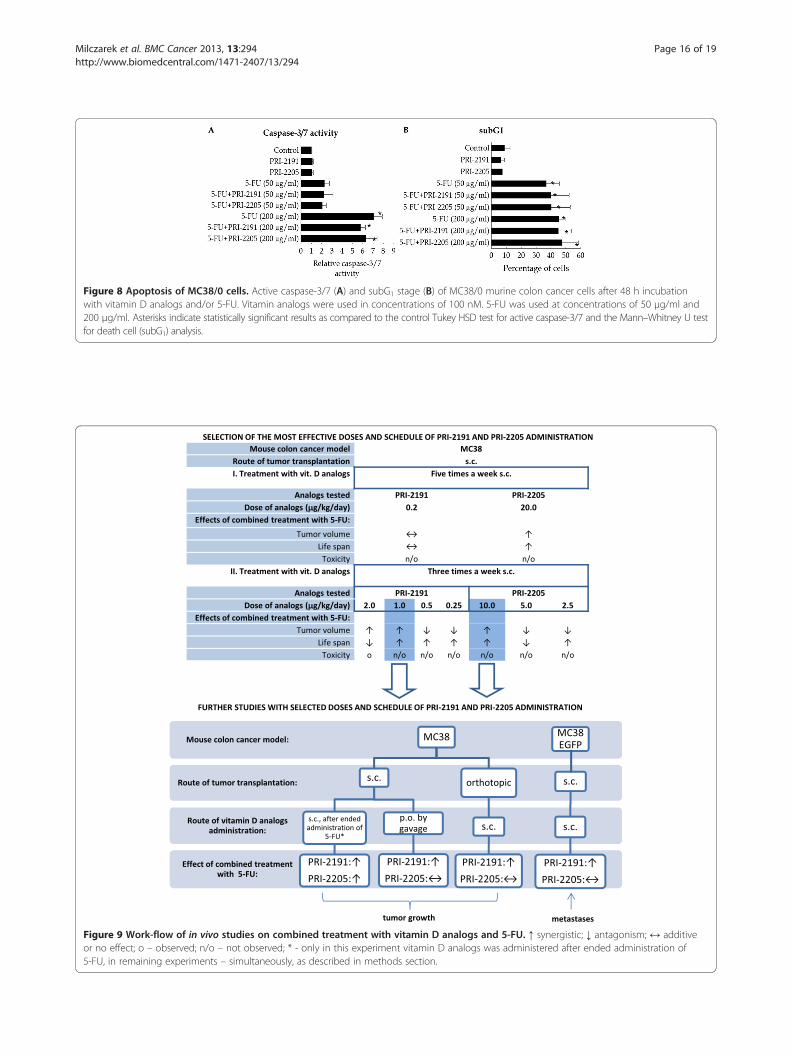
Figure 8 Apoptosis of MC38/0 cells. Active caspase-3/7 (A) and subG1 stage (B) of MC38/0 murine colon cancer cells after 48 h incubationwith vitamin D analogs and/or 5-FU. Vitamin analogs were used in concentrations of 100 nM. 5-FU was used at concentrations of 50 μg/ml and200 μg/ml. Asterisks indicate statistically significant results as compared to the control Tukey HSD test for active caspase-3/7 and the Mann–Whitney U testfor death cell (subG1) analysis.
Figure 9 Work-flow of in vivo studies on combined treatment with vitamin D analogs and 5-FU. ↑ synergistic; ↓ antagonism; ↔ additiveor no effect; o – observed; n/o – not observed; * - only in this experiment vitamin D analogs was administered after ended administration of5-FU, in remaining experiments – simultaneously, as described in methods section.
Milczarek et al. BMC Cancer 2013, 13:294 Page 16 of 19http://www.biomedcentral.com/1471-2407/13/294

Milczarek et al. BMC Cancer 2013, 13:294 Page 17 of 19http://www.biomedcentral.com/1471-2407/13/294
Our combined treatment regimen was also success-fully applied in the orthotopic model of colon cancer;thus, in an environment similar to natural. Moreover,the vitamin D analogs applied following the administra-tion completion of 5-FU could prolong the antitumoractivity of this drug. However, better results were in gen-eral observed for PRI-2191 than PRI-2205. Our in vitrostudies on HT-29 colon cancer cells showed thatPRI-2191 acts similarly to calcitriol or PRI-2201(calcipotriol), whereas the mode of action of PRI-2205 dif-fers. Also, our previous studies showed that PRI-2205appeared to be less potent in the induction of cancer celldifferentiation in vitro as compared to calcitriol orPRI-2191 [12,20]. Moreover, this is in accordance with theresults of ERK1/2 phosphorylation from our previousin vivo studies on HT-29 tumors, where analog PRI-2191used alone increased the level of p-ERK1/2, which is notobserved in tumors from mice treated with PRI-2205 [20].Thymidylate synthase (TS) is an important target for
5-FU. Improved 5-FU activity might be achieved, e.g.,by increasing and prolonging TS inhibition and preven-tion of TS induction [29]. Our in vitro studies on HT-29cells showed that PRI-2191 – the more active analog –used along with 5-FU caused accumulation of cells inthe G0/G1 cell cycle phase with a parallel decreasing ofcells in the S stage. It is possible that this action ofPRI-2191 is responsible, in part, for an increased sensi-tivity of colon cancer to 5-FU, because the expression ofTS is the highest during S phase progression and de-creases when the cells do not proliferate [29]. A differentmechanism was observed in the case of analog PRI-2205applied along with 5-FU. PRI-2205 combined with 5-FUsignificantly increases cell percentage in the S cell cyclephase compared to 5-FU applied alone, which indicatesthat one of the direct mechanisms of combination ther-apy may in part be attributable to the synergistic effectof both compounds. Liu et al. reported that calcitriolsuppressed the expression of TS in colon cancer cellsand promoted a cytotoxic response to 5-FU [24]. Ourfurther studies on MC38/0 cells have shown thatPRI-2205 indicates a tendency to enhancing cell deathinduction by 5-FU but in parallel decreases the activityof caspase-3/7 compared to 5-FU, while 5-FU signifi-cantly induces its activity. A similar effect was de-scribed by Koren et al. after simultaneous incubation ofHT-29 cells with H2O2 and calcitriol. The increasedcytotoxic effect of combined treatment was shown, butthe activity of caspase-3 was decreased by calcitriolcompared to H2O2 [30].The differentiation of colonic epithelial cells may be
regulated by extracellular Ca2+ and the CaSR. Moreover,the induction of E-cadherin may be an important mech-anism underlying the chemopreventive action of Ca2+
and calcitriol in colon cancer [31-33]. This is in
accordance with our studies, in which PRI-2191, similarto calcitriol, promoted the expression of E-cadherin onHT-29 cells in in vitro culture when used along with5-FU. On the other hand, PRI-2205 lacked this activity.We also observed that 5-FU combined with PRI-2191causes a significant decrease in colon cancer metastasisto lymph nodes compared to 5-FU applied alone. Pub-lished data from clinical studies showed that E-cadherinexpression was significantly downregulated in gastro-intestinal tumors with distant metastases [34]. Our ex-periments showed that the antimetastatic potency ofPRI-2191 combined with 5-FU is correlated with an in-crease in E-cadherin expression in vitro. On the otherhand, a different mechanism of antimetastatic activitymay also be considered. Our previous studies showedthat the in vivo inhibition of mouse Lewis lung (LLC)tumor metastasis after treatment with calcitriol and ana-log PRI-2191 was a consequence of diminished integrinαvβ3 expression [35].As we previously showed, both analogs used in in vivo
studies were less toxic than calcitriol. However,PRI-2191 in therapeutic doses induces a rise in serumcalcium levels [19], which is not observed in the case ofPRI-2205 [12], these results are confirmed in the com-bined treatment with 5-FU (Table 7). The slight increasein serum calcium levels by analog PRI-2191 without anyevidence of toxicity may favorably affect cell-cell adhe-sion mediated by E-cadherin which is dependent on ad-equate calcium levels [31,32].A number of in vitro and in vivo studies have shown
that calcitriol and its analogs enhance the antiproliferativeand antitumor efficacy of multiple chemotherapeuticagents in different cancer models [17,36-39]. It has beendemonstrated that calcitriol combined with cisplatincauses a higher tumor growth inhibition than cisplatinalone in the treatment of mice bearing Y-79 human ret-inoblastoma [40], as well as Lewis lung carcinoma tumors[15]. However, in both studies combined therapy inducedsignificant toxicity. Neither combination used in our stud-ies caused toxicity, expressed as a body weight decrease.Thus, similar to oxaliplatin and irinotecan [20], which areagents used as a components of FOLFOX (5-fluorouracil,folinic acid and oxaliplatin) and FOLFOXIRI (5-fluoroura-cil/leucovorin, oxaliplatin, and irinotecan) colon cancertreatment protocols in clinics [41], 5-FU could be safelyused in combined treatment with vitamin D analogs.
ConclusionIn conclusion, both vitamin D analogs PRI-2191 andPRI-2205 enhance the antitumor and antimetastatic ac-tivity of 5-FU and capecitabine in colon cancer in vivo.The combined therapy, in the optimal treatmentschedule, induces a synergistic effect and does notshow toxicity. Moreover, both analogs prolong the

Milczarek et al. BMC Cancer 2013, 13:294 Page 18 of 19http://www.biomedcentral.com/1471-2407/13/294
antitumor effect of 5-FU after the completion of drugadministration. These effects are correlated with a pro-differentiating potential of analog PRI-2191, whereasanalog PRI-2205 acts through a different mechanism,which requires further investigation.Our data suggest that these vitamin D analogs might
be potentially applied to clinical use in order to enhancethe anticancer effect of 5-FU and also prolong its activityin patients suffering from colorectal cancer. However,the attention should be paid on such conclusions, be-cause, the relationship between vitamin D and canceris not easy to interchangeable interpretation. A lot ofstudies have shown a relationship between vitamin Dand cancer, however the direct movement from preclin-ical to clinical conditions is impossible [42]. The dosageof vitamin D analogs appears to be one of the importantreasons of indirect translation of preclinical to clinicalresults. In our studies, lower doses of PRI-2191 act an-tagonistically with 5-FU (summarized in Figure 9).In clinical studies, in general, single agent vitamin D hasnot been directly associated with objective tumor re-sponse in phase I–II clinical trials. It is probably the re-sult of the fact, that very few clinical studies have beenconducted using doses attaining the maximum tolerateddose. There are also questions, if there is known themaximal tolerated dose for patients [43]. Second import-ant clue, is the heterogeneity of patient tumors, whichfrequently lead to misinterpretation of the results. Theanimal studies are conducted on particular histologicaltype of tumor, and some clinical studies showed, thatconsideration of the histological type of tumors duringinterpretation of the results may change the conclusions[44]. As it is concluded in many papers analyzingthe preclinical and clinical studies on vitamin D com-pounds, the further studies should be pointed to preclinicalanalysis of the possible large panel of vitamin D targets andthe clinical studies should be better planned to eliminatethe known defects of those conducted [42,43].
Abbreviations5-FU: 5-Fluorouracil; TGI: Tumor growth inhibition; %H TGI: Hypotheticaltumor growth inhibition; ILS: Increase of life span; %H ILS: Hypotheticalincrease of life span; TGD: Tumor growth delay; i.p: Intraperitoneally;i.v: Intravenously; s.c: Subcutaneously; i.i: Orthotopically; p.o: Orally.
Competing interestsThe authors declare that they have no competing interests.
Authors’ contributionParticipated in research design: JW, MM, AK. Conducted experiments: JW,MM, MP. Contributed new reagents or analytic tools: AK, MM, MP. Performeddata analysis: MM, MP. Wrote or contributed to the writing of themanuscript: JW, MM, AK, MP. All authors read and approved the finalmanuscript.
AcknowledgmentsThis project was supported by the Ministry of Science and Higher Education[Grants No. PBZ-MNiI-1/1/2005, No. N N401 014535].
The authors would like to thank Marcin Poręba and Marcin Drąg (Division ofBioorganic Chemistry, Wroclaw University of Technology) for providing thecaspase substrate and for their help in the caspase activity assay.
Author details1Department of Experimental Oncology, Ludwik Hirszfeld Institute ofImmunology and Experimental Therapy, Polish Academy of Sciences, R. WeiglaSt. 12, Wroclaw 53-114, Poland. 2Division of Medicinal Chemistry andMicrobiology, Wroclaw University of Technology, Wroclaw, Poland.3Pharmaceutical Research Institute, L. Rydygiera St. 8, Warsaw 01-793, Poland.
Received: 18 March 2013 Accepted: 12 June 2013Published: 18 June 2013
References1. Ferlay J, Shin HR, Bray F, Forman D, Mathers C, Parkin DM: Estimates of
worldwide burden of cancer in 2008: GLOBOCAN 2008. Int J Cancer 2010,127:2893–2917.
2. Gorham ED, Garland CF, Garland FC, Grant WB, Mohr SB, Lipkin M, et al:Vitamin D and prevention of colorectal cancer. J Steroid Biochem Mol Biol2005, 97:179–194.
3. Giovannucci E: Strengths and limitations of current epidemiologicstudies: vitamin D as a modifier of colon and prostate cancer risk.Nutr Rev 2007, 65:S77–S79.
4. Holt PR: New insights into calcium, dairy and colon cancer. World JGastroenterol 2008, 14:4429–4433.
5. Tangpricha V, Spina C, Yao M, Chen TC, Wolfe MM, Holick MF: Vitamin Ddeficiency enhances the growth of MC-26 colon cancer xenografts inBalb/c mice. J Nutr 2005, 135:2350–2354.
6. Gonzalez-Sancho JM, Larriba MJ, Ordonez-Moran P, Palmer HG, Munoz A:Effects of 1α,25-dihydroxyvitamin D3 in human colon cancer cells.Anticancer Res 2006, 26:2669–2681.
7. Murillo G, Matusiak D, Benya RV, Mehta RG: Chemopreventive efficacy of25-hydroxyvitamin D3 in colon cancer. J Steroid Biochem Mol Biol 2007,103:763–767.
8. Eelen G, Verlinden L, De CP, Vandewalle M, Bouillon R, Verstuyf A: VitaminD analogs and coactivators. Anticancer Res 2006, 26:2717–2721.
9. Saito T, Okamoto R, Haritunians T, O’Kelly J, Uskokovic M, Maehr H, et al:Novel Gemini vitamin D3 analogs have potent antitumor activity.J Steroid Biochem Mol Biol 2008, 112:151–156.
10. Smith DC, Johnson CS, Freeman CC, Muindi J, Wilson JW, Trump DL: APhase I trial of calcitriol (1,25-dihydroxycholecalciferol) in patients withadvanced malignancy. Clin Cancer Res 1999, 5:1339–1345.
11. Chodynski M, Wietrzyk J, Marcinkowska E, Opolski A, Szelejewski W, KutnerA: Synthesis and antiproliferative activity of side-chain unsaturated andhomologated analogs of 1,25-dihydroxyvitamin D2. (24E)-(1S)-24-Dehydro-24a-homo-1,25-dihydroxyergocalciferol and congeners. Steroids2002, 67:789–798.
12. Wietrzyk J, Chodynski M, Fitak H, Wojdat E, Kutner A, Opolski A: Antitumorproperties of diastereomeric and geometric analogs of vitamin D3.Anticancer Drugs 2007, 18:447–457.
13. Opolski A, Wietrzyk J, Siwinska A, Marcinkowska E, Chrobak A, Radzikowski C,et al: Biological activity in vitro of side-chain modified analogues ofcalcitriol. Curr Pharm Des 2000, 6:755–765.
14. Wietrzyk J, Milczarek M, Kutner A: The effect of combined treatment onhead and neck human cancer cell lines with novel analogs of calcitrioland cytostatics. Oncol Res 2007, 16:517–525.
15. Wietrzyk J, Nevozhay D, Filip B, Milczarek M, Kutner A: The antitumor effectof lowered doses of cytostatics combined with new analogs of vitaminD in mice. Anticancer Res 2007, 27:3387–3398.
16. Wietrzyk J, Nevozhay D, Milczarek M, Filip B, Kutner A: Toxicity andantitumor activity of the vitamin D analogs PRI-1906 and PRI-1907 incombined treatment with cyclophosphamide in a mouse mammarycancer model. Cancer Chemother Pharmacol 2008, 62:787–797.
17. Pelczynska M, Switalska M, Maciejewska M, Jaroszewicz I, Kutner A, OpolskiA: Antiproliferative activity of vitamin D compounds in combination withcytostatics. Anticancer Res 2006, 26:2701–2705.
18. Opolski A, Wietrzyk J, Chrobak A, Marcinkowska E, Wojdat E, Kutner A, et al:Antiproliferative activity in vitro of side-chain analogues of calcitriolagainst various human normal and cancer cell lines. Anticancer Res 1999,19:5217–5222.

Milczarek et al. BMC Cancer 2013, 13:294 Page 19 of 19http://www.biomedcentral.com/1471-2407/13/294
19. Wietrzyk J, Pelczynska M, Madej J, Dzimira S, Kusnierczyk H, Kutner A, et al:Toxicity and antineoplastic effect of (24R)-1,24-dihydroxyvitamin D3
(PRI-2191). Steroids 2004, 69:629–635.20. Milczarek M, Rosinska S, Psurski M, Maciejewska M, Kutner A, Wietrzyk J:
Combined colonic cancer treatment with vitamin D analogs andirinotecan or oxaliplatin. Anticancer Res 2013, 33:433–444.
21. Grem JL: Mechanisms of action and modulation of fluorouracil. SeminRadiat Oncol 1997, 7:249–259.
22. Longley DB, Harkin DP, Johnston PG: 5-fluorouracil: mechanisms of actionand clinical strategies. Nat Rev Cancer 2003, 3:330–338.
23. Wang X, Chen W, Singh N, Promkan M, Liu G: Effects of potential calciumsensing receptor inducers on promoting chemosensitivity of humancolon carcinoma cells. Int J Oncol 2010, 36:1573–1580.
24. Liu G, Hu X, Chakrabarty S: Vitamin D mediates its action in human coloncarcinoma cells in a calcium-sensing receptor-dependent manner:downregulates malignant cell behavior and the expression ofthymidylate synthase and survivin and promotes cellular sensitivity to5-FU. Int J Cancer 2010, 126:631–639.
25. Pajtasz-Piasecka E, Szyda A, Rossowska J, Krawczenko A, Indrova M,Grabarczyk P, et al: Loss of tumorigenicity of murine colon carcinomaMC38/0 cell line after transduction with a retroviral vector carryingmurine IL-12 genes. Folia Biol (Praha) 2004, 50:7–14.
26. Peters GJ, van der Wilt CL, van Moorsel CJ, Kroep JR, Bergman AM, AcklandSP: Basis for effective combination cancer chemotherapy withantimetabolites. Pharmacol Ther 2000, 87:227–253.
27. Ekici OD, Li ZZ, Campbell AJ, James KE, Asgian JL, Mikolajczyk J, et al:Design, synthesis, and evaluation of aza-peptide Michael acceptors asselective and potent inhibitors of caspases-2, -3, -6, -7, -8, -9, and −10.J Med Chem 2006, 49:5728–5749.
28. Turna A, Pekcolaklar A, Metin M, Yaylim I, Gurses A: The effect of season ofoperation on the survival of patients with resected non-small cell lungcancer. Interact Cardiovasc Thorac Surg 2012, 14:151–155.
29. Peters GJ, Backus HH, Freemantle S, van TB, Codacci-Pisanelli G, van der WiltCL, et al: Induction of thymidylate synthase as a 5-fluorouracil resistancemechanism. Biochim Biophys Acta 2002, 1587:194–205.
30. Koren R, Wacksberg S, Weitsman GE, Ravid A: Calcitriol sensitizes coloncancer cells to H2O2-induced cytotoxicity while inhibiting caspaseactivation. J Steroid Biochem Mol Biol 2006, 101:151–160.
31. Bhagavathula N, Hanosh AW, Nerusu KC, Appelman H, Chakrabarty S, VaraniJ: Regulation of E-cadherin and beta-catenin by Ca2+ in colon carcinomais dependent on calcium-sensing receptor expression and function. Int JCancer 2007, 121:1455–1462.
32. Chakrabarty S, Radjendirane V, Appelman H, Varani J: Extracellular calciumand calcium sensing receptor function in human colon carcinomas:promotion of E-cadherin expression and suppression of β-catenin/TCFactivation. Cancer Res 2003, 63:67–71.
33. Chakrabarty S, Wang H, Canaff L, Hendy GN, Appelman H, Varani J: Calciumsensing receptor in human colon carcinoma: interaction with Ca2+ and1,25-dihydroxyvitamin D3. Cancer Res 2005, 65:493–498.
34. Ding J, Zhang Z, Pan Y, Liao G, Zeng L, Chen S: Expression andsignificance of twist, E-cadherin, and N-cadherin in gastrointestinalstromal tumors. Dig Dis Sci 2012, 57:2318–2324.
35. Wietrzyk J, Filip B, Milczarek M, Kłopotowska D, Maciejewska M, DabrowskaK, et al: The influence of 1,25-dihydroxyvitamin D3 and 1,24-dihydroxyvitamin D3 on αvβ3 integrin expression in cancer cell lines.Oncol Rep 2008, 20:941–952.
36. Ma Y, Trump DL, Johnson CS: Vitamin D in combination cancer treatment.J Cancer 2010, 1:101–107.
37. Hershberger PA, Yu WD, Modzelewski RA, Rueger RM, Johnson CS, TrumpDL: Calcitriol (1,25-dihydroxycholecalciferol) enhances paclitaxelantitumor activity in vitro and in vivo and accelerates paclitaxel-inducedapoptosis. Clin Cancer Res 2001, 7:1043–1051.
38. Cho YL, Christensen C, Saunders DE, Lawrence WD, Deppe G, Malviya VK,et al: Combined effects of 1,25-dihydroxyvitamin D3 and platinum drugson the growth of MCF-7 cells. Cancer Res 1991, 51:2848–2853.
39. Siwinska A, Opolski A, Chrobak A, Wietrzyk J, Wojdat E, Kutner A, et al:Potentiation of the antiproliferative effect in vitro of doxorubicin,cisplatin and genistein by new analogues of vitamin D. Anticancer Res2001, 21:1925–1929.
40. Kulkarni AD, van Ginkel PR, Darjatmoko SR, Lindstrom MJ, Albert DM: Use ofcombination therapy with cisplatin and calcitriol in the treatment of
Y-79 human retinoblastoma xenograft model. Br J Ophthalmol 2009,93:1105–1108.
41. Masi G, Loupakis F, Pollina L, Vasile E, Cupini S, Ricci S, et al: Long-termoutcome of initially unresectable metastatic colorectal cancer patientstreated with 5-fluorouracil/leucovorin, oxaliplatin, and irinotecan(FOLFOXIRI) followed by radical surgery of metastases. Ann Surg 2009,249:420–425.
42. Mocellin S: Vitamin D and cancer: deciphering the truth. Biochim BiophysActa 1816, 2011:172–178.
43. Krishnan AV, Trump DL, Johnson CS, Feldman D: The role of vitamin D incancer prevention and treatment. Rheum Dis Clin North Am 2012,38:161–178.
44. Raimondi S, Johansson H, Maisonneuve P, Gandini S: Review and meta-analysis on vitamin D receptor polymorphisms and cancer risk.Carcinogenesis 2009, 30:1170–1180.
doi:10.1186/1471-2407-13-294Cite this article as: Milczarek et al.: Vitamin D analogs enhance theanticancer activity of 5-fluorouracil in an in vivo mouse colon cancermodel. BMC Cancer 2013 13:294.
Submit your next manuscript to BioMed Centraland take full advantage of:
• Convenient online submission
• Thorough peer review
• No space constraints or color figure charges
• Immediate publication on acceptance
• Inclusion in PubMed, CAS, Scopus and Google Scholar
• Research which is freely available for redistribution
Submit your manuscript at www.biomedcentral.com/submit