vitamin d resistance in chronic kidney disease (ckd) d resistance in chronic kidney disease (ckd)...
TRANSCRIPT
Parikh et al. BMC Nephrology 2014, 15:47http://www.biomedcentral.com/1471-2369/15/47
RESEARCH ARTICLE Open Access
Vitamin D resistance in chronic kidney disease(CKD)Amay Parikh1, Herbert S Chase2, Linda Vernocchi1 and Leonard Stern1*
Abstract
Background: Previous studies have shown that treatment with ergocalciferol in patients with CKD stage 3 + 4 isnot effective with less than 33% of patients achieving a 25-OH vitamin D target of >30 ng/ml. The aim of this studywas to test the response to cholecalciferol in CKD. We attempted to replete 25-OH vitamin D to a target level of40–60 ng/ml using the response to treatment and PTH suppression as an outcome measure.
Methods: This retrospective cohort study identified patients (Stages 2–5 and Transplant) from 2001–2010 whoregistered at the Chronic Kidney Disease Clinic. Patients received cholecalciferol 10,000 IU capsules weekly as initialtherapy. When levels above 40 ng/ml were not achieved, doses were titrated up to a maximum of 50,000 IUweekly. Active vitamin D analogs were also used in some Stage 4–5 CKD patients per practice guidelines. Patientsreaching at least one level of 40 ng/mL were designated RESPONDER, and if no level above 40 ng/mL they weredesignated NON-RESPONDER. Patients were followed for at least 6 months and up to 5 years.
Results: 352 patients were included with a mean follow up of 2.4 years. Of the CKD patients, the initial 25-OHvitamin D in the NON-RESPONDER group was lower than the RESPONDER group (18 vs. 23 ng/ml) (p = 0.03).Among all patients, the initial eGFR in the RESPONDER group was significantly higher than the NON-RESPONDERgroup (36 vs. 30 ml/min/1.73 m2) (p < 0.001). Over time, the eGFR of the RESPONDER group stabilized or increased(p < 0.001). Over time, the eGFR in the NON-RESPONDER group decreased toward a trajectory of ESRD. Proteinuriadid not impact the response to 25-OH vitamin D replacement therapy. There were no identifiable variablesassociated with the response or lack of response to cholecalciferol treatment.
Conclusions: CKD patients treated with cholecalciferol experience treatment resistance in raising vitamin D levelsto a pre-selected target level. The mechanism of vitamin D resistance remains unknown and is associated withprogressive loss of eGFR. Proteinuria modifies but does not account for the vitamin D resistance.
Keywords: Cholecalciferol, Vitamin D deficiency, Parathyroid hormone, Chronic kidney disease, Kidney transplant
BackgroundVitamin D [25(OH)D] insufficiency and secondary hyper-parathyroidism is widely prevalent in patients with chronickidney disease [1] including patients who have received arenal transplant [2]. The Kidney Disease Outcomes Qual-ity Initiative (K/DOQI) guidelines recommend measuringPTH and initiating treatment of vitamin D insufficiencystarting with CKD stage 3 [3].Dietary intake of vitamin D may originate from either
plant based sources (ergocalciferol, D2) or animal basedsources (cholecalciferol, D3). The metabolic pathways of
* Correspondence: [email protected], Columbia University, New York, NY, USAFull list of author information is available at the end of the article
© 2014 Parikh et al.; licensee BioMed Central LCommons Attribution License (http://creativecreproduction in any medium, provided the orDedication waiver (http://creativecommons.orunless otherwise stated.
activating vitamin D2 or D3 are essentially the same.Active vitamin D2 and D3 both target the vitamin Dreceptor initiating vitamin D regulated gene trans-cription [4]. Achieving optimal levels of serum 25(OH)D remains a challenge in patients with advanced chronickidney disease. Not only is 25-OH vitamin D purportedto have its own beneficial effects, but metabolism to cal-citriol has been shown to reduce PTH levels [1,5]. TheNational Kidney Foundation (NKF) guidelines state thatoptimal 25(OH) D levels should be greater than 30 ng/mLand should be repleted with oral ergocalciferol [3] while theAmerican Journal of Nutrition guidelines recommend levelsgreater than 40 ng/mL [6,7]. There are no specific re-commendations in the NKF/KDOQI guidelines regarding
td. This is an Open Access article distributed under the terms of the Creativeommons.org/licenses/by/2.0), which permits unrestricted use, distribution, andiginal work is properly credited. The Creative Commons Public Domaing/publicdomain/zero/1.0/) applies to the data made available in this article,
Parikh et al. BMC Nephrology 2014, 15:47 Page 2 of 10http://www.biomedcentral.com/1471-2369/15/47
vitamin D insufficiency for kidney transplant recipients orpatients receiving dialysis. There is not much data availableto suggest exactly how much ergocalciferol should beadministered [8-10] and whether such therapy impactsvitamin D and serum PTH concentrations. Zissman et al.studied the response to ergocalciferol for CKD Stages 3 and4 [11]. They found a partial reduction in PTH for patientswith Stage 3 CKD but not Stage 4 when repleted with ergo-calciferol. Less than 33% of patients achieved a 25-vitaminD (Vit D) target of >30 ng/ml and 50% did not respond totreatment [11]. Al-Aly et al. found similar findings andnoted a similar lack of response to ergocalciferol [5].Cholecalciferol (D3) is presumably more potent than
ergocalciferol (D2) [4,12,13]. The aim of this study wasto test the response to cholecalciferol in CKD. Similar tothat observed for ergocalciferol, suggested dosages forrepletion vary [14,15]. We attempted to replete 25-OHvitamin D levels to a target of 40–60 ng/mL using theresponse to treatment and PTH suppression as outcomemeasures. The working hypothesis for this study wasthat patients with more advanced CKD require signifi-cantly higher dosages to achieve the pre-selected bloodlevel target. Patients with better kidney function wouldrespond more readily to vitamin D supplementationcompared to those with poorer function.
MethodsPatients570 patients were identified from 2001 to 2010 who reg-istered at the Chronic Kidney Disease (CKD) Clinic atColumbia University Medical Center (CUMC). Patientdata measurements were electronically extracted fromthe clinical data warehouse (CDW), the research databaseof the Columbia University Medical Center (CUMC) ofthe New York Presbyterian Hospital (NYPH) system. Dataextracted from this cohort included demographic infor-mation, serum creatinine, 25-OH and 1,25-OH vitamin Dmeasurements, calcium, albumin, parathyroid hormonelevels, proteinuria levels (24 hr. urine protein or protein/creatinine ratio) and other laboratory tests. Diabetes andhypertension status was assigned from ICD-9 coding.
Study designPatients received vitamin D repletion with cholecalcif-erol 10,000 IU capsules weekly. Patients also receiveddietary counseling to reduce daily calcium intake to lessthan 500 mg and daily phosphate intake to less than1000 mg. At each patient follow-up visit, the medicationlist was reviewed and compliance was confirmed. Whenlevels of 25-OH vitamin D above 40 ng/mL were notachieved, doses were titrated upwards over 6 months toa maximum of 50,000 IU weekly. A step-wise protocolwas followed to increase the dosage of cholecalciferol.Follow-up, repeat vitamin D levels, and dose adjustment
as needed was performed at regular intervals concurrentwith clinic visits at 1–4 month intervals. 25-OH vitamin Dlevels were measured by a chemiluminescent immuno-assay (ARUP Laboratories; Salt Lake City, UT). The samelaboratory and assay were used throughout the studyperiod.Responsiveness, as measured by the change in 25-OH
vitamin D level, was the primary outcome measure.PTH suppression was a secondary outcome measure.Safety markers included hypercalcemia and hyperpho-sphatemia. eGFR was calculated for patients using the 4variable modified diet in renal disease (MDRD) equationand extracted creatinine and demographic values [16].
Study populationPatients who maintained 25-OH vitamin D levels ≥40 ng/mL and never fell below this level were designated RE-PLETE. When a patient’s 25-OH vitamin D level fell below40 ng/mL, the patient was followed until a response wasnoted (i.e. 25-OH vitamin D level ≥ 40 ng/mL). A responsewas defined as achieving a level of at least 40 ng/mL in aperiod no less than 6 months. Patients achieving vitaminD levels of ≥40 ng/ml with cholecalciferol were designatedas RESPONDER. If the vitamin D level did not reach 40,the patient was designated NON-RESPONDER. Patientswere excluded who had only one measurement of 25-OHvitamin D during the study period, or who had no follow-up beyond 6 months.
Statistical analysisChi-squared or Fisher’s exact test was used to comparecategorical variables; t-test or Wilcoxon Rank Sum testwas used to compare continuous variables. Significancewas defined at a 95% confidence level for two-tail meas-urement. Longitudinal effects of 25-OH vitamin D wereanalyzed by a linear mixed effect model with randomintercept, which accounted for individual differences inthe initial outcome value. Analysis was performed toadjust for varying follow-up times. A time adjusted vari-able was introduced to reduce observer bias whichdecreased the N. Repeated measures ANOVA was notutilized due to missing values, unequal time differencesbetween data points, and the inability to adjust for anumber of covariates.Statistical analysis was performed using SAS version
9.2 (SAS Institute Inc., Cary, NC). Columbia Univer-sity Medical Center Institutional Review Board, FWA#00002636, approved this protocol (IRB Protocol #AAAD8498) in compliance with the Helsinki Declarationfrom 8/6/09 to present. There was no interaction or inter-vention with any human subjects. This data is not publiclyavailable. The IRB granted a waiver of consent for thisretrospective cohort study.

Parikh et al. BMC Nephrology 2014, 15:47 Page 3 of 10http://www.biomedcentral.com/1471-2369/15/47
ResultsOf the 570 patients identified from 2000 to 2010 who weretreated at the Chronic Kidney Disease Clinic, 127 wereinitially excluded (Figure 1). 8 patients were analyzed whoconsistently maintained 25-OH vitamin D levels > 40 ng/mL. These patients were designated REPLETE. Over the10 year period, the 25-OH vitamin D level fell below40 ng/mL in 221 patients, and was successfully repletedwith cholecalciferol supplementation. These patients weredesignated RESPONDER. Supplementation with cholecal-ciferol failed to achieve a level of 40 ng/ml or greater in169 patients who were designated NON-RESPONDER.Table 1 shows the distribution of patients between 3
groups: CKD, post-transplant (TXP), and renal replace-ment therapy (RRT). Statistically significant differencesbetween groups were observed in age (p < 0.001) and forhypertensive renal disease (p = 0.03).Table 2 shows the distribution of patients by vitamin D
status. Of the 309 CKD patients, 54.7% of CKD patientsare RESPONDER compared to 42.7% NON-RESPONDER(p < 0.001). 81.4% of the 43 TXP patients were RESPON-DER compared to 18.6% NON-RESPONDER. Among allpatients, unadjusted initial values for eGFR, Albumin,Phosphate, and 1, 25-OH vitamin D were significantly dif-ferent between the RESPONDER and NON-RESPONDERgroups. In the patients with CKD stages 3–5, the unadjus-ted initial mean eGFR and 25-OH vitamin D were lowerin the NON-RESPONDER compared to the RESPONDER(p < 0.03 and p < 0.001, respectively). Table 3 shows theinitial values of the subset of patients for whom protein-uria data was available. Significant differences in initial
Figure 1 Study flow diagram.
mean 25-OH vitamin D (p = 0.02) and mean proteinuria(p < 0.004) were noted.Figure 2 depicts the relationship between log eGFR ver-
sus time in the RESPONDER and NON-RESPONDERgroups for patients where the 25-OH vitamin D was below40 ng/mL for CKD patients Stage 3–5 (N = 240) and treat-ment was initiated. In this model adjusted for Age, Sex,Race, Diabetes status and Hypertension status, the initialeGFR is not different between the NON-RESPONDERand RESPONDER groups (p = 0.77). Over the follow-uptime the eGFR for NON-RESPONDER (N = 108) is lowerand declines over time (coefficient −0.007) compared toRESPONDER with a higher eGFR which increases overtime (coefficient 0.004) (p < 0.001). No differences in thedistribution of CKD stages between the two groups werenoted (p = 0.21).Figure 3 depicts the relationship between PTH versus
time in the RESPONDER and NON-RESPONDERgroups for patients where the 25-OH vitamin D wasbelow 40 ng/mL for CKD patients Stage 3–5 (N = 212)and treatment was initiated. There is no difference inPTH over time between the NON-RESPONDER andRESPONDER (p =NS). Similarly, there is no differencebetween 1, 25-OH vitamin D levels versus time and25-OH vitamin D when the level was below 40 for CKDpatients Stage 3–5 and treatment was initiated (figure notshown).Figure 4 illustrates the Log Proteinuria over Time by
Group for CKD patients with eGFR < 60 ml/min/1.73 m2.Proteinuria increases for 11 months in the NON-RE-SPONDER and decreases for 21 months in the

Table 1 Demographics
CKD Transplant RRT P value‡
N 309 43 46
Males 162 (52.4%) 24 (55.8%) 25 (54.4%) 0.90
Age† 66·4 (14.2) 51·6 (11.9) 58.1 (17.5) <0.001
Diabetes, only 1 (0.3%) 0 (0·0%) 0 (0.0%) 1.0
Hypertension, only 123 (39.8%) 25 (58.1%) 15 (32.6%) 0.03
Diabetic and hypertensive 171 (55.3%) 18 (41.9%) 29 (63.0%) 0.12
Non-diabetic and non-hypertensive 14 (4.5%) 0 (0.0%) 2 (4.4%) 0.44†Categorical variables are expressed as count (%); age as mean (SD); all other continuous variables as median (25th percentile-75th percentile).‡Based on chi-squared or Fisher’s exact test for categorical variables; t-test or Wilcoxon Rank Sum test for continuous variables.
Parikh et al. BMC Nephrology 2014, 15:47 Page 4 of 10http://www.biomedcentral.com/1471-2369/15/47
RESPONDER. Figure 5 shows the Log eGFR vs. Log Pro-teinuria by Group for CKD patients with eGFR < 60 ml/min/1.73 m2. For all levels of proteinuria, the eGFR islower in the NON-RESPONDER compared to the RE-SPONDER group.
DiscussionVitamin D insufficiency is highly prevalent in the CKDpopulation. Across the United States prevalence rangesup to 70% [1]. This is consistent with our study in which98% of all patients studied had vitamin D levels less than40 ng/mL. Zisman suggested target levels of vitamin D in-adequately suppress PTH in patients with CKD especially
Table 2 Primary and secondary outcomes
Non-responder Respond
N 169 221
Males 86 (50.9%) 119 (53.9
Age† 62.5 (15·5) 64.9 (14
Diabetes, only 0 (0.0%) 1 (0.45%
Hypertension, only 62 (36.7%) 97 (43.9
Diabetic and hypertensive 102 (60.4%) 113 (51.1
Non-diabetic and non-hypertensive 5 (3.0%) 10 (4.5%
CKD 132 (78.1%) 169 (76.5
Transplant 8 (4.7%) 35 (15.8
RRT 29 (17.2%) 17 (7.7%
eGFR 30.0 (22.0) 36·0 (27
PTH 99.0 (137.0) 86·5(102
Albumin 3.9 (0.6) 4·3 (0.4
Initial mean phosphate 3.8 (1.1) 3·5 (0.9
Initial mean 1,25-OH vitamin D 20.5 (18.0) 30.0 (19
For CKD stage
Initial mean eGFR 28·3 (11.5) 31·5 (11
Initial mean 25-OH vitamin D 18·3 (9.0) 23·6 (9.†Categorical variables are expressed as count (%); age as mean (SD); all other contin‡Based on chi-squared or Fisher’s exact test for categorical variables; t-test or Wilcox†At time of 25-vitamin D grouping.‡Prior to 25-vitamin D grouping.The boldface items indicate values less than 0.05.
those with more advanced impairment in kidney function[11]. In the absence of published, definitive evidence basedguidelines, our clinical practice guideline was to targetvitamin D to levels of 40–60 ng/mL. This target waschosen based on a review of consensus opinions in thenephrology and endocrine communities [17,18]. We notedthat this was exceedingly difficult to achieve in our po-pulation, which included both CKD and post-transplantpatients.One aim of our study was to demonstrate that chole-
calciferol is a more potent vitamin D precursor therapyin CKD patients compared to ergocalciferol, which wasused in previous studies [11]. What we uncovered is a
er Replete P Value‡ (responder vs. non-responders)
8
%) 6 (75.0%) 0.56
·9) 64.5 (18·0) 0.12
) 0 (0.0%) 1.0
%) 4 (50.0%) 0.15
%) 3 (37.5%) 0.07
) 1 (12.5%) 0.43
%) 8 (100.0%)
<0.001%) 0 (0.0%)
) 0 (0.0%)
.0) <0.001
.0) 0.08
) <0.001
) <0.001
.0) <0.001
s 3–5 only
.2) 0.0253
1) <0.001
uous variables as median (25th percentile-75th percentile).on Rank Sum test for continuous variables.

Table 3 Outcomes by group
Non-responder Responder P value
*Initial Mean 25VitD 19 ng/mL 23 ng/mL 0.02
*Initial Mean Proteinuria 1.47 g/day 0.89 g/day <0.004
*Initial Mean eGFR (ml/min/1·73 m2) 27 31 0.09
*Initial PTH 110 pg/mL 100 pg/mL 0.38
*Not adjusted for follow-up time. Closely concurrent lab values were measured with initial 25-OH Vitamin D.The boldface items indicate values less than 0.05.
Parikh et al. BMC Nephrology 2014, 15:47 Page 5 of 10http://www.biomedcentral.com/1471-2369/15/47
new concept of vitamin D treatment resistance in CKDwhich we suggest is associated with a progressive declinein renal function over time. Our study has demonstratedthat CKD patients treated with cholecalciferol, similar toergocalciferol, experience treatment resistance in raisingvitamin D levels to a pre-selected target level. Althoughmultiple studies have suggested that cholecalciferol ismore potent [19,20], we could not find evidence that theresponse to treatment with cholecalciferol is any betterthan with ergocalciferol. We did however, find two dis-tinct groups in our population: 1) those who respondedto treatment (RESPONDER) and 2) those who did not(NON-RESPONDER). Among all the patients, initiallaboratory values which were unadjusted for follow-upfor the NON-RESPONDER group were significant for a
Figure 2 Log eGFR over time by group for CKD Stages 3–5. Log eGFRN = 108 Non-responders and 132 Responders. No differences in the distribu
lower initial eGFR, higher PTH, lower albumin, higherphosphate, and lower 1, 25-OH vitamin D.Similar to other studies with ergocalciferol, our popu-
lation received a vigorous supplementation protocol withcholecalciferol. This regimen resulted in a significant in-crease in vitamin D levels only for a specific population:RESPONDER. This group has unique characteristicscompared to those whose levels did not increase to thepre-selected target level of greater than 40 ng/mL. In theCKD Stage 3–5 group, unadjusted initial mean eGFR and25-OH vitamin D levels were higher in the RESPONDERgroup. When adjusted for time for CKD patients with aneGFR < 60 ml/min/1.73 m2 the initial eGFR in RESPON-DER and NON-RESPONDER groups is indistinguishable(Figure 2). The data suggests that the eGFR for the
over Time by Group for CKD patients with eGFR < 60 ml/min/1.73 m2.tion of CKD stages between the two groups were noted (p = 0.21).
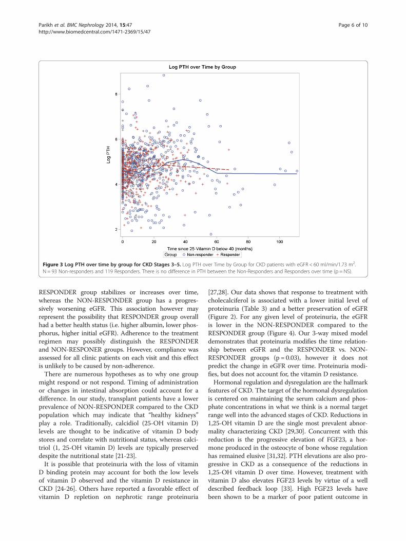
Figure 3 Log PTH over time by group for CKD Stages 3–5. Log PTH over Time by Group for CKD patients with eGFR < 60 ml/min/1.73 m2.N = 93 Non-responders and 119 Responders. There is no difference in PTH between the Non-Responders and Responders over time (p = NS).
Parikh et al. BMC Nephrology 2014, 15:47 Page 6 of 10http://www.biomedcentral.com/1471-2369/15/47
RESPONDER group stabilizes or increases over time,whereas the NON-RESPONDER group has a progres-sively worsening eGFR. This association however mayrepresent the possibility that RESPONDER group overallhad a better health status (i.e. higher albumin, lower phos-phorus, higher initial eGFR). Adherence to the treatmentregimen may possibly distinguish the RESPONDERand NON-RESPONER groups. However, compliance wasassessed for all clinic patients on each visit and this effectis unlikely to be caused by non-adherence.There are numerous hypotheses as to why one group
might respond or not respond. Timing of administrationor changes in intestinal absorption could account for adifference. In our study, transplant patients have a lowerprevalence of NON-RESPONDER compared to the CKDpopulation which may indicate that “healthy kidneys”play a role. Traditionally, calcidiol (25-OH vitamin D)levels are thought to be indicative of vitamin D bodystores and correlate with nutritional status, whereas calci-triol (1, 25-OH vitamin D) levels are typically preserveddespite the nutritional state [21-23].It is possible that proteinuria with the loss of vitamin
D binding protein may account for both the low levelsof vitamin D observed and the vitamin D resistance inCKD [24-26]. Others have reported a favorable effect ofvitamin D repletion on nephrotic range proteinuria
[27,28]. Our data shows that response to treatment withcholecalciferol is associated with a lower initial level ofproteinuria (Table 3) and a better preservation of eGFR(Figure 2). For any given level of proteinuria, the eGFRis lower in the NON-RESPONDER compared to theRESPONDER group (Figure 4). Our 3-way mixed modeldemonstrates that proteinuria modifies the time relation-ship between eGFR and the RESPONDER vs. NON-RESPONDER groups (p = 0.03), however it does notpredict the change in eGFR over time. Proteinuria modi-fies, but does not account for, the vitamin D resistance.Hormonal regulation and dysregulation are the hallmark
features of CKD. The target of the hormonal dysregulationis centered on maintaining the serum calcium and phos-phate concentrations in what we think is a normal targetrange well into the advanced stages of CKD. Reductions in1,25-OH vitamin D are the single most prevalent abnor-mality characterizing CKD [29,30]. Concurrent with thisreduction is the progressive elevation of FGF23, a hor-mone produced in the osteocyte of bone whose regulationhas remained elusive [31,32]. PTH elevations are also pro-gressive in CKD as a consequence of the reductions in1,25-OH vitamin D over time. However, treatment withvitamin D also elevates FGF23 levels by virtue of a welldescribed feedback loop [33]. High FGF23 levels havebeen shown to be a marker of poor patient outcome in

Figure 4 Log Proteinuria over time by group for CKD patients with eGFR < 60 ml/min/1·73 m2. Log Proteinuria over Time by Group forCKD patients with eGFR < 60 ml/min/1.73 m2. N = 69 Non-responders and 76 Responders. (p < 0.05). Proteinuria increases for 11 months inNon-responders. Proteinuria decreases for 21 months in Responders.
Parikh et al. BMC Nephrology 2014, 15:47 Page 7 of 10http://www.biomedcentral.com/1471-2369/15/47
both CKD and ESRD [34-36]. However, a recent animalstudy in a CKD model showed that targeted reduction inFGF23 resulted in increased mortality further complicat-ing our understanding of this complex physiology [37]. Inlarge data base studies of patients with CKD and ESRD,treatment with vitamin D has been shown to promote asurvival advantage [38-42]. Calcitriol in high dose hasbeen shown to promote vascular calcification in animalmodels of CKD [43-45] and a recent meta-analysis sug-gests that low-dose paricalcitol may do the same [46].Thus the clinician in practice is faced with the clinicalconundrum and safety concern of how much vitamin D toadminister to patients with vitamin D insufficiency and toaim for what vitamin D target level in order to achieve thebest clinical outcome. In our current understanding of thiscomplex system, treatment with 25 vitamin D precursorsand active vitamin D analogues is initiated to target PTHprimarily to limit the progressive bone injury that occurswith elevated PTH levels. The PTH target is also a matterof much debate in the nephrology community [47].The understanding of the interaction between active
and inactive forms of vitamin D and PTH has improvedimmensely with the identification of FGF-23 [48], Klotho[49,50], and CYP27B1 [51]. Some have suggested thathigh doses of vitamin D supplementation similar to what
was administered in our study induce the catabolic cyto-chrome P-450 enzyme CYP24A1 [52]. This may explainwhy high dose vitamin D supplementation and previousattempts of vitamin D repletion at lower dosage havefailed [53]. Another potential mechanism which en-hances vitamin D catabolism relates to the progressiveelevation of the phosphaturic FGF23 noted in CKD.Both the administration of vitamin D and an elevatedFGF23 in CKD have the potential to hyper-catabolizevitamin D in all forms. Therefore, an elevated FGF-23may limit the response to a preselected vitamin D targetlevel (i.e. vitamin D resistance) through this hypercatab-olism mechanism.Limitations of the study include the retrospective nature
of the study design. Despite our best efforts to ascertainmedication compliance, some patients may still have nottaken their cholecalciferol. Seasonal differences were notinvestigated due to the long follow-up period of eachpatient. Cumulative doses of cholecalciferol given werenot available. Weight, changes in weight, or the role ofobesity in both groups was not ascertained. Given the lownumbers of patients receiving home hemodialysis (3), out-patient center hemodialysis (23), and peritoneal dialysis(20) in our study, they were grouped together as renalreplacement therapy and excluded from the study analysis.

Figure 5 Log eGFR vs. log proteinuria by group for CKD patients with eGFR < 60. Log eGFR vs. Log Proteinuria by Group for CKD patientswith eGFR < 60 ml/min/1.73 m2. N = 47 Non-responders and N = 45 Responders. For all levels of proteinuria, eGFR is lower in the Non-responders.
Parikh et al. BMC Nephrology 2014, 15:47 Page 8 of 10http://www.biomedcentral.com/1471-2369/15/47
Conclusions could not adequately be drawn regardingthese groups however the benefits of supplementationhave been previously suggested in the PD population [6].Information regarding malabsorption syndromes (suchas by gastric bypass) of the patients in the study areunknown.Multiple non-renal associations have been described
between vitamin D: cardiovascular disease [54], immunefunction [55], asthma [56], cancer [57], and autoimmunediseases [58]. Low 25-OH vitamin D has also been sug-gested as a marker of mortality in the critical care setting[59]. Whether vitamin D becomes a marker of nutritionor of something more, monitoring of blood levels andtargeted replacement therapy in the CKD population maygive a hint of future CKD status. Our study is unable toidentify suitable markers that could advance the state ofknowledge and distinguish which patients will be in afuture RESPONDER group and which will not.
ConclusionThe clinical implications of a replete 25-OH vitamin Dlevel remain unknown. Responders appear to stabilize orincrease their eGFR over time, however the Non-Re-sponders display a deterioration of eGFR over time. Pro-teinuria modifies the response to cholecalciferol initiallybut does not predict the change in eGFR over time. Themechanism of the treatment resistance (RESPONDER vs.
NON-RESPONDER) is also unknown and could be causedby a change in vitamin D metabolism (decreased synthesisor increased catabolism). Both of these effects could bemediated by the progressive increase of FGF-23 levelsobserved in CKD. Further research is necessary to test thishypothesis and to identify predictive variables that couldbe used for prognostic guidance.
AbbreviationsCKD: Chronic kidney disease; NKF: National kidney foundation; KDOQI: Kidneydisease outcomes quality initiative; PTH: Parathyroid hormone; ICD-9: International classification of diseases ninth revision; eGFR: Estimatedglomerular filtration rate; MDRD: Modified diet in renal disease;TXP: Transplant; RRT: Renal replacement therapy; FGF23: Fibroblast growthfactor 23.
Competing interestsThe authors declare that they have no competing interests.
Authors’ contributionsAP performed the literature search, study design, data collection, dataanalysis, data interpretation, and writing. HC performed data collection, dataanalysis, data interpretation, writing. LV participated in data collection,data analysis, and writing. LS performed literature search, study design,data analysis and interpretation, and writing. All authors had access to the dataand a role in writing the manuscript. All authors read and approved thefinal manuscript.
AcknowledgementsStatistical analysis was performed by Emilia Bagiella, PhD (ColumbiaUniversity) and Helena Chang (Columbia University).Results were presented at American Society of Nephrology (ASN) RenalWeek 2010 in Denver, CO and ASN Renal Week 2011 in Philadelphia, PA.

Parikh et al. BMC Nephrology 2014, 15:47 Page 9 of 10http://www.biomedcentral.com/1471-2369/15/47
This study was supported by a generous donation from the NortilloFoundation. The funding source has had no role in the writing or any otheraspect of the manuscript. No payment has been received to write this article.
Author details1Nephrology, Columbia University, New York, NY, USA. 2BiomedicalInformatics, Columbia University, New York, NY, USA.
Received: 28 October 2013 Accepted: 12 March 2014Published: 19 March 2014
References1. LaClair RE, Hellman RN, Karp SL, Kraus M, Ofner S, Li Q, Graves KL, Moe SM:
Prevalence of calcidiol deficiency in CKD: a cross-sectional study acrosslatitudes in the United States. Am J Kidney Dis 2005, 45(6):1026–1033.
2. Stavroulopoulos A, Cassidy MJ, Porter CJ, Hosking DJ, Roe SD: Vitamin Dstatus in renal transplant recipients. Am J Transplant 2007,7(11):2546–2552.
3. K/DOQI clinical practice guidelines for bone metabolism and disease inchronic kidney disease. Am J Kidney Dis 2003, 42(4 Suppl 3):S1–S201.
4. Bodyak N, Ayus JC, Achinger S, Shivalingappa V, Ke Q, Chen YS, Rigor DL,Stillman I, Tamez H, Kroeger PE, Wu-Wong RR, Karumanchi SA, Thadhani R,Kang PM: Activated vitamin D attenuates left ventricular abnormalitiesinduced by dietary sodium in Dahl salt-sensitive animals. Proc Natl AcadSci U S A 2007, 104(43):16810–16815.
5. Al-Aly Z, Qazi RA, Gonzalez EA, Zeringue A, Martin KJ: Changes in serum25-hydroxyvitamin D and plasma intact PTH levels following treatmentwith ergocalciferol in patients with CKD. Am J Kidney Dis 2007,50(1):59–68.
6. Heaney RP, Holick MF: Why the IOM recommendations for vitamin D aredeficient. J Bone Miner Res 2011, 26(3):455–457.
7. Holick MF: The vitamin D epidemic and its health consequences.J Nutr 2005, 135(11):2739S–2748S.
8. Shah BR, Finberg L: Single-day therapy for nutritional vitamin D-deficiency rickets: a preferred method. J Pediatr 1994, 125(3):487–490.
9. Adams JS, Kantorovich V, Wu C, Javanbakht M, Hollis BW: Resolution ofvitamin D insufficiency in osteopenic patients results in rapid recoveryof bone mineral density. J Clin Endocrinol Metab 1999, 84(8):2729–2730.
10. Malabanan A, Veronikis IE, Holick MF: Redefining vitamin D insufficiency.Lancet 1998, 351(9105):805–806.
11. Zisman AL, Hristova M, Ho LT, Sprague SM: Impact of ergocalciferoltreatment of vitamin D deficiency on serum parathyroid hormoneconcentrations in chronic kidney disease. Am J Nephrol 2007, 27(1):36–43.
12. Trang HM, Cole DE, Rubin LA, Pierratos A, Siu S, Vieth R: Evidence thatvitamin D3 increases serum 25-hydroxyvitamin D more efficiently thandoes vitamin D2. Am J Clin Nutr 1998, 68(4):854–858.
13. Houghton LA, Vieth R: The case against ergocalciferol (vitamin D2) as avitamin supplement. Am J Clin Nutr 2006, 84(4):694–697.
14. Heaney RP, Davies KM, Chen TC, Holick MF, Barger-Lux MJ: Human serum25-hydroxycholecalciferol response to extended oral dosing withcholecalciferol. Am J Clin Nutr 2003, 77(1):204–210.
15. Vieth R, Chan PC, MacFarlane GD: Efficacy and safety of vitamin D3 intakeexceeding the lowest observed adverse effect level. Am J Clin Nutr 2001,73(2):288–294.
16. Levey AS, Stevens LA, Schmid CH, Zhang YL, Castro AF, 3rd, Feldman HI,Kusek JW, Eggers P, Van Lente F, Greene T, Coresh J, Ckd EPI: A newequation to estimate glomerular filtration rate. Ann Intern Med 2009,150(9):604–612.
17. Holick MF, Binkley NC, Bischoff-Ferrari HA, Gordon CM, Hanley DA, HeaneyRP, Murad MH, Weaver CM: Evaluation, treatment, and prevention ofvitamin D deficiency: an Endocrine Society clinical practice guideline.J Clin Endocrinol Metab 2011, 96(7):1911–1930.
18. Tuohimaa P, Tenkanen L, Ahonen M, Lumme S, Jellum E, Hallmans G, StattinP, Harvei S, Hakulinen T, Luostarinen T, Dillner J, Lehtinen M, Hakama M:Both high and low levels of blood vitamin D are associated with ahigher prostate cancer risk: a longitudinal, nested case–control study inthe Nordic countries. Int J Cancer 2004, 108(1):104–108.
19. Heaney RP, Recker RR, Grote J, Horst RL, Armas LA: Vitamin D(3) is morepotent than vitamin D(2) in humans. J Clin Endocrinol Metab 2011,96(3):E447–E452.
20. Armas LA, Hollis BW, Heaney RP: Vitamin D2 is much less effective thanvitamin D3 in humans. J Clin Endocrinol Metab 2004, 89(11):5387–5391.
21. Felsenfeld AJ, Rodriguez M: Phosphorus, regulation of plasma calcium,and secondary hyperparathyroidism: a hypothesis to integrate ahistorical and modern perspective. J Am Soc Nephrol 1999,10(4):878–890.
22. Schmidt-Gayk H, Bouillon R, Roth HJ: Measurement of vitamin D and itsmetabolites (calcidiol and calcitriol) and their clinical significance. ScandJ Clin Lab Invest Suppl 1997, 227:35–45.
23. Slatopolsky E, Brown A, Dusso A: Pathogenesis of secondaryhyperparathyroidism. Kidney Int Suppl 1999, 73:S14–S19.
24. Robinson AB, Thierry-Palmer M, Gibson KL, Rabinovich CE: Disease activity,proteinuria, and vitamin D status in children with systemic lupuserythematosus and juvenile dermatomyositis. J Pediatr 2012,160(2):297–302.
25. Fowlkes JL, Bunn RC, Cockrell GE, Clark LM, Wahl EC, Lumpkin CK, ThrailkillKM: Dysregulation of the intrarenal vitamin D endocytic pathway in anephropathy-prone mouse model of type 1 diabetes. Exp Diabetes Res2011, 2011:269378.
26. Thrailkill KM, Jo CH, Cockrell GE, Moreau CS, Fowlkes JL: Enhancedexcretion of vitamin D binding protein in type 1 diabetes: a role invitamin D deficiency? J Clin Endocrinol Metab 2011, 96(1):142–149.
27. Kuhlmann A, Haas CS, Gross ML, Reulbach U, Holzinger M, Schwarz U,Ritz E, Amann K: 1,25-Dihydroxyvitamin D3 decreases podocyte loss andpodocyte hypertrophy in the subtotally nephrectomized rat. Am J PhysiolRenal Physiol 2004, 286(3):F526–F533.
28. Agarwal R, Acharya M, Tian J, Hippensteel RL, Melnick JZ, Qiu P, Williams L,Batlle D: Antiproteinuric effect of oral paricalcitol in chronic kidneydisease. Kidney Int 2005, 68(6):2823–2828.
29. Pavik I, Jaeger P, Ebner L, Wagner CA, Petzold K, Spichtig D, Poster D,Wuthrich RP, Russmann S, Serra AL: Secreted Klotho and FGF23 inchronic kidney disease Stage 1 to 5: a sequence suggested from across-sectional study. Nephrol Dial Transplant 2013, 28(2):352–359.
30. Russo D, Battaglia Y: Clinical significance of FGF-23 in patients with CKD.Int J Nephrol 2011, 2011:364890.
31. Silver J, Naveh-Many T: FGF-23 and secondary hyperparathyroidism inchronic kidney disease. Nat Rev Nephrol 2013, 9(11):641–649.
32. Kovesdy CP, Quarles LD: Fibroblast growth factor-23: what we know, whatwe don't know, and what we need to know. Nephrol Dial Transplant 2013,28(9):2228–2236.
33. Liu S, Tang W, Fang J, Ren J, Li H, Xiao Z, Quarles LD: Novel regulators ofFgf23 expression and mineralization in Hyp bone. Mol Endocrinol 2009,23(9):1505–1518.
34. Negri AL: Fibroblast growth factor 23: associations with cardiovasculardisease and mortality in chronic kidney disease. Int Urol Nephrol 2014,46(1):9–17.
35. Kendrick J, Cheung AK, Kaufman JS, Greene T, Roberts WL, Smits G,Chonchol M: FGF-23 associates with death, cardiovascular events,and initiation of chronic dialysis. J Am Soc Nephrol 2011,22(10):1913–1922.
36. Jean G, Terrat JC, Vanel T, Hurot JM, Lorriaux C, Mayor B, Chazot C: Highlevels of serum fibroblast growth factor (FGF)-23 are associated withincreased mortality in long haemodialysis patients. Nephrol DialTransplant 2009, 24(9):2792–2796.
37. Shalhoub V, Shatzen EM, Ward SC, Davis J, Stevens J, Bi V, Renshaw L,Hawkins N, Wang W, Chen C, Tsai MM, Cattley RC, Wronski TJ, Xia X, Li X,Henley C, Eschenberg M, Richards WG: FGF23 neutralization improveschronic kidney disease-associated hyperparathyroidism yet increasesmortality. J Clin Investig 2012, 122(7):2543–2553.
38. Shoben AB, Rudser KD, de Boer IH, Young B, Kestenbaum B: Association oforal calcitriol with improved survival in nondialyzed CKD. J Am SocNephrol 2008, 19(8):1613–1619.
39. Kovesdy CP, Ahmadzadeh S, Anderson JE, Kalantar-Zadeh K: Association ofactivated vitamin D treatment and mortality in chronic kidney disease.Arch Intern Med 2008, 168(4):397–403.
40. Teng M, Wolf M, Ofsthun MN, Lazarus JM, Hernan MA, Camargo CA Jr,Thadhani R: Activated injectable vitamin D and hemodialysis survival: ahistorical cohort study. J Am Soc Nephrol 2005, 16(4):1115–1125.
41. Teng M, Wolf M, Lowrie E, Ofsthun N, Lazarus JM, Thadhani R: Survival ofpatients undergoing hemodialysis with paricalcitol or calcitriol therapy.N Engl J Med 2003, 349(5):446–456.

Parikh et al. BMC Nephrology 2014, 15:47 Page 10 of 10http://www.biomedcentral.com/1471-2369/15/47
42. Kalantar-Zadeh K, Kuwae N, Regidor DL, Kovesdy CP, Kilpatrick RD,Shinaberger CS, McAllister CJ, Budoff MJ, Salusky IB, Kopple JD: Survivalpredictability of time-varying indicators of bone disease in maintenancehemodialysis patients. Kidney Int 2006, 70(4):771–780.
43. Niederhoffer N, Bobryshev YV, Lartaud-Idjouadiene I, Giummelly P, Atkinson J:Aortic calcification produced by vitamin D3 plus nicotine. J Vasc Res 1997,34(5):386–398.
44. Niederhoffer N, Lartaud-Idjouadiene I, Giummelly P, Duvivier C, Peslin R,Atkinson J: Calcification of medial elastic fibers and aortic elasticity.Hypertension 1997, 29(4):999–1006.
45. Lopez I, Aguilera-Tejero E, Mendoza FJ, Almaden Y, Perez J, Martin D,Rodriguez M: Calcimimetic R-568 decreases extraosseous calcificationsin uremic rats treated with calcitriol. J Am Soc Nephrol 2006,17(3):795–804.
46. Han T, Rong G, Quan D, Shu Y, Liang Z, She N, Liu M, Yang B, Cheng G,Lv Y, Stern L: Meta-analysis: the efficacy and safety of paricalcitol for thetreatment of secondary hyperparathyroidism and proteinuria in chronickidney disease. BioMed Res Int 2013, 2013:11.
47. Pontoriero G, Cozzolino M, Locatelli F, Brancaccio D: CKD patients:the dilemma of serum PTH levels. Nephron Clin Pract 2010,116(4):c263–c268.
48. Kocelak P, Olszanecka-Glinianowicz M, Chudek J: Fibroblast growth factor23–structure, function and role in kidney diseases. Adv Clin Exp Med 2012,21(3):391–401.
49. Komaba H, Fukagawa M: Vitamin D and secreted Klotho: a long-awaitedpanacea for vascular calcification? Kidney Int 2012, 82(12):1248–1250.
50. Komaba H, Fukagawa M: The role of FGF23 in CKD–with or withoutKlotho. Nat Rev Nephrol 2012, 8(8):484–490.
51. Bailey R, Cooper JD, Zeitels L, Smyth DJ, Yang JH, Walker NM, Hypponen E,Dunger DB, Ramos-Lopez E, Badenhoop K, Nejentsev S, Todd JA:Association of the vitamin D metabolism gene CYP27B1 with type 1diabetes. Diabetes 2007, 56(10):2616–2621.
52. Masuda S, Byford V, Arabian A, Sakai Y, Demay MB, St-Arnaud R, Jones G:Altered pharmacokinetics of 1alpha,25-dihydroxyvitamin D3 and25-hydroxyvitamin D3 in the blood and tissues of the 25-hydroxyvitaminD-24-hydroxylase (Cyp24a1) null mouse. Endocrinology 2005,146(2):825–834.
53. Vande Griend JP, McQueen RB, Linnebur SA, Vondracek SF: Prescriptionergocalciferol dosing for vitamin D repletion: a retrospective evaluation.Pharmacotherapy 2012, 32(2):135–141.
54. Gonzalez-Parra E, Rojas-Rivera J, Tunon J, Praga M, Ortiz A, Egido J: VitaminD receptor activation and cardiovascular disease. Nephrol Dial Transplant2012, 27(Suppl 4):iv17–iv21.
55. Arnson Y, Itzhaky D, Mosseri M, Barak V, Tzur B, Agmon-Levin N, Amital H:Vitamin D inflammatory cytokines and coronary events: a comprehensivereview. Clin Rev Allergy Immunol 2013, 45(2):236–247.
56. Mai XM, Langhammer A, Camargo CA Jr, Chen Y: Serum 25-hydroxyvitamin D Levels and incident asthma in adults: the HUNT study.Am J Epidemiol 2012, 176(12):1169–1176.
57. Garland CF, Gorham ED, Mohr SB, Garland FC: Vitamin D for cancerprevention: global perspective. Ann Epidemiol 2009, 19(7):468–483.
58. Agmon-Levin N, Theodor E, Segal RM, Shoenfeld Y: Vitamin D in systemicand organ-specific autoimmune diseases. Clin Rev Allergy Immunol 2013,45(2):256–266.
59. Shelley BG, Quasim T, Kinsella J, Talwar D, McMillan DC: Low-serum25-hydroxyvitamin D reflects severity of illness in critically ill patients.Crit Care Med 2012, 40(8):2530. author reply 2530–2532.
doi:10.1186/1471-2369-15-47Cite this article as: Parikh et al.: Vitamin D resistance in chronic kidneydisease (CKD). BMC Nephrology 2014 15:47.
Submit your next manuscript to BioMed Centraland take full advantage of:
• Convenient online submission
• Thorough peer review
• No space constraints or color figure charges
• Immediate publication on acceptance
• Inclusion in PubMed, CAS, Scopus and Google Scholar
• Research which is freely available for redistribution
Submit your manuscript at www.biomedcentral.com/submit