vol. 20 weekly issue 17 30 april 2015 · chikungunya outbreak in montpellier, france, september to...
TRANSCRIPT

www.eurosurveillance.org
Vol. 20 | Weekly issue 17 | 30 April 2015
E u r o p e ’ s j o u r n a l o n i n f e c t i o u s d i s e a s e e p i d e m i o l o g y, p r e v e n t i o n a n d c o n t r o l
Rapid communications
Emergence of enterovirus D68 in Denmark, June 2014 to February 2015 2by SE Midgley, CB Christiansen, MW Poulsen, CH Hansen, TK Fischer
Surveillance and outbreak reports
Chikungunya outbreak in Montpellier, France, September to October 2014 8by E Delisle, C Rousseau, B Broche, I Leparc-Goffart, G L’Ambert, A Cochet, C Prat, V Foulongne, JB Ferré, O Catelinois, O Flusin, E Tchernonog, IE Moussion, A Wiegandt, A Septfons, A Mendy, MB Moyano, L Laporte, J Maurel, F Jourdain, J Reynes, MC Paty, F Golliot
Cheese-related listeriosis outbreak, Portugal, March 2009 to February 2012 14by R Magalhães, G Almeida, V Ferreira, I Santos, J Silva, MM Mendes, J Pita, G Mariano, I Mâncio, MM Sousa, J Farber, F Pagotto, P Teixeira
Research articles
Monitoring meticillin resistant Staphylococcus aureus and its spread in Copenhagen, Denmark, 2013, through routine whole genome sequencing 20by MD Bartels, H Larner-Svensson, H Meiniche, K Kristoffersen, K Schønning, JB Nielsen, SM Rohde, LB Christensen, AW Skibsted, JO Jarløv, HK Johansen, LP Andersen, IS Petersen, DW Crook, R Bowden, K Boye, P Worning, H Westh
2 www.eurosurveillance.org
Rapid communications
Emergence of enterovirus D68 in Denmark, June 2014 to February 2015
S E Midgley ([email protected])1, C B Christiansen2, M W Poulsen1, C H Hansen1, T K Fischer1,3
1. Section for Virus Surveillance and Research, Department of Microbiological Diagnostics and Virology, Statens Serum Institut, Copenhagen, Denmark
2. Department of Clinical Microbiology, Rigshospitalet, Copenhagen, Denmark3. Center for Global Health, Clinical Institute, Syddansk University, Odense, Denmark
Citation style for this article: Midgley SE, Christiansen CB, Poulsen MW, Hansen CH, Fischer TK. Emergence of enterovirus D68 in Denmark, June 2014 to February 2015. Euro Surveill. 2015;20(17):pii=21105. Available online: http://www.eurosurveillance.org/ViewArticle.aspx?ArticleId=21105
Article submitted on 20 April 2015 / published on 30 April 2015
From June 2014 through February 2015, respiratory samples from 130 Danish patients were screened for enterovirus D68 (EV-D68). Fourteen EV-D68 cases were detected, of which 12 presented with respira-tory symptoms, and eight had known underlying dis-ease. The median age of EV-D68 cases was three years (interquartile range: 0–30 years). Acute flaccid paraly-sis (AFP) was not detected although Danish EV-D68 strains showed > 98% nt identity with EV-D68-strains from AFP cases from the United States and France.
This study reports the burden and characteristics of enterovirus D68 (EV-D68) disease in Denmark from June 2014 through February 2015. A retro- and prospec-tive EV-D68 surveillance study was implemented at the National Danish World Health Organization (WHO) Reference Laboratory for Poliovirus at Statens Serum Institut (SSI) Copenhagen, in September 2014 as a result of the extensive outbreak of severe respiratory disease caused by EV-D68 in the United States (US) [1] and Canada [2] that started in July 2014. Surveillance for EV-D68 in respiratory samples has continued in Denmark since then. A number of neurological cases associated with EV-D68 have also been reported [3-5], as well as a small number of likely EV-D68 associated fatalities [6].
Laboratory investigation In the study, we included a total of 1,322 samples, pre-dominantly of respiratory origin, but also cerebrospinal fluid and unspecified swabs from patients from gen-eral practitioners (GPs) (26%) and hospital inpatients (74%). Samples were submitted to SSI for diagnostic testing for respiratory viruses, and respiratory sam-ples of both GP (2%) and hospital origin (98%) were submitted for EV genotyping as part of the national EV surveillance [7], between 1 June 2014 and 28 February 2015. Informed consent from patients was not required according to Danish legislation regarding use of sam-ples collected for surveillance purposes. Human rhino-virus (HRV) ribonucleic acid (RNA) was detected using an in-house real-time RT (reverse transcriptase)-PCR
assay, and EV RNA was detected using primers described previously [8], both assays targeting the 5’non-translated region (NTR) and expected to detect all HRV and EV genotypes. A total of 130 samples rep-resenting 119 individuals tested positive for either HRV, EV, or HRV and EV. Of the 130 samples, 61 (47%) were EV-positive only, 41 (31.5%) were HRV positive only, and 28 (21.5%) were both EV and HRV positive (Table 1). Nasopharyngeal secretion was the most com-monly submitted sample material, followed by tracheal secretion.
Ninety-two samples were from children (range 0–15 years of age) and 38 samples were from adults (range 21–88 years of age). The sex distribution was slightly
Table 1Clinical samples screened for enterovirus D68, Denmark, 1 June 2014 to 28 February 2015 (n=130)
Sample materialDiagnostic finding
TotalEV EV and HRV HRV
BAL 1 0 5 6Biopsya 4 0 0 4CSF 2 0 0 2Expectorate 2 2 0 4Nasopharyngeal secretion 28 13 11 52Swabb 1 4 3 8Tracheal secretion 17 5 10 32Unspecified 6 4 12 22Total 61 28 41 130
BAL: bronchoalveolar lavage; CSF: cerebrospinal fluid; EV: enterovirus; HRV: human rhinovirus.
a Biopsy materials included lung and lymph node tissue.
b Swabs were taken from unspecified locations and tongue.

3www.eurosurveillance.org
skewed as 51/92 of children were male and 22/38 adults were female.
All samples that tested positive for EV and/or HRV RNA in the diagnostic test were screened with an EV-D68 specific real-time RT-PCR [9]. As part of the Danish enterovirus surveillance system all EV positive sam-ples were further characterised at the National WHO Reference Laboratory for Poliovirus at SSI, using the routine genotyping assay which amplifies part of the VP2 or VP1 region [10,11] followed by Sanger sequenc-ing [7]. Fourteen patients were identified as EV-D68-positive (Table 2).
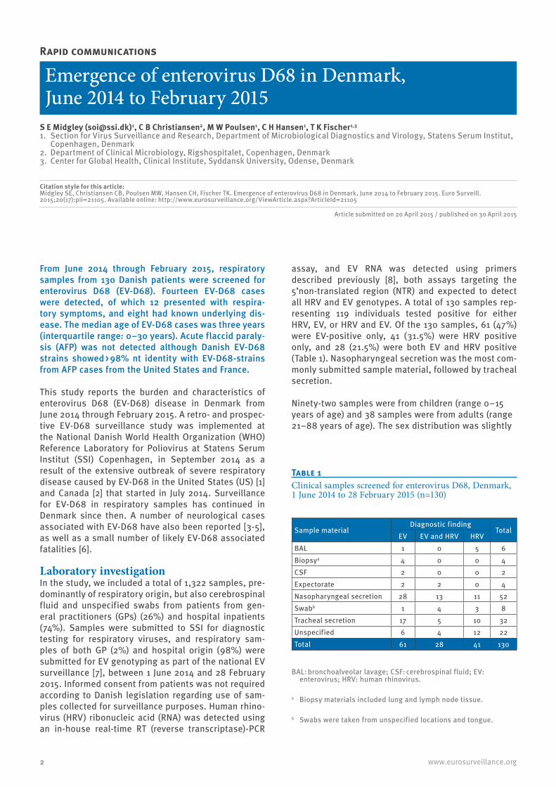
Twelve of these cases were detected using the EV-D68 specific real-time RT-PCR, two were detected by sequencing the amplicon from the VP2 PCR (data not shown). Eight were EV- and HRV-positive in the initial diagnostic test, the remaining six were EV-positive. All EV-D68 cases were detected between September and November 2014, which corresponds to the main peak of the EV season in Denmark 2014 (Figure 1, Table 2). Information on duration of illness was available for 10 patients, and patients with underlying conditions had a longer duration of illness (three weeks or longer,
Table 2Enterovirus D68-positive cases Denmark, detected between September and November 2014 (n=14)
Case Clinical information Underlying disease Age Sampling date Sample material Diagnostic findings
1Pneumonia and
respiratory failure lasting four weeks
Pulmonary defect and asthma 2 24 Sep 2014 Tracheal secretion EV and HRV-positive
2Acute bronchitis, repeated admissions during a three-
month period
Pulmonary defect and/or chronic lung disease 0 30 Sep 2014 Tracheal secretion EV- and HRV-
positive
3 Cough and fever Malignancy 1 9 Oct 2014 Nasopharyngeal secretion EV-positive
4 Pneumonia Pulmonary defect and/or chronic lung disease 7 13 Oct 2014 Nasopharyngeal
secretion EV-positive
5 Acute respiratory failure Cardiac disease and/or chronic lung disease 4 14 Oct 2014 Nasopharyngeal
secretionEV- and HRV
positive
6 Coughing and fever lasting three weeks
Cardiac disease and/or chronic lung disease 0 20 Oct 2014 Nasopharyngeal
secretion EV-positive
7
Asthmatic wheezing for seven days, not
responding to standard inhalation treatment
None 2 24 Oct 2014 Nasopharyngeal secretion EV-positive
8 Low gradea fever for four days None 3 27 Oct 2014 Nasopharyngeal
secretionEV- and HRV-
positive
9
Acute onset, throat pain, coughing, muscle pain, fatigue, fever 38–39 °C, severe congestion and
runny nose, lasting eight days
None 61 3 Nov 2014 Expectorate EV- and HRV-positive
10 Mild pharyngitis for five days None 30 4 Nov 2014 Unspecified EV- and HRV-
positive
11 Cough and fever for one week None 0 26 Nov 2014 Tracheal secretion EV- and
HRV-positive
12
Cough and fever for two weeks; acute breathing difficulties resulting in CPAP treatment upon
hospital admission (nine days).
None 68 26 Nov 2014 Unspecified EV- and HRV-positive
13 Cough, congestion and fever lasting 21 days Malignancy 3 26 Nov 2014 Nasopharyngeal
secretion EV-positive
14 Asthmatic cough and fever for four weeks Malignancy 5 27 Nov 2014 Nasopharyngeal
secretion EV-positive
CPAP: continuous positive airway pressure; EV: enterovirus; HRV: human rhinovirus.a Fever <38°C.
4 www.eurosurveillance.org
compared with four to 14 days for patients with no underlying condition).
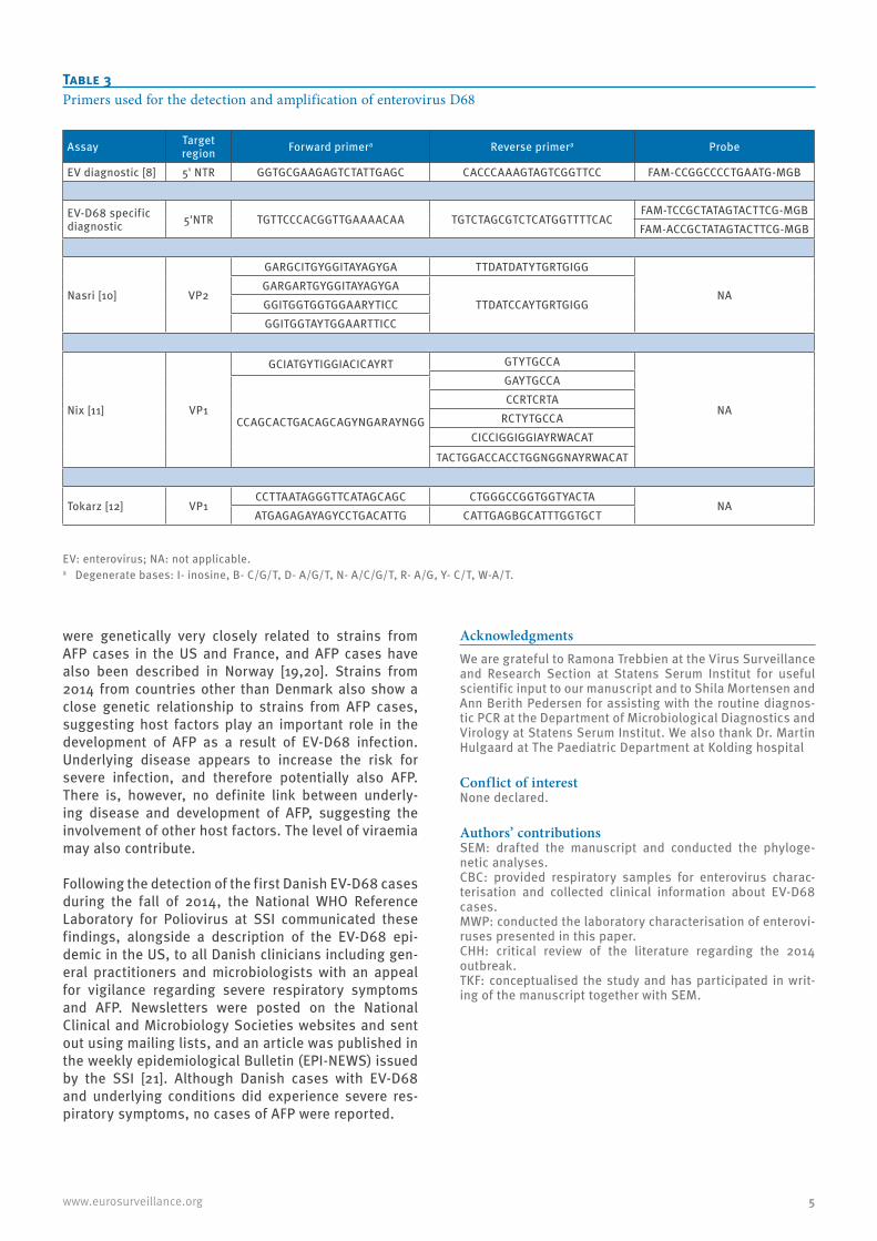
All EV-D68-positive samples from 2014, as well as historical samples, were characterised using an assay specific for the VP1 region of EV-D68 [12] and included in the phylogenetic analysis.All PCR amplicons were sequenced using the forward and reverse primers from the VP1 and VP2 assays on an ABI 3500 automated sequencer (Applied Biosystems). Sequences were assembled in BioNumerics v6.6 (Applied Maths BV) and genotyped using BLAST anal-ysis in the genotyping database and on GenBank. Sequences were aligned with published EV-D68 sequences downloaded from GenBank using the SSE v1.1 software [13]. The most appropriate phylogenetic model for the data was found using the model test function in MEGA6 [14], and phylogenetic analysis was carried out using maximum likelihood with the Kimura 2 parameter algorithm, gamma distribution, invariable sites and 1,000 bootstrap replications, also in MEGA6. Sequences obtained in this study were submitted to GenBank, accession numbers KP729103-KP729109, and KR108018 - KR108026. All primers used in this study can be seen in Table 3.
Phylogenetic analysesAll but two samples amplified successfully in the EV-D68 specific VP1 assay, producing sequences of around 800 nt. The two samples which could not be characterised using VP1 were the same two that failed to amplify in the real-time assay, and were confirmed to be EV-D68 by VP2 sequence analysis. Four historical EV-D68-positive samples were successfully amplified in the EV-D68 specific VP1 assay. BLAST analysis of the Danish VP1 sequences revealed that 6/12 of the Danish
EV-D68 strains showed > 98% homology in 100% of the sequence (between 716 and 839 nt in length) with the US 2014 outbreak strains, and 2/12 strains showed > 98% homology with a EV-D68 strain from an acute flaccid paralysis (AFP) case in France case (LN626610). The remaining four Danish 2014 strains shared > 98% homology with other French 2014 EV-D68 strains. Phylogenetic analysis of the 12 EV-D68 VP1 sequences identified eight clusters containing Danish strains of EV-D68 (Figure 2). Eleven of the Danish strains from 2014 cluster within clade B as described by Tokarz et al., 2012 [12], one clusters within clade A. Strains from 2008, 2010, and 2013 cluster within clade A.
DiscussionEV-D68 which is a member of the large picornaviri-dae family of viruses, has primarily been associated with mild to severe respiratory infections [15,16]. Historically, EV-D68 was only sporadically detected worldwide during the usual EV seasonal epidemics, but since 2008 the EV-D68 has occasionally given rise to larger outbreaks globally [15,17], although no previous outbreak has seen neither the same scale nor severity as the North American EV-D68 outbreak in 2014 [1]. Shortly after the North American outbreak was announced in July, EV-D68 cases were detected in Europe, and the European Centre for Disease Prevention and Control (ECDC) issued a rapid risk assessment on 26 September 2014 [18]. Denmark joined an initiative started by the European Society for Clinical Virology (ESCV) to investigate the prevalence of EV-D68 in the European region in a retro- and prospective study cov-ering June through November 2014.
In Denmark, ca 10% of respiratory samples tested between June 2014 and February 2015 were positive for EV and/or HRV, and of these 11% were determined to be EV-D68 by a combination of 5´NTR, VP1 and VP2 PCR and sequencing. This is a comparable detection rate to that described in other countries during the same time period [19]. No EV-D68 was detected in samples that were HRV-positive only, suggesting that the EV and HRV diagnostic result for EV-D68 cases is due to cross-reaction in the HRV assay. However, only 29.6% of double positive EV and HRV cases were associated with EV-D68. Other respiratory EV may also cross-react in an HRV assay in the 5’NTR, and, as a consequence, diagnostic laboratory reports from SSI for samples which are EV and HRV positive now contain a comment stating that this result may be due to infection with a virus detectable using both assays, rather than a dou-ble infection. The number of samples with this result has historically been small at SSI; should it continue to increase beyond the 10% rate identified in this study, the HRV assay may need to be revised.
Not all EV-D68 samples were detected using real-time and VP1 PCR, illustrating the difficulties in detecting and genotyping EVs in general due to the high level of diversity within this family of viruses. Danish strains
Figure 1Seasonal distribution of cases of enterovirus and enterovirus D68 infection, Denmark, 2014
EV: enterovirus.Samples taken between January and May 2014 were not screened
for the presence of enterovirus D68.
0
1
2
3
4
5
6
7
0
10
20
30
40
50
60
EV-D
68 c
ases
EV c
ases
2014
EV cases EV-D68 cases
Jan
Feb
Mar
Apr
May
Jun
Jul
Aug
Sep
Oct
Nov
Dec
5www.eurosurveillance.org
were genetically very closely related to strains from AFP cases in the US and France, and AFP cases have also been described in Norway [19,20]. Strains from 2014 from countries other than Denmark also show a close genetic relationship to strains from AFP cases, suggesting host factors play an important role in the development of AFP as a result of EV-D68 infection. Underlying disease appears to increase the risk for severe infection, and therefore potentially also AFP. There is, however, no definite link between underly-ing disease and development of AFP, suggesting the involvement of other host factors. The level of viraemia may also contribute.
Following the detection of the first Danish EV-D68 cases during the fall of 2014, the National WHO Reference Laboratory for Poliovirus at SSI communicated these findings, alongside a description of the EV-D68 epi-demic in the US, to all Danish clinicians including gen-eral practitioners and microbiologists with an appeal for vigilance regarding severe respiratory symptoms and AFP. Newsletters were posted on the National Clinical and Microbiology Societies websites and sent out using mailing lists, and an article was published in the weekly epidemiological Bulletin (EPI-NEWS) issued by the SSI [21]. Although Danish cases with EV-D68 and underlying conditions did experience severe res-piratory symptoms, no cases of AFP were reported.
Acknowledgments We are grateful to Ramona Trebbien at the Virus Surveillance and Research Section at Statens Serum Institut for useful scientific input to our manuscript and to Shila Mortensen and Ann Berith Pedersen for assisting with the routine diagnos-tic PCR at the Department of Microbiological Diagnostics and Virology at Statens Serum Institut. We also thank Dr. Martin Hulgaard at The Paediatric Department at Kolding hospital
Conflict of interestNone declared.
Authors’ contributionsSEM: drafted the manuscript and conducted the phyloge-netic analyses.CBC: provided respiratory samples for enterovirus charac-terisation and collected clinical information about EV-D68 cases.MWP: conducted the laboratory characterisation of enterovi-ruses presented in this paper.CHH: critical review of the literature regarding the 2014 outbreak.TKF: conceptualised the study and has participated in writ-ing of the manuscript together with SEM.
Table 3Primers used for the detection and amplification of enterovirus D68
Assay Target region Forward primera Reverse primera Probe
EV diagnostic [8] 5' NTR GGTGCGAAGAGTCTATTGAGC CACCCAAAGTAGTCGGTTCC FAM-CCGGCCCCTGAATG-MGB
EV-D68 specific diagnostic 5'NTR TGTTCCCACGGTTGAAAACAA TGTCTAGCGTCTCATGGTTTTCAC
FAM-TCCGCTATAGTACTTCG-MGBFAM-ACCGCTATAGTACTTCG-MGB
Nasri [10] VP2
GARGCITGYGGITAYAGYGA TTDATDATYTGRTGIGG
NAGARGARTGYGGITAYAGYGA
TTDATCCAYTGRTGIGGGGITGGTGGTGGAARYTICCGGITGGTAYTGGAARTTICC
Nix [11] VP1
GCIATGYTIGGIACICAYRT GTYTGCCA
NA
GAYTGCCA
CCAGCACTGACAGCAGYNGARAYNGG
CCRTCRTARCTYTGCCA
CICCIGGIGGIAYRWACAT
TACTGGACCACCTGGNGGNAYRWACAT
Tokarz [12] VP1CCTTAATAGGGTTCATAGCAGC CTGGGCCGGTGGTYACTA
NAATGAGAGAYAGYCCTGACATTG CATTGAGBGCATTTGGTGCT
EV: enterovirus; NA: not applicable.a Degenerate bases: I- inosine, B- C/G/T, D- A/G/T, N- A/C/G/T, R- A/G, Y- C/T, W-A/T.

6 www.eurosurveillance.org
Figure 2Phylogenetic analysis of enterovirus D68 VP1 sequences
The phylogenetic tree was constructed by maximum likelihood, with the Kimura 2-parameter algorithm, with gamma distribution, invariable sites and 1,000 bootstrap replicates, using MEGA6. Only bootstrap values > 70% are indicated. Branch lengths are drawn to the indicated scale, proportion of nt substitutions per site. Sequences were 731 nt in length. Reference sequences were downloaded from GenBank; all complete or near complete genomes (n = 50, downloaded 14 Apr 2015), as well as partial VP1 sequences > 700 nt in length with complete overlap with the sequences from this study (n = 232, downloaded 14 Apr 2015) were included, resulting in a dataset of 294 sequences. GenBank reference sequences and sequences from this study are identified by their accession numbers. Country of origin and detection year is specified after the sequence ID (where available) for references and study samples within defined clusters. Four EV-D68 sequences from the Danish genotyping database, identified between 2008 and 2013, were also included in the analysis. Sequences from this study are identified with filled circles and bold text. Reference sequences from known acute flaccid paralysis cases are identified with bold italics.
LN681325LN681323LN681334LN681330LN681329KP729103 DENMARK 2014KP729109 DENMARK 2014KR108024 DENMARK 2014
LN681324LN681340LN681335
KP745769KP745768
KP729108 DENMARK 2014KP729104 DENMARK 2014
KM851230KM851229
KP114665LN681333
LN681338LN681326
KC763167KF254922
LN681319LN681318LN681321LN626610KP729105 DENMARK 2014
LN681320KR108026 DENMARK 2014
KC763169KC763164KF254913
KF254918KF254921KF254923KF254917KF254919KF254920
JF896311AB614431AB614430AB614432AB614437AB614433AB614409AB614427
AB902837JF896304
JF896306JF896307
JF896309JF896308JF896310JF896305JF896303JF896302
KM892501KP240936
KP114664JX898785
KM892499KM892498
KC763162KP153544KP153542KP153545
KP153541KP153539KP153543KP153546KP153540
KP114663KP745767KP745756KP100794LN681332
LN681337LN681327
KP745765KP745754KP114662KP745751KM851226
KR108023 DENMARK 2014KP745758KP745763KP745752
KP745753KM881710KM851225
KP745759KP745770
KR108025 DENMARK 2014KP729107 DENMARK 2014KR108022 DENMARK 2014
KP745757KM851228KP745761KP745755KP745760KP100793
KP745762KP100792KP100796KP100795KP745766KP745764KM851227KM892502
AB861414KJ472886
JX101799JX101795
JX101800AY426494AY426493
AY426496AY426495
AY426492AY426491
AB667890AB667891AB667889AB667888AB667887AB667886AB667885AB667893AB667894AB667892
KC763171KC763168
KC763173KC763172KC763158KC763160
KC763157AB667895
AB601881AB601880
KM892497AB601885JX101786
AB601883AB601872AB601884AB601882
AB601877AB601876AB601879AB601875AB614428AB601874AB601878AB601873
AB614439AB614410AB614429AB614423AB614438AB614436AB614435AB614434AB614424AB614422AB614421AB614420AB614419AB614418AB614417AB614416AB614415AB614414AB614413AB614412AB614411AB614407AB614406AB614426AB614425AB614408
AY426497EF107098
JX101801AY426498
JX101798JX101797JX101796
AY426499LN681317LN681316LN681331
KP729106 DENMARK 2014KP153538LN681328KR108021 DENMARK 2014
LN681336KM851231
JX898786KF254914
JX898784KF726085AB667899
JX101802AB667896
AY426500LN681339
LN681322JX101792JX101790JX101793JX101791JX101794AB667897AB667898
KM892500KJ472878
JX101789JX101788
JX101846JX101812JX101805JX101806JX101807
JX101810KC763170
KR108018 DENMARK 2014KC763161
KC763165KC763166KC763163
KJ472882KJ472885
KJ472880KJ472883KJ472884
AB861413JX101787
JF896301JF896299
KR108019 DENMARK 2014JF896300KF254916KC763159KF254915
AB920412KC763177
JF896289AB614444AB614443
AB614442AB614441KC763176KC763175
JF896290JF896292JF896291
KR108020 DENMARK 2014JF896293
JF896294JF896295
JX101803JF896312JF896297JQ713904JQ713912JQ713911JX070222JQ713913
JQ713909JQ713907JQ713910
JQ713905JQ713908JQ713906AB614440
JX101814JX101811JX101804JF896296KC763174JX101809JX101813
JX101808JF896288JF896287
AY426490AY426489
AY426531AY355268
AY426486AY426488
AY426487
100
96
73
71
100
100
97
98
93
93
93
94
96
100
87
90
98
74
100
70
93
70
100
95
80
87
80
99
100
96
71
92
100
83
90
97
97
85
95
99
82
77
73
81
73
78
100
99
100
97
96
98
94
78
90
98
91
78
83
99
77
90 78
94
84
96
80
90
96
0,02
Tokarz lineage A
Tokarz lineage B
Tokarz lineage C
LN681325 FRANCE 2014LN681323 FRANCE 2014LN681334 FRANCE 2014
LN681330 FRANCE 2014LN681329 FRANCE 2014
KP729103 DENMARK 2014KP729109 DENMARK 2014
KR108024 DENMARK 2014LN681324 FRANCE 2014
LN681340 FRANCE 2014LN681335 FRANCE 2014
KP745769 USA 2014KP745768 USA 2014
KP729108 DENMARK 2014KP729104 DENMARK 2014
KM851230 USA 2014KM851229 USA 2014
KP114665 CANADA 2014LN681333 FRANCE 2014
LN681338 FRANCE 2014LN681326 FRANCE 2014
KC763167 ITALY 2012KF254922 SPAIN 2012
LN681319 FRANCE 2014LN681318 FRANCE 2014LN681321 FRANCE 2014
LN626610 FRANCE 2014 AFPKP729105 DENMARK 2014
LN681320 FRANCE 2014KR108026 DENMARK 2014
KC763169 ITALY 2012KC763164 ITALY 2012
KF254913 SPAIN 2013KF254918 SPAIN 2012
KF254921 SPAIN 2012KF254923 SPAIN 2013
KF254917 SPAIN 2012KF254919 SPAIN 2012KF254920 SPAIN 2012
78
94
90
96
97
98
0.002
KM892501 USA 2013 AFP
KP240936 CHINA 2014
KP114664 CANADA 2014
JX898785 CHINA 2011
KM892499 USA 2013 AFP
KM892498 USA 2013 AFP
KC763162 ITALY 2012
KP153544 ITALY 2014
KP153542 ITALY 2014
KP153545 ITALY 2014
KP153541 ITALY 2014
KP153539 ITALY 2014
KP153543 ITALY 2014
KP153546 ITALY 2014
KP153540 ITALY 2014
KP114663 CANADA 2014
KP745767 USA 2014
KP745756 USA 2014
KP100794 USA 2013 AFP
LN681332 FRANCE 2014
LN681337 FRANCE 2014
LN681327 FRANCE 2014
KP745765 USA 2014
KP745754 USA 2014
KP114662 CANADA 2014
KP745751 USA 2014
KM851226 USA 2014
KR108023 DENMARK 2014
KP745758 USA 2014
KP745763 USA 2014
KP745752 USA 2014
KP745753 USA 2014
KM881710 USA 2014
KM851225 USA 2014
KP745759 USA 2014
KP745770 USA 2014
KR108025 DENMARK 2014
KP729107 DENMARK 2014
KR108022 DENMARK 2014
KP745757 USA 2014
KM851228 USA 2014
KP745761 USA 2014
KP745755 USA 2014
KP745760 USA 2014
KP100793 USA 2014 AFP
KP745762 USA 2014
KP100792 USA 2014 AFP
KP100796 USA 2014 AFP
KP100795 USA 2014 AFP
KP745766 USA 2014
KP745764 USA 2014
KM851227 USA 2014
KM892502 USA 2014 AFP
100
78
94
84
90
80
96
96
0.002
JX101789 SENEGAL 2010
JX101788 SENEGAL 2010
JX101846 USA 2009
JX101812 USA 2009
JX101805 USA 2009
JX101806 USA 2009
JX101807 USA 2009
JX101810 USA 2009
KC763170 ITALY 2008
KR108018 DENMARK 2008
KC763161 ITALY 2008
KC763165 ITALY 2008
KC763166 ITALY 2008
KC763163 ITALY 2008
KJ472882 KENYA 2010
KJ472885 KENYA 2010
KJ472880 KENYA 2008
KJ472883 KENYA 2010
KJ472884 KENYA 2010
AB861413 PHILIPPINES 2011
JX101787 SENEGAL 2010
JF896301 NETHERLANDS 2010
JF896299 NETHERLANDS 2010
KR108019 DENMARK 2010
JF896300 NETHERLANDS 2010
KF254916 SPAIN 2013
KC763159 ITALY 2012
KF254915 SPAIN 2013
AB920412 JAPAN 2013
KC763177 ITALY 2010
JF896289 NETHERLANDS 2010
AB614444 JAPAN 2010
AB614443 JAPAN 2010
AB614442 JAPAN 2010
AB614441 JAPAN 2010
KC763176 ITALY 2010
KC763175 ITALY 2010
JF896290 NETHERLANDS 2010
JF896292 NETHERLANDS 2010
JF896291 NETHERLANDS 2010
KR108020 DENMARK 2010
JF896293 NETHERLANDS 2010
JF896294 NETHERLANDS 2010
JF896295 NETHERLANDS 2010
JX101803 USA 2009
JF896312 NETHERLANDS 2010
JF896297 NETHERLANDS 2010
JQ713904 NEW ZEALAND 2010
JQ713912 NEW ZEALAND 2010
JQ713911 NEW ZEALAND 2010
JX070222 NEW ZEALAND 2010
JQ713913 NEW ZEALAND 2010
JQ713909 NEW ZEALAND 2010
JQ713907 NEW ZEALAND 2010
JQ713910 NEW ZEALAND 2010
JQ713905 NEW ZEALAND 2010
JQ713908 NEW ZEALAND 2010
JQ713906 NEW ZEALAND 2010
AB614440 JAPAN 2010
JX101814 USA 2009
JX101811 USA 2009
JX101804 USA 2009
JF896296 NETHERLANDS 2010
KC763174 ITALY 2010
JX101809 USA 2009
JX101813 USA 2009
JX101808 USA 2009
JF896288 NETHERLANDS 2010
JF896287 NETHERLANDS 2010
100
98
87
93
74
70
93
90
100
96
98
0.002
LN681317 FRANCE 2014
LN681316 FRANCE 2014
LN681331 FRANCE 2014
KP729106 DENMARK 2014
KP153538 ITALY 2014
LN681328 FRANCE 2014
KR108021 DENMARK 2013
LN681336 FRANCE 2014
KM851231 USA 2014
JX898786 CHINA 2012
KF254914 SPAIN 2013
JX898784 CHINA 2010
KF726085 CHINA 2008
AB667899 JAPAN 2008
JX101802 SOUTH AFRICA 2000
AB667896 JAPAN 2006
AY426500 USA
96
73
71
100
100
95
0.005

7www.eurosurveillance.org
References1. Midgley CM, Jackson MA, Selvarangan R, Turabelidze G,
Obringer E, Johnson D, et al. Severe respiratory illness associated with enterovirus D68 - Missouri and Illinois, 2014. MMWR Morb Mortal Wkly Rep. 2014;63(36):798-9. PMID:25211545
2. National Collaborating Centre for Infectious Diseases (NCCID). Disease debrief: EV-D68. Winnipeg, Manitoba: NCCID. [Accessed 21 Nov 2014]. Available from: http://www.nccid.ca/disease-debrief-ev-d68#Q1.
3. Pastula DM, Aliabadi N, Haynes AK, Messacar K, Schreiner T, Maloney J, et al.; Centers for Disease Control and Prevention (CDC). Acute neurologic illness of unknown etiology in children - Colorado, August-September 2014. MMWR Morb Mortal Wkly Rep. 2014;63(40):901-2. PMID:25299607
4. British Columbia Centre for Disease Control. Emerging respiratory virus bulletin: MERS-CoV, Influenza A(H7N9) and A(H3N2)v, Enterovirus D68. British Columbia: Centre for Disease Control. 4 Oct 2014. Available from: http://www.bccdc.ca/NR/rdonlyres/88FD3DD4-BEB0-4F29-93C0-6093A7AFBD4B/0/Full_ERVUpdate20141004.pdf
5. Lang M, Mirand A, Savy N, Henquell C, Maridet S, Perignon R, et al. Acute flaccid paralysis following enterovirus D68 associated pneumonia, France, 2014. Euro Surveill. 2014;19(44):20952. http://dx.doi.org/10.2807/1560-7917.ES2014.19.44.20952 PMID:25394254
6. Centers for Disease Control and Prevention. Non-Polio Enterovirus: Enterovirus D68. Atlanta: National Center for Immunization and Respiratory Diseases (NCIRD), Division of Viral Diseases, Centers for Disease Control and Prevention. Updated 23 Mar 2015. Available from: http://www.cdc.gov/non-polio-enterovirus/about/ev-d68.html
7. Fischer TK, Nielsen AY, Sydenham TV, Andersen PH, Andersen B, Midgley SE. Emergence of enterovirus 71 C4a in Denmark, 2009 to 2013. Euro Surveill. 2014;19(38):20911. http://dx.doi.org/10.2807/1560-7917.ES2014.19.38.20911 PMID:25306878
8. Nielsen AC, Böttiger B, Midgley SE, Nielsen LP. A novel enterovirus and parechovirus multiplex one-step real-time PCR-validation and clinical experience. J Virol Methods. 2013;193(2):359-63. http://dx.doi.org/10.1016/j.jviromet.2013.06.038 PMID:23845901
9. Poelman R, Schölvinck EH, Borger R, Niesters HGM, van Leer-Buter C. The emergence of enterovirus D68 in a Dutch University Medical Center and the necessity for routinely screening for respiratory viruses. J Clin Virol. 2015;62:1-5. http://dx.doi.org/10.1016/j.jcv.2014.11.011 PMID:25542461
10. Nasri D, Bouslama L, Omar S, Saoudin H, Bourlet T, Aouni M, et al. Typing of human enterovirus by partial sequencing of VP2. J Clin Microbiol. 2007;45(8):2370-9. http://dx.doi.org/10.1128/JCM.00093-07 PMID:17537940
11. Nix WA, Oberste MS, Pallansch MA. Sensitive, seminested PCR amplification of VP1 sequences for direct identification of all enterovirus serotypes from original clinical specimens. J Clin Microbiol. 2006;44(8):2698-704. http://dx.doi.org/10.1128/JCM.00542-06 PMID:16891480
12. Tokarz R, Firth C, Madhi SA, Howie SR, Wu W, Sall AA, et al. Worldwide emergence of multiple clades of enterovirus 68. J Gen Virol. 2012;93(Pt 9):1952-8. http://dx.doi.org/10.1099/vir.0.043935-0 PMID:22694903
13. Simmonds P. SSE: a nucleotide and amino acid sequence analysis platform. BMC Res Notes. 2012;5(1):50. http://dx.doi.org/10.1186/1756-0500-5-50 PMID:22264264
14. Tamura K, Stecher G, Peterson D, Filipski A, Kumar S. MEGA6: Molecular Evolutionary Genetics Analysis version 6.0. Mol Biol Evol. 2013;30(12):2725-9. http://dx.doi.org/10.1093/molbev/mst197 PMID:24132122
15. Meijer A, van der Sanden S, Snijders BE, Jaramillo-Gutierrez G, Bont L, van der Ent CK, et al. Emergence and epidemic occurrence of enterovirus 68 respiratory infections in The Netherlands in 2010. Virology. 2012;423(1):49-57. http://dx.doi.org/10.1016/j.virol.2011.11.021 PMID:22177700
16. Renois F, Bouin A, Andreoletti L. Enterovirus 68 in pediatric patients hospitalized for acute airway diseases. J Clin Microbiol. 2013;51(2):640-3. http://dx.doi.org/10.1128/JCM.02640-12 PMID:23224095
17. Meijer A, Benschop KS, Donker GA, van der Avoort HG. Continued seasonal circulation of enterovirus D68 in the Netherlands, 2011-2014. Euro Surveill. 2014;19(42):20935. http://dx.doi.org/10.2807/1560-7917.ES2014.19.42.20935 PMID:25358039
18. Eurosurveillance editorial team. ECDC’s latest publications. Euro Surveill. 2014;19(39):20915. Available from: http://www.eurosurveillance.org/ViewArticle.aspx?ArticleId=20915
19. Bragstad K, Jakobsen K, Rojahn AE, Skram MK, Vainio K, Holberg-Petersen M, et al. High frequency of enterovirus D68
in children hospitalised with respiratory illness in Norway, autumn 2014. Influenza Other Respi Viruses. 2015;9(2):59-63.
20. Pfeiffer HC, Bragstad K, Skram MK, Dahl H, Knudsen PK, Chawla MS, et al. Two cases of acute severe flaccid myelitis associated with enterovirus D68 infection in children, Norway, autumn 2014. Euro Surveill. 2015;20(10):21062. http://dx.doi.org/10.2807/1560-7917.ES2015.20.10.21062 PMID:25788251
21. Fischer TK, Midgley SE, Poulsen MW, Andersen B, Christiansen CB, Andersen PH. Outbreak of enterovirus D68 in children in USA and Canada, and circulation in Denmark. EPI-NYT 2014;49. Available from: http://www.ssi.dk/pdf.ashx?title=No-49---2014---EPI-NEWS&url=http%3a%2f%2fwww.ssi.dk%2fEnglish%2fNews%2fEPI-NEWS%2f2014%2fNo+49+-+2014.aspx%3fpdf%3d1
8 www.eurosurveillance.org
Surveillance and outbreak reports
Chikungunya outbreak in Montpellier, France, September to October 2014
E Delisle ([email protected])1, C Rousseau1, B Broche2, I Leparc-Goffart3, G L’Ambert4, A Cochet1, C Prat3, V Foulongne5, J B Ferré4, O Catelinois1, O Flusin3, E Tchernonog5, I E Moussion2, A Wiegandt2, A Septfons6, A Mendy2, M B Moyano2, L Laporte2, J Maurel2, F Jourdain7, J Reynes5, M C Paty7, F Golliot1
1. Regional office of the French Institute for Public Health Surveillance (Cire Languedoc-Roussillon), Montpellier, France2. Regional Health Agency of Languedoc-Roussillon, Montpellier, France3. Institut de Recherche Biomédicale des Armées, National Reference Laboratory for arboviruses, Marseille, France4. Entente Interdépartementale pour la Démoustication du littoral Méditerranéen (EID Méditerranée), Public mosquito control
operator, Montpellier, France5. Montpellier University Hospital, Montpellier, France6. French Institute for Public Health Surveillance (Institut de Veille Sanitaire, InVS), Saint-Maurice, France7. National Centre of Expertise on Vectors, Montpelier, France
Citation style for this article: Delisle E, Rousseau C, Broche B, Leparc-Goffart I, L’Ambert G, Cochet A, Prat C, Foulongne V, Ferré JB, Catelinois O, Flusin O, Tchernonog E, Moussion IE, Wiegandt A, Septfons A, Mendy A, Moyano MB, Laporte L, Maurel J, Jourdain F, Reynes J, Paty MC, Golliot F. Chikungunya outbreak in Montpellier, France, September to October 2014. Euro Surveill. 2015;20(17):pii=21108. Available online: http://www.eurosurveillance.org/ViewArticle.aspx?ArticleId=21108
Article submitted on 24 December 2014 / published on 30 April 2015
In October 2014, an outbreak of 12 autochthonous chikungunya cases, 11 confirmed and 1 probable, was detected in a district of Montpellier, a town in the south of France colonised by the vector Aedes albopic-tus since 2010. A case returning from Cameroon living in the affected district was identified as the primary case. The epidemiological investigations and the repeated vector control treatments performed in the area and around places frequented by cases helped to contain the outbreak. In 2014, the chikungunya and dengue surveillance system in mainland France was challenged by numerous imported cases due to the chikungunya epidemic ongoing in the Caribbean Islands. This first significant outbreak of chikungunya in Europe since the 2007 Italian epidemic, however, was due to an East Central South African (ECSA) strain, imported by a traveller returning from West Africa. Important lessons were learned from this episode, which reminds us that the threat of a chikungunya epi-demic in southern Europe is real.
BackgroundChikungunya (CHIK), as well as dengue virus which shares the same vector, has been identified as a threat for mainland France for several years, as all the prereq-uisites for autochthonous transmission of the virus are present in southern regions [1,2]. The mosquito vector, Aedes albopictus, first introduced in southern France in 2004, was established in 18 districts in 2014 [3]; and the virus is regularly introduced by viraemic travel-lers returning from endemic or epidemic countries to mainland France where the population is naive for chi-kungunya virus (CHIKV). This situation led to a first epi-sode of autochthonous transmission of chikungunya in south-east France in 2010 [4]. Other countries in the south of Europe are also threatened by CHIK and den-gue virus as the vector Ae. albopictus is established in
large part around Mediterranean Sea and continues to disseminate further each year. This was demonstrated when an epidemic occurred in Italy in 2007, with more than 200 cases infected in three months [5].
In mainland France, since 2006, a chikungunya and dengue preparedness and contingency plan is imple-mented every year in vector-colonised districts. This plan aims to prevent the transmission and dissemi-nation of these viruses [3,6]. It describes five risk levels defined according to entomological and epide-miological surveillance results. All clinically-suspected imported cases must be reported to the regional health authority in addition to mandatory notification of con-firmed cases. Epidemiological investigations are then implemented as well as appropriate vector control measures in each location visited by patients during their period of viraemia. In addition, a network of labo-ratories practicing chikungunya and dengue diagnosis provides daily reports of their results to the French Institute of Public Health Surveillance regional boards. This enables identifying imported and autochthonous cases not already notified. A national database col-lects all suspected and confirmed cases identified dur-ing the season.
Here we report the epidemiological and entomological investigations of an outbreak of chikungunya which took place in Montpellier from September to October 2014 and try to explain the reasons why it occurred. Montpellier is a town of 400,000 inhabitants, located on the Mediterranean coast. Ae. Albopictus, identi-fied in 2010, has progressively colonised the whole conurbation.
9www.eurosurveillance.org
The alertOn 1 October 2014, a probable autochthonous chi-kungunya case was identified in Montpellier (case 1, index case). The patient, a woman in her 40s living near Montpellier, had developed 13 days earlier, on 18 September, a sudden fever, incapacitating arthral-gia, myalgia and a rash. She had not travelled abroad during the two weeks before symptoms onset but had experienced multiple mosquito bites on 14 September in a residential area of Montpellier. She consulted her general practitioner (GP) who prescribed several sero-logical tests, including for CHIKV. Analyses performed on a serum sample taken five days after symptoms onset identified IgM antibodies to CHIKV, that were confirmed by the National Reference Centre (NRC) in Marseille, on 13 October. Real-time reverse transcrip-tion PCR (RT-PCR) was negative for this sample.
While waiting for a second blood-sample to confirm the CHIKV infection by seroconversion, four additional sus-pected autochthonous chikungunya cases were notified on 16 October by the Infectious and Tropical Diseases Department of Montpellier University Hospital (UH) to the regional health authority. These cases (Cases 2 to 5) were adults from a same family and had not travelled abroad recently. They were not linked to the index case. They successively developed acute fever, painful joints and a rash between 20 September and 12 October (Figure).
They lived in a house located ca 150 m from the place where Case 1 (index case) experienced mosquito bites. On 17 October, a chikungunya real-time RT-PCR was positive for one of the family members at the Montpellier UH virology laboratory. The diagnosis of chikungunya was confirmed for all the family cases and Case 1 by the NRC, respectively on the 20 October and the 22 October (real-time RT-PCR and seroconver-sion, Table 1).
These geographically and temporally linked cases formed a cluster in a distinct area of Montpellier. According to the national contingency plan against the spread of CHIK and dengue viruses, these findings prompted immediate epidemiological and entomologi-cal investigations in order to contain the transmission.
Methods
Case definitionsFor the investigation, the following case definition was applied. From 1 September to 30 November 2014, in Montpellier and conurbation:
• An autochthonous suspected case of chikungunya was defined by sudden onset of fever (≥ 38.5 °C) and arthralgia, not explained by another medical condition in a person without a history of foreign travel within 15 days prior to symptoms onset.
• An autochthonous probable case was defined as a suspected case with an epidemiological link to
a confirmed case or a suspected case with posi-tive chikungunya tests (real-time RT-PCR or serol-ogy) performed by a private laboratory, or hospital laboratory.
• An autochthonous confirmed case was defined as a suspected case with positive laboratory tests (real-time RT-PCR or serology positive for IgM and IgG antibodies to CHIKV) performed by the NRC or a suspected case with an epidemiological link to a confirmed case and a serology positive for IgM antibodies to CHIKV performed by the NRC.
Epidemiological investigationsImmediately after the identification of Case 1 (index case), the enhanced surveillance database was ana-lysed to identify a potential primary case. Among all the imported chikungunya cases identified during the 2014 season, cases that had symptoms onset after 28 July and lived near or visited the same places as Case 1 were listed.
The time after 28 July was considered as the maximal period for viral transmission from symptoms onset of the primary case to symptoms onset of the second case, considering a mosquito lifespan of maximum one month, human incubation of maximum 12 days and human viraemia of seven days.
Active case finding of suspected autochthonous cases was implemented in Montpellier and conurbation: (i) a door-to-door investigation was conducted in the neigh-bourhood of the autochthonous cases’ residences (200m radius). Nearly 1,000 households were targeted, representing ca 2,250 inhabitants; (ii) information on the event and a request to immediately notify all sus-pected autochthonous cases to the regional health authority was sent to all GPs and laboratories estab-lished in the Montpellier conurbation as well as all six emergency medical services (including the Montpellier UH). In addition, health professionals established within a 2km radius of the cases’ residences were con-tacted by telephone (30 GPs and 11 laboratories).
For each new suspected case identified, blood sam-ples were collected and analysed by the NRC. Real-time RT-PCR was performed on samples collected within eight days after symptoms onset and serology on sam-ples collected after day 5 of symptoms onset.
Entomological investigationsEntomological investigations were carried out inside the primary case’s property, as soon as this case was identified, and in the affected area, in order to estimate vector densities, using BG Sentinel adult mosquito traps. Prospection of mosquito breeding sites was con-ducted concomitantly to the door-to-door case finding.
10 www.eurosurveillance.org
Results
Epidemiological investigations
Primary caseAn imported case living in the same area as the five autochthonous cases was identified in the enhanced surveillance database. The patient, who had returned from Cameroon on 29 August, had symptoms onset two days later. He consulted at the Montpellier UH and was diagnosed on 5 September by the laboratory of virology via chikungunya-specific real-time RT-PCR on a serum sample at day 3 post symptoms onset (Figure, Table 1). He was immediately notified to the regional health authority. Entomological investigators, respon-sible for interventions at that time, could not enter his
property and did not identify the vector in the vicinity of the case’s residence. No mosquito-control treatment was performed then.
Autochthonous casesThe door-to-door case finding and notification by health professionals of suspected autochthonous cases enabled the detection of 20 autochthonous sus-pected cases of chikungunya in the neighbourhood of Cases 1 to 5 (200m radius). Six cases were laboratory-confirmed by the NRC (Table 1) and the results were negative for 14. All six confirmed cases had been iden-tified through the door-to-door investigations. At the time of investigation, five had fully recovered and for sixth case, the symptoms started on the very day of the investigation (Figure). Moreover, epidemiological
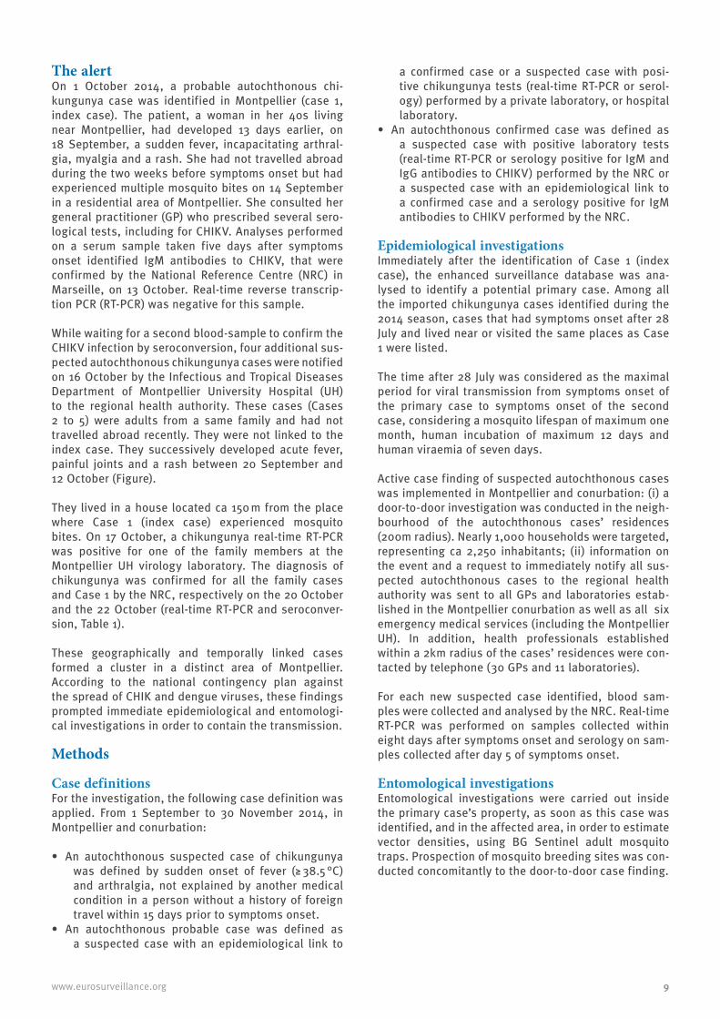
FigureTimeline of symptoms onset for imported and autochthonous cases of chikungunya and epidemiological features, Montpellier, France, September–October 2014 (n = 13)
NRC: National Reference Center; RT-PCR: reverse transcriptase-PCR; UH: University Hospital.Cases numbered by order of identification.Source : French Institute for Public Health Surveillance (Institut de veille sanitaire), 2014.
a Possible period of viral transmission from infected vector (infected from the primary case):- mosquitoes biting the primary case between the first day and the last day of his viraemic period, extrinsic incubation period: seven days [8], mosquito lifespan: 10 days [9].
76543211
27 28 29 30 31 1 2 3 4 5 6 7 8 9 10 11 12 13 14 15 16 17 18 19 20 21 22 23 24 25 26 27 28 29 30 1 2 3 4 5 6 7 8 9 10 11 12 13 14 15 16 17 18 19 20 21 22 23 24 25 26 27 28 29 30 31 1 2 3 4 5 6
410
511
2
September October November
6sesac suonothcotuA
121
98
73
P Primary caseConfirmed autochthonous caseProbable autochthonous case
F Family case
Travel in CameroonAverage incubation period (3 days) [7]Maximal incubation period (12 days) [7] Viraemic period (- one day / + seven days from date of symptoms onset)Date of symptoms onsetPossible period of viral transmission from infected vectors
(infected from the primary case)a
Primary case negative entomological
investigation
Case 1 (index case) Identification by health
authorities
Vector control treatments in the cluster area
Case 1 NRC laboratory confirmation /Door-to-door case finding
Family cluster NRC laboratory
confirmation
Case 5 Real-time RT-PCR (+)
by the Montpellier UH
Family cluster (Case 2 to 5)
Notification to health authorities
Primary case notification to
health authorities
Primarycase
F FF FPF

11www.eurosurveillance.org
investigations conducted among the family cases (cases 2 to 5) identified an additional family member as a probable case, but this patient did not consent to being tested.
Altogether, 12 autochthonous chikungunya cases were identified: 11 confirmed and one probable. For three confirmed cases, the CHIKV was detected (real-time RT-PCR positive) and for eight, IgM with or without IgG antibodies against CHIKV were detected (Table 1). The CHIKV sequence data obtained from sera of two autoch-thonous cases indicated that the CHIKV infection was due to a strain belonging to the East Central South African (ECSA) genotype harbouring the E1-A226V adaptive mutation (data not shown). Sequencing per-formed from the primary case serum showed same results.
All cases lived in or had visited the same area of Montpellier, a square of side 250m enclosing small buildings and individual houses with many gardens. The median age was 59 years (range 22–80). Half of the cases (6/12) were women. The date of symptoms onset ranged from 8 September 2014 to 22 October 2014. All cases presented with fever and incapacitat-ing arthralgia, mainly in the hands or feet. A rash was present for 10/12 (Table 2), which appeared mainly after fever onset (median two days; range 0–5). Nine of the 12 cases were free of general symptoms (fever, rash, myalgia etc.) within eight days (range 4–21) but seven cases still suffered from persistent joint pains two months after symptoms onset. All cases consulted
their GPs while symptomatic but chikungunya diagno-sis was suspected only for one (Case 1, index case). Five were referred to the Montpellier UH where the diagnosis was suspected for four of them (Cases 2 to 5) and laboratory tests carried out.
Entomological investigation and control measuresFollowing the identification of Case 1 (index case) and immediately after the primary case had been identi-fied, entomological investigations were repeated in mid-October in the neighbourhood of the primary case residence. They showed very high densities of Aedes albopictus larvae and adults (average of 30 mosquitoes per BG-Sentinel trap per day), promoted by numerous gardens located in the area providing mosquito breed-ing and resting sites.
A mosquito-control treatment was therefore performed three days later (Figure) in the outbreak area (± 150m radius circle around the residences of the primary case, of the family cases and the place visited by case 1), with Ultra Low Volume spraying of deltamethrin (Cerathrin and Aqua-KOthrine 2 and 1g of active sub-stance.ha − 1, respectively). This operation was repeated twice, five days and 11 days later, within a larger area (± 250m radius circle around the case’s residences), with consideration to sensitive persons and activities as well as respective institutions in the vicinity such as nursery, kindergarten, elementary and high schools. Following the first insecticide treatment, the vector population declined drastically in the area: from 243
Table 1Laboratory investigation of imported and autochthonous cases of chikungunya by the National Reference Centre, Montpellier, France, September–October 2014 (n = 13)
Case number Date ofsymptoms onset Date of samplinga
Serological tests for chikungunya Chikungunya real-time RT- PCRIgM IgG
Importedb 30 Aug 2014 D3 ND Positive
1c 18 Sep 2014D5 Positive Negative Negative
D29 Positive Positive ND2 20 Sep 2014 D28 Positive Positive ND3 24 Sep 2014 D24 Positive Positive ND4 09 Oct 2014 D7 Positive Positive Negative5 12 Oct 2014 D4 ND Positive6 08 Sep 2014 D4 ND Positive7 14 Sep 2014 D51 Positive Positive ND8 14 Sep 2014 D51 Positive Positive ND9 16 Sep 2014 D6 Positive Negative Negative
10 11 Oct 2014D5 Positive Negative Negative
D23 Positive Positive ND11 22 Oct 2014 D0 ND Positive12 20 Sep 2014 ND ND ND
ND: not done; RT-PCR: reverse transcriptase-PCR.a Day post symptom onset. b Primary case.c Index case.

12 www.eurosurveillance.org
Ae. albopictus collected per day with 8 BG Sentinel traps 24h before the treatment to eight per day after the treatment. Mosquito-control treatment was also performed around places (± 150m radius circle) fre-quented by each case during viraemia.
The numerous mosquito breeding sites identified in the neighbourhood houses during the door-to-door inspection were eliminated by the Entente Interdépartementale pour la Démoustication (EID) vec-tor control professionals, involving each inhabitant for pedagogical purposes.
Before the fogging of insecticide, an information leaflet was distributed in all mail-boxes inside the treatment area, concerning the epidemiological situation, the aim of the treatment and with explanation how to limit contact with insecticides, and giving recommendations about community mosquito control and protection i.e. maintaining gardens regularly and drying containers filled with water, using repellent, wearing long-sleeved clothes, etc.
DiscussionThis outbreak involving 12 autochthonous chikungu-nya cases is the first significant outbreak in Europe since the 2007 epidemic in Emilia Romagna, Italy [5]. It started as all the conditions for autochthonous trans-mission of CHIKV were met in a densely populated neighbourhood of Montpellier with high densities of Aedes albopictus.
The absence of immediate vector control treatment around the primary case’s residence where the vector was initially not identified, and the delay in identify-ing the first autochthonous cases enabled the estab-lishment of a CHIKV transmission cycle, which involves several generations of mosquitoes (incubation 2–12 days [7], extrinsic incubation period 4–13 days [8], mosquito lifespan 10-30 days [9]). This delay is due to the fact that chikungunya is rare in mainland France
and except for case 1, none of the GPs consulted by cases suspected the disease.However, there was a prompt response following the alert. The epidemiological investigations and the repeated vector control treatments performed in the area and around places frequented by cases helped to contain the outbreak: the number of cases was prob-ably curtailed and no spread beyond the affected area was identified. Only one case (case 11) presented with an onset of symptoms three days after the first treat-ment in the area. The effectiveness of vector control measures was also suggested by the results of trap-ping before and after treatments. The beginning of autumn and weather conditions in October might also have contributed to the end of the outbreak: the cooler temperatures were unfavourable to virus transmission and vector activity, the shorter daylight period induced egg diapauses and a decrease in vector density. In this outbreak involving a few cases, no socioeconomic fac-tor was identified as favouring the transmission.
The outbreak occurred as the ongoing chikungunya epi-demic in the Caribbean Islands [10] alerted the French public health authorities and challenged the national surveillance system [3,11,12]. Four hundred and five of the 449 (90%) imported cases of chikungunya identi-fied during the 2014 season in the French vector-col-onised districts were returning from the French West Indies, where the Asian genotype of CHIKV circulates [13]. No autochthonous transmission had been identi-fied from those imported cases. The primary case in the outbreak, however, was a viraemic traveller return-ing from Cameroon, infected by a strain belonging to the ECSA genotype with the E1-A226V adaptive muta-tion. These observations raise questions about the adaptation of the Asian genotype CHIKV strain circulat-ing in the Caribbean to Aedes albopictus [14].
This outbreak, like the previous autochthonous cases of chikungunya and dengue which occurred in south-ern France since 2010 [4,15-17], highlights that autoch-thonous transmission of vector-borne diseases is possible and can lead to outbreaks in France, under favourable climatic and entomological conditions. Repeated episodes of transmission of chikungunya or dengue viruses are likely to occur in the future, espe-cially as the vector is spreading further each year: in 2014, in France, more than 14 million residents live in areas colonised by Aedes albopictus, including many densely-populated cities.
This episode, detected and contained early when the number of cases was still limited, shows the impor-tance of the French contingency plan, whose purpose is, more than to avoid autochthonous transmission, to contain them. The plan organises epidemiological and entomological surveillances, facilitates the coordina-tion of investigations and mosquito control activities, allows the anticipation of necessary resources in case of an outbreak and provides regulatory tools. This plan is evaluated and adapted every year in order to
Table 2Distribution of symptoms among autochthonous cases of chikungunya, Montpellier, France, September–October 2014 (n = 12)
Symptoms Number of casesFever > 38.5 °C 12 Arthralgia 12 feet / ankles 10 hands / wrists 9 Rash 10 Myalgia 7 Diarrhoea 3 Headache 2 Back pain 1
13www.eurosurveillance.org
maintain its efficiency. However, the involvement of the population and health professionals is a key point for success. Thus, given the observations stated dur-ing the investigations, the following actions need to be pursued in the French vector- colonised areas to improve public health response: (i) increasing popu-lation awareness regarding the risk of chikungunya and dengue and consequently improving prevention (from individual protection to breeding sites control) in order to limit vector-borne transmission; (ii) increas-ing awareness of physicians and laboratories regard-ing the possibility of autochthonous transmission, the appropriate laboratory diagnosis tools and the notifica-tion of cases; (iii) maintaining a coordinated approach and a close collaboration between epidemiological and entomological surveillance, as well as a concerted pre-paredness of the various parties such as national and local health authorities, vector control professionals, national laboratory before the start of the season in order to facilitate rapid response.
ConclusionThis outbreak, following importation of an ECSA CHIKV strain by a traveller returning from Cameroon, is the first significant one in mainland France. Such a local circulation of the virus was not unexpected and the national contingency plan showed its effectiveness in controlling the outbreak. However, some weaknesses, in vector control measures around the primary case and awareness of health professionals, facilitated the spread of the virus. Contingency plan and epide-miological and entomological surveillance stakeholder preparedness is necessary to implement rapid and proportionate measures of surveillance and control. Awareness of health professionals and the community about vector control and disease symptoms need to be strengthened.
AcknowledgementsThe biomedical laboratories Cerba (Saint-Ouen l’Aumone) and Biomnis (Lyon). The private laboratories and the general practitioners of the Hérault district; C Ricoux, InVS, France and S Guglielmi, C Salvio, S Lesterle, O Puech, ARS, France and C Tizon, J Vidal, T Perimentel, F Richard, L Vançon, S Estaran EID Méditerranée, France for contributing to the field epidemiological investigations; vector control professionals at EID Méditerranée; D Bouillin and F Entezam, ARS, France; E Couturier and H De Valk, InVS, France, for reviewing the manuscript. The patients and their relatives approached for this investigation.
Conflict of interestNone declared.
Authors’ contributionsContribution to the epidemiological investigations: E Delisle, C Rousseau, MC Paty, A Cochet, O Catelinois, F Golliot, A Septfons, A Mendy, MB Moyano, L Laporte, J Maurel. Interviews of the patients: A Mendy, MB Moyano, L Laporte, J Maurel, E Delisle. Diagnosis of the family cases
and notification to health authorities: E Tchernonog, J Reynes, V Foulogne. Implementation of the control measures: B Broche. A Wiegandt, I Estève-Moussion. Coordination of the control meas-ures: B Broche. Entomological investigations and coordination of vector control treatment: G L’Ambert, JB Ferré. Laboratory inves-tigations: I Leparc-Goffart, C Prat, O Flusin. Coordination of the epidemiological investigations: E Delisle, C Rousseau (regional level), MC Paty (national level). Supervisor of the investigations: F Golliot. Drafted the manuscript: E Delisle, C Rousseau, MC Paty. Contribution to the writing of the paper: A Cochet, O Catelinois, G L’Ambert, F Jourdain, I Leparc-Goffart, A Wiegandt.
References1. Fontenille D, Failloux AB, Romi R. Should we expect Chik and
dengue in southern Europe? In: Takken W, Knols BGJ, editors. Emerging pests and vector-borne diseases in Europe. Vol 1. Wageningen Academic Publishers;2007.
2. Tomasello D, Schlagenhauf P. Chikungunya and dengue autochthonous cases in Europe, 2007-2012. Travel Med Infect Dis. 2013;11(5):274-84.http://dx.doi.org/10.1016/j.tmaid.2013.07.006 PMID:23962447
3. Paty MC, Six C, Charlet F, Heuzé G, Cochet A, Wiegandt A, et al. Large number of imported chikungunya cases in mainland France, 2014: a challenge for surveillance and response. Euro Surveill. 2014;19(28):20856. http://dx.doi.org/10.2807/1560-7917.ES2014.19.28.20856 PMID:25060572
4. Grandadam M, Caro V, Plumet S, Thiberge JM, Souarès Y, Failloux AB, et al. Chikungunya virus, southeastern France. Emerg Infect Dis. 2011;17(5):910-3.http://dx.doi.org/10.3201/eid1705.101873 PMID:21529410
5. Rezza G, Nicoletti L, Angelini R, Romi R, Finarelli AC, Panning M, et al.; CHIKV study group. Infection with chikungunya virus in Italy: an outbreak in a temperate region. Lancet. 2007;370(9602):1840-6.http://dx.doi.org/10.1016/S0140-6736(07)61779-6 PMID:18061059
6. Ministère des Affaires Sociales et de la Santé. Guide relatif aux modalités de mise en œuvre du plan anti-dissémination du chikungunya et de la dengue en métropole. [Chikungunya and dengue preparedness and response plan to monitor and prevent the risk of dissemination in mainland France]. Paris: Ministère des Affaires Sociales et de la Santé; 2014. French. [Accessed 11 November 2014]. Available from: http://circulaire.legifrance.gouv.fr/pdf/2014/05/cir_38279.pdf
7. Burt FJ, Rolph MS, Rulli NE, Mahalingam S, Heise MT. Chikungunya: a re-emerging virus. Lancet. 2012;379(9816):662-71.http://dx.doi.org/10.1016/S0140-6736(11)60281-X PMID:22100854
8. Christofferson RC, Chisenhall DM, Wearing HJ, Mores CN. Chikungunya viral fitness measures within the vector and subsequent transmission potential. PLoS ONE. 2014;9(10):e110538.http://dx.doi.org/10.1371/journal.pone.0110538 PMID:25310016
9. Hawley WA. The biology of Aedes albopictus. J Am Mosq Control Assoc Suppl. 1988;1(suppl):1-39. PMID:3068349
10. Ledrans M, Cassadou S, Boucau S, Huc-Anaïs P, Leparc-Goffart I, Prat C, et al. Émergence du chikungunya dans les départements français d’Amérique: organisation et résultats de la surveillance épidémiologique, avril 2014. [Emergence of chikungunya in the French overseas territories of the Americas: organization and results of epidemiological surveillance, April 2014]. Bull Epidémiol Hebd (Paris). 2014;(21-22):368-79. French. Available from: http://www.invs.sante.fr/beh/2014/21-22/2014_21-22_1.html
11. European Centre for Disease Prevention and Control (ECDC). Chikungunya outbreak in Caribbean region. Stockholm: ECDC; 2014 June. Available from: http://ecdc.europa.eu/en/publications/Publications/chikungunya-caribbean-june-2014-risk-assessment.pdf
12. Noël H, Rizzo C. Spread of chikungunya from the Caribbean to mainland Central and South America: a greater risk of spillover in Europe? Euro Surveill. 2014;19(28):20855. http://dx.doi.org/10.2807/1560-7917.ES2014.19.28.20855 PMID:25060570
13. Leparc-Goffart I, Nougairede A, Cassadou S, Prat C, de Lamballerie X. Chikungunya in the Americas. Lancet. 2014;383(9916):514.http://dx.doi.org/10.1016/S0140-6736(14)60185-9 PMID:24506907
14. Weaver SC. Arrival of chikungunya virus in the new world: prospects for spread and impact on public health. PLoS Negl Trop Dis. 2014;8(6):e2921. http://dx.doi.org/10.1371/journal.pntd.0002921 PMID:24967777
15. La Ruche G, Souarès Y, Armengaud A, Peloux-Petiot F, Delaunay P, Desprès P, et al. First two autochthonous dengue virus infections in metropolitan France, September 2010. Euro Surveill. 2010;15(39):pii=:19676.
16. Marchand E, Prat C, Jeannin C, Lafont E, Bergmann T, Flusin O, et al. Autochthonous case of dengue in France, October 2013. Euro Surveill. 2013;18(50):20661.http://dx.doi.org/10.2807/1560-7917.ES2013.18.50.20661 PMID:24342514
17. Giron S, Rizzi J, Leparc-Goffart I, Septfons A, Tine R, Cadiou B, et al. Nouvelles apparitions de cas autochtones de dengue en région Provence-Alpes-Côte d’Azur, France, août-septembre 2014. [New occurrence of autochthonous cases of dengue fever in southeast France, August-September 2014]. Bull Epidemiol Hebd (Paris). 2015; (13-14):217-23. Available from: http://www.invs.sante.fr/beh/2015/13-14/2015_13-14_3.html

14 www.eurosurveillance.org
Surveillance and outbreak reports
Cheese-related listeriosis outbreak, Portugal, March 2009 to February 2012
R Magalhães1, G Almeida1, V Ferreira1, I Santos1, J Silva1, M M Mendes2, J Pita2, G Mariano2, I Mâncio2, M M Sousa3, J Farber4, F Pagotto4, P Teixeira ([email protected])1
1. Centro de Biotecnologia e Química Fina – Laboratório Associado, Escola Superior de Biotecnologia, Universidade Católica Portuguesa/Porto, Porto, Portugal
2. Autoridade de Segurança Alimentar e Económica, Lisbon, Portugal3. Administração Regional de Saúde de Lisboa e Vale do Tejo, Lisbon, Portugal4. Listeriosis Reference Centre for Canada, Bureau of Microbial Hazards, Health Canada, Ottawa, Ontario, Canada
Citation style for this article: Magalhães R, Almeida G, Ferreira V, Santos I, Silva J, Mendes MM, Pita J, Mariano G, Mâncio I, Sousa MM, Farber J, Pagotto F, Teixeira P. Cheese-related listeriosis outbreak, Portugal, March 2009 to February 2012. Euro Surveill. 2015;20(17):pii=21104. Available online: http://www.eurosurveillance.org/ViewArticle.aspx?ArticleId=21104
Article submitted on 20 June 2014 / published on 30 April 2015
In Portugal, listeriosis has been notifiable since April 2014, but there is no active surveillance programme for the disease. A retrospective study involving 25 national hospitals led to the detection of an outbreak that occurred between March 2009 and February 2012. The amount of time between the start of the outbreak and its detection was 16 months. Of the 30 cases of listeriosis reported, 27 were in the Lisbon and Vale do Tejo region. Two cases were maternal/neonatal infec-tions and one resulted in fetal loss. The mean age of the non-maternal/neonatal cases was 59 years (stand-ard deviation: 17); 13 cases were more than 65 years-old. The case fatality rate was 36.7%. All cases were caused by molecular serogroup IVb isolates indistin-guishable by pulsed-field gel electrophoresis and ribotype profiles. Collaborative investigations with the national health and food safety authorities identi-fied cheese as the probable source of infection, traced to a processing plant. The magnitude of this outbreak, the first reported food-borne listeriosis outbreak in Portugal, highlights the importance of having an effective listeriosis surveillance system in place for early detection and resolution of outbreaks, as well as the need for a process for the prompt submission of Listeria monocytogenes isolates for routine laboratory typing.
IntroductionListeria monocytogenes is an intracellular bacterial pathogen of humans and a variety of animal species. In humans, L. monocytogenes infections are mainly food-borne and can cause an invasive and often fatal dis-ease in pregnant women and their fetuses, newborns, elderly people and immunocompromised individuals, with a case fatality rate of up to 30% [1]. The incidence of listeriosis increased in several European coun-tries between 2009 and 2013 (such as Germany, the Netherlands, Spain and the United Kingdom [1,2]) and, was the most frequent cause of hospitalisation and death (15.6%) due to the consumption of contaminated food in Europe in 2013 [2]. This increase reinforces the
need for each country to establish enhanced molecu-lar surveillance of listeriosis for efficient outbreak detection, investigation and control, as carried out by PulseNet USA or the Centre National de Référence des Listeria, Institut Pasteur, Paris, for example [3,4]. A sim-ilar programme for listeriosis surveillance at European Union level by harmonising methodological variables such as case definition, laboratory procedures and reporting systems is crucial. A pilot project was con-ducted by the European Centre for Disease Prevention and Control (ECDC) between January and March 2013 aiming to evaluate a Listeria external quality assurance scheme for the typing of L. monocytogenes that cov-ered pulsed-field gel electrophoresis (PFGE) method and serological typing (both as a phenotypic and a multiplex polymerase chain reaction (PCR)-based method) [5]. Results demonstrated that the majority (59%) of the participating laboratories were able to produce a PFGE gel of sufficiently high quality and the average score for serotyping among the participants was 94% and 97% for traditional and multiplex PCR based methods, respectively; however, higher quality could be achieved through trouble-shooting assistance and training.
In the absence of an active surveillance system for listeriosis at a national level, a collaborative study between the Listeria Research Centre of Escola Superior de Biotecnologia (LRCESB) and 25 of the major national hospitals (on a voluntary basis), covering about 90% of the population, was established in 2003 with the aim of obtaining epidemiological data on human listeriosis cases in Portugal and characterising clinical isolates of L. monocytogenes both phenotypically and genetically.In 2003, the incidence of listeriosis was 0.14 cases per 100,000 population [6]. An increase was reported between 2003 and 2007, i.e. it was 0.23 cases per 100,000 inhabitants for the year 2007 [7]. As a result of this study, an increase in the number of listeriosis cases was detected between January and July 2010, particularly in the Lisbon and Vale do Tejo region
15www.eurosurveillance.org
that corresponds to 13% of the total area of mainland Portugal and 34% of the total population (3.6 mil-lion inhabitants) [8], representing the first detected outbreak of listeriosis in Portugal. Here we describe the outbreak, as well as give details of the investiga-tions carried out in order to determine the source of infection.
Methods
Case definitionA listeriosis case was defined as a non-maternal/neonatal (non-MN) patient who met the laboratory criteria or a mother with a laboratory-confirmed lis-teriosis infection in her fetus, stillborn or newborn, as described in the Commission Decision of 28/IV/2008 [9]. Cases (laboratory confirmed with unknown clinical criteria) were detected through voluntary reporting by hospitals to the LRCESB in Porto.
If the pathogen was isolated from a pregnant woman and her newborn, stillborn or fetus, this was counted as a single case. Information regarding the sex and age of the patient, underlying pathology (if present), the tissue or fluid from which the bacteria were isolated and the year of isolation was reported.
Culture collectionHospitals sent isolates of L. monocytogenes to LRCESB for species confirmation and typing. Species confir-mation was performed by carbohydrate fermentation (rhamnose, xylose and mannitol) and Christie Atkins Munch-Petersen (CAMP) test [10]. Confirmed isolates of L. monocytogenes were stored in tryptic soy broth with 30% (v/v) glycerol at –80 °C in the culture collec-tion of the LRCESB.
Molecular-serotypingMolecular serotype of L. monocytogenes isolates was determined by multiplex PCR according to Doumith et al. [11]. This assay differentiates five major subtypes, each representing more than one serotype: geno-sero-group IVb (serotypes 4b, 4d and 4e), geno-serogroup IIa (serotypes 1/2a and 3a), geno-serogroup IIb (sero-types 1/2b, 3b and 7), geno-serogroup IIc (serotypes 1/2c and 3c) and geno-serogroup IVa (serotypes 4a and 4c).
Pulsed-field gel electrophoresisPFGE typing was performed according to the stand-ard CDC PulseNet protocol [12] using the restriction enzymes AscI and ApaI and gel run in CHEF III DR System (Bio-Rad, Laboratories, Hercules, CA, United States). Salmonella enterica serovar Braenderup H9812 (ATCC) DNA digested with XbaI was used as a reference size standard. Cluster analysis of the PFGE types was per-formed with the GelCompar software (Applied Maths, Sint-Martens-Latem, Belgium) by the unweighted pair group method with average linkages (UPGMA), using the Dice coefficient, and visually validated.
RibotypingAutomated ribotyping was performed using the restric-tion enzyme EcoRI and the RiboPrinter microbial char-acterisation system (Qualicon Inc., Wilmington, DE, United States), as previously described [13,14].
Outbreak investigationThe outbreak was investigated by the national health (Direção Geral de Saúde and Administração Regional de Saúde de Lisboa e Vale do Tejo) and food safety (Autoridade de Segurança Alimentar e Económica) authorities in collaboration with LRCESB.
A standardised questionnaire (adapted from a Canadian listeriosis outbreak, kindly supplied by Dr Jeff Farber of the Public Health Agency of Canada) was administered by the national health authority to patients diagnosed with listeriosis or their families (face-to-face interview) concerning their diet histories in the two months before symptom onset, with reference to the type of food con-sumed and household shopping patterns.
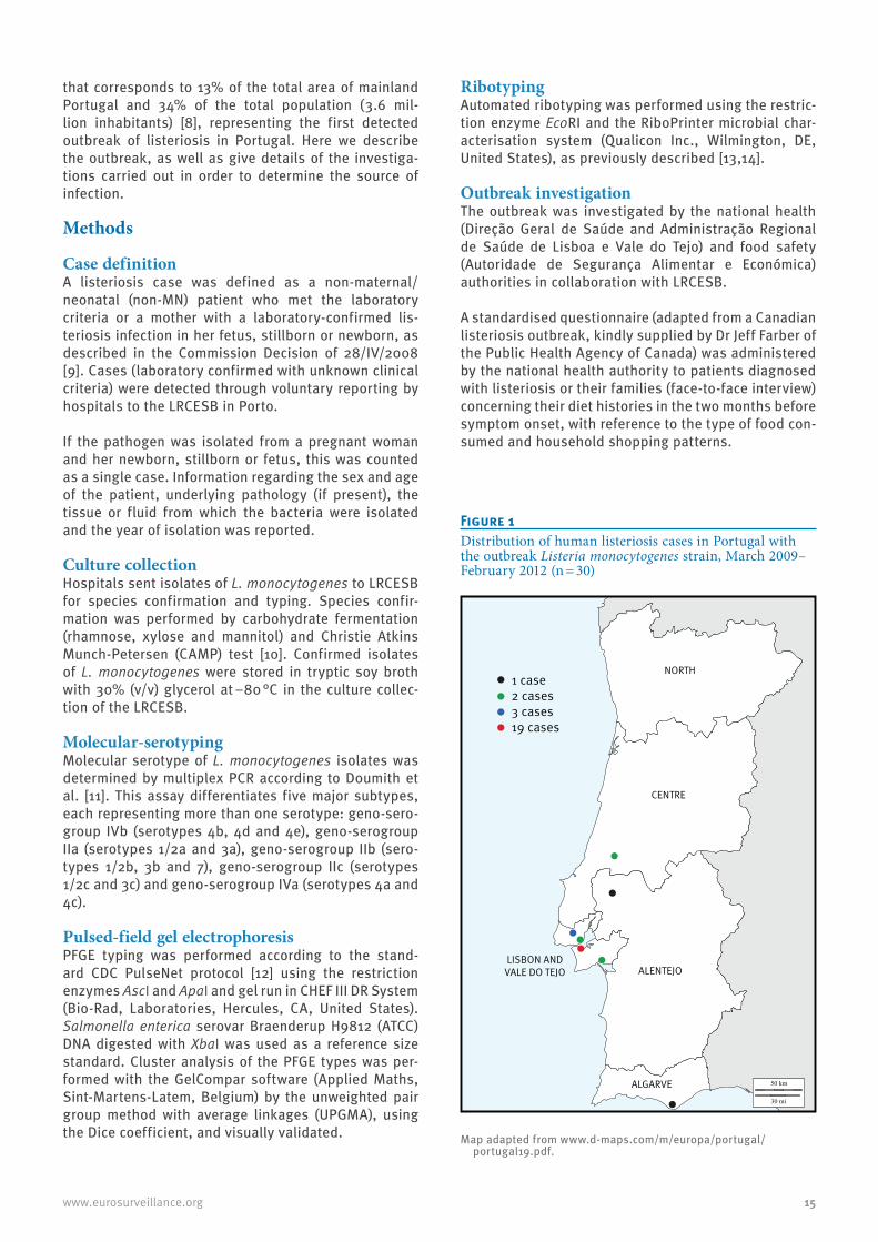
Figure 1Distribution of human listeriosis cases in Portugal with the outbreak Listeria monocytogenes strain, March 2009–February 2012 (n = 30)
NORTH
CENTRE
ALENTEJO
ALGARVE
LISBON ANDVALE DO TEJO
1 case2 cases3 cases19 cases
Map adapted from www.d-maps.com/m/europa/portugal/portugal19.pdf.

16 www.eurosurveillance.org
Analysis of food products and environmental samples was conducted by the food safety authority. L. monocy-togenes isolates from food and environmental samples were sent to LRCESB for typing.
International enquiryTo determine if the outbreak-associated strain of L. monocytogenes had been recovered from clinical or food samples from other countries, the PFGE type was communicated and compared with those of L. mono-cytogenes isolates in databases in France (Centre National de Référence des Listeria, Institut Pasteur), Canada (Listeriosis Reference Centre, Health Canada) and United States (Food Microbe Tracker, Food Safety Laboratory, Cornell University).
Results
Recognition of the outbreakBetween January and July 2010, a high number of lis-teriosis cases was observed (40 cases compared with 20 cases observed during all of 2009) [15], particularly in the Lisbon and Vale do Tejo region, where the major-ity of the cases were reported. Molecular typing of the 40 L. monocytogenes clinical isolates revealed that 18 serotype IVb isolates presented the same PFGE type and ribotype, which had been observed for five isolates recovered in 2009, four of which were in the Lisbon and Vale do Tejo region (in March, April and September) and one in the Centre region (in July) (Figures 1 and 2). This PFGE type was not found in the databases searched. In July 2010, the national health and food safety authorities were alerted to the increased number of cases and an outbreak investigation was initiated. A public health alert was issue to national hospitals requesting prompt notification and reporting of cases.
LRCESB continued to receive clinical isolates for typ-ing. Continued monitoring detected two more cases with the outbreak strain in November 2010, and three more cases in January, February and March 2011 (two in the Lisbon and Vale do Tejo region and one in the Algarve). Thereafter, in February 2012, there were two new cases with the same strain in the Lisbon and Vale do Tejo region. The total number of outbreak cases between March 2009 and February 2012 was 30.
Trace-back and investigation of the food sourceAnalysis of the epidemiological questionnaires pointed to different types and sizes of food retailers and iden-tified the following as possible sources of infection: cheeses (cured cheese and queijo fresco, made from pasteurised cow and goat milk), ice cream, ham and fermented sausages. On the basis of data gathered concerning the type of establishments where the food products were purchased, as well as the geographi-cal location of the cases, suspected foods and foods commonly associated with listeriosis, the food safety authority inspected 42 food retailers and collected 103 samples for analysis (51 meat products, 24 dairy products, 13 ready-to-eat foods and 15 environmental swabs). L. monocytogenes was detected in four sam-ples collected at a retailer: three from queijo fresco and one from a swab taken from a ham slicing-machine; one queijo fresco sample contained counts of L. mono-cytogenes greater than 100 colony-forming units/g.
PFGE typing revealed that isolates recovered from two queijo fresco samples of different brands from the same retailer showed the same PFGE type as the clini-cal isolates with the outbreak strain. Further investiga-tion of the processing plants where these cheeses had been produced involved collecting and testing environ-mental and cheese samples. The outbreak strain was
Figure 2Human listeriosis cases with the 2010 Listeria monocytogenes outbreak strain, Portugal, March 2009–February 2012 (n = 30)
0
1
2
3
4
Jan Feb Mar Apr May Jun Jul Aug Sept Oct Nov Dec Jan Feb Mar Apr May Jun Jul Aug Sept Oct Nov Dec Jan Feb Mar Apr May Jun Jul Aug Sept Oct Nov Dec Jan Feb2009 2010 2011 2012
Lisbon and Vale do Tejo regionCentre regionAlgarve region
Num
ber o
f cas
es
Year
All except three cases were in the Lisbon and Vale do Tejo region (two were in Centre region, one case occurred in the Algarve region). Information on the number of listeriosis cases caused by Listeria monocytogenes strains with non-outbreak pulsed-field gel electrophoresis types for each year is available from Magalhães et al. [15].

17www.eurosurveillance.org
detected in L. monocytogenes isolates from cheese samples from one of the two processing plants investi-gated (located in the Alentejo region) (Figure 3). Thus, cheeses produced by this plant were considered the probable source of the outbreak; cross-contamination between products or contamination from the environ-ment, or both, may have occurred at retail level, as both suspected brands of queijo fresco were sold in the same market. As a result of these findings, the food safety authority recalled both products and more sam-ples from the processing plant were analysed. Cheeses made with pasteurised cow and goat milk collected at the processing plant tested positive for L. monocy-togenes and the collected isolates had the same PFGE pattern as the outbreak strain. Subsequently, in March 2011 the processing plant voluntarily suspended its activities during 15 days. After appropriate cleaning and disinfection measures, intensified product and environmental sampling was carried out. No positive samples were detected and products were allowed to be sold in the marketplace. Samples were then col-lected monthly by the food safety authority and no fur-ther positive samples have been detected.
Characteristics of listeriosis outbreak-associated casesOf the 30 cases, two were MN cases, both of which occurred in 2010 (Table).
One MN case resulted in stillbirth and the other MN case involved a newborn with unknown outcome. For the 28 non-MN cases, isolates were collected from blood (n = 16), cerebrospinal fluid (n = 10) and from both blood and cerebrospinal fluid (n = 2). The mean age of the 27 non-MN cases with a reported age was 58.9 years (standard deviation: 17); the median was 64 years (range: 15–83); 13 non-MN cases were older than 65 years. The ratio of male:female non-MN cases was 22:6. Information was available for 20 non-MN patients with underlying conditions (e.g. diabetes mellitus, can-cer, hepatitis, human immunodeficiency syndrome (HIV) infection/acquired immunodeficiency syndrome
(AIDS)). For seven non-MN cases, no such information was available. The absence of known predisposing condition was reported for one 15 year-old patient. The overall case fatality rate, for MN and non-MN cases, was 37% (11/30).
DiscussionAs there is no active surveillance programme for lis-teriosis in Portugal, outbreak detection is extremely difficult. The incubation period of the infection can be very long, up to 70 days, which makes it difficult to find a link between cases [1]. Detection of the outbreak
Figure 3Pulsed-field gel electrophoresis type of the Listeria monocytogenes outbreak strain from 2010 in Portugal, using ApaI and AscI restriction enzymes
A: L. monocytogenes isolate from a listeriosis case from a hospital in the Lisbon and Vale do Tejo region (February 2010); B: L. monocytogenes isolate from a cheese sample collected at a retailer selected on the basis of the results of the epidemiological questionnaires (October 2010), C: L. monocytogenes isolate from a contaminated cheese sample collected at a processing plant located in the Alentejo region and identified by trace-back investigations (March 2011); S: Salmonella Braenderup size control.
S A B C
ApaI AscI
Table Listeriosis outbreak-associated cases, Portugal, March 2009–February 2012 (n = 30)
DataNumber of cases
2009 2010 2011 2012 TotalsClinical formNon-MN 5 18 3 2 28MN 0 2 0 0 2SexFemale 2 5 1 0 8Male 3 15 2 2 22Age of non-MN cases (years)< 65 3 8 2 1 14≥ 65 2 10 1 0 13Age unknown 0 0 0 1 1Clinical sample of non-MN casesBlood 4 10 1 1 16CSF 1 6 2 1 10Blood and CSF 0 2 0 0 2Fatal outcomeNon-MN 3 7 NA NA 10MN NA 1 NA NA 1
CSF: cerebrospinal fluid; MN: maternal/neonatal cases; NA: not available; non-MN: non-maternal/neonatal cases.

18 www.eurosurveillance.org
reported here was due mainly to retrospective inves-tigations. The amount of time between the presumed onset of the outbreak (March 2009) and its recognition was extremely long (16 months). This long delay ampli-fied the magnitude of the outbreak, leading to a high number of cases (n = 30) and a high case-fatality rate (36.7%). Underestimation of the number of cases is likely, as many cases usually go unreported and unrec-ognised, since patient data and strains are voluntarily reported and listeriosis is an infrequent disease [2], with clinical symptoms that are difficult to identify [16].
Typing of clinical and food isolates of L. monocytogenes by molecular techniques, such as PFGE, was essential for the identification of cheese of a specific brand as being the most probable source of contamination. Although a cheese from another producer was contami-nated at retail by a strain with the outbreak-associated PFGE type, this was probably a result of cross-contam-ination since no positive samples were detected in the processing plant. Increased risk of cross-contamination of ready-to-eat foods by L. monocytogenes in a retail environment has been demonstrated in several studies [17-20]. For example, a quantitative risk assessment conducted by Endrikat et al. suggested that ready-to-eat deli meats sliced at a retailer are five times more likely to cause listeriosis than pre-packaged products (per annum basis) [21].
Additional information is needed for a better under-standing of the risk factors and for the development of improved strategies for controlling L. monocytogenes in these environments.
The long duration of this outbreak (March 2009 to February 2012) is noteworthy and reinforces the impor-tance of setting up an effective multidisciplinary team able to help ensure rapid notification of cases and the prompt submission of L. monocytogenes isolates for routine laboratory typing.
Of the 28 non-MN cases, 13 were 65 years of age or older and at least 20 cases presented an underlying condi-tion. In European countries with established surveil-lance programmes, such as France, Germany and the United Kingdom, the incidence of listeriosis is reported to be increasing and the distribution of cases is shift-ing, primarily affecting elderly persons and those with predisposing medical conditions, leading to a high case fatality rate [1,2]. This is of concern as life expectancy increases, including for those who are immunocompro-mised (e.g. those with AIDS, under immunosuppres-sive therapy for cancer) [22]. In addition, food habits are changing worldwide, with an increasing demand for processed ready-to-eat foods [23]. Therefore, it is likely that there will be an increased risk of food-borne listeriosis.
Data gathered from the surveillance of human dis-ease and also from all stages in the food production chain should be continuously collected and analysed
to understand the ecology of L. monocytogenes and its routes of transmission. This will be crucial for develop-ing enhanced strategies to control this organism and contribute to a decrease in the incidence of food-borne listeriosis in Portugal.
Acknowledgments This work was supported by funding from the National Funds from the Fundação para a Ciência e a Tecnologia (FCT) through project Pest-OE/EQB/LA0016/2011 and thorough project KLEAR - Knowledge and tools for safety, quality and competitiveness in the food chain (no. NORTE-07-0124-FEDER-000052) financed by the North Portugal Regional Operational Programme (ON.2 – O Novo Norte), under the National Strategic Reference Framework (NSRF), through the European Regional Development Fund (ERDF), as well as by National Funds through the FCT. Financial support for au-thors Rui Magalhães and Vânia Ferreira was provided by FCT through doctoral fellowship SFRH/BD/71704/2010 and post-doctoral fellowship SFRH/BPD/72617/2010, respectively.
We kindly acknowledge the following national hospitals for prompting reporting of listeriosis cases and clinical data: Centro Hospitalar do Baixo Vouga, Entidade Pública Empresarial (EPE; Sónia Ferreira, Raquel Diaz, Elmano Ramalheira), Centro Hospitalar do Barlavento Algarvio, EPE; Centro Hospitalar Lisboa Central, EPE (Cristina Marcelo, João Marques, Isabel Daniel, Isabel Peres, Margarida F. Pinto, Odete Chantre); Centro Hospitalar Lisboa Norte, EPE (J. Melo Cristino); Centro Hospitalar de Lisboa Ocidental, EPE (Judite Batista, Teresa Marques); Centro Hospitalar do Porto (Helena Ramos, Paulo Pereira); Centro Hospitalar de Setúbal, EPE (Jesuina Duarte, Luisa Espadinha, Andrea Bernardo, Inês Tapadinhas); Centro Hospitalar do Tâmega e Sousa, EPE (Isaura Terra, Flávio Assunção, Mariana Viana); Centro Hospitalar e Universitário de Coimbra, EPE (Henrique Manuel Madeira Resende Oliveira, Luísa Boaventura); Centro Hospitalar de Vila Nova de Gaia/Espinho, EPE (Paulo Lopes, Vânia Soares); Hospital Beatriz Ângelo & Hospital de Cascais Dr. José de Almeida (Laura Brum); Hospital de Braga (Maria Alberta Faustino); Hospital Distrital de Santarém, EPE (Paula Pinto); Hospital Dr. Nélio Mendonça, Madeira (Teresa Afonso); Hospital Espírito Santo, EPE (Adriana Coutinho); Hospital de Faro, EPE; Hospital Garcia da Horta (José Diogo, Ana Rodrigues, Isabel Nascimento); Hospital Militar de Belém (João Lago); Hospital Pedro Hispano (Maria Antónia Read); Hospital Prof. Doutor Fernando Fonseca (Luisa Sancho, Teresa Sardinha); Hospital de São João, EPE (Tiago Guimarães, Maria Dolores Pinheiro, Manuela Ribeiro); Serviço de Patologia Clínica Hospital CUF Descobertas & CML Dr Germano de Sousa (M. Favila Menezes, M. J. Sousa, J. Germano de Sousa); Serviço de Patologia Clínica (Laboratório de Microbiologia), Hospital de Curry Cabral (Helena Peres, Teresa Pina).
The authors would also like to acknowledge Professor Martin Wiedmann from Cornell University, NY, United States, and Dr Alexandre Leclercq from the WHO Collaborating Centre for Listeria, Institut Pasteur, Paris, France, for comparing the pulsotype of the outbreak strain with those deposited in in-ternational databases. Advice on outbreak investigation by Professor Martin Wiedmann is also gratefully acknowledged.
Conflict of interestNone declared.
19www.eurosurveillance.org
Authors’ contributionsMaria Manuela Sousa was responsible for the collection of epidemiological data and for the link between hospitals and Centro de Biotecnologia e Química Fina (CBQF). Autoridade de Segurança Alimentar e Económica (ASAE) team was in charge of collection and analysis of food products and en-vironmental samples. CBQF team was responsible for col-lecting clinical isolates supplied by the hospitals and food isolates supplied by ASAE. CBQF and Listeriosis Reference Centre for Canada teams were responsible for characterisa-tion of the isolates. All authors participated in the analysis and interpretation of data. Rui Magalhães, Gonçalo Almeida, Vânia Ferreira and Paula Teixeira drafted the manuscript. Jeffrey Farber and Franco Pagotto critically reviewed the draft manuscript and provided substantive input. All authors approved the final version.
References1. Allerberger F, Wagner M. Listeriosis: a resurgent foodborne
infection. Clin Microbiol Infect. 2010;16(1):16-23. http://dx.doi.org/10.1111/j.1469-0691.2009.03109.x PMID:20002687
2. European Food Safety Authority (EFSA) and European Centre for Disease Prevention and Control (ECDC). The European Union summary report on trends and sources of zoonoses, zoonotic agents and food-borne outbreaks in 2013. EFSA Journal. 2015;13(1):3991. doi:10.2903/j.efsa.2015.3991
3. Gerner-Smidt P, Hise K, Kincaid J, Hunter S, Rolando S, Hyytiä-Trees E, et al. PulseNet USA: a five-year update. Foodborne Pathog Dis. 2006;3(1):9-19. http://dx.doi.org/10.1089/fpd.2006.3.9 PMID:16602975
4. Goulet V, Jacquet C, Martin P, Vaillant V, Laurent E, de Valk H. Surveillance of human listeriosis in France, 2001-2003. Euro Surveill. 2006;11(6):79-81. PMID:16801697
5. European Centre for Disease Prevention and Control (ECDC). External quality assurance scheme for Listeria monocytogenes typing. Stockholm: ECDC; 2014. Available from: http://ecdc.europa.eu/en/publications/Publications/EQA-listeria-monocytogenes.pdf
6. Almeida GN, Gibbs PA, Hogg TA, Teixeira PC. Listeriosis in Portugal: an existing but under reported infection. BMC Infect Dis. 2006;6(1):153-6. http://dx.doi.org/10.1186/1471-2334-6-153 PMID:17054782
7. Almeida G, Morvan A, Magalhães R, Santos I, Hogg T, Leclercq A, et al.; Research Team. Distribution and characterization of Listeria monocytogenes clinical isolates in Portugal, 1994-2007. Eur J Clin Microbiol Infect Dis. 2010;29(10):1219-27. http://dx.doi.org/10.1007/s10096-010-0988-x PMID:20563829
8. Instituto Nacional de Estatística (Statistics Portugal). Lisbon: Statistics Portugal. [Accessed 6 Jan 2015]. Available from: http://www.ine.pt
9. European Commission. Commission Decision of 28/IV/2008 amending Decision 2002/253/EC laying down case definitions for reporting communicable diseases to the Community network under Decision No 2119/98/EC of the European Parliament and of the Council. Luxembourg: Publications Office of the European Union; 2008. Available from: http://ec.europa.eu/health/ph_threats/com/docs/1589_2008_en.pdf
10. McKellar RC. Use of the CAMP test for identification of Listeria monocytogenes. Appl Environ Microbiol. 1994;60(12):4219-25. PMID:7811061
11. Doumith M, Buchrieser C, Glaser P, Jacquet C, Martin P. Differentiation of the major Listeria monocytogenes serovars by multiplex PCR. J Clin Microbiol. 2004;42(8):3819-22. http://dx.doi.org/10.1128/JCM.42.8.3819-3822.2004 PMID:15297538
12. Graves LM, Swaminathan B. PulseNet standardized protocol for subtyping Listeria monocytogenes by macrorestriction and pulsed-field gel electrophoresis. Int J Food Microbiol. 2001;65(1-2):55-62. http://dx.doi.org/10.1016/S0168-1605(00)00501-8 PMID:11322701
13. Allerberger F, Fritschel SJ. Use of automated ribotyping of Austrian Listeria monocytogenes isolates to support epidemiological typing. J Microbiol Methods. 1999;35(3):237-44. http://dx.doi.org/10.1016/S0167-7012(99)00025-1 PMID:10333075
14. Bruce JL, Hubner RJ, Cole EM, McDowell CI, Webster JA. Sets of EcoRI fragments containing ribosomal RNA sequences are conserved among different strains of Listeria monocytogenes. Proc Natl Acad Sci USA. 1995;92(11):5229-33. http://dx.doi.org/10.1073/pnas.92.11.5229 PMID:7539145
15. Magalhães R, Ferreira V, Santos I, Almeida G, Teixeira P; Research Team. Genetic and phenotypic characterization of Listeria monocytogenes from human clinical cases that occurred in Portugal between 2008 and 2012. Foodborne Pathog Dis. 2014;11(11):907-16. http://dx.doi.org/10.1089/fpd.2014.1806 PMID:25361468
16. Scallan E, Hoekstra RM, Angulo FJ, Tauxe RV, Widdowson MA, Roy SL, et al. Foodborne illness acquired in the United States--major pathogens. Emerg Infect Dis. 2011;17(1):7-15. http://dx.doi.org/10.3201/eid1701.P11101 PMID:21192848
17. Gombas DE, Chen Y, Clavero RS, Scott VN. Survey of Listeria monocytogenes in ready-to-eat foods. J Food Prot. 2003;66(4):559-69. PMID:12696677
18. Sauders BD, Sanchez MD, Rice DH, Corby J, Stich S, Fortes ED, et al. Prevalence and molecular diversity of Listeria monocytogenes in retail establishments. J Food Prot. 2009;72(11):2337-49. PMID:19903398
19. Hoelzer K, Oliver HF, Kohl LR, Hollingsworth J, Wells MT, Wiedmann M. Structured expert elicitation about Listeria monocytogenes cross-contamination in the environment of retail deli operations in the United States. Risk Anal. 2012;32(7):1139-56. http://dx.doi.org/10.1111/j.1539-6924.2011.01729.x PMID:22053817
20. Hoelzer K, Sauders BD, Sanchez MD, Olsen PT, Pickett MM, Mangione KJ, et al. Prevalence, distribution, and diversity of Listeria monocytogenes in retail environments, focusing on small establishments and establishments with a history of failed inspections. J Food Prot. 2011;74(7):1083-95. http://dx.doi.org/10.4315/0362-028X.JFP-10-567 PMID:21740710
21. Endrikat S, Gallagher D, Pouillot R, Hicks Quesenberry H, Labarre D, Schroeder CM, et al. A comparative risk assessment for Listeria monocytogenes in prepackaged versus retail-sliced deli meat. J Food Prot. 2010;73(4):612-9. PMID:20377948
22. Rechel B, Grundy E, Robine JM, Cylus J, Mackenbach JP, Knai C, McKee M. Ageing in the European Union. Lancet 2013;381(9874):1312-22. doi:10.1016/S0140-6736(12)62087-X
23. Broglia A, Kapel C. Changing dietary habits in a changing world: emerging drivers for the transmission of foodborne parasitic zoonoses. Vet Parasitol. 2011;182(1):2-13. http://dx.doi.org/10.1016/j.vetpar.2011.07.011 PMID:21835548

20 www.eurosurveillance.org
Research articles
Monitoring meticillin resistant Staphylococcus aureus and its spread in Copenhagen, Denmark, 2013, through routine whole genome sequencing
M D Bartels ([email protected])1,2, H Larner-Svensson2,3, H Meiniche3, K Kristoffersen3, K Schønning1,4, J B Nielsen1,3, S M Rohde3, L B Christensen3, A W Skibsted3, J O Jarløv5, H K Johansen6, L P Andersen7, I S Petersen5, D W Crook8, R Bowden9, K Boye1, P Worning3, H Westh1,3,4
1. Department of Clinical Microbiology, Hvidovre University Hospital, Denmark2. These authors contributed equally to this work3. MRSA Knowledge Center, Hvidovre University Hospital, Denmark4. Institute of Clinical Medicine, University of Copenhagen, Denmark5. Department of Clinical Microbiology, Herlev University Hospital, Denmark6. Department of Clinical Microbiology, Rigshospitalet, Denmark7. Department of Infection control, Rigshospitalet, Denmark8. Infectious Diseases and Microbiology Unit John Radcliffe Hospital, Oxford, United Kingdom9. Wellcome Trust Centre for Human Genetics, University of Oxford, Oxford, United Kingdom
Citation style for this article: Bartels MD, Larner-Svensson H, Meiniche H, Kristoffersen K, Schønning K, Nielsen JB, Rohde SM, Christensen LB, Skibsted AW, Jarløv JO, Johansen HK, Andersen LP, Petersen IS, Crook DW, Bowden R, Boye K, Worning P, Westh H. Monitoring meticillin resistant Staphylococcus aureus and its spread in Copenhagen, Denmark, 2013, through routine whole genome sequencing. Euro Surveill. 2015;20(17):pii=21112. Available online: http://www.eurosurveillance.org/ViewArticle.aspx?ArticleId=21112
Article submitted on 12 March 2014 / published on 30 April 2015
Typing of meticillin resistant Staphylococcus aureus (MRSA) by whole genome sequencing (WGS) is per-formed routinely in Copenhagen since January 2013. We describe the relatedness, based on WGS data and epidemiological data, of 341 MRSA isolates. These comprised all MRSA (n = 300) identified in Copenhagen in the first five months of 2013. Moreover, because MRSA of staphylococcal protein A (spa)-type 304 (t304), sequence type (ST) 6 had been associated with a continuous neonatal ward outbreak in Copenhagen starting in 2011, 41 t304 isolates collected in the city between 2010 and 2012 were also included. Isolates from 2013 found to be of t304, ST6 (n=14) were com-pared to the 41 earlier isolates. In the study, isolates of clonal complex (CC) 22 were examined in detail, as this CC has been shown to include the hospital-acquired epidemic MRSA (EMRSA-15) clone. Finally, all MRSA ST80 were also further analysed, as repre-sentatives of an important community-acquired MRSA in Europe. Overall the analysis identified 85 spa-types and 35 STs from 17 CCs. WGS confirmed the related-ness of epidemiologically linked t304 neonatal out-break isolates. Several non-outbreak related patients had isolates closely related to the neonatal isolates suggesting unrecognised community chains of trans-mission and insufficient epidemiological data. Only four CC22 isolates were related to EMRSA-15. No com-munity spread was observed among the 13 ST80 iso-lates. WGS successfully replaced conventional typing and added information to epidemiological surveil-lance. Creation of a MRSA database allows clustering of isolates based on single nucleotide polymorphism
(SNP) calling and has improved our understanding of MRSA transmission.
IntroductionWhole genome sequencing (WGS) is expected to trans-form the practice of clinical microbiology and infection control [1,2]. Advances in WGS technology and extended multiplexing on desktop-based WGS machines has reduced sequencing cost to approximately EUR 100 per genome. While detailed bioinformatics analysis remains a challenge, commercial software and web-based solutions are now available for performing mul-tilocus sequence typing and screening for gene-based antibiotic resistance on the WGS data [3].
The sequence-based typing method staphylococ-cal protein A (spa)-typing of meticillin-resistant Staphylococcus aureus (MRSA) has been performed since 2003 at the MRSA Knowledge Center, Department of Clinical Microbiology, at Hvidovre Hospital [4]. In January 2013, this Sanger sequencing method was replaced by WGS of all MRSA isolates and we rou-tinely produce 24 full genomes twice a week. From WGS data, script-based bioinformatics programmes are used to identify mecA, mecC, nuc, ccr and Panton–Valentine leukocidin (PVL) genes as well as the argi-nine catabolic mobile element (ACME) genes (arcA to arcD). Bioinformatics is also used to determine direct repeat units (dru) types and multilocus sequence types (MLST). Furthermore, single nucleotide polymorphism (SNP) analysis is used routinely to compare related-ness of MRSA isolates.

21www.eurosurveillance.org
In this study, the overall relatedness of all consecu-tive MRSA isolates from the first five months of 2013 (n = 300) in Copenhagen, Denmark, was assessed by SNP analysis. In addition, 41 isolates of spa-type (t) 304 (t304) from 2010 to 2012 were whole genome sequenced, and compared with 14 of the isolates from 2013, that were determined to be t304, ST6. A continu-ous outbreak has been caused by t304, ST6 in neo-natal wards in Copenhagen since 2011, and the t304 isolates from prior to 2013 were included to study the
t304, ST6 isolates both before and during the outbreak period in more detail. All clonal complex (CC) 22 iso-lates were selected for detailed analysis as this CC contains the globally important international hospital-acquired MRSA (HA-MRSA) clone epidemic MRSA-15 (EMRSA-15) [5]. Finally, all ST80 isolates (European clone) were described in more detail as representatives of an important community-acquired MRSA (CA-MRSA) in Europe [6].
Table Typing information on meticillin resistant Staphylococcus aureus isolates derived from analysing whole genome sequence data and number and proportion of isolates obtained by screening, Copenhagen, Denmark, 2013a (n=341)
Clonal complex
Isolates N
Sequence types (N)
Spa-types t(N)
Panton–Valentine leukocidin
N (%)
Obtained by screening
N (%)
1 20 ST1 (12); ST1_SLV (1); ST772 (6); ST2689 (1)
t127 (10); t177 (1); t345 (1); t386 (1); t657(4); t5388 (1); t5608 (1); t7358
(1)7 (35) 8 (40)
5 45ST5 (30); ST5_SLV (1); ST105 (1); ST149 (6); ST149_SLV (2); ST225
(2); ST1457 (1); ST2626 (2)
t002 (33); t003 (2); t062 (3); t067 (1); t105 (1); t442 (1); t579 (2); t688
(1); t854 (1)6 (13) 27 (60)
6 56b ST6 t304 (55); t104 (1) 0 (0) 40 (71)
8 59 ST8
t008 (28); t024 (17); t118 (1); t121 (1); t304 (3); t801 (1); t596 (1); t1476
(1); t1774 (2); t2849 (1); t3240 (1); t6197 (1); t13119 (1)b
26 (44) 30 (51)
22 29 ST22 (20); ST22_SLV (1); ST1327 (6); ST2371 (2)
t005 (8); t022 (1); t032 (1)d; t223 (6); t670 (1); t852 (5); t1328 (1); t2834 (1); t4326 (2); t11808 (2);
t12093(1)
10 (34) 19 (66)
30 42 ST30 (36); ST36 (1); ST1456 (4)t019 (29); t021 (4); t253 (2); t685 (1); t964 (1); t1133 (2); t1752 (2);
t2872 (1)38 (90) 18 (43)
45 13 ST45 (9); ST46 (2); ST508 (1)t015 (4); t026 (1); t050 (1); t069 (1); t116 (1); t230 (1); t583 (1); t728 (1);
t2988 (2)0 (0) 7 (54)
59 9 ST59 (8); ST59_SLV (1) t437 (8); t3513 (1) 6 (67) 5 (56)
72 18 ST72 (16); ST72_SLV (2) t148 (5); t324 (8); t791 (3); t2409 (1); t11917 (1) 2 (11) 14 (78)
80 17 ST80 (13); ST80_SLV (4) t044 (11); t131 (1); t376 (3); t1028 (2) 17 (100) 14 (82)
88 10 ST88 (9); ST859 (1) t325 (1): t690 (2); t729 (1); t1855 (1); t5147 (5) 2 (20) 2 (20)
97 1 ST97 t267 0 (0) 0 (0)130 1 ST130 t1048 0 (0) 0 (0)152 2 ST152 t355 (1); t454 (1) 1 (50) 1 (50)228 1 ST111 t041 0 (0) 0 (0)239 1 ST239 t138 0 (0) 1 (100)398 5 ST398 t011 (1); t034 (4) 0 (0) 2 (40)
Not known 6 ST140 (2); ST1245 (2); ST1633_SLV; ST1943 (1) t296 (2); t355 (1); t843 (2); t978 (1) 1 (17) 4 (67)
MLSTc failure 7 MLST failurec t024 (2); t062 (2); t304 (1); t437 (1) 1 (14) 2 (29)Total 341 – – 117 (34) 194 (57)
MLST: multilocus sequence type; SLV: single locus variant; Spa-type: staphylococcal protein A-type; ST: sequence type.
a Whole genome sequencing was conducted in 2013 on the 341 isolates. Of these, 300 had been obtained in 2013, while 41 were selected t304, ST6 isolates from the years 2010 to 2012.
b 41 of these isolates were from the years 2010 to 2012.c MLST failure: In seven cases a ST could not be obtained due to a gene being in two contigs, the identification of a new allele or a new
combination of alleles leading to a novel ST not listed in the MLST database.d This isolate was included as a ST22 isolate having six of the alleles in ST22, but a missing glpF gene.

22 www.eurosurveillance.org
Figure 1Phylogenetic tree of all meticillin resistant Staphylococcus aureus isolates found in Copenhagen, Denmark, January–May 2013 (n=300)
CC: clonal complex; PVL: Panton–Valentine leukocidin.
Six of the larger clonal complexes (CC) are indicated at the end of branches, as well as the percentage of PVL-positive isolates within each CC. The length of the scale bar corresponds to 6,000 single nucleotide polymorphisms (SNPs).
6000.0
CC6
PVL+ 0%
CC80
PVL+ 100%
CC5
CC22
CC30
CC8
PVL+ 13%
PVL+ 34%
PVL+ 90%
PVL+ 44%

23www.eurosurveillance.org
Methods
SettingThe Capital Region of Denmark manages 12 hospi-tals and the general practice healthcare services of the region. This study covers the region’s 1.73 mil-lion inhabitants. MRSA isolates are from individual patients. They are identified in clinical or screening samples submitted to the three Departments of Clinical Microbiology in the Capital Region. WGS is only per-formed at Hvidovre Hospital and all MRSA isolates from the two other Departments of Clinical Microbiology are sent to Hvidovre Hospital for WGS.
Isolates and patient dataEach cultured isolate is immediately confirmed to be MRSA with an in-house multiplex real-time polymerase chain reaction (PCR) that can detect the presence of
nuc, mecA and mecC (data not shown). In this study, a total of 341 MRSA-confirmed isolates were whole genome sequenced. Three-hundred consecutive iso-lates were collected between 1 January 2013 and 31 May 2013, while 41 isolates of t304, ST6 were collected in the period from 2010 to 2012. The latter 41 isolates were originally spa-typed by Sanger sequencing and were whole genome sequenced in 2013.
According to the national MRSA guidelines all patients seen in a hospital are asked for MRSA risk factors. These include hospitalisation abroad within the last six months, previous MRSA positivity, contact to a MRSA positive person and contact with pigs within the last six months. In our internal microbiology database, a case report form is generated for each patient. The report contains information on date of sampling, sub-mitting hospital unit or general practitioner, sample site, known MRSA contacts, household relations and antibiogram of the isolate. Sample types were cat-egorised as being either from infection or screening. Screening samples were from hospitalised patients having risk factors as mentioned above, household contacts of MRSA patients and/or outbreak investiga-tions in hospitals and nursing homes.
Whole genome sequencing proceduresEach confirmed MRSA was whole genome sequenced on a MiSeq (Illumina, United States (US)). The current workload is a four-day set-up. DNA concentrations were normalised using a Qubit (Invitrogen, United Kingdom (UK)). Libraries were made with Nextera XT DNA sample preparation kit (Illumina, US), genomes multiplexed to 24 isolates per run and sequenced with 2 x 150 bp paired-end reads. For the detection of sin-gle nucleotide variants relative to the reference, we used a reference-based mapping approach via Stampy (Lunter G). Reads were mapped to a USA300 reference sequence (US300_TCH1516) using Stampy v1.0.11 with no BWA pre-mapping and an expected substitution rate of 0.01 [7]. Variants were called using SAMtools v0.1.12 mpileup command with options –M0 -Q30 –q30 –o40 –e20 –h100 –m2 –D –S. The genome was assembled using Velvet v1.0.11 [8], with hash (kmer) size and cov-erage parameters optimised to give the highest num-ber of bases in contigs with length greater than 1 kb. In cases were a spa-type could not be generated, WGS was repeated.
Staphylococcal protein A-types, sequence types and Panton–Valentine leukocidin genesAn additional script has been developed in-house to analyse MRSA genomes for mecA, mecC, nuc, ccr genes, spa-type, MLST, dru types and PVL. Spa-types and ST were called from the assembled contigs by comparison to the published types on the SpaServer [9] and the S. aureus MLST.net database [10]. A CC was assigned based on the MLST.net webpage. Each iso-late was reported in the Laboratory Information System with confirmation of being an MRSA, spa-type, ST and presence or absence of PVL genes.
Figure 2Unrooted neighbour joining tree of meticillin resistant Staphylococcus aureus isolates of staphylococcal protein A-type 304, sequence type 6, Copenhagen, Denmark, 2010–2013 (n=55)
The circle marks 49 isolates that include the 34 neonatal outbreak isolates and 15 isolates with between four and 36 single nucleotide polymorphisms (SNPs) to the closest neonatal isolates. These 49 isolates are further presented in Figure 3. The length of the scale bar corresponds to 100 SNPs.
100.0

24 www.eurosurveillance.org
Phylogenetic analysesPhylogenetic analyses were performed using a dis-tance method, based on pairwise nucleotide sequence alignments for the S. aureus core genome, as defined by USA300, among all strains. Phylogeny was inferred by neighbour-joining analysis as implemented in SplitsTree v4.11.3 [11]. Tree drawing was managed with FigTree [12].
Staphylococcal protein A-type 304 analysisTo illustrate the value of WGS in outbreak investigation 14 t304 isolates obtained in 2013 and 41 t304 isolates from 2010 to 2012 were sequenced. t304 has been associated with a neonatal intensive care unit outbreak spreading to several hospitals in Copenhagen mainly in 2011 and 2012. Both isolates from persons with and without known contact to the neonatal units were included. Epidemiological data for all persons were registered and compared with the findings in the SNP analysis.
Analysis of sequence type 80 and clonal complex 22 isolatesTo study the import and spread of international well known MRSA clones, we analysed in depth CC22 iso-lates and their relationship to a representative of the international, HA-MRSA clone, EMRSA-15. The reference isolate ERR017169 of ST22-A2 was included as a qual-ity control (QC) isolate for EMRSA-15 [5]. All CC22 iso-lates were also screened for a 1bp deletion in the ureC gene, an identifier of EMRSA-15 according to a recently published paper [5]. The European clone (ST80, PVL positive) was studied more in detail as a representa-tive of a typical CA-MRSA. This clone is known to be very homogeneous and therefore a QC isolate was not included [13]. Epidemiological data for all persons were registered and the results of the analysis of these data were compared with the findings in the SNP analysis.
Figure 3Unrooted neighbour joining tree of meticillin resistant Staphylococcus aureus isolates of staphylococcal protein A-type 304, sequence type 6, that were implicated in a neonatal outbreak (n=34) as well as closely related isolates (n=15), Copenhagen, Denmark, 2010–2013
The isolates depicted in the tree are those marked in Figure 2. The numbers at the end of branches are the internal isolate numbers. The 34 isolates epidemiologically related to the neonatal outbreak are marked with asterisks (*). The length of the scale bar corresponds to five single nucleotide polymorphisms (SNPs).
5 SNPs
M2916M2182*
HL384*
M2649
HL388*
M2569
HL397*
M2646*
HL391*
M2910
M2024*
M2947M2055*
M2901
M2865
HL386*
M2840*
HL396*
M2320*
H1266*
H1211*
M3028
HL392*
HL382*RH86*
M2960
RH152*
RH84*
M1828
M2886
M2339*
M2053*
HL393*
H1186*
M2879
M2025*
HL389*
M2648
HL401*
RH80*
M2984
HL390*
H1267*
M2054*
RH83*
M2948
H1265*
M2297*
HL398*
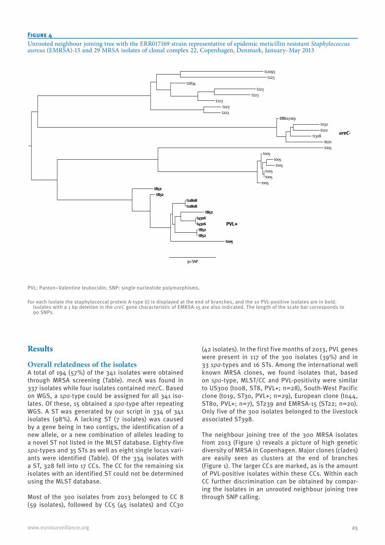
25www.eurosurveillance.org
Results
Overall relatedness of the isolatesA total of 194 (57%) of the 341 isolates were obtained through MRSA screening (Table). mecA was found in 337 isolates while four isolates contained mecC. Based on WGS, a spa-type could be assigned for all 341 iso-lates. Of these, 15 obtained a spa-type after repeating WGS. A ST was generated by our script in 334 of 341 isolates (98%). A lacking ST (7 isolates) was caused by a gene being in two contigs, the identification of a new allele, or a new combination of alleles leading to a novel ST not listed in the MLST database. Eighty-five spa-types and 35 STs as well as eight single locus vari-ants were identified (Table). Of the 334 isolates with a ST, 328 fell into 17 CCs. The CC for the remaining six isolates with an identified ST could not be determined using the MLST database. Most of the 300 isolates from 2013 belonged to CC 8 (59 isolates), followed by CC5 (45 isolates) and CC30
(42 isolates). In the first five months of 2013, PVL genes were present in 117 of the 300 isolates (39%) and in 33 spa-types and 16 STs. Among the international well known MRSA clones, we found isolates that, based on spa-type, MLST/CC and PVL-positivity were similar to US300 (t008, ST8, PVL+; n=28), South-West Pacific clone (t019, ST30, PVL+; n=29), European clone (t044, ST80, PVL+; n=7), ST239 and EMRSA-15 (ST22; n=20). Only five of the 300 isolates belonged to the livestock associated ST398.
The neighbour joining tree of the 300 MRSA isolates from 2013 (Figure 1) reveals a picture of high genetic diversity of MRSA in Copenhagen. Major clones (clades) are easily seen as clusters at the end of branches (Figure 1). The larger CCs are marked, as is the amount of PVL-positive isolates within these CCs. Within each CC further discrimination can be obtained by compar-ing the isolates in an unrooted neighbour joining tree through SNP calling.
Figure 4Unrooted neighbour joining tree with the ERR017169 strain representative of epidemic meticillin resistant Staphylococcus aureus (EMRSA)-15 and 29 MRSA isolates of clonal complex 22, Copenhagen, Denmark, January–May 2013
PVL: Panton–Valentine leukocidin; SNP: single nucleotide polymorphisms.
For each isolate the staphylococcal protein A-type (t) is displayed at the end of branches, and the 10 PVL-positive isolates are in bold. Isolates with a 1 bp deletion in the ureC gene characteristic of EMRSA-15 are also indicated. The length of the scale bar corresponds to 90 SNPs.
90 SNP
PVL+
ureC-
ERR017169
t223
t005
t223
t005
t005
t005
t852
t223
t005
t670
t2834
t005
t4326
t022
t005
t852
t852
t005
t852
t223
t223
t1328
t11808
t032
t852
t11808
t4326
t223
t12093

26 www.eurosurveillance.org
Staphylococcal protein A-type 304 analysisHealthcare-associated MRSA outbreaks are rare in Denmark, and the only clusters identified as HA-MRSA were t304, ST6, PVL-negative, associated with a
neonatal outbreak spreading in neonatal wards in the Capital Region (Figure 2 and Figure 3) and t024, ST8, PVL-negative, a clone that has been healthcare associ-ated since 2003 [14]. t304, ST6 in neonates and their relatives were, apart from one case in February 2013, all identified either in the last seven months of 2011 (n = 7) or in 2012 (n = 26) . Comparing the 55 t304, ST6 isolates from Copenhagen in the period from 2010 to 2013, the epidemiologically related outbreak isolates (n = 34) were genetically closely related, having from 1 to 10 SNPs compared with the closest related outbreak isolates. t304 isolates from the same neonatal ward were in most cases even closer related with a maxi-mum of five SNPs separating them. The t304 patients who were not epidemiologically related to the out-break (n = 21), were in six cases clearly different from the outbreak strains with between 67 and more than 200 SNPs separating them from the outbreak strains. Seven isolates had between 11 to 36 SNPs compared with the closest related of the outbreak isolates, while eight isolates only had from four to ten SNPs (Figure 2 and Figure 3).
Clonal complex 22 analysisAn expansion of the CC22 isolate tree is shown in Figure 4 and compared with a ST22 isolate representa-tive of EMRSA-15 [5]. The 29 Danish CC22 isolates are diverse and include ten different spa-types. Ten of these isolates were from patients living in four differ-ent households. Based on SNP calling, the clusters of these isolates within households differed between 0 to 14 SNPs, while epidemiologically unrelated isolates differed at between 69 to 1,207 SNPs. PVL was found in ten of the 29 isolates (32%) including all t852, t4326, t11808 and one of eight t005 isolates. Four of the 29 isolates had the 1 bp deletion in ureC described as typ-ical for EMRSA-15 and were the isolates closest related to the English prototype ST22 isolate ERR017169 based on SNP calling. These four patients had been hospital-ised before the finding of MRSA, two of them in Italy and Spain and two in Denmark. Only one of the remain-ing 25 CC22 patients had been hospitalised before the finding of MRSA. Nineteen of the 29 patients were not of ethnic Danish origin, and one of the five ethnic Danes had recently travelled in India. We assume that most of these CC22 isolates were acquired through international travel.
Sequence type 80 analysisA total of 13 isolates belonging to the European clone are presented in Figure 5. They were all PVL-positive and represented four different spa-types of which t044 accounted for seven isolates. Two of the t044 isolates differing with 59 SNPs were from two persons living in a three-generation household. SNP analysis revealed that the 59 SNPs were dispersed throughout the genome, and could therefore not be explained by a single recombination event. The rest of the t044 iso-lates were from patients not epidemiologically related, and they differed with 130 to 365 SNPs. There were three t376 isolates, which were from a father and his
Figure 5Unrooted neighbour joining tree of meticillin resistant Staphylococcus aureus isolates of sequence type 80 found in Copenhagen, Denmark, January–May 2013 (n=13)
*Same household, 59 SNPs between the two isolates. **Father and two daughters.
SNPs: single nucleotide polymorphisms.
The staphylococcal protein A-type (t) of each isolate is displayed at the end of branches. The length of the scale bar corresponds to 100 SNPs.
t1028
t1028
t376**
t376**
t376**
t044
t044
t044
t044
t044*
t044*
t044
t131
100.0

27www.eurosurveillance.org
two daughters and which had a maximum of 10 SNPs between them. Two isolates of t1028 had only 17 SNPs between them, but we have not identified an epidemio-logical link between these patients.
DiscussionThe emergence of predominantly CA-MRSA was first recognised in Denmark in 2003 [14]. Since 2003 the number of new MRSA patients in Copenhagen has increased 20-fold from 33 to 674 cases in 2012 [4]. The genetic diversity that was already seen in the first years of the MRSA epidemic has increased over the years, and in 2012, 142 different spa-types were found in Copenhagen (data not shown). This diversity is mainly related to travel and tourism e.g. cases where Danish families visit relatives in countries where MRSA is prevalent in the community, and acquisition in persons working with healthcare abroad [15]. The high diversity based on spa-types indicates a low level of endemic transmission, and could be further differentiated in this study, based on WGS. Cases imported through hospitalisation abroad are routinely found through screening of these patients when they are transferred to Danish hospitals. In general, MRSA is first isolated in clinical samples and subsequent ring screening usu-ally identifies carriers in their households. However, there is an under-identification of MRSA as a number of patients have had recurrent skin and soft tissue infec-tions before they are sampled. This means that some patients who only have a single infection or short-term carriage state and then clear MRSA are not identified.
Livestock associated MRSA, ST398, accounted for less than 2% of MRSA in Copenhagen, a capital city sur-rounded by a large suburban area with little livestock production. However, ST398 was the most common clone in the whole of Denmark in 2012 accounting for 232 of 1,556 isolates (15%), predominantly seen in regions with a large pig production [4].
Two UK studies on neonatal MRSA outbreaks have con-firmed the usefulness of WGS in outbreak investiga-tions, and showed that the number of SNPs between outbreak related isolates were less than 15 [16,17], except in one case where a mutation in the mutS gene was explained as the reason for more divergence. Furthermore, Harris et al. [16] found additionally ten isolates that were initially not considered part of a particular outbreak, but were closely related to out-break strains by WGS, and retrospective epidemiologi-cal links were found in most cases. In our study, WGS could confirm the relatedness between 34 t304 iso-lates that were epidemiologically related to the neona-tal outbreak in Copenhagen. Six isolates were clearly different from the outbreak strains, and this was also consistent with patient data. Fifteen isolates, that were not considered outbreak-related based on epidemio-logical data, differed in eight cases with only four to 10 SNPs to the closest related outbreak strains, and in seven cases with 11 to 36 SNPs (Figure 3). Since 11 of these 15 isolates were obtained in 2013, we hypothesise
that a community transmission chain might exist, but that the epidemiological link between patients is miss-ing. WGS seems promising in outbreak investigations, and might add information on the spread of outbreak clones outside the hospital setting, where the connec-tion between patients is frequently not clear.
The CC22 isolates found were diverse and only four isolates were similar to the English prototype CC22, EMRSA-15. Two of these four isolates were imported from Spain and Italy. In 2012, t032, CC22 accounted for 89 isolates and was the fifth most common MRSA type in Denmark representing almost 6% of new MRSA cases [4], but only seven of these 89 isolates were from the Capital Region [18].
The typical CA-MRSA ST80 was only found in 13 patients in the five month period. There is evidence of repeated introduction of this clone into Denmark with only one case of possible community spread, t1028, based on the number of SNPs (Figure 5). Although there is no consensus on how many SNPs define whether isolates are related or unrelated [19], it was surprising that two household isolates of ST80 had as many as 59 SNPs between them. A closer look at this household revealed that this family of three generations immigrated to Denmark five years prior, and presumably have been MRSA carriers for many years. According to Holden et al. [5], SNPs are estimated to occur in the core genome at a rate of 1.3 x 10 −6 substitutions per nucleotide per year or three to four SNPs per year. Based on the rela-tively few closely related isolates in our study, we won-der if we will have a congruent SNP evolution for all MRSA clones.
Our policy until the end of 2012 has been to spa-type all MRSA using differences in spa-types to delineate different clones e.g. t024 different from t008 [20]. However, when less common spa-types were observed the link to international clones was often not seen. For example, the rare t1133, ST1456, PVL-positive , with only 11 isolates in the SpaServer database, was not easily associated to the South-West Pacific clone (t019, ST30, PVL+) before the neighbour joining tree clustered them together. Currently, the spa-type, ST and absence/presence of PVL are routinely found from the laboratory information system. The cost of WGS is steadily going down and today it would be more expen-sive for us to perform traditional Sanger sequencing for spa-typing and MLST. Furthermore, the WGS platform is universally applicable and is now routinely used in other healthcare-associated outbreak investigations.
A disadvantage is the slower turnaround time in our WGS-setup before typing results can be delivered. WGS is run in batches of 24 isolates, typically twice weekly. This means that from the date of sample to a WGS typ-ing result can be up to 12 days or 4–8 days after the isolate is received for WGS. The current MLST S. aureus database contains fewer isolates and types than the spa server database and this leads to a number of

28 www.eurosurveillance.org
isolates only having six known alleles and one novel allele. In these cases our software calls all the STs with six matches, and we manually assign the ST as a single locus variant (SLV) of the most prevalent ST.
The data presented in this study show that after build-ing up a local MRSA whole genome database, real time WGS gives additional information to the evolution and spread of MRSA. The method can be used to investi-gate outbreaks, but epidemiological data still need to be included. More insight into how to interpret the relatedness between isolates based on SNP analysis is needed as well as improved software designed for per-sons without bioinformatics skills.
Acknowledgments We are grateful for the continuous help and support given to us by Elizabeth Batty, Tanya Golubchik, NDM Experimental Medicine Division, University of Oxford and Camilla Ip, Wellcome Trust Centre for Human Genetics, Oxford, UK. We thank the staff at the Departments of Clinical Microbiology at Hvidovre Hospital, Herlev Hospital, and Rigshospitalet and the Infection Control Organizations at Hvidovre Hospital, Herlev Hospital, Bispebjerg Hospital, Frederiksberg Hospital, Hillerød Hospital and Rigshospitalet. HKJ was supported by a clinical research grant from The Novo Nordisk Foundation.
Conflict of interestNone declared.
Authors’ contributionsMDB, HLS and HW: Contributed substantially to the design, the analysis and the interpretation of data. Drafted the work and revised it for important intellectual content. HM, KK, KS, JBN, SMR, LBC, AJWS, JOJ, HKJ, LPA, ISP, DWC, RB, KB, PW: Contributed substantially to the interpretation of data. Revised the work critically for important intellectual content.
References1. Le VT, Diep BA. Selected insights from application of whole-
genome sequencing for outbreak investigations. Curr Opin Crit Care. 2013;19(5):432-9. http://dx.doi.org/10.1097/MCC.0b013e3283636b8c PMID:23856896
2. Reuter S, Ellington MJ, Cartwright EJ, Köser CU, Török ME, Gouliouris T, et al. Rapid bacterial whole-genome sequencing to enhance diagnostic and public health microbiology. JAMA Intern Med. 2013;173(15):1397-404. http://dx.doi.org/10.1001/jamainternmed.2013.7734 PMID:23857503
3. Zankari E, Hasman H, Cosentino S, Vestergaard M, Rasmussen S, Lund O, et al. Identification of acquired antimicrobial resistance genes. J Antimicrob Chemother. 2012;67(11):2640-4. http://dx.doi.org/10.1093/jac/dks261 PMID:22782487
4. The Danish Integrated Antimicrobial Resistance Monitoring and Research Programme (DANMAP) Reports. [Accessed 5 Dec 2013]. Available from: http: www.danmap.org/Downloads/Reports.aspx
5. Holden MT, Hsu LY, Kurt K, Weinert LA, Mather AE, Harris SR, et al. A genomic portrait of the emergence, evolution, and global spread of a methicillin-resistant Staphylococcus aureus pandemic. Genome Res. 2013;23(4):653-64. http://dx.doi.org/10.1101/gr.147710.112 PMID:23299977
6. Otter JA, French GL. Molecular epidemiology of community-associated meticillin-resistant Staphylococcus aureus in Europe. Lancet Infect Dis. 2010;10(4):227-39. http://dx.doi.org/10.1016/S1473-3099(10)70053-0 PMID:20334846
7. Didelot X, Bowden R, Wilson DJ, Peto TE, Crook DW. Transforming clinical microbiology with bacterial genome sequencing. Nat Rev Genet. 2012;13(9):601-12. http://dx.doi.org/10.1038/nrg3226 PMID:22868263
8. Zerbino DR, Birney E. Velvet: algorithms for de novo short read assembly using de Bruijn graphs. Genome Res. 2008;18(5):821-9. http://dx.doi.org/10.1101/gr.074492.107 PMID:18349386
9. Ridom SpaServer. [Accessed 5 Dec 2013]. Available from: http: www.spaserver.ridom.de/
10. Multi Locus Sequence Typing (MLST). [Accessed 5 Dec 2013]. Available from: http: www.mlst.net/
11. Huson DH, Bryant D. Application of phylogenetic networks in evolutionary studies. Mol Biol Evol. 2006;23(2):254-67. http://dx.doi.org/10.1093/molbev/msj030 PMID:16221896
12. FigTree. [Accessed 5 Dec 2013]. Available from: http: tree.bio.ed.ac.uk/software/figtree/
13. Monecke S, Coombs G, Shore AC, Coleman DC, Akpaka P, Borg M, et al. A field guide to pandemic, epidemic and sporadic clones of methicillin-resistant Staphylococcus aureus. PLoS ONE. 2011;6(4):e17936. http://dx.doi.org/10.1371/journal.pone.0017936 PMID:21494333
14. Bartels MD, Boye K, Rhod Larsen A, Skov R, Westh H. Rapid increase of genetically diverse methicillin-resistant Staphylococcus aureus, Copenhagen, Denmark. Emerg Infect Dis. 2007;13(10):1533-40. http://dx.doi.org/10.3201/eid1310.070503 PMID:18258003
15. Böcher S, Gervelmeyer A, Monnet DL, Mølbak K, Skov RL; Danish CA-MRSA Study Group. Methicillin-resistant Staphylococcus aureus: risk factors associated with community-onset infections in Denmark. Clin Microbiol Infect. 2008;14(10):942-8. http://dx.doi.org/10.1111/j.1469-0691.2008.02055.x PMID:18752595
16. Harris SR, Cartwright EJ, Török ME, Holden MT, Brown NM, Ogilvy-Stuart AL, et al. Whole-genome sequencing for analysis of an outbreak of meticillin-resistant Staphylococcus aureus: a descriptive study. Lancet Infect Dis. 2013;13(2):130-6. http://dx.doi.org/10.1016/S1473-3099(12)70268-2 PMID:23158674
17. Köser CU, Holden MT, Ellington MJ, Cartwright EJ, Brown NM, Ogilvy-Stuart AL, et al. Rapid whole-genome sequencing for investigation of a neonatal MRSA outbreak. N Engl J Med. 2012;366(24):2267-75. http://dx.doi.org/10.1056/NEJMoa1109910 PMID:22693998
18. Böcher S, Skov RL, Knudsen MA, Guardabassi L, Mølbak K, Schouenborg P, et al. The search and destroy strategy prevents spread and long-term carriage of methicillin-resistant Staphylococcus aureus: results from the follow-up screening of a large ST22 (E-MRSA 15) outbreak in Denmark. Clin Microbiol Infect. 2010;16(9):1427-34. http://dx.doi.org/10.1111/j.1469-0691.2010.03137.x PMID:20041904
19. Lindsay JA. Evolution of Staphylococcus aureus and MRSA during outbreaks. Infect Genet Evol. 2014;21:548-53. http://dx.doi.org/10.1016/j.meegid.2013.04.017 PMID:23665384
20. Larsen AR, Goering R, Stegger M, Lindsay JA, Gould KA, Hinds J, et al. Two distinct clones of methicillin-resistant Staphylococcus aureus (MRSA) with the same USA300 pulsed-field gel electrophoresis profile: a potential pitfall for identification of USA300 community-associated MRSA. J Clin Microbiol. 2009;47(11):3765-8. http://dx.doi.org/10.1128/JCM.00934-09 PMID:19759225