volume 27, n umber 3, 2011 - american red cross€¦ · · 2012-06-09volume 27, number 3, 2011...
TRANSCRIPT

Vo lu m e 27, Nu m b e r 3 , 2011

109 Advert isements
113 instruct ions for Authors
ImmunohematologyVolume 27, Number 3, 2011
C O N T EN TS
83 cAse report
Persistent complement-dependent Anti-AnWj in a lymphoproliferative disorder: a case study and reviewG. Grigoriadis, J. Condon, K. Green, M.A. Anderson, M. Borosak, and E. Wood
104 orig inAl report
Should we be screening for anti-Jsa?N.M. Nikolis, F.N. Boctor, W.A. Heaton, and J. Martone
89 rev iew
The Indian blood group systemQ. Xu
94 report
The legacy of Charles R. Drew, MD, CM, MDSc.B.A. Wilson, W.G. O’Connor, and M.S. Willis
101 cAse report
An unusual case of an acute hemolytic transfusion reaction caused by an autoanti-IN. Win, M. Needs, S. Rahman, P. Gold, and S. Ward
107 Announcements

Immunohematology is published quarterly (March, June, September, and December) by the American Red Cross, National Headquarters, Washington, DC 20006.
Immunohematology is indexed and included in Index Medicus and MEDLINE on the MEDLARS system. The contents are also cited in the EBASE/Excerpta Medica and Elsevier
BIOBASE/Current Awareness in Biological Sciences (CABS) databases.
The subscription price is $40.00 (U.S.) and $50.00 (foreign) per year.
Subscriptions, Change of Address, and Extra Copies:
Immunohematology, P.O. Box 40325 Philadelphia, PA 19106
Or call (215) 451-4902
Web site: www.redcross.org/immunohematology
Copyright 2011 by The American National Red Cross ISSN 0894-203X
editor- in-chief
Sandra Nance, MS, MT(ASCP)SBBPhiladelphia, Pennsylvania
mAnAging editor
Cynthia Flickinger, MT(ASCP)SBBPhiladelphia, Pennsylvania
senior medicAl editor
Geralyn M. Meny, MDPhiladelphia, Pennsylvania
technicAl editor
Dawn M. Rumsey, ART (CSMLT)Glen Allen, Virginia
Assoc iAte medicAl editors
David Moolten, MDPhiladelphia, Pennsylvania
Ralph R. Vassallo, MDPhiladelphia, Pennsylvania
editor iAl AssistAnt
Sheetal Patel
copy editor
Mary L. Tod
proofreAder
Lucy Oppenheim
product ion AssistAnt
Marge Manigly
electronic publ isher
Paul Duquette
editor iAl boArd
Patricia Arndt, MT(ASCP)SBBPomona, California
James P. AuBuchon, MDSeattle, Washington
Martha R. Combs, MT(ASCP)SBBDurham, North Carolina
Geoffrey Daniels, PhDBristol, United Kingdom
Anne F. Eder, MDWashington, District of Columbia
George Garratty, PhD, FRCPathPomona, California
Brenda J. Grossman, MDSt. Louis, Missouri
Christine Lomas-Francis, MScNew York City, New York
Paul M. Ness, MDBaltimore, Maryland
Joyce Poole, FIBMSBristol, United Kingdom
Mark Popovsky, MDBraintree, Massachusetts
Marion E. Reid, PhD, FIBMSNew York City, New York
S. Gerald Sandler, MDWashington, District of Columbia
Jill R. Storry, PhD Lund, Sweden
David F. Stroncek, MDBethesda, Maryland
emeritus editor
Delores Mallory, MT(ASCP) SBBSupply, North Carolina
on our cover
Charles Drew teaching interns and residents during rounds at Freedmen’s Hospital, ca. 1947Original Repository: Howard University, Moorland-Spingarn Research Center, Charles R. Drew Papers. Used with permission of Scurlock Studio Records, Archives Center, National Museum of American History, Smithsonian Institution.
The boys whom we are now helping to train, I believe, in time will constitute my greatest contribution to medicine.
Charles R. Drew, MD, CM, MDSc, 1947

IMMUNOHEMATOLOGY, Volume 27, Number 3, 2011 83
Persistent complement-dependent anti-AnWj in a lymphoproliferative disorder: a case study and reviewG. Grigoriadis, J. Condon, K. Green, M.A. Anderson, M. Borosak, and E. Wood
AnWj is a high-incidence antigen present on the red blood cells (RBCs) of greater than 99 percent of the general population. A 58-year-old man underwent autologous hematopoietic stem cell transplantation (HSCT) for stage IVa mantle cell lymphoma. This procedure was complicated by failure to engraft, necessitating ongoing support with blood components. After a 2-month period of uneventful transfusion support, the patient experienced increasingly severe reactions with fever and evidence of intravascular hemolysis, including hemoglobinuria. Testing revealed a complement-dependent anti-AnWj. Phenotyping confirmed the AnWj– phenotype. Anti-AnWj was persistent despite immunosuppression, including treatment with allogeneic HSCT. Of interest, the pathogenesis of the downregulation of the graft AnWj in this patient is unclear. Immunohematology 2011;27:83–88.
Key Words: anti-AnWj, lymphoma, hemolysis
AnWj is a high-incidence antigen present on the red blood cells (RBCs) of greater than 99 percent of the population. Its molecular basis is not yet known. An antibody to this antigen, anti-AnWj, has been described in extremely rare serum samples as a result of an acquired AnWj– phenotype in individuals with lymphoproliferative disorders.1–5 In several cases, successful treatment of the underlying disease has resulted in disappearance of the antibody.1,4 Since it was first recognized, there have been several reports that have addressed transfusion in the presence of anti-AnWj, with a range of clinical outcomes.1,4–9 We report a case of persistent acquired complement-dependent anti-AnWj in the context of an underlying lymphoproliferative disorder unresponsive to immunosuppression including allogeneic hematopoietic stem cell transplantation (HSCT).
Case Report
A 58-year-old man diagnosed with stage IVa mantle cell lymphoma achieved complete remission after chemotherapy and proceeded to an autologous HSCT. Unfortunately, the autologous HSCT was complicated by failure to engraft that necessitated transfusion support with both RBCs
and platelets. Bone marrow biopsy revealed a hypoplastic marrow with features consistent with severe aplasia without evidence of residual lymphoma. The cause of the aplasia was likely iatrogenic, although a stromal defect could not be excluded. Three months later, episodes of hemoglobinuria after transfusion of RBCs and intermittent mild transfusion reactions with evidence of biochemical hemolysis and a positive hemosiderin test were noted (Fig. 1); however, posttransfusion testing performed on four donor units, transfused on June 20, July 4, July 30, and August 24, 2008, and on antibody screening cells was unremarkable. These reactions culminated in a severe hemolytic transfusion reaction on August 24, when transfusion of 150 mL of blood resulted in chills,
Case RepoRt
Figure 1. Results of laboratory testing including bilirubin (μmol/L) and hemoglobin (Hb; g/L) that demonstrate evidence of biochemical hemolysis (bilirubin peaks) without a unsustained increase in Hb following red blood cell (RBC) transfusion with AnWj+ units. Interspersed are transfusions with AnWj+ units (arrows) that did result in an incremental increase in Hb without overt evidence of hemolysis. The severity of the transfusion reactions increased over this time period. Posttransfusion testing was unremarkable (see text).
84 IMMUNOHEMATOLOGY, Volume 27, Number 3, 2011
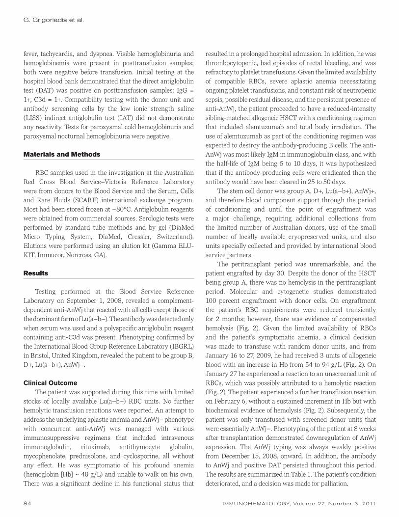
fever, tachycardia, and dyspnea. Visible hemoglobinuria and hemoglobinemia were present in posttransfusion samples; both were negative before transfusion. Initial testing at the hospital blood bank demonstrated that the direct antiglobulin test (DAT) was positive on posttransfusion samples: IgG = 1+; C3d = 1+. Compatibility testing with the donor unit and antibody screening cells by the low ionic strength saline (LISS) indirect antiglobulin test (IAT) did not demonstrate any reactivity. Tests for paroxysmal cold hemoglobinuria and paroxysmal nocturnal hemoglobinuria were negative.
Materials and Methods
RBC samples used in the investigation at the Australian Red Cross Blood Service–Victoria Reference Laboratory were from donors to the Blood Service and the Serum, Cells and Rare Fluids (SCARF) international exchange program. Most had been stored frozen at –80°C. Antiglobulin reagents were obtained from commercial sources. Serologic tests were performed by standard tube methods and by gel (DiaMed Micro Typing System, DiaMed, Cressier, Switzerland). Elutions were performed using an elution kit (Gamma ELU-KIT, Immucor, Norcross, GA).
Results
Testing performed at the Blood Service Reference Laboratory on September 1, 2008, revealed a complement-dependent anti-AnWj that reacted with all cells except those of the dominant form of Lu(a–b–). The antibody was detected only when serum was used and a polyspecific antiglobulin reagent containing anti-C3d was present. Phenotyping confirmed by the International Blood Group Reference Laboratory (IBGRL) in Bristol, United Kingdom, revealed the patient to be group B, D+, Lu(a–b+), AnWj–.
Clinical OutcomeThe patient was supported during this time with limited
stocks of locally available Lu(a–b–) RBC units. No further hemolytic transfusion reactions were reported. An attempt to address the underlying aplastic anemia and AnWj– phenotype with concurrent anti-AnWj was managed with various immunosuppressive regimens that included intravenous immunoglobulin, rituximab, antithymocyte globulin, mycophenolate, prednisolone, and cyclosporine, all without any effect. He was symptomatic of his profound anemia (hemoglobin [Hb] ~ 40 g/L) and unable to walk on his own. There was a significant decline in his functional status that
resulted in a prolonged hospital admission. In addition, he was thrombocytopenic, had episodes of rectal bleeding, and was refractory to platelet transfusions. Given the limited availability of compatible RBCs, severe aplastic anemia necessitating ongoing platelet transfusions, and constant risk of neutropenic sepsis, possible residual disease, and the persistent presence of anti-AnWj, the patient proceeded to have a reduced-intensity sibling-matched allogeneic HSCT with a conditioning regimen that included alemtuzumab and total body irradiation. The use of alemtuzumab as part of the conditioning regimen was expected to destroy the antibody-producing B cells. The anti-AnWj was most likely IgM in immunoglobulin class, and with the half-life of IgM being 5 to 10 days, it was hypothesized that if the antibody-producing cells were eradicated then the antibody would have been cleared in 25 to 50 days.
The stem cell donor was group A, D+, Lu(a–b+), AnWj+, and therefore blood component support through the period of conditioning and until the point of engraftment was a major challenge, requiring additional collections from the limited number of Australian donors, use of the small number of locally available cryopreserved units, and also units specially collected and provided by international blood service partners.
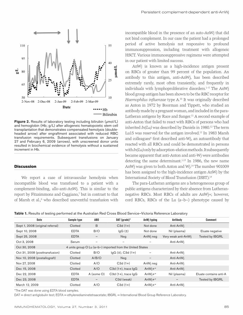
The peritransplant period was unremarkable, and the patient engrafted by day 30. Despite the donor of the HSCT being group A, there was no hemolysis in the peritransplant period. Molecular and cytogenetic studies demonstrated 100 percent engraftment with donor cells. On engraftment the patient’s RBC requirements were reduced transiently for 2 months; however, there was evidence of compensated hemolysis (Fig. 2). Given the limited availability of RBCs and the patient’s symptomatic anemia, a clinical decision was made to transfuse with random donor units, and from January 16 to 27, 2009, he had received 3 units of allogeneic blood with an increase in Hb from 54 to 94 g/L (Fig. 2). On January 27 he experienced a reaction to an unscreened unit of RBCs, which was possibly attributed to a hemolytic reaction (Fig. 2). The patient experienced a further transfusion reaction on February 6, without a sustained increment in Hb but with biochemical evidence of hemolysis (Fig. 2). Subsequently, the patient was only transfused with screened donor units that were essentially AnWj–. Phenotyping of the patient at 8 weeks after transplantation demonstrated downregulation of AnWj expression. The AnWj typing was always weakly positive from December 15, 2008, onward. In addition, the antibody to AnWj and positive DAT persisted throughout this period. The results are summarized in Table 1. The patient’s condition deteriorated, and a decision was made for palliation.
G. Grigoriadis et al.
IMMUNOHEMATOLOGY, Volume 27, Number 3, 2011 85
Persistent complement-dependent anti-AnWj
Discussion
We report a case of intravascular hemolysis when incompatible blood was transfused to a patient with a complement-binding, allo-anti-AnWj. This is similar to the report by Fitzsimmons and Caggiano,7 but in contrast to that of Marsh et al.,6 who described uneventful transfusion with
incompatible blood in the presence of an auto-AnWj that did not bind complement. In our case the patient had a prolonged period of active hemolysis not responsive to profound immunosuppression, including treatment with allogeneic HSCT. Various immunosuppressive regimens were attempted in our patient with limited success.
AnWj is known as a high-incidence antigen present on RBCs of greater than 99 percent of the population. An antibody to this antigen, anti-AnWj, has been described extremely rarely, most often transiently, and frequently in individuals with lymphoproliferative disorders.1–5 The AnWj blood group antigen has been shown to be the RBC receptor for Haemophilus influenzae type A.10 It was originally described as Anton in 1972 by Boorman and Tippett, who studied an antibody made by a pregnant woman, and included in the para-Lutheran antigens by Race and Sanger.11 A second example of anti-Anton that failed to react with RBCs of persons who had inherited In(Lu) was described by Daniels in 1980.12 The term Lu15 was reserved for the antigen involved.12 In 1983 Marsh and colleagues6 first described anti-Wj, an autoantibody that reacted with all RBCs and could be demonstrated in persons with In(Lu) only by adsorption-elution methods. It subsequently became apparent that anti-Anton and anti-Wj were antibodies detecting the same determinant.1,13 In 1986, the new name AnWj was given to both Anton and Wj.14 The number 901009 has been assigned to the high-incidence antigen AnWj by the International Society of Blood Transfusion (ISBT).14
The para-Lutheran antigens are a heterogeneous group of public antigens characterized by their absence from Lutheran-negative RBCs. Most RBCs of adults are AnWj+; however, cord RBCs, RBCs of the Lu (a–b–) phenotype caused by
Table 1. Results of testing performed at the Australian Red Cross Blood Service–Victoria Reference Laboratory
Date Sample type ABO DAT (grade)* AnWj typing Antibody Comment
Sept 1, 2008 (original referral) Clotted B C3d (1+) Not done Anti-AnWj
Sept 10, 2008 EDTA B/O lgG (±) Not done Nil (plasma) Eluate negative
Sept 25, 2008 EDTA — Neg AnWj neg Very weak anti-AnWj Tested by IBGRL
Oct 3, 2008 Serum – – – Anti-AnWj
Oct 30, 2008 4 units group O Lu (a–b–) imported from the United States
Oct 31, 2008 (posttransfusion) Clotted B/O lgG (±), C3d (1+) – Anti-AnWj
Nov 10, 2008 (postallograft) Clotted A/B/O Neg – Anti-AnWj
Nov 27, 2008 Clotted A/O C3d (1+) AnWj neg Anti-AnWj
Dec 15, 2008 Clotted A/O C3d (1+), trace IgG AnWj+w Anti-AnWj
Dec 22, 2008 EDTA A (some O) C3d (1+), trace IgG AnWj+w Nil (plasma) Eluate contains anti-A
Dec 23, 2008 EDTA – C3d (weak) AnWj+w – Tested by IBGRL
March 12, 2009 Clotted A/O C3d (1+) AnWj+w Anti-AnWj
*The DAT was done using EDTA blood samples.DAT = direct antiglobulin test; EDTA = ethylenediaminetetraacetate; IBGRL = International Blood Group Reference Laboratory.
Figure 2. Results of laboratory testing including bilirubin (μmol/L) and hemoglobin (Hb; g/L) after allogeneic hematopoietic stem cell transplantation that demonstrates compensated hemolysis (double-headed arrow) after engraftment associated with reduced RBC transfusion requirements. Subsequent transfusions on January 27 and February 6, 2009 (arrows), with unscreened donor units resulted in biochemical evidence of hemolysis without a sustained increment in Hb.

86 IMMUNOHEMATOLOGY, Volume 27, Number 3, 2011
G. Grigoriadis et al.
the dominant suppressor gene In(Lu), and RBCs from rare individuals with anti-AnWj in their serum essentially type as AnWj–.15 Poole and Giles16 in 1982 demonstrated that the Anton “para-Lutheran” antigen is associated with the Lutheran blood group system only by In(Lu), a gene independent of the Lutheran locus. Several examples of anti-AnWj have been documented to be autoantibodies; their appearance was associated with transient depression of AnWj on the RBC.1,6,7 The AnWj– phenotype is usually considered to be an acquired phenomenon. In 1991, Poole and colleagues17 described a family showing inheritance of the Anton blood group antigen, AnWj, and independence of AnWj from Lutheran.
Although hemolytic transfusion reactions have been attributed to anti-AnWj, predicting events for individual patients is difficult. Given the limited availability of AnWj– RBCs, predicting the hemolytic potential by in vitro and in vivo assays in each case may be considered in the event of a requirement for transfusion. Several reports have addressed transfusion in the presence of anti-AnWj.1,4–9 In these reports, the antibody was an autoantibody, with the results of transfusing incompatible blood varying from asymptomatic to fever, chills, and intravascular hemolysis with hemoglobinuria and hemoglobinemia. Whitsett et al.,4 using chromium studies, revealed that allogeneic AnWj+ RBCs had shortened survival with two components identified; 24 percent of RBCs were eliminated rapidly (within 24 hours), with an additional 24 percent of RBCs being eliminated more slowly (6 days). This pattern is usually observed when there are both IgG and IgM antibodies present, or when the IgG antibodies fix complement.4 The shortened survival of allogeneic blood of similar phenotype may explain the report of transfusion reactions explained by autoanti-AnWj. Erythrocyte survival studies may be useful in cases of anti-AnWj to assist in predicting acute hemolytic reactions,5,9,18 especially when antigen-negative donor units may not be available; however, these studies are not routinely accessible and often are not available in the timeframe required for urgent transfusion decision making.
The molecular basis for the high-incidence AnWj is not yet known. The acquired form of the AnWj– phenotype is a transient depression of AnWj, usually with a concomitant alloanti-AnWj, and is extremely rare. It has been described in individuals with lymphoproliferative disorders. The pathogenesis of the downregulation of AnWj is unclear; however, in several cases successful treatment of the underlying disease resulted in disappearance of the antibody and reinstatement of AnWj.1,4 Transient depression of RBC antigens has been described; such cases include loss of Kell
and Lutheran during consecutive relapses of autoimmune thrombocytopenia.19 Various mechanisms have been postulated and include antigen shedding, defective insertion of the antigen into the RBC membrane, glycosylation, or blocking with downregulation of antigen copy number.19 Further work needs to be done to identify the mechanism of this downregulation.
CD44 is postulated to be the location of AnWj. CD44 is part of the collagen family of receptors and has an important role in hyaluronic acid binding, lymphocyte homing, leukocyte activation, and cell adhesion.20 It is ubiquitously expressed.20 Spring et al.21 demonstrated that the Indian blood group antigen is located on CD44. The molecular basis of the Ina/Inb polymorphism is a result of a single point mutation in the CD44 gene.22 A case report of a 9-year-old with a novel form of congenital dyserythropoietic anemia associated with a deficiency of erythrocyte CD44 lacked In antigens and happened to be Co(a–b–) and AnWj–.23 A phenotypic associ-ation of AnWj with CD44 was described, and unlike Ina/Inb, AnWj is resistant to trypsin, chymotrypsin, neuraminidase, and papain.24 The protease cleavage sites on CD44 were localized by monoclonal anti-CD44 antibodies that predict that AnWj is located in a region near the lipid bilayer.15 This region provides several sites for potential posttranslational modification.15 The lack of AnWj on CD44-deficient cells and the appearance of AnWj on Jurkat cells transfected with wild-type CD44 cDNA lends support to AnWj being located on CD44.25 It has been suggested that the structure of the antigen depends on posttranslational modification of the protein by a particular glycosylation.15 This may explain the conversion in newborn infants from AnWj– to AnWj+ within the first 50 days of life. This conversion was seen to take only 1 day to complete. This was not attributable to replacement of AnWj– cells with AnWj+ cells from the bone marrow but caused by an extrinsic factor.26 However, no such factor could be isolated from the serum of the neonates. The mechanism that enables a RBC population to express antigen within days is not understood but may in part be caused by the potential for posttranslational modification as discussed earlier.
Interestingly, AnWj is the putative receptor for fimbriae-bearing strains of H. influenzae.10 Elucidation of the nature of AnWj is of considerable interest because as a receptor for H. influenzae, a major cause of respiratory infections worldwide, it may serve as a putative therapeutic target. H. influenzae agglutinated RBCs of all types except those from individuals with In(Lu)-acquired AnWj– phenotype or from cord blood samples.10 Epithelial cells from an individual with inherited AnWj– phenotype failed to bind H. influenzae, but cells from

IMMUNOHEMATOLOGY, Volume 27, Number 3, 2011 87
individuals with the acquired phenotype, in which cells other than erythrocytes express AnWj, were capable of binding H. influenzae.27 It is postulated that the low levels of AnWj in the acquired AnWj– phenotype, In(Lu), and cord blood are sufficient for binding of H. influenzae.27
The few cases in the literature that describe anti-AnWj describe it as a transient phenomenon, typically in the setting of an underlying lymphoproliferative disorder. In our case the patient had a prolonged period of active hemolysis unresponsive to profound immunosuppression that included an allogeneic HSCT. The pathogenesis of the downregulation of AnWj in this setting remains unclear. These situations present a major management challenge for transfusion support in patients who have antibodies to high-incidence antigens whose clinical significance is unclear. Caution is required in their transfusion management.
Acknowledgments
The assistance from the UK NHS Blood and Transplant and the American Red Cross in provision of reference testing and rare RBC units, the laboratory testing by The Alfred Blood Bank, and the management and care of the patient by the staff at The Alfred Hospital are gratefully acknowledged. We also thank Dr. Amanda Davis for transfusion assistance with the patient.
References
1. Mannessier L, Rouger P, Johnson CL, Mueller KA, Marsh WL. Acquired loss of red-cell Wj antigen in a patient with Hodgkin’s disease. Vox Sang 1986;50:240–4.
2. Harris T, Steiert S, Marsh WL, Berman LB. A Wj-negative patient with anti-Wj. Transfusion 1986;26:117.
3. Magrin G, Harrison C. One hour 51Cr survival in a patient with An-Wj (abstract). 20th Congress of the International Society of Blood Transfusion, (London) 1988:228.
4. Whitsett CF, Hare VW, Oxendine SM, Pierce JA. Autologous and allogeneic red cell survival studies in the presence of autoanti-AnWj. Transfusion 1993;33:845–7.
5. Kaneko H, Oki M, Shimura K, Taniwaki M, Ohkawara Y. Anti-AnWj antibody in a case with non-Hodgkin lymphoma. Int J Hematol 2008;88:246–7.
6. Marsh WL, Brown PJ, DiNapoli J, et al. Anti-Wj: an autoantibody that defines a high-incidence antigen modified by the In(Lu) gene. Transfusion 1983;23:128–30.
7. Fitzsimmons J, Caggiano V. Autoantibody to a high-frequency Lutheran antigen associated with immune hemolytic anemia and a hemolytic transfusion episode (abstract). Transfusion 1981;21:612.
Persistent complement-dependent anti-AnWj
8. de Man AJ, van Dijk BA, Daniels GL. An example of anti-AnWj causing haemolytic transfusion reaction. Vox Sang 1992;63: 238.
9. Stowers RE, Richa EM, Stubbs JR, Moore SB. RBC transfusion in a patient with anti-AnWj: a case report. Immunohematology 2007;23:55–8.
10. van Alphen L, Poole J, Overbeeke M. The Anton blood group antigen is the erythrocyte receptor for Haemophilus influenzae. FEMS Microbiol Lett 1986;37:69–71.
11. Boorman KE, Tippett P. Unpublished observations, 1972. Cited in Race RR, Sanger R. Blood groups in man. 6th ed. Oxford, UK: Blackwell Scientific Publications, 1975.
12. Daniels GL. Blood group antigens of the high frequency: a serological and genetical study. PhD thesis, University of London, 1980.
13. Poole J, Giles C. Anton and Wj, are they related? (letter). Transfusion 1985;25:443.
14. Daniels G. Human blood groups. 2nd ed. Oxford, UK: Blackwell Science Ltd, 2002.
15. Issitt PD, Anstee DJ. Applied blood group serology. 4th ed. Durham, NC: Montgomery Scientific Publications, 1998.
16. Poole J, Giles CM. Observations on the Anton antigen and antibody. Vox Sang 1982;43:220–2.
17. Poole J, Levene C, Bennett M, Sela R, van Alphen L, Spruell PJ. A family showing inheritance of the Anton blood group antigen AnWj and independence of AnWj from Lutheran. Transfus Med 1991;1:245–51.
18. van Gaalen FA, Zanin DEA, Brand A. Erythrocyte survival tests in cases of anti-AnWj antibodies. Vox Sang 2009; 97:275–6.
19. Williamson LM, Poole J, Redman C, et al. Transient loss of proteins carrying Kell and Lutheran red cell antigens during consecutive relapses of autoimmune thrombocytopenia. Br J Haematol 1994;87:805–12.
20. Haynes BF, Liao HX, Patton KL. The transmembrane hyaluronate receptor (CD44): multiple functions, multiple forms. Cancer Cells 1991;3:347–50.
21. Spring FA, Dalchau R, Daniels GL, et al. The Ina and Inb blood group antigens are located on a glycoprotein of 80,000 MW (the CDw44 glycoprotein) whose expression is influenced by the In(Lu) gene. Immunology 1988;64:37–43.
22. Telen MJ, Udani M, Washington MK, Levesque MC, Lloyd E, Rao N. A blood group-related polymorphism of CD44 abolishes a hyaluronan-binding consensus sequence without preventing hyaluronan binding. J Biol Chem 1996; 271:7147–53.
23. Parsons SF, Jones J, Anstee DJ, et al. A novel form of congenital dyserythropoietic anemia associated with deficiency of erythroid CD44 and a unique blood group phenotype [In(a–b–), Co(a–b–)]. Blood 1994;83:860–8.
24. Daniels G. Effect of enzymes on and chemical modifications of high frequency red cell antigens. Immunohematology 1992;8:53–7.
25. Telen MJ, Rao N, Udani M, et al. Relationship of the AnWj blood group antigen to expression of CD44 (abstract). Transfusion 1993; 33(Suppl 9S):48S.
26. Poole J, van Alphen L. Haemophilus influenzae receptor and the AnWj. Transfusion 1988;28:289.

88 IMMUNOHEMATOLOGY, Volume 27, Number 3, 2011
G. Grigoriadis et al.
27. van Alphen L, van Ham M, Geelen-van den Broek L, Pieters T. Relationship between secretion of the Anton blood group antigen in saliva and adherence of Haemophilus influenzae to oropharynx epithelial cells. FEMS Microbiol Immunol 1989;1:357–62.
George Grigoriadis, PhD (corresponding author), Senior Lecturer, Australian Centre for Blood Diseases, Monash University, Alfred Medical Research and Education Precinct, 89 Commercial Road, Melbourne, Victoria 3004, Australia, Jennifer Condon, BAppSc,
Senior Scientist, and Kate Green, BAppSc., Scientist, Australian Red Cross Blood Service, Melbourne, Victoria, Australia, Mary Ann Anderson, MBBS, Haematology Registrar, Department of Clinical Haematology & BMT Service, The Royal Melbourne Hospital, Parkville, Victoria, Australia, Marija Borosak, MBBS, Transfusion Medicine Specialist, and Erica Wood, MBBS, Transfusion Medicine Specialist, Transfusion Medicine Services, Australian Red Cross Blood Service, Melbourne, Victoria, Australia.
Important Notice About Manuscripts for Immunohematology
Please e-mail all manuscripts to [email protected] is on the Web!
www.redcross.org/immunohematology
For more information, send an e-mail to [email protected]
For information concerning the National Reference
Laboratory for Blood Group Serology, including the American
Rare Donor Program, contact Sandra Nance, by phone at
(215) 451-4362, by fax at (215) 451-2538, or by e-mail at
Attention: SBB and BB Students
You are eligible for a free 1-year subscription to
Immunohematology.
Ask your education supervisor to submit the name and
complete address for each student and the inclusive dates
of the training period to [email protected]

IMMUNOHEMATOLOGY, Volume 27, Number 3, 2011 89
The Indian blood group systemQ. Xu
The Indian blood group system (ISBT: IN/023) consists of two antithetical antigens: Ina (IN1), which is present in approximately 10 percent of some Arab populations and in 3 percent of Bombay Indians, and its allelic antigen Inb (IN2), an antigen of high incidence in all populations. In 2007, two new high-incidence antigens were identified as belonging to the IN blood group system, namely IN3 (INFI) and IN4 (INJA). The antigens in this system are located on CD44, a single-pass membrane glycoprotein that is encoded by the CD44 gene on chromosome 11 at position p13. The biologic function of CD44 is as a leukocyte homing receptor and cellular adhesion molecule. The Ina and Inb polymorphism represents a 252G>C substitution of CD44, encoding R46P, and lack of IN3 and IN4 results from homozygosity for mutations encoding H85Q and T163R in the CD44 gene. The high-frequency antigen AnWj (901009) has not been assigned to the Indian system, but either is located on an isoform of CD44 or is closely associated with it. Immunohematology 2011;27:89–93.
Key Words: blood groups, Indian, AnWj, antigens, antibodies, CD44 molecule, molecular genetics
History
The first antigen of the Indian blood group system, Ina, was described in 1973 by Badakere et al.1,2 and named for its finding in an Indian population. Ina is a low-incidence antigen that was present on the red blood cells (RBCs) of about 3 percent of random Indian donors from Bombay.1,2 Two years later, Giles3 noted that the Pakistan-origin serum Salis, which is an antibody to a high-incidence antigen that had remained unsolved for 12 years since its discovery in 1963, had an antithetical relationship to anti-Ina and therefore named it anti-Inb. The Indian antigens were formally approved to be an independent blood group system by the International Society of Blood Transfusion (ISBT) Committee on Terminology for RBC surface antigens in 1995.4 There were only two antigens, Ina and Inb, in the Indian system until 2007, when two new high-incidence antigens were identified as belonging to the IN blood group system, namely IN3 (INFI) and IN4 (INJA).5
In 1988, Spring and colleagues6 demonstrated by immunoblotting that Ina and Inb were carried on the leukocyte homing receptor and cellular adhesion molecule, CD44, which was encoded by CD44 located on chromosome 11 at 11p13. The Ina/Inb polymorphism arises from the substitution of one amino acid, from Pro to Arg at position 46 of the CD44
protein, which resulted from a single mutation G>C in CD44 at position 252.7
Between 1972 and 1982, many antibodies were found to define the same high-incidence antigen, named Anton.8–10 In 1983 an autoantibody, anti-Wj, was found to detect the same specificity as anti-Anton11; the antigen later was renamed AnWj.12 AnWj is located in the 901 ISBT high-incidence antigen series. There is strong evidence that AnWj might be carried on an isoform of CD44, although it is not formally included in the Indian system.13–15
Nomenclature
In 1995, the Indian blood group system was formally recognized as an independent blood group system and given conventional and ISBT terminology.4 Ina and Inb were given the antigen numbers IN1 and IN2. In 2007, two new antigens, IN3 and IN4, were added to this system (Table 1).5
Genetics and Inheritance
Ina is a low-incidence antigen. Of 1749 Indians, 51 (3%) were In(a+).2 A higher incidence of Ina was found in 59 of 557 (10.6%) Iranians and 29 of 246 (11.8%) Arabs in Bombay.16 Based on the incidence of Ina, the In(a+b–) phenotype would be expected to be extremely rare in certain populations. However, 2 of 700 Indian blood donors were In(b–), whereas 2 of 251 donors from the Asian immigrant population of northern England were In(a+b–), showing a greater incidence of In(a+b–) than that expected from the incidence of Ina (Table 2).2,16–19 From the limited number of families reported, Ina and Inb appear to be inherited as codominant autosomal alleles.2,3,20 When a girl with In(a–b–) phenotype experienced
Review
Table 1. Indian blood group system and AnWj antigens
Historical name Conventional name ISBT symbol ISBT No.Ina IN1 023001
Salis Inb IN2 023002
INFI IN3 023003
INJA IN4 023004
Anton(Lu15) and Wj AnWj AnWj 901009
ISBT = International Society of Blood Transfusion.

90 IMMUNOHEMATOLOGY, Volume 27, Number 3, 2011
Q. Xu
congenital dyserythropoietic anemia that was associated with a deficiency of erythrocyte CD44, her RBCs also typed as Co(a–b–) and AnWj–, and were weak for LWa and LWb. The patient’s parents and sister were In(a–b+), giving no evidence as to whether the phenotype In(a–b–) is inherited or related to her disease.13
The rare AnWj– phenotype is usually acquired and may be transient, but in 1991 Poole et al.21 showed that it could also be inherited. RBCs from two of seven siblings of an AnWj– Arab woman with anti-AnWj were AnWj–. The consanguineous parents and the six children of the proposita were AnWj+, suggesting that the AnWj– phenotype in this family results from homozygosity of a rare recessive gene. The family study demonstrated that AnWj is not controlled by LU, or by ABO, MNS, RH, KEL, FY, JK, XG, or XK. Screening of RBCs from 2400 American donors for AnWj revealed three Lunull In(Lu) samples whose RBCs initially failed to react with anti-AnWj but could be shown to bind that antibody and yield it on elution; however, no AnWj– RBCs with normal Lutheran antigens were found.22
Molecular Basis
Ina and Inb are carried on the CD44 glycoprotein, which is encoded by CD44.7 CD44 is located on the short arm of chromosome 11 (11p13),23 spans 50 to 60 kb of DNA, and consists of 20 exons.24 CD44 exists as multiple isoforms, arising partly from alternative splicing of at least 10 of the 20 exons during processing of the CD44 transcript and partly from variation in glycosylation.24,25 CD44H (hemopoietic), the standard form of the molecule, is the CD44 isoform present on RBCs and leukocytes. CD44H comprises the products of exons 1 through 5, 15 through 17, and 19 (Fig. 1). Exons 6 through 14 are alternatively spliced in and encode CD44 isoforms found on other cells. The exons encoding the distal-link protein homology domain are conserved in all forms of
the molecule. Tissue-specific variants are created by splicing in exons that enlarge the proximal extracellular domain. Alternative splicing of exons encoding the cytoplasmic domain has also been reported.24,26
The Ina/Inb polymorphism results from a single base change in exon 2 of CD44 encoding an amino acid substitution; the common allele, Inb, has G252 and encodes Arg46; Ina has C252 and encodes Pro46.7 The Jurkat human leukemia cell line was transfected with CD44 complementary DNA (cDNA) constructs from Inb or Ina alleles, or from the Inb allele with a G252C change introduced by site-directed mutagenesis. All three types of transfectant produced CD44 protein, but only cells transfected with the wild-type cDNA Inb sequence expressed Inb. Other changes detected in some In(a+b–) individuals (T322C, silent; T370C, silent; A441C, Tyr109Ser; A831G, Glu239Gly) do not appear to affect expression of the Indian antigens.27
Two novel antigens, IN3 (INFI) and IN4 (INJA), of high incidence have been identified in the Indian blood group system. Lack of IN3 and IN4 results from homozygosity for mutations encoding H85Q and T163R in the CD44 gene, respectively. Anti-IN3 and anti-IN4 have been produced by three and two individuals, respectively, whose RBCs lack the corresponding antigen.5
Biochemistry
Spring and colleagues6 used immunoblotting with eluates of human anti-Ina and anti-Inb to show that Ina and Inb were located on a molecule of an apparent molecular weight of 80 kDa in RBC membranes. In the same paper, these authors
Table 2. Incidence of Ina and Inb, and phenotypes in different populations2,16–19
Populations In(a+) In(b+)
Phenotypes
In(a+b–) In(a–b+) In(a+b+)
Caucasians 0.1% 99% Rare 99.9% <0.1%
Asians and Blacks 0.1%
Indians in Bombay 2.9%
Indians (South Asians) 4.0% 96% Rare 96% 4%
Asian Immigrants in Northern England
4.0%
Iranians 10.6% Rare 90% 10%
Arabs 11.8% Rare 90% 10%
Figure 1. Structure of the CD44 gene.18,24 A minor hemopoietic form of CD44 results from the splicing in of the product of exon 18. This results in a form of CD44 with a cytoplasmic tail of only 3 amino acids. are constitutive exons; are alternative exons; indicate the 3′ noncoding region.
Alternative splicing of exons
mRNA encoding predominant hematopoietic form of CD44
Encode extracellular
domainExon 19 encodes
cyoplasmic domain
Alternative donor splice site
Exon 17 encodes transmembrane
domain
CD44 gene

IMMUNOHEMATOLOGY, Volume 27, Number 3, 2011 91
Indian blood group system: a review
showed that this molecule was identical to a glycoprotein known as CD44. CD44 had been defined by a cluster analysis of murine monoclonal antibodies. The first monoclonal anti-CD44 to be reported was F10.44.2, an antibody originally described as reactive with an antigen on human brain cells, granulocytes, and T lymphocytes.28 Spring et al.6 showed that CD44 purified using F10.44.2 would bind human anti-Inb. Since that time, CD44 has been extensively studied, and it is now established as a major adhesion molecule present on the surface of numerous different cell types.
The molecular basis of the Ina/Inb polymorphism was not established until 1996, when Telen et al.7 reported that a single point mutation in CD44 (G252 to C) resulted in a single amino acid substitution in the polypeptide chain of the CD44 molecule (Arg46 to Pro). The presence of Arg46 correlated with Inb expression, and Pro46, with Ina expression. Formal proof that Arg46 is the critical residue for Inb was obtained by expression studies. Jurkat cells were transfected with wild-type cDNA having Arg46, cDNA from an In(a+b–) individual having Pro46, and wild-type cDNA in which Arg46 was changed to Pro by site-directed mutagenesis. The latter construct was necessary because the cDNA from the In(a+b–) individual shows five nucleotide sequences that were changed from that of the wild type, and three of these predicted single amino acid differences (Arg46 to Pro, Tyr109 to Ser, and Glu239 to Gly). It was therefore necessary to examine the possible role of Arg46Pro in the absence of the other changes. When the transfected cells were tested with human anti-Inb by flow cytometry, only the cells expressing the wild-type cDNA were reactive. These results establish that Arg46 is critical for Inb expression, but because the transfected cells were not tested with anti-Ina, a role for the other amino acid changes (at residues 109 and 239) in the structure of Ina cannot formally be excluded. The tertiary structure of CD44 is not known, but models of the protein deduced from available structural information suggest that residues 109 and 239 are likely to be distant from Pro46 and are therefore unlikely to be involved in Ina expression (Fig. 2). Ina is rare outside the indigenous peoples of India, Pakistan, and the Middle East. If the wild-type cDNA used in these studies was derived from an individual of a different ethnic group, it is possible that some or all of the other amino acid sequence changes observed are a reflection of the different ethnic origins of the cDNAs.18
Antibodies in the System
Indian antigens appear to be good immunogens. Thirty of 39 In(a–) donors immunized for anti-D production with D+
In(a+) RBCs made anti-Ina.29 Anti-Ina has also been produced in response to transfusion of a unit of In(a+) blood.30 One anti-Inb was produced in an untransfused woman during her first pregnancy.20 Anti-Ina and anti-Inb agglutinate antigen-positive RBCs directly, although the strength of the reaction is generally enhanced by antiglobulin.3,17,29
Radiolabelled In(a+) RBCs were eliminated from the circulation of two individuals with anti-Ina within 20 minutes, suggesting the potential for a transfusion reaction.29 There is one case of anti-Inb causing an immediate hemolytic transfusion reaction after infusion of 50 mL of incompatible blood.31 Reduced in vivo survival of In(b+) cells in a patient with anti-Inb was observed at 24 hours.20 Neither anti-Ina nor anti-Inb has been reported to cause hemolytic disease of the fetus and newborn (HDFN). In(b+) cord cells from babies born to mothers with immunoglobulin G1 anti-Inb do not usually give a positive direct antiglobulin test (DAT), and anti-Inb cannot usually be detected in the infants’ sera.17,20,32 In one case with maternal anti-Inb of high titer, RBCs obtained from the baby yielded a positive DAT, and anti-Inb could be eluted from them.33 However, there was no sign of hemolytic disease of the newborn, and it is postulated that binding of anti-Inb to CD44 on fetal monocytes and macrophages could have a blocking effect on FcγR1.33
Clinical Significance
Anti-Ina has not been reported to cause a hemolytic transfusion reaction so far, perhaps because the In(a–) phenotype is predominant in all populations and patients with anti-Ina are unlikely to be transfused with In(a+) blood. However, in vivo survival studies showed radiolabelled In(a+) RBCs to be rapidly removed in two patients with anti-Ina, suggesting an occurrence of a hemolytic transfusion reaction.29
Figure 2. Model of CD44H.27 The extracellular domain consists of two regions: a membrane-proximal region containing one N-glycosylation site (N), several O-glycosylation sites (O), and several Ser-Gly chondroitin sulfate linkage sites (SG); and a distal region containing five N-glycosylation sites and six cysteine residues (C), which suggests the presence of three disulfide bonds (S–S). The position of the amino acid substitution responsible for the Ina/Inb polymorphism is shown.

92 IMMUNOHEMATOLOGY, Volume 27, Number 3, 2011
Q. Xu
Joshi et al.31 reported an immediate hemolytic transfusion reaction in a patient with anti-Inb who was transfused with 50 mL of In(b+) blood. Another case report on anti-Inb and in vivo cell survival showed destruction of transfused In(b+) RBCs by the antibody.20
There is no reported HDFN caused by either anti-Ina or anti-Inb antibody so far. The finding that anti-Inb cannot cause HDFN perhaps can be attributed to the fact that anti-Inb may have been adsorbed by CD44 from placental or fetal tissues. CD44 is also involved in binding hyaluronan, collagen, fibronectin, and laminin and plays a varied clinical role.
Antibodies to AnWj have been reported to cause severe hemolytic transfusion reactions or immune hemolytic anemia in cases in which the patients had Waldenstrom macroglobulinemia, non-Hodgkin lymphoma, and ovary carcinoma.34–36 In in vivo RBC survival studies, the AnWj antibodies were demonstrated to cause accelerated clearance of AnWj+ RBCs.37 Because AnWj was expressed very weakly on the RBCs of an individual with the dominant gene In(Lu), if available, In(Lu) Lunull cells might be expected to survive better in a patient with anti-AnWj than random AnWj+ RBCs.18 AnWj was also reported to be the receptor for Haemophilus influenzae, and anti-AnWj inhibited agglutination of AnWj+ RBCs by the bacteria.
There is no indication that anti-AnWj could cause HDFN. In the first family in which the recessive type of AnWj– was recognized, both the proposita and her sister had anti-AnWj in their sera. They had no transfusion history, but both had been pregnant. The proposita’s anti-AnWj did not cause HDFN in any of her children. Their brother, with AnWj– RBCs and no transfusion history, did not produce anti-AnWj in his serum.21 Anti-AnWj could be stimulated by low-level AnWj on neonatal RBCs, demonstrating that it is a good immunogen.
Summary
The Indian blood group system is a polymorphism in people from the Indian and Arabian populations. The blood group phenotypes in this system are attributable to mutations in the gene for CD44. Immediate hemolytic transfusion reaction has been reported in patients with anti-Inb antibodies, but as yet no HDFN case has been reported to be caused by antibody in this system. Variants in the system are found through identification of serologic anomalies in transfusion reference laboratories and can be valuable in providing information about the structures and functions of cell surface molecules.
References
1. Badakere SS, Joshi SR, Bhatia HM, Desai PK, Giles CM, Goldsmith KL. Evidence for a new blood group antigen in the Indian population (a preliminary report). Indian J Med Res 1973;61:563.
2. Badakere SS, Parab BB, Bhatia HM. Further observations on the Ina (Indian) antigen in Indian populations. Vox Sang 1974;26:400–3.
3. Giles CM. Antithetical relationship of anti-Ina with the Salis antibody. Vox Sang 1975;29:73–6.
4. Daniels GL, Anstee DJ, Cartron JP, et al. Blood group terminology 1995. ISBT Working Party on Terminology for Red Cell Surface Antigens. Vox Sang 1995;69:265–79.
5. Poole J, Tilley L, Warke N, et al. Two missense mutations in the CD44 gene encode two new antigens of the Indian blood group system [published correction appears in Transfusion 2007;47:1741]. Transfusion 2007;47:1306–11.
6. Spring FA, Dalchau R, Daniels GL, et al. The Ina and Inb blood group antigens are located on a glycoprotein of 80,000 MW (the CDw44 glycoprotein) whose expression is influenced by the In(Lu) gene. Immunology 1988;64:37–43.
7. Telen MJ, Udani M, Washington MK, Levesque MC, Lloyd E, Rao N. A blood group-related polymorphism of CD44 abolishes a hyaluronan-binding consensus sequence without preventing hyaluronan binding. J Biol Chem 1996; 271:7147–53.
8. Race RR, Sanger R. Blood groups in man. 6th ed. Oxford: Blackwell Scientific Publications, 1975.
9. Daniels GL. Blood group antigens of high frequency: a serological and genetical study. PhD thesis, University of London, 1980.
10. Poole J, Giles CM. Observations on the Anton antigen and antibody. Vox Sang 1982;43:220–2.
11. Marsh WL, Brown PJ, Dinapoli J, et al. Anti-Wj: an autoantibody that defines a high-incidence antigen modified by the In(Lu) gene. Transfusion 1983;23:128–30.
12. Poole J, Giles C. Anton and Wj, are they related (letter)? Transfusion 1985;25:443.
13. Parsons SF, Jones J, Anstee DJ, et al. A novel form of congenital dyserythropoietic anemia associated with deficiency of erythroid CD44 and a unique blood group phenotype [In(a–b–), Co(a–b–)]. Blood 1994;83:860–8.
14. Rao N, Udani M, Telen MJ. Demonstration by monoclonal antibody immobilization of erythrocyte antigens and dot blot that both the In and AnWj blood group antigens reside on CD44 [abstract]. Transfusion 1994;34:255.
15. Telen MJ, Rao N, Udani M, Liao H-X, Haynes BF. Relationship of the Wj blood group antigen to expression of CD44 (abstract). Transfusion 1993;33:48S.
16. Badakere SS, Vasantha K, Bhatia HM, et al. High frequency of Ina antigen among Iranians and Arabs. Hum Hered 1980;30:262–3.
17. Longster GH, North DI, Robinson EA. Four further examples of anti-Inb detected during pregnancy. Clin Lab Haematol 1981;3:351–6.
18. Issitt PD, Anstee DJ. Applied blood group serology. 4th ed. Durham, NC: Montgomery Scientific Publications, 1998.

IMMUNOHEMATOLOGY, Volume 27, Number 3, 2011 93
Indian blood group system: a review
19. Reid ME, Lomas-Francis C. The blood group antigen factsbook. 2nd ed. San Diego, CA: Academic Press, 2003.
20. Ferguson DJ, Gaal HD. Some observations on the Inb antigen and evidence that anti-Inb causes accelerated destruction of radiolabeled red cells. Transfusion 1988;28:479–82.
21. Poole J, Levene C, Bennett M, Sela R, van Alphen L, Spruell PJ. A family showing inheritance of the Anton blood group antigen AnWj and independence of AnWj from Lutheran. Transfus Med 1991;1:245–51.
22. Lukasavage T. Donor screening with anti-AnWj (letter). Immunohematology 1993;9:112.
23. Goodfellow PN, Banting G, Wiles MV, et al. The gene, MIC4, which controls expression of the antigen defined by monoclonal antibody F10.44.2, is on human chromosome 11. Eur J Immunol 1982;12:659–63.
24. Screaton GR, Bell MV, Jackson DG, Cornelis FB, Gerth U, Bell JI. Genomic structure of DNA encoding the lymphocyte homing receptor CD44 reveals at least 12 alternatively spliced exons. Proc Natl Acad Sci U S A 1992;89:12160–4.
25. Tölg C, Hofmann M, Herrlich P, Ponta H. Splicing choice from ten variant exons establishes CD44 variability. Nucleic Acids Res 1993;21:1225–9.
26. Goldstein LA, Zhou DF, Picker LJ, et al. A human lymphocyte homing receptor, the hermes antigen, is related to cartilage proteoglycan core and link proteins. Cell 1989;56:1063–72.
27. Daniels G. Human blood groups. 2nd ed. Oxford: Blackwell Science, 2002.
28. Dalchau R, Kirkley J, Fabre JW. Monoclonal antibody to a human brain-granulocyte-T lymphocyte antigen probably homologous to the W 3/13 antigen of the rat. Eur J Immunol 1980;10:745–9.
29. Bhatia HM, Badakere SS, Mokashi SA, Parab BB. Studies on the blood group antigen Ina. Immunol Commun 1980;9: 203–15.
30. Joshi SR, Gupta D, Choudhury RK, Choudhury N. Transfusion-induced anti-Ina following a single-unit transfusion. Transfusion 1993;33:444.
31. Joshi SR. Immediate haemolytic transfusion reaction due to anti-Inb. Vox Sang 1992;63:232–3.
32. Sosler SD, Saporito C, Perkins JT, Unger PJ, Orlina AR. The clinical and serologic behavior of another example of anti-Inb (letter). Transfusion 1989;29:465.
33. Garner SF, Devenish A. Do monocyte ADCC assays accurately predict the severity of hemolytic disease of the newborn caused by antibodies to high-frequency antigens? Immunohematology 1996;12:20–6.
34. Magrin G, Harrison C. One hour 51Cr survival in a patient with anti-Wj (abstract). Proceedings of the 20th Congress of the International Society of Blood Transfusion (London, 1988). 1988:228.
35. de Man AMJ, van Dijk BA, Daniels GL. An example of anti-AnWj causing haemolytic transfusion reaction (letter). Vox Sang 1992;63:238.
36. Fitzsimmons J, Caggiano V. Autoantibody to a high frequency Lutheran antigen associated with immune hemolytic anemia and a hemolytic transfusion episode (abstract). Transfusion 1981;21:612.
37. Whitsett CF, Hare VW, Oxendine SM, Pierce JA. Autologous and allogeneic red cell survival studies in the presence of autoanti-AnWj. Transfusion 1993;33:845–7.
Qun Xu, PhD, Director, Blood Group Reference and Research Laboratory, Shandong Blood Centre, 22 Shanshi Eastroad, Jinan 250014, Shandong Province, P.R. China.
Attention: State Blood Bank Meeting Organizers
If you are planning a state meeting and would like copies of Immunohematology for distribution, please send request, 4 months in advance, to [email protected]
Notice to Readers
All articles published, including communications and book reviews, reflect the opinions of the authors and do not necessarily reflect the official policy of the American Red Cross.

94 IMMUNOHEMATOLOGY, Volume 27, Number 3, 2011
The legacy of Charles R. Drew, MD, CM, MDScB.A. Wilson, W.G. O’Connor, and M.S. Willis
April 2011 marked the 70th anniversary of the establishment of the American Red Cross Blood Services (ARCBS). In this report, we present a biography of Dr. Charles Drew, the first medical director of the ARCBS. Although many may recognize Dr. Charles Drew for this position, the research and training that led him to be uniquely qualified to take this position may not be as well known. We present his professional training, his research on blood preservation and distribution, and his service to the larger medical community and country. Lastly, we address the many myths that have arisen over the years since his untimely death at the age of 45 on April 1, 1950, and present the legacy of Dr. Charles Drew that has largely been unknown to the greater medical and scientific community. Immunohematology 2011;27:94–100.
April 2011 marked the 70th anniversary of the establishment of the American Red Cross Blood Services (ARCBS). In this review, Dr. Charles Drew’s role as the first medical director of the ARCBS is reviewed, as are his life and untimely death at the age of 45 on April 1, 1950.
Early Life and Formal Education
Charles R. Drew was born on June 3, 1904, in Washington, D.C., to Nora Rosella Drew and Richard Thomas. Drew grew up in a middle-class family in the interracial neighborhood of Foggy Bottom. He attended the segregated Paul Laurence Dunbar High School, where he excelled in a variety of sports, including competitive swimming, football, basketball, and track. He graduated in 1922, having earned an athletic scholarship to attend Amherst College in Massachusetts.1 Drew continued his exceptional athletic performance in college as the only freshman to win a major letter, and later as the recipient of the Thomas W. Ashley Memorial Trophy for his contributions to athletics. In 1925, he received an honorable mention as an All-American halfback in the eastern division. Drew graduated from Amherst College with a bachelor’s degree (B.A.) in 1926. From 1926 to 1928, Drew served as the Director of Athletics and Instructor of Biology and Chemistry at Morgan College in Baltimore, Maryland.2 Then in 1928, Drew was accepted into medical school at McGill University in Montreal, Quebec, Canada. Drew was awarded the M.D., C.M. degrees (Doctor of Medicine and Master of Surgery [Chirurgie] degrees) from McGill University in 1933.
Professional Training
Excellence of performance will transcend artificial barriers created by man.
Attributed to Charles Drew by his trainees3
After graduating from McGill University School of Medicine, Drew completed an internship at the Royal Victoria Hospital (1933–1934), followed by an additional year of residency training in internal medicine at the Montreal General Hospital (1934–1935).4 Drew then served as an Instructor of Pathology at Howard University in Washington, D.C., from 1933 to 1936, and as a resident in surgery at Howard University’s Freedmen’s Hospital from 1936 to 1938.5 From 1938 to 1940 Drew was a resident in surgery at New York City’s Presbyterian Hospital as well as a General Education Board Fellow in surgery at Columbia University under the mentorship of Dr. Allen Whipple and Dr. John Scudder. Dr. Scudder introduced Drew to the new concepts of fluid and electrolyte therapy in the treatment of shock, as well as blood preservation in the blood bank at the Columbia-Presbyterian Medical Center.5,6 Drew passed the American Board of Surgery examinations during his last months in New York with Dr. Whipple’s sponsorship. His outstanding performance on the oral part of the examination, in which he lectured on fluid balance and shock management, was well recognized in surgical circles. He became the first African American to be appointed an examiner of the American Board of Surgery.7
Drew’s Studies on Blood Preservation and Distribution and His Service to the American Red Cross
As you know, there is no scientific basis for the separation of the bloods of different races except on the basis of the individual blood types or groups.
Charles Drew1,8
In 1937, Bernard Fantus established the first hospital blood storage and distribution center at the Cook County Hospital in Chicago, where he coined the term blood bank.9,10 However,
RepoRt

IMMUNOHEMATOLOGY, Volume 27, Number 3, 2011 95
Charles Drew legacy
the model for the first blood banks began during World War I when combat injuries provided insight into blood’s functions. Oswald H. Robertson, a physician volunteering with the U.S. Army during World War I, is credited with establishing the first blood-collection storage device along the front lines in France.10 At the Battle of Cambrai, Robertson constructed an ice chest from two ammunition cases that he used to store blood. He transported 22 units of blood to a casualty clearing station to treat wounded Canadian soldiers who were determined to be in advanced shock and not surgical candidates.10 Nine of the 20 soldiers receiving transfusions lived.10 Also during this time, Captain Gordon R. Ward, a surgeon with the British Royal Army Medical Corps, recommended the use of plasma in patients wounded in combat to treat shock caused by rapid blood loss.11 During the 1930s, early blood banks were formed using the knowledge gained from Russian researchers, who performed experiments using cadaver blood in transfusions, and research by British and American scientists.11
John Elliott, a laboratory chief at Rowan Hospital in Salisbury, North Carolina, researched early methods to separate plasma during the 1930s. Plasma was the first blood product tested for blood banking in the United States because plasma did not have to be typed (at that time).11 Moreover, it had increased resistance to degradation during handling, and it could be stored for long periods.11 Elliott contacted the ARC to promote the use of plasma on a large scale after reading that the organization was conducting pilot blood collections. Elliott also gave a plasma sample to Dr. Scudder at Columbia University, who was on the board of the Blood Betterment Association and who was Charles Drew’s mentor.11
In 1938, Drew began working with Dr. Scudder, whose research initially focused on the dynamics of fluid loss and blood volume related to shock and later focused on the preservation of blood.9 Drew’s Doctorate of Science dissertation, entitled “Banked Blood: A Study in Blood Preservation,” was completed in the spring of 1940.4,11 On reading Drew’s dissertation today, it is impressive for its detailed history of blood transfusions and the evolution of blood banks. Several experimental studies are described that address the physiologic changes that occur in the electrolytes of plasma in stored blood as well as the cellular changes in heparinized blood.11 In particular, the toxicity caused by increasing levels of potassium in stored blood is discussed in relation to various blood preservatives.12 Drew also studied the chemical and biologic changes that occur during blood storage in different containers. Additionally, Drew designed experiments that evaluated the effects of trauma, specifically in the form of shaking blood, for varying times.12 These results had direct implications for the transport
and storage of blood during World War II, which the United States joined shortly after the completion of Drew’s work. For example, Drew’s dissertation included an outline for establishing the experimental blood bank at the Presbyterian Hospital by incorporating several aspects from his research into the logistics of running a blood bank. Drew meticulously described the processes of blood typing donors and drawing blood.12 He listed both donor and recipient statistics regarding the indications for transfusion as well as the types of reactions, including urticaria, fever, chills, and jaundice.12 In the first 400 transfusions, there were 60 reactions, which was a 15 percent reaction rate.12
Drew also investigated the cellular and protein changes in the recipients after the blood transfusions.12,13 More specifically, he examined the increases in protein content, and the changes in hemoglobin, red blood cell count, and cell volume,12,13 Overall, Drew concluded that blood could be stored for a week under his conditions; however, he observed that blood
Portrait of Dr. Charles Drew. Painted by Betsy Graves Reyneau (1884–1964), sponsored by the Harmon Foundation, a New York organization devoted to the support and promotion of African Americans. American Red Cross Museum Collection.

96 IMMUNOHEMATOLOGY, Volume 27, Number 3, 2011
B. Wilson et al.
could become toxic as a result of increasing potassium levels in the plasma.12–15 He identified that the addition of glucose helped maintain a near-neutral pH, which lengthened the life of the red blood cells.12,15 Moreover, Drew determined that blood storage could be improved by minimizing the interface between the cells and the plasma.12,16 Drew believed that the blood bank’s functions could best be achieved when all of its activities were centered in one facility, and he included a floor plan of a center in his dissertation. Drew’s research on blood, as elucidated in his dissertation, served as a foundation for his later work with the ARC.
During the latter part of 1940, the threat of joining World War II served as the impetus for the U.S. military to determine the utility of using plasma in combat. At this time, the ARC and the Blood Transfusion Betterment Association, a privately run blood service based in New York City, were researching the ability to process plasma on a national scale.4 In August 1940, the ARC and the Blood Transfusion Betterment Association collectively formed the Blood for Britain project.11 The Blood for Britain project was a humanitarian project sponsored by the ARC, which was organized to prepare and ship large quantities of liquid plasma to England. The project included eight hospitals in the New York area. After blood was collected from donors, it was sent to a central laboratory to remove the plasma. Because plasma is inherently susceptible to bacterial contamination, the plasma was then shipped to refrigerated warehouses, and then overseas. In September 1940, Drew was offered the position of Medical Director of the Blood for Britain project. He took a leave of absence from Howard University to contribute to this project. Drew’s attention to detail extended to each step of the plasma processing as Medical Director of the Blood for Britain project. This included testing plasma at regular intervals for 3 weeks to screen for bacteria at all intervals of growth, as well as mandating that testing be performed at one central laboratory, which eliminated the contamination that had been found in one of the earlier shipments to England.9 Drew, along with Tracy Voorhees, submitted a proposal for a 3-month pilot program in December 1940 for the mass production of dried plasma. In his final report for the Blood for Britain project, Drew emphasized the importance of continuing plasma research in New York.9 Based on his prior work and expertise, Drew was asked to extend his leave from Howard University to serve as director for the New York pilot program.17 In January 1941, the ARC established a program at Columbia University’s Presbyterian Hospital for the U.S. Army and Navy for the mass production of plasma. Drew began to research a system to procure and prepare dried plasma based on his own studies and the findings of others.
The pilot program that he proposed aimed to mass produce dried plasma. For the first time, mobile blood units were used to collect blood, and Drew supervised the first mobile run into Farmingdale, Long Island.9 The concept of donating blood to unknown recipients without financial consideration was new, and unexpectedly successful, at this time.5
Drew continued to serve as the medical director for the ARC pilot program (Blood for Britain) until April 1, 1941.9,18 In February 1941, Drew was officially appointed medical director of the first ARBCS. During the 8 months that Drew served as both the acting and appointed medical director of the ARCBS and pilot program (Blood for Britain), he was able to establish and supervise a blood-collection program in addition to directing the preparation of dried plasma. The pilot blood plasma program that Drew had envisioned served as a model for the organization and operation of the ARC in the national blood plasma program.9 Drew returned from his leave of absence from teaching at Howard University in April 1941. In October 1941, he was named the Chair of the Department of Surgery and Chief Surgeon at Howard University’s Freedmen’s Hospital, a position he held until his death.18
Myths regarding Dr. Drew’s reasons for leaving the ARC have been written for years, as recently as 1996.19,20 In fact, the rumors are so prevalent on the Internet, it takes a fair amount of reading of the established literature to distinguish fact from fiction. One account published in 1979 states that Drew “was no longer medical supervisor of the blood program—for an ironic reason. By order of the armed forces, the Red Cross had adopted a policy under which only Caucasian blood would be acceptable for later administration to members of the military forces.…Dr. Drew could not sit still for this affront both to his race and to science. He subsequently resigned from the Red Cross.…”17 In reality, Drew served the ARC (i.e., beyond its identity as the Blood for Britain project) for only 3 months, from January to April 1941. He left before any ARC centers beyond New York City were opened. As late as August 18, 1941, ARC rules were in place to accept blood from both men and women and from people of all races.21 However, later that year, after Drew returned to Howard University, the ARC gave in to the demands of the strictly segregated U.S. Army, which first refused blood donations from and later segregated blood from African American donors.21 Therefore, the segregation policy was not implemented for some time after Dr. Drew left the ARC; an official statement of his rationale for leaving was never made.9,21

IMMUNOHEMATOLOGY, Volume 27, Number 3, 2011 97
Charles Drew legacy
Service after the American Red Cross
The boys whom we are now helping to train, I believe, in time will constitute my greatest contribution to medicine.
Charles Drew, 1947. From a letter written to
President of Amherst College Charles W. Cole3,9
The surgical resident training program at Freedmen’s Hospital was established in 1936 under the leadership of Dr. Edward Lee Howes.22 Howard created its residency program as a means to increase the viability of its medical education, because African American medical graduates had limited opportunities to obtain clinical appointments in the United States. When Drew returned to Freedmen’s Hospital in April 1941, his commitment was to teaching. His infallible drive, compassion, and concern for his residents extended to every aspect of their scholastic education and personal well-being. He relentlessly worked to secure internships, and he encouraged specialty training for his best students at white medical institutions once they had completed their education. Unfortunately, it was not until after Drew’s death that more opportunities for African American doctors to gain admittance to residency programs became available.9
Drew was a member of the American-Soviet Medical Society for several years.9 He was named a fellow in the International College of Surgeons in 1946. Drew received honorary Doctor of Science degrees from Virginia State College in 1945 and from Amherst College, his alma mater, in 1947.17,18 In the summer of 1949, he served as a surgical consultant to the Surgeon General of the U.S. Army and toured hospitals in occupied Europe to implement improvements in medical care and instruction.18 In recognition of his work with plasma, Drew was awarded the Spingarn Medal in 1944 by the National Association for the Advancement of Colored People (NAACP).9 As Drew had envisioned, one of his greatest achievements was that he taught more than half of the African American surgeons receiving certification papers from the American Board of Surgery between 1941 and 1950.22
The Segregation of Blood
As blood bank practice developed throughout the 1950s, studies were published that were interpreted by some to support the practice of segregating blood, a practice the ARC adopted in its early days because of its relationship with the U.S. Army.9,19 A highly publicized Associated Press release on November 7, 1959, reported that “[a] noted medical research
and blood specialist says transfusions are safer for patients when blood of their own race is used.”23 The story covered a presentation made at the 12th annual meeting of the American Association of Blood Banks in Chicago, Illinois, on November 6, 1959, at which the lead author, Dr. John Scudder, suggested a scientific rationale for segregating blood based largely on racial differences found in newly discovered antigens, such as Kidd and Duffy.23 And Dr. Scudder, Drew’s previous scientific mentor at Columbia, was very open about his opinions. Another 1959 article in the New York Times read, “Blood Expert Says Transfusion between Races May Be Perilous,”24 based on Dr. Scudder’s opinions.25 (A more detailed account of Dr. Scudder’s controversial ideas on blood segregation can be found in several well-documented analyses.23,26) Drew’s opposing views on the segregation of blood were made clear in a communication with Dr. Scudder in 1944, in which he suggested the concept of segregation came from “Army attitude” toward segregation.9,19 Although the ARC started segregating blood in 1941, it was not until 1950 that the ARC leadership voted to discontinue racial designations on donor medical records.27 Even 10 years after Drew’s death and after the practice of blood segregation had been given up in 1960, Dr. Scudder was still a proponent of the practice. For example, Dr. Scudder was invited to give the 1960 Charles R. Drew Memorial Lecture at Tuskegee Institute (founded in 1880 by Booker T. Washington) in Alabama. Here he ironically took the opportunity to give the valedictory lecture entitled “Practical Genetic Concepts in Modern Medicine,” recalling many of the themes of his 1959 talk in Chicago regarding the medical benefits of segregating the blood supply.26
Death
The success of the group as a whole is the basis for any tradition which we may create. In such traditions lies the sense of discipleship and the inspiration which serves as a guide for those who come after, so that each man’s job is not just his job alone but a part of a greater job whose horizons we at present can only dimly imagine for they are beyond our view.
Letter to his resident Jack White, 19467
At a time when segregation permeated all aspects of society, including the medical profession, Drew traveled to a medical conference held annually at the John A. Andrew Hospital in Tuskegee, Alabama, for rural African American residents who lived in regions of the Deep South.3,9 The free

98 IMMUNOHEMATOLOGY, Volume 27, Number 3, 2011
B. Wilson et al.
medical clinics that were offered at the conference were attended by predominately African American physicians from across the country. Drew attended this conference annually and enjoyed the opportunity to provide education for young surgeons.9 After working a long day that began at 6:30 am and concluded with hospital rounds at 11 pm, Drew and three other physicians—Samuel Bullock, a Howard medical professor, and Walter R. Johnson and John R. Ford, both surgery interns from Howard University—began driving to Tuskegee, Alabama, for this medical conference in the early morning hours of April 1, 1950.
After driving through the night and alternating drivers, Drew began driving Dr. Bullock’s Buick Roadmaster around 7:30 am.9 As they drove along a stretch of NC 49 north of the town of Haw River in Alamance County, North Carolina, Drew apparently fell asleep at the wheel. Years later Dr. Ford reconstructed the events at the time of the accident around 7:50 am. He recalled the wheels on the right side of the car hitting the shoulder and Dr. Bullock yelling out, “Hey Charlie!”9 Drew pulled the wheel sharply to the left causing the car to roll over while traveling more than 70 miles per hour. As the car flipped over toward the passenger side, both the driver and passenger doors opened.9
The car turned over a second time, and Drew was halfway thrown from the car with his head, thorax, and left leg sustaining serious trauma from the impact.9 Johnson and Bullock were relatively uninjured; however, Ford was ejected from the car and sustained a concussion, fractures of the left humerus and scapula, and severe injuries to his right knee.9 Drew and Ford were transported to the emergency room at Alamance General Hospital, the only hospital in Alamance County, 40 minutes after the accident. Drew was still alive at the time of arrival and was immediately seen by Dr. George Carrington, a surgeon on staff who recognized him. Carrington was joined by three on-call surgeons: Dr. Harold Kernodle, an orthopedic surgeon, and Drs. Charles Kernodle and Ralph Brooks, both general surgeons.9
Charles Kernodle recalled that he recognized that Drew had sustained a fatal injury after examining him. Drew’s eyes were fixed and dilated, and there was evidence that he was in shock. Fluids were started, and the doctors at Alamance called Duke University to consult with specialists to see whether they could recommend any other measures in Drew’s treatment.9 Kernodle recalled that they would have transferred Drew to Duke University if they believed he could survive the 45-minute to 1-hour trip. However, given the severity of Drew’s injuries and condition, they decided that the transfer was not feasible.9 Drew was pronounced dead at 10:10 am
in the emergency room, as he did not live long enough to be admitted to the hospital. Drew had endured a closed head injury, severe soft-tissue injuries, and a crushed chest injury, which resulted in obstruction of the superior vena cava and avulsion of the hepatic veins.2 Drew’s death certificate, which was signed by Harold Kernodle, lists the following injuries from the automobile accident that led to his death: (1) brain injury, (2) internal hemorrhage lungs, and (3) multiple extremities injuries.9 John Ford was admitted to the hospital and remained there for 2 days. During his treatment he was assigned to a room in the basement, which was reserved for African American patients. At Alamance General Hospital the treatment of admitted patients was segregated by race as in other hospitals throughout the country at the time; however, both African American and white patients were treated in the emergency room.9
Drew’s body was taken to the Sharpe Memorial Chapel in Burlington, North Carolina, and from there, the McGuire Funeral Home transported him back to Washington.9 His body lay in state for public viewing at Howard University’s Andrew Rankin Memorial Chapel from April 4 until the day of his funeral service on April 5, 1950. The service was held at the Nineteenth Street Baptist Church, the church Drew and his family had attended his entire life.9 Drew was 45 years old at the time of his death, and he was survived by his wife, Lenore, and four children: Bebe Roberta, Charlene Rosella, Rhea Sylvia, and Charles Richard, Jr., ages 9, 8, 6, and 4, respectively, at the time of his death.
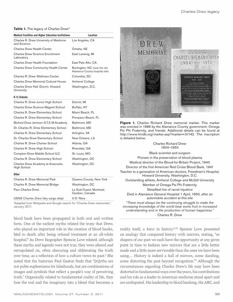
Drew was awarded a posthumous fellowship in the American College of Surgeons, an organization to which he had not previously been allowed admission.9 At least twelve K–12 schools and six medical facilities and higher education institutions have been named in honor of Dr. Drew (Table 1). In 1981, the U.S. Postal Service issued a 35-cent stamp in honor of Drew as part of the “Great American Series.”5 The Charles Drew University of Medicine and Science was established in 1966 in Los Angeles, California, as a private medical and health sciences institution with the goal of providing education to medical professionals serving the underprivileged. There is a Charles R. Drew Memorial Bridge in Washington, D.C., and one of the boroughs of Montreal, where he attended McGill University, has been named after him (Parc Charles-Drew, in Le Sud-Ouest). A marker was erected in 1986 by the Alamance County Historic Properties Commission, the Omega Psi Phi Fraternity, and Drew’s friends at the site of Drew’s accident in Haw River, North Carolina (Fig. 1).
Unfortunately, in the decades since Drew’s death, myths related to the circumstances of his death and work with the
IMMUNOHEMATOLOGY, Volume 27, Number 3, 2011 99
blood bank have been propagated in both oral and written form. One of the earliest myths related the irony that Drew, who played an important role in the creation of blood banks, bled to death after being refused treatment at an all-white hospital.9 As Drew biographer Spencie Love related, although these myths and legends were not true, they were altered and extrapolated on, often obscuring and obliterating the truth over time, as a reflection of how a culture views its past.9 She noted that the historian Paul Gaston finds that “[m]yths are not polite euphemisms for falsehoods, but are combinations of images and symbols that reflect a people’s way of perceiving truth.” Organically related to fundamental reality of life, they fuse the real and the imaginary into a blend that becomes a
reality itself, a force in history.9,28 Spencie Love presented an analogy that compared history with mirrors, stating, “as shapers of our past we each have the opportunity at any given point in time to fashion new mirrors that are a little better made and a little more serviceable than the ones we have been using…. History is indeed a hall of mirrors, some dazzling, some distorting the past beyond recognition.”9 Although the circumstances regarding Charles Drew’s life may have been distorted in fundamental ways over the years, his contributions and his role as a leader in American medicine stand apart and are undisputed. His leadership in blood banking, the ARC, and
Charles Drew legacy
Table 1. The legacy of Charles Drew*
Medical Facilities and Higher Education Institutions Location
Charles R. Drew University of Medicine and Science
Los Angeles, CA
Charles Drew Health Center Omaha, NE
Charles Drew Science Enrichment Laboratory
East Lansing, MI
Charles Drew Health Foundation East Palo Alto, CA
Charles Drew Community Health Center Burlington, NC (near the old Alamance County hospital site)
Charles R. Drew Wellness Center Columbia, SC
Charles Drew Memorial Cultural House Amherst College
Charles Drew Hall (Dorm), Howard University
Washington, D.C.
K-12 Schools
Charles R. Drew Junior High School Detroit, MI
Charles Drew Science Magnet School Buffalo, NY
Charles R. Drew Elementary School Miami Beach, FL
Charles R. Drew Elementary School Pompano Beach, FL
Bluford Drew Jemison S.T.E.M Academy Baltimore, MD
Dr. Charles R. Drew Elementary School Baltimore, MD
Charles R. Drew Elementary School Arlington, VA
Dr. Charles Drew Elementary School New Orleans, LA
Charles R. Drew Charter School Atlanta, GA
Charles R. Drew High School Riverdale, GA
Compton-Drew Middle School ILC St. Louis, MO
Charles R. Drew Elementary School Washington, DC
Charles Drew Academy at Anacostia High School
Washington, DC
Other
Charles R. Drew Memorial Park Queens County, New York
Charles R. Drew Memorial Bridge Washington, DC
Parc Charles-Drew Le Sud-Ouest, Montreal, Quebec, Canada
USNS Charles Drew (dry cargo ship) U.S. Navy
*Adapted from Wikipedia and Google search for “Charles Drew memorials,” January 2011.
Figure 1. Charles Richard Drew memorial marker. This marker was erected in 1986 by the Alamance County government, Omega Psi Phi Fraternity, and friends. Additional details can be found at http://www.hmdb.org/marker.asp?marker=31142. The inscription is detailed below.
Charles Richard Drew1904–1950
Black scientist and surgeonPioneer in the preservation of blood plasma
Medical director of the Blood-for-Britain Project, 1940Director of the first American Red Cross Blood Bank, 1941
Teacher to a generation of American doctors, Freedmen’s Hospital, Howard University, Washington, D.C.
Outstanding athlete, Amherst College and McGill UniversityMember of Omega Psi Phi Fraternity
Steadfast foe of racial injusticeDied in Alamance General Hospital 1 April, 1950, after an
automobile accident at this site“There must always be the continuing struggle to make the
increasing knowledge of the world bear some fruit in increased understanding and in the production of human happiness.”
Charles R. Drew

100 IMMUNOHEMATOLOGY, Volume 27, Number 3, 2011
the larger medical community and country are undeniable. This is the legacy that needs to be shared.
Acknowledgments
The authors wish to thank Connor S. Willis for his inspiring curiosity about Dr. Drew, which became the impetus for writing this manuscript. We also thank him for his assistance with the memorial photographs taken in Burlington, North Carolina.
References
1. Along the N.A.A.C.P. Battlefront. The Crisis 1944;51(5): 166–7.
2. Hoover EL. Dr. Charles R. Drew: facts, fiction and legend. J Natl Med Assoc 2005;97(6):837–8.
3. Cornwell EE 3rd, Chang DC, Leffall LD Jr. The Southern Surgical Association History of Medicine Scholarship presentation: Dr. Charles Drew, a surgical pioneer. Ann Surg 2006;243(5):612–8.
4. Wynes CE. Charles Richard Drew: the man and the myth. Urbana, IL: University of Illinois Press; 1988.
5. Yancey AG Sr. US postage stamp issued in honor of Charles R. Drew, MD, MDSc. J Natl Med Assoc 1982;74(6):561–5.
6. Scudder J. Practical genetic concepts in modern medicine. J Natl Med Assoc 1960;52:266–77.
7. U.S. National Library of Medicine. Profiles in Science. The Charles R. Drew Papers. http://profiles.nlm.nih.gov/BG/Views/Exhibit/narrative/surgery.html. Accessed January 21, 2011.
8. Current Biography: Who’s News and Why, 1944. Vol 5. New York, NY: H.W. Wilson Company; 1945:180.
9. Love S. One blood: the death and resurrection of Charles R. Drew. Chapel Hill, NC: University of North Carolina Press; 1996.
10. Hess JR, Schmidt PJ. The first blood banker: Oswald Hope Robertson. Transfusion 2000;40(1):110–3.
11. Starr DP. Blood: an epic history of medicine and commerce. New York, NY: Alfred A. Knopf, Inc.; 1998.
12. Drew CR. Banked blood: a study in blood preservation [PhD dissertation]. New York, NY: Columbia University; 1940.
13. Scudder J, Smith ME, Drew CR. Plasma potassium content of cardiac blood at death. Am J Physiol 1939;126:337–40.
14. Scudder J, Drew CR, Corcoran DR, Bull DC. Studies in blood preservation: I. Repartition of potassium in cells and plasma. JAMA 1939;112(22):2263–71.
15. Scudder J, Drew CR, Tuthill E, Smith ME. Newer knowledge of blood transfusions. Bull N Y Acad Med 1941;17(5): 373–98.
16. Scudder J, Bishop K, Drew CR. Studies in blood preservation: the shape of the container. JAMA 1940;115(4):290.
17. Parks D. Charles Richard Drew, MD 1904–1950. J Natl Med Assoc 1979;71(9):893–5.
18. Cobb WM. Charles Richard Drew, MD 1904–1950. J Natl Med Assoc 1950;42(4):238–46.
19. Schmidt PJ. Charles Drew, a legend in our time. Transfusion 1997;37(2):234–6.
20. Reitman J. Bad blood: crisis in the American Red Cross. New York, NY: Kensington Books; 1996.
21. Kendrick DB. Blood program in World War II. Washington, DC: Office of the Surgeon General Department of the Army; 1964;112:306.
22. Jones RF. The surgical resident training program at Freedmen’s hospital. J Natl Med Assoc 1960;52:187–93.
23. Anonymous. Scudder blood transfusion controversy. J Natl Med Assoc 1960;52:56–8.
24. Wehrwein AC. Blood expert says transfusions between races may be perilous. New York Times November 7, 1959: A1, A3.
25. Scudder J, Wigle WD. Safer transfusions through appreciation of variants in blood group antigens in Negro and white blood donors, including two case reports showing development of anti-Jka; anti-Jkb; anti-S and anti-Fya iso-antibodies. J Natl Med Assoc 1960;52:75–80.
26. Kenny MG. A question of blood, race, and politics. J Hist Med Allied Sci 2006;61(4):456–91.
27. Guglielmo TA. “Red Cross, double cross”: race and America’s World War II–era blood donor service. J Am History 2010;97(1):63–90.
28. Gaston PM. The New South creed: a study in Southern mythmaking. New York, NY: Alfred A. Knopf; 1970.
Bryan A. Wilson,* BS, Department of Molecular Medicine and Translational Sciences, Wake Forest University School of Medicine, Winston-Salem, NC; Wendi G. O’Connor,* MA, MD, and Monte S. Willis, MD, PhD (corresponding author), McAllister Heart Institute, University of North Carolina at Chapel Hill, 2340B Medical Biomolecular Research Building, 103 Mason Farm Road, Chapel Hill, NC 27599-7525, and Department of Pathology & Laboratory Medicine, University of North Carolina, Chapel Hill, NC.
*Both authors contributed equally to this work.
B. Wilson et al.
Notice to ReadersImmunohematology is printed on acid-free paper.
For information concerning Immunohematology or the Immunohematology Methods and Procedures manual, contact us by e-mail at [email protected]

IMMUNOHEMATOLOGY, Volume 27, Number 3, 2011 101
An unusual case of an acute hemolytic transfusion reaction caused by an autoanti-IN. Win, M. Needs, S. Rahman, P. Gold, and S. Ward
In general, naturally occurring cold autoagglutinins react optimally at low temperatures. We describe a young child who experienced an acute hemolytic transfusion reaction by an unusual autoanti-I. The IgM autoanti-I was detected at 4°C (titer 256) and also reacted at 30°C. This case highlights the potential hazard of transfusing units of blood immediately upon removal from the blood refrigerator, especially into neonates and children of small stature. Immunohematology 2011;27:101–103.
Key Words: autoanti-I, acute hemolytic transfusion reaction, cold autoagglutinins
In general, naturally occurring cold autoagglutinins react optimally at low temperatures (at or below 4°C) with a titer below 64, and with low thermal amplitude (maximal thermal range at 20°C or less).1 Most cold autoagglutinins are benign, usually IgM antibodies, and can be detected by direct saline agglutination tests. Clinically significant cold autoagglutinins usually react at a thermal amplitude of 30°C or above and may activate complement.
Autoanti-I is commonly found to be the specificity implicated in cold hemagglutinin disease (CHD), and also after mycoplasma infection. In CHD, the cold agglutinin titer is usually higher than 1000 when tested at 4°C.2
We report an acute hemolytic transfusion reaction (AHTR) in a child of small stature caused by the transfusion of two units of blood immediately upon removal from a temperature-controlled blood refrigerator, with no warming of the units to ambient room temperature. Extensive laboratory investigations revealed that the antibody implicated was an unusual autoanti-I reacting at 30°C.
This case highlights the potential hazard of transfusing cold units of blood taken from a temperature-controlled refrigerator to neonates and children of small stature.
Case Report
A 9-year-old boy, with acute lymphoblastic leukemia (ALL), was admitted to a district hospital with febrile neutropenia. He received intravenous (IV) antibiotics and his fever improved, but no organism was identified. His hemoglobin (Hb) fell to
7.3 g/dL (normal range, 12.2–14.2 g/dL), with a hematocrit of 19.5%, and transfusion was requested. The patient’s blood group was group A, D+, with no atypical alloantibodies detected in the plasma. The direct antiglobulin test (DAT) was positive (2+ with anti-C3d only). Two units of crossmatched compatible (by indirect antiglobulin technique [IAT] at 37°C) group A, D+, K– red blood cells (RBCs) were issued.
The first unit was taken from the temperature-controlled blood refrigerator and was immediately transfused without any adverse reactions. The second unit was also transfused without waiting for the unit to reach ambient room temperature. While receiving the second unit, the patient had classic signs of an AHTR: high fever of 40°C, rigors, loin pain, sweating, and tachycardia. The transfusion was stopped immediately (about 100 mL had been infused), and IV normal saline, hydrocortisone, and chlorpheniramine were given with oral acetaminophen. The patient also passed a quantity of dark urine soon after cessation of the transfusion, and the left kidney was tender on palpation. There was a rise in bilirubin level from 25 μmol/L to 93 μmol/L (normal, 2–17 μmol/L). The blood film showed some RBC agglutination and some atypical lymphocytes.
In view of the severe nature of the reaction and hemoglobinuria, the patient was urgently transferred to the teaching hospital the same day. The patient remained on IV fluid and antibiotics. The blood samples (pretransfusion and posttransfusion samples taken at room temperature) were referred to the reference laboratory for investigation. The Hb level the next day was 9.6 g/dL, the hemoglobinuria subsided quickly, and the bilirubin dropped to 44 μmol/L. There were no long-term clinically adverse sequelae, and the child was discharged from the hospital the following day with a Hb level of 9.2 g/dL.
A culture from the blood units showed no growth. The patient’s mycoplasma screening and Paul-Bunnell test for glandular fever were negative.
Materials and Methods
IATs were carried out using gel column agglutination (DiaMed Micro-ID System, BioRad Laboratories, Switzerland)
Case RepoRt

102 IMMUNOHEMATOLOGY, Volume 27, Number 3, 2011
N. Win et al.
at the reference and hospital laboratories. Direct saline agglutination test and titration studies were carried out by standard tube methods, with the reactants warmed to the desired incubation temperature before mixing.
In view of the DAT findings (see Results), the hospital was asked to provide more warm-separated serum from whole blood, kept strictly at 37°C, to investigate for indirect Donath-Landsteiner test, using the method previously described.3
Results
Both pretransfusion and posttransfusion samples were examined macroscopically after centrifugation. The color of the supernatant plasma from the pretransfusion sample was normal (straw colored), whereas that from the posttransfusion sample was dirty brown.
Initial serologic results at the reference laboratory for both pretransfusion and posttransfusion samples showed the patient to be group AB, Rh phenotype DCcEe, and K+, with a positive DAT with anti-IgG (1+), anti-IgA (1+), anti-IgM (3+), anti-C3c (1+), and anti-C3d (3+). The reverse ABO grouping results of the plasma was consistent with group O. None of these results were reliable, however, as positive (1+) reactions were seen in all the columns containing an inert control. This suggested the presence of a cold-reactive antibody.
These results were discrepant with those found at the hospital. Although tests there were also carried out by column agglutination technology, the samples tested at the reference laboratory had been stored at 4°C, whereas those at the hospital had been tested without cooling beyond laboratory temperature. This allowed for more of the autoantibody to be adsorbed onto the RBC surface, thus giving false-positive results.
After washing of the RBCs with phosphate-buffered saline (PBS), warmed to 37°C, the tests were repeated, and the results showed the samples to be group A, probable Rh genotype R1R1, K–. The reverse grouping, also incubated at 37°C, showed the expected negative reaction with A1 RBCs and the expected positive reaction with group B RBCs. The DAT was positive (3+) with anti-C3d only.
No RBC alloantibodies were detected by the IAT at 37°C both in the pretransfusion and posttransfusion samples. Repeat crossmatches by the IAT using pretransfusion plasma samples and RBCs from the two transfused units were compatible.
As the initial tests suggested the presence of a cold-reactive antibody, titration studies at different temperatures were undertaken by saline direct agglutination. Testing was performed using the plasma from the pretransfusion samples, after warming of the patient’s RBCs to ensure that optimal amounts of the autoantibody were eluted back into the plasma, as shown in Table 1.
The results indicate the presence of a low-titer, high thermal amplitude (reacting at 30°C) cold-reactive antibody with anti-I specificity. See Table 1. Reactions with papain-treated RBCs (not shown) were positive, thus ruling out a specificity of anti-Pr. The indirect Donath-Landsteiner test was negative, which ruled out anti-P associated with paroxysmal cold hemoglobinuria.
Discussion
In general, naturally occurring cold-reactive autoanti-bodies have a low titer (<64) at 4°C, and have a thermal amplitude of less than 20°C.1 The most common cold autoagglutinins are directed against the I blood group system.4
Table 1. Specificity and thermal amplitude of cold antibody*
Plasma dilutions
Temperature Cells Neat 1:2 1:4 1:8 1:16 1:32 1:64 1:128 1:256 1:512
4°C Group O adult cells (H+I+) 5+ 5+ 5+ 5+ 3+ 3+ 3+ 2+ 1+ 0
Group A1 adult cells (H–I+) 5+ 5+ 5+ 5+ 3+ 3+ 3+ 2+ 1+ 0
Group O ii adult cells (H+I–) 5+ 5+ 5+ 5+ 4+ 3+ 3+ 2+ 1+ 0
23°C Group O adult cells (H+I+) 5+ 4+ 3+ 2+ 1+ 0 0 0 0 0
Group A1 adult cells (H–I+) 5+ 4+ 3+ 2+ 1+ 0 0 0 0 0
Group O ii adult cells (H+I–) 4+ 3+ 1+ 0 0 0 0 0 0 0
30°C Group O adult cells (H+I+) 1+ 0 0 0 0 0 0 0 0 0
Group A1 adult cells (H–I+) 1+ 0 0 0 0 0 0 0 0 0
Group O ii adult cells (H+I–) 0 0 0 0 0 0 0 0 0 0
*Using different red cell phenotypes, plasma dilutions were tested by direct saline agglutination method to identify the specificity and thermal amplitude of the cold antibody.

IMMUNOHEMATOLOGY, Volume 27, Number 3, 2011 103
Acute HTR caused by autoanti-I
We describe a young child who experienced an AHTR caused by an unusual cold-reactive autoantibody. In our case, the anti-I IgM cold antibody was detected at 4°C (titer 256), and the antibody reacted at 30°C. In general, anti-I is commonly found in patients with CHD, and the autoantibody titer is typically much higher (>1000) at 4°C.2 This was not a case of CHD, as there was no laboratory evidence of autohemolysis before the transfusion. Also the reaction was transient in nature.
Although both units were transfused immediately upon removal from the refrigerator, it was during transfusion of the second unit that the AHTR occurred. It is conjectured that the first unit lowered the peripheral body temperature, and the second unit further reduced it to a level at which the autoanti-I caused the AHTR.
Cases of autoanti-IH causing AHTR have been well described,5,6 but to our knowledge, there was only one reported case of autoanti-I with a wide thermal range causing intravascular hemolysis.7 In our case, autoanti-I reacted at 30°C, and this lends weight to the importance of knowing the thermal range of the antibody, rather than the specificity or titer.
British Committee for Standards in Haematology (BCSH) guidelines for the administration of blood and blood components stated that “the transfusion of blood and blood components should begin as soon as possible after delivery to the ward or operating theatre.”8 There is no clear guidance or clarification for the administration of RBCs in neonates and children.9 Fatal hypothermic reactions have been reported after receiving blood transfused immediately upon removal from a blood refrigerator.10
We report a rare, unusual case of AHTR caused by autoanti-I; this case highlights the potential hazard of transfusing units of blood immediately upon removal from the blood refrigerator, especially into neonates and children of small stature.
References
1. Dacie JV. The haemolytic anaemias, congenital and acquired, part II. The auto-immune haemolytic anaemias, 2nd ed. London: Churchill, 1962:460.
2. Petz LD, Garratty G. Differential diagnosis of immune hemolytic anemias. In: Petz LD, Garratty G. Immune hemolytic anemias, 1st ed. New York, NY: Churchill Livingstone, 1980:185–231.
3. Win N, Stamps R, Knight R. Paroxysmal cold haemoglobinuria/Donath-Landsteiner test. Transfus Med 2005;15:254.
4. Issitt PD, Anstee DJ. Applied blood group serology. 4th ed. Durham, NC: Montgomery Scientific Publications, 1998: 277–91.
5. Darabi K, Makar RS. Acute hemolysis of transfused A2 red cells by an auto-HI antibody. Transfusion 2008;48:964–8.
6. Campbell SA, Shirey RS, King KE, Ness PM. An acute hemolytic transfusion reaction due to anti-IH in a patient with sickle cell disease. Transfusion 2000;40:828–31.
7. Issitt PD. Applied blood group serology. 3rd ed. Durham, NC: Montgomery Scientific Publications, 1985:547.
8. [No authors listed]. The administration of blood and blood components and the management of transfused patients. British Committee for Standards in Haematology, Blood Transfusion Task Force. Royal College of Nursing and the Royal College of Surgeons of England. Transfus Med 1999;9:227–38.
9. Gibson BE, Todd A, Roberts I, et al. Transfusion guidelines for neonates and older children. Brit J Haematol 2004;134: 433–53.
10. Rosenfield RE, Jagathambal E. Transfusion therapy for autoimmune hemolytic anemia. Semin Hematol 1976;13: 311–21.
Nay Win, MBBS, FRCP, FRCPath, CTM (Edin) (corresponding author), Consultant Haematologist, Malcolm Needs, Reference Service Manager, and Sally Rahman, Specialist Biomedical Scientist, Red Cell Immunohaematology, NHSBT—Tooting Centre, 75, Cranmer Terrace, Tooting, United Kingdom, SW17 0RB; and Philip Gold, Transfusion Laboratory Manager, and Dr. Susan Ward, Consultant Haematologist, Queen Mary’s Hospital, Sidcup, Kent, United Kingdom.

104 IMMUNOHEMATOLOGY, Volume 27, Number 3, 2011
We analyzed our historic patient database at North Shore University Hospital and determined both the overall frequency of anti-Jsa and the frequency at which it was detected in combination with other alloantibodies to red blood cell (RBC) antigens. Screening cells used currently are negative for Jsa. Our data suggest that anti-Jsa would not be detected in 30 to 40 percent of patients in which it is the sole antibody present. Since 1996 the antibody was only detected when other antibodies were found in the screening process. We are exposing 1.7 percent of our patients (90 patients/year) to Jsa. The clinical significance of anti-Jsa is apparent with previous literature, and our conclusion is that additional studies should be performed to determine whether Jsa should be included in current antibody screening cells. Immunohematology 2011;27:104–106.
Key Words: Jsa, antibody, antigen, low prevalence antigens, antibody screening
Jsa is generally considered a low-prevalence blood group antigen. Jsa was first identified in 1958. At that time, the red cell antigen was named Sutter after the patient whose serum demonstrated the antibody. In 1965, Jsa and Jsb were identified and categorized under the Kell blood group system. The Kell blood group system is the most immunogenic after the ABO and Rh blood group systems.1
Less than 0.01 percent of Caucasians are found to be positive for Jsa. However, 19.5 percent of African Americans are positive for Jsa. Issitt1 has reported that in 1000 random black donors, 7 were Js(a+b–), 152 were Js(a+b+), and 842 were Js(a–b+). Anti-Jsa is seen infrequently because it has been hypothesized that fewer donors are positive for Jsa, considering that fewer blacks donate blood.1 In the New York City area, approximately 8 percent of the donor population is African American, as per the New York Blood Center’s (NYBC’s) statistics.2 At North Shore University Hospital (NSUH) we obtain close to 97 percent of our blood from the NYBC. At approximately 26,000 RBC transfusions per year, 1.7 percent of our patients are transfused with RBCs positive for the Jsa antigen. The NYBC has also reported that the incidence of detecting anti-Jsa in their antibody workups has increased from 14 (1980–1984) to 86 (2005–2009) in an increasingly diverse population of blood donors.3
Most examples of anti-Jsa occur when the patient has had a RBC transfusion or pregnancy, but the antibody has also been discovered without an allogeneic RBC exposure.
The antibody has been demonstrated to cause hemolytic transfusion reactions4 and hemolytic disease of the newborn (HDN), both mild and severe.5 In the last 15 years, reagent RBCs used to screen for blood group antibodies were negative for Jsa, minimizing the detection of the antibody in our patient population.6,7 Therefore, the antibody would be detected only when other antibodies are detected in the screening process. The antibody would be mostly detected in Caucasians because they have the lowest prevalence of the antigen and are more likely to develop anti-Jsa.8,9
The racial distribution of the patients at NSUH who displayed anti-Jsa was as follows: 17 Caucasian (50%); 4 African American (12%); 4 Asian (12%); 3 Hispanic (9%); and 6 unknown or other (17%).
Materials and Methods
We performed a retrospective study of our transfused patient population and also of the antibodies identified during the same time frame. Using the blood bank’s computer system, we generated a general database scan report to identify the patients who had been assigned a computer flag or antibody alert for anti-Jsa for the period of January 1, 1989, to April 6, 2010. We reviewed each patient for the following information: dates of positive antibody screen results and panel results, antibody identification result(s), and race (Table 1).
Results
The total number of patients during the period 1989 to the present who had anti-Jsa present in their serum was 34. Using this information, we determined the prevalence of the antibody when detected with at least one other antibody and when solely detected. Sixty-eight percent of these patients had anti-Jsa present in combination with another antibody. Thirty-two percent of these patients had anti-Jsa solely present. For the period 1989 to 1995, anti-Jsa was found both in combination and solely (58% and 42%, respectively). For the period 1996 to the present, 100 percent of the examples of anti-Jsa were found only in combination with other antibodies (Table 2, Fig. 1). We also generated a report for the total number of patients transfused within the same period. We
Should we be screening for anti-Jsa?N.M. Nikolis, F.N. Boctor, W.A. Heaton, and J. Martone
oRiginal RepoRt

IMMUNOHEMATOLOGY, Volume 27, Number 3, 2011 105
found that 0.08 percent of our transfused patients had anti-Jsa detected in their serum during the period 1989 to 1995 and only 0.01 percent of our transfused patients had anti-Jsa detected in their serum during the period 1996 to the present (Table 2).
Our antibody screening methodology during the years has been as follows: 1989 to 1994, test tube low-ionic-strength
saline; 1995 to 2008, gel; and 2009 to present, Galileo Echo Instrumentation, Immucor, Norcross, Georgia 30091.
Discussion
Screening cells currently used are negative for Jsa. Based on our data, it appears that anti-Jsa would not be detected
Table 1. Results of anti-Jsa occurrences in our patient population
1st admissionDate of 1st positive
ABSC/panel ABID resultDate of 2nd positive
ABSC/panel ABID resultDate of 3rd positive
ABSC/panel ABID result
1/18/83 1/14/90 Anti-Jsa/anti-P1 n/a n/a n/a n/a
3/11/85 1/25/89 Anti-Jsa/anti-K n/a n/a n/a n/a
8/30/88 2/21/89 Nonspecific 10/4/89 Anti-Jsa n/a n/a
2/2/89 10/16//94 Anti-K 1/20/00 Anti-Jsa 2/28/00 Anti-V
5/3/89 12/8/94 Anti-Jsa 12/12/94 Anti-E n/a n/a
6/5/89 6/5/89 Anti-Jsa n/a n/a n/a n/a
1/6/90 1/23/90 Anti-Jsa n/a n/a n/a n/a
1/8/90 2/6/93 Anti-K 2/6/93 Anti-Jsa/anti-V n/a n/a
9/6/90 10/28/93 Anti-Jsa n/a n/a n/a n/a
12/25/90 2/12/96 Anti-Jsa/anti-K n/a n/a n/a n/a
5/29/91 7/16/91 Anti-Jsa 7/18/91 Anti-E n/a n/a
9/16/91 11/1/91 Anti-C 2/15/95 Anti-Jsa 4/14/95 and 9/6/96 Anti-e/anti-K
10/8/91 10/8/91 Anti-Jsa n/a n/a n/a n/a
2/13/92 6/29/94 Anti-Jsa, anti-Kpa, anti-S, anti-K, anti-D
7/20/94 Anti-C 12/7/94 Anti-Fya
4/6/92 4/28/92 Cold 2/5/93 Anti-Jsa 2 n/a
1/23/93 9/12/94 Anti-K/anti-Kpa/anti-E 11/1/94 Anti-c 11/2/94 Anti-Jsa
7/16/93 10/25/93 Nonspecific 10/27/93 Anti-Jsa n/a n/a
9/25/93 10/16/93 Anti-Jsa n/a n/a n/a n/a
10/24/93 1/8/94 Anti-K 10/31/94 Anti-D 11/1/94 and 4/25/95 Anti-Jsa/anti-E/anti-C
10/25/93 10/25/93 Nonspecific 8/5/95 Anti-Jsa/anti-K 9/22/98 Nonspecific
4/24/94 4/24/94 Anti-Leb 11/23/97 Anti-Jsa n/a n/a
5/5/94 5/5/94 Anti-Jsa n/a n/a n/a n/a
11/10/94 11/10/94 Autoanti-I 7/12/97 Anti-Jsa n/a n/a
12/12/94 3/10/95 Anti-Jsa n/a n/a n/a n/a
3/22/95 7/19/95 Anti-Jsa n/a n/a n/a n/a
4/24/95 4/24/95 Nonspecific 6/14/95 Anti-Jsa/anti-E/ anti-K
n/a n/a
5/25/95 5/25/95 Anti-Jsa/anti-K n/a n/a n/a n/a
9/26/95 9/27/95 Anti-Jsa n/a n/a n/a n/a
2/10/96 2/11/96 Anti-Jsa/anti-K n/a n/a n/a n/a
2/21/97 2/21/97 Anti-Jsa/anti-D/ nonspecific
n/a n/a n/a n/a
4/30/97 5/2/97 Anti-Jsa/cold n/a n/a n/a n/a
6/12/97 6/17/97 Anti-Jsa/anti-K n/a n/a n/a n/a
8/11/97 10/4/97 Anti-Jsa/anti-K n/a n/a n/a n/a
6/8/98 6/9/98 Anti-Fya/anti-E 8/28/01 Anti-M 9/5/01, 12/16/03 and 6/10/04
Anti-Fyb/anti-K/anti-Jsa
ABSC = antibody screen, ABID = antibody identification, n/a = not applicable
Anti-Jsa screening
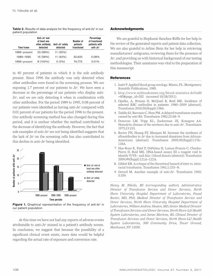
106 IMMUNOHEMATOLOGY, Volume 27, Number 3, 2011
N. Nikolis et al.
in 40 percent of patients in which it is the sole antibody present. Since 1996 the antibody was only detected when other antibodies were found in the screening process. We are exposing 1.7 percent of our patients to Jsa. We have seen a decrease in the percentage of our patients who display anti-Jsa, and we are only detecting it when in combination with other antibodies. For the period 1989 to 1995, 0.08 percent of our patients were identified as having anti-Jsa compared with 0.01 percent of our patients for the period 1996 to the present. Our antibody screening method has also changed during this period, and it is unclear whether the method contributed to the decrease of identifying the antibody. However, the fact that sole examples of anti-Jsa are not being identified suggests that the lack of Jsa on the screening cells has also contributed to this decline in anti-Jsa being identified.
At this time we have not had any reports of adverse events attributable to anti-Jsa missed in a patient’s antibody screen. In conclusion, we suggest that because the possibility of a significant clinical event exists, more data would be helpful regarding the actual rate of exposure and conversion rate.
Acknowledgments
We are grateful to Stephanie Sanchez-Riffle for her help in the review of the generated reports and patient data collection. We are also grateful to Arline Stein for her help in retrieving manufacturers’ antigrams, reviewing them for the presence of Jsa, and providing us with historical background of our testing methodologies. Their assistance was vital to the preparation of this manuscript.
References
1. Issitt P. Applied blood group serology. Miami, FL: Montgomery Scientific Publications, 1985.
2. http://www.nybloodcenter.org/blood-statistics.do?sid0 =85&page_id=202 (accessed 10/28/2011).
3. Opalka, A, Strauss D, McQuail B, Reid ME. Incidence of selected RBC antibodies in patients: 1980–2009 (abstract). Transfusion 2010;50(Suppl):138A.
4. Taddie SJ, Barrasso C, Ness PM. A delayed transfusion reaction caused by anti-K6. Transfusion 1982;22:68–9.
5. Donovan LM, Tripp KL, Zuckerman JE, Konugres AA. Hemolytic disease of the newborn due to anti-Jsa. Transfusion 1973;13:153.
6. Boctor FN, Huang ST, Marques M. Increase the incidence of alloantibodies to Jsa due to increased donations from African-Americans (abstract). Transfusion 2005;45(Suppl):117A–118A.
7. Hue-Roye K, Patel P, DePalma H, Lomas-Francis C, Charles-Pierre D, Reid ME. DNA-based assays fill a reagent void to identify V/VS– and Js(a–) blood donors (abstract). Transfusion 2009;49(Suppl):121A–122A.
8. Giblett ER. A critique of the theoretical hazard of inter vs. intra-racial transfusion. Transfusion 1961;1:233–8.
9. Gerard M. Another example of anti-Jsa. Transfusion 1965; 5:359.
Nancy M. Nikolis, BS (corresponding author), Administrative Director of Transfusion Service and Donor Services, North Shore University Hospital Department of Laboratories, Fouad Boctor, MD, PhD, Medical Director of Transfusion Service and Donor Services, North Shore University Hospital Department of Laboratories, William Andrew Heaton, MD, Senior Medical Director of Transfusion Services and Donor Services, North Shore-LIJ Health System Laboratories, and James Martone, BS, Clinical Director of Transfusion Services and Donor Services, North Shore-LIJ Health System Laboratories, 300 Community Drive, Tower Ground, Manhasset, NY 11030.
Table 2. Results of data analysis for the frequency of anti-Jsa in our patient population
Time frame
Anti-Jsa and at least one
other antibody detected
Anti-Jsa solely detected
Number of patients
transfused
Percentage of transfused patients with
anti-Jsa
1989–present 23 (68%) 11 (32%)
1989–1995 15 (58%) 11 (42%) 30,620 0.08%
1996–present 8 (100%) 0 (0%) 74,773 0.01%
Figure 1. Graphical representation of the frequency of anti-Jsa in our patient population
25
20
15
10
5
0
Anti-Jsa and at least one other antibody detected
Anti-Jsa solely detected
1989–present 1989–1995 1996–present
Num
ber o
f pat
ient
s w
ith a
nti-
Jsa
Time periods

IMMUNOHEMATOLOGY, Volume 27, Number 3, 2011 107
Specialist in Blood Bank (SBB) ProgramThe Department of Transfusion Medicine, National Institutes of Health, is accepting applications for its 1-year Specialist in Blood Bank Technology Program. Students are federal employees who work 32 hours/week. This program introduces students to all areas of transfusion medicine including reference serology, cell processing, HLA, and infectious disease testing. Students also design and conduct a research project. NIH is an Equal Opportunity Organization. Application deadline is December 31, 2011, for the July 2012 class. See www.cc.nih.gov/dtm > education for brochure and application. For further information contact Karen M. Byrne at (301) 451-8645 or [email protected]
announCements
Free Classified Ads and Announcements
Immunohematology will publish classified ads and announcements (SBB schools, meetings, symposia, etc.) without charge.
Deadlines for receipt of these items are as follows:
Deadlines
1st week in January for the March issue 1st week in July for the September issue
1st week in April for the June issue 1st week in October for the December issue
E-mail or fax information to [email protected] or phone (215) 451-2538
The editorial staff of Immunohematology welcomes manuscripts pertaining to blood group serology and education for consideration for publication. We are especially interested in case reports, papers on platelet and white cell serology, scientific articles covering original investigations, and papers on new methods for use in the blood bank. Deadlines for receipt of manuscripts for consideration for the March, June, September, and December
issues are the first weeks in November, February, May, and August, respectively. For instructions for scientific articles, case reports, and review articles, see Instructions for Authors in every issue of Immunohematology or on the Web at www.redcross.org/immunohematology. Include fax and phone numbers and e-mail address with all manuscripts and correspondence. E-mail all manuscripts to [email protected]
Manuscripts

108 IMMUNOHEMATOLOGY, Volume 27, Number 3, 2011
Announcements, cont.

IMMUNOHEMATOLOGY, Volume 27, Number 3, 2011 109
adveRtisements
Masters (MSc) in Transfusion and Transplantation Sciences at
The University of Bristol, England
Applications are invited from medical or science graduates for the Master of Science (MSc) degree in Transfusion and Transplantation Sciences at the University of Bristol. The course starts in October 2012 and will last for 1 year. A part-time option lasting 2 or 3 years is also available. There may also be opportunities to continue studies for PhD or MD following the MSc. The syllabus is organized jointly by The Bristol Institute for Transfusion Sciences and the University of Bristol, Department of Pathology and Microbiology. It includes:
• Scientificprinciplesoftransfusionandtransplantation
•Clinicalapplicationsoftheseprinciples
• Practicaltechniquesintransfusionandtransplantation
• Principlesofstudydesignandbiostatistics
•Anoriginalresearchproject
Application can also be made for Diploma in Transfusion and Transplantation Science or a Certificate in Transfusion and Transplantation Science.
The course is accredited by the Institute of Biomedical Sciences.
Further information can be obtained from the Web site:http://ibgrl.blood.co.uk/MSc/MscHome.htm
For further details and application forms please contact:
Dr Patricia Denning-Kendall
University of Bristol
Paul O’Gorman Lifeline Centre
Department of Pathology and Microbiology
Southmead Hospital
Westbury-on-Trym, Bristol BS10 5NB, England
Fax +44 1179 595 342, Telephone +44 1779 595 455, e-mail: [email protected].

110 IMMUNOHEMATOLOGY, Volume 27, Number 3, 2011
Advertisements, cont.
Diagnostic testing for:• Neonatal alloimmune thrombocytopenia (NAIT)• Posttransfusion purpura (PTP)• Refractoriness to platelet transfusion• Heparin-induced thrombocytopenia (HIT)• Alloimmune idiopathic thrombocytopenia purpura (AITP)
Medical consultation available
Test methods:• GTI systems tests — detection of glycoprotein-specific platelet antibodies — detection of heparin-induced antibodies (PF4 ELISA)• Platelet suspension immunofluorescence test (PSIFT)• Solid phase red cell adherence (SPRCA) assay• Monoclonal immobilization of platelet antigens (MAIPA)• Molecular analysis for HPA-1a/1b
For Further inFormation, contact:
Platelet Serology Laboratory (215) 451-4205
Janet Demcoe (215) 451-4914 [email protected]
Sandra Nance (215) 451-4362 [email protected]
American Red Cross Biomedical ServicesMusser Blood Center
700 Spring Garden StreetPhiladelphia, PA 19123-3594
National Platelet Serology Reference Laboratory
CLIA licensed
Our laboratory specializes in granulocyte antibody detection and granulocyte antigen typing.
Indications for granulocyte serology testing include:• Alloimmune neonatal neutropenia (ANN)• Autoimmune neutropenia (AIN)• Transfusion-related acute lung injury (TRALI)
Methodologies employed:• Granulocyte agglutination (GA)• Granulocyte immunofluorescence by flow cytometry (GIF)• Monoclonal antibody immobilization of neutrophil antigens
(MAINA)
TRALI investigations also include:• HLA (PRA) Class I and Class II antibody detection
For Further inFormation, contact:
Neutrophil Serology Laboratory (651) 291-6797
Randy Schuller (651) 291-6758 [email protected]
American Red Cross Biomedical ServicesNeutrophil Serology Laboratory
100 South Robert StreetSt. Paul, MN 55107
National Neutrophil Serology Reference Laboratory
CLIA licensed

IMMUNOHEMATOLOGY, Volume 27, Number 3, 2011 111
Advertisements, cont.
Antibody identification and problem resolution
HLA-A, B, C, and DR typing
HLA-disease association typing
Paternity testing/DNA
For inFormation, contact:
Mehdizadeh Kashi
at (503) 280-0210
or write to:
Tissue Typing Laboratory
American Red Cross Biomedical Services
Pacific Northwest Region
3131 North Vancouver
Portland, OR 97227
Reference and Consultation Services
CLIA licensed, ASHI accredited
IgA and anti-IgA testing are available to do the
following:
• Identify IgA-deficient patients
• Investigate anaphylactic reactions
• Confirm IgA-deficient donors
Our ELISA for IgA detects protein to 0.05 mg/dL.
For additional inFormation contact:
Janet Demcoe at (215) 451-4914,
or e-mail:
or write to:
American Red Cross Biomedical Services
Musser Blood Center
700 Spring Garden Street
Philadelphia, PA 19123-3594
ATTN: Janet Demcoe
IgA/Anti-IgA Testing
CLIA licensed
• Effective tool for screening large volumes of donors
• Gel diffusion test that has a 15-year proven track record:
Approximately 90 percent of all donors identified as
IgA deficient by this method are confirmed by the more
sensitive testing methods
For additional inFormation:
Kathy Kaherl
at (860) 678-2764
e-mail:
or write to:
Reference Laboratory
American Red Cross Biomedical Services
Connecticut Region
209 Farmington Ave.
Farmington, CT 06032
Donor IgA Screening
CLIA licensed
Immunohematology Reference LaboratoryAABB, ARC, New York State, and CLIA licensed
24-hour phone number: (215) 451-4901
Fax: (215) 451-2538
American Rare Donor Program24-hour phone number:
(215) 451-4900Fax: (215) 451-2538
ImmunohematologyPhone, business hours:
(215) 451-4902Fax: (215) 451-2538
Quality Control of Cryoprecipitated–AHFPhone, business hours:
(215) 451-4903Fax: (215) 451-2538
National Reference Laboratory for Blood Group Serology

112 IMMUNOHEMATOLOGY, Volume 27, Number 3, 2011
Advertisements, cont.
What is a certified Specialist in Blood Banking (SBB)?• Someone with educational and work experience qualifications that successfully passes the American Society for Clinical Pathology (ASCP)
board of certification (BOC) examination for the Specialist in Blood Banking.• This person will have advanced knowledge, skills, and abilities in the field of transfusion medicine and blood banking.
Individuals who have an SBB certification serve in many areas of transfusion medicine: • Serve as regulatory, technical, procedural, and research advisors• Perform and direct administrative functions • Develop, validate, implement, and perform laboratory procedures• Analyze quality issues preparing and implementing corrective actions to prevent and document nonconformances• Design and present educational programs• Provide technical and scientific training in transfusion medicine• Conduct research in transfusion medicine
Who are SBBs?Supervisors of Transfusion Services Executives and Managers of Blood Centers LIS Coordinators EducatorsSupervisors of Reference Laboratories Research Scientists Consumer Safety OfficersQuality Assurance Officers Technical Representatives Reference Lab Specialists
Why become an SBB?Professional growth Job placement Job satisfaction Career advancement
How does one become an SBB?CAAHEP-accredited SBB Technology program or grandfather the exam based on ASCP education and experience criteria.
Fact: In recent years, a greater percentage of individuals who graduate from CAAHEP-accredited programs pass the SBB exam compared to individuals who grandfather the exam. The BEST route for obtaining an SBB certification is to attend a CAAHEP-accredited Specialist in Blood Bank Technology Program.
Which approach are you more compatible with?
Contact the following programs for more information:
Additional information can be found by visiting the following Web sites: www.ascp.org, www.caahep.org, and www.aabb.org
Program Contact Name Phone Contact Email Contact WebsiteOnsite or Online Program
Walter Reed Army Medical Center William Turcan 202-782-6210 [email protected] www.militaryblood.dod.mil/fellow Onsite
American Red Cross, Southern California Region Michael Coover 909-859-7496 [email protected] none Onsite
ARC-Central OH Region Joanne Kosanke 614-253-2740 x 2270 [email protected] none Onsite
Blood Center of Wisconsin Lynne LeMense 414-937-6403 [email protected] www.bcw.edu Onsite
Community Blood Center/CTS Dayton, Ohio Nancy Lang 937-461-3293 [email protected] www.cbccts.org/education/sbb.html Online
Gulf Coast School of Blood Bank Technology Clare Wong 713-791-6201 [email protected] http://giveblood.org/index.php?page=sbb-distance-program Online
Hoxworth Blood Center/University of Cincinnati Susan Wilkinson 513-558-1275 [email protected] [email protected]
www.hoxworth.org Onsite
Indiana Blood Center Jayanna Slayten 317-916-5186 [email protected] www.indianablood.org Online
Johns Hopkins Hospital Lorraine Blagg 410-502-9584 [email protected] http://pathology.jhu.edu/department/divisions/tranfusion/sbb2.cfm
Onsite
Medical Center of Louisiana Karen Kirkley 504-903-3954 [email protected] none Onsite
NIH Clinical Center Department of Transfusion Medicine Karen Byrne 301-496-8335 [email protected] www.cc.nih.gov/dtm/research/sbb.html Onsite
Rush University Veronica Lewis 312-942-2402 [email protected] www.rushu.rush.edu/catalog/acadprograms/chs/cls/clssbbcert.html
Online
Transfusion Medicine Academic Center at Florida Blood Services
Marjorie Doty 727-568-5433 x 1514 [email protected] www.fbsblood.org Online
University Health System and Affiliates, San Antonio Linda Myers 210-731-5526 [email protected] www.sbbofsa.org Onsite
University of Texas Medical Branch at Galveston Janet Vincent 409-772-3055 [email protected] www.utmb.edu/sbb Online
University of Texas SW Medical Center LeAnne Hutson 214-648-1780 [email protected] http://utsouthwestern.edu/mls Online
Becoming a Specialist in Blood Banking (SBB)
Revised August 2009

IMMUNOHEMATOLOGY, Volume 27, Number 3, 2011 113
I. GENERAL INSTRUCTIONSBefore submitting a manuscript, consult current issues of Immunohematology for style. Double-space throughout the manuscript. Number the pages consecutively in the upper right-hand corner, beginning with the title page.
II. SCIENTIFIC ARTICLE, REVIEW, OR CASE REPORT WITH LITERATURE REVIEW
A. Each component of the manuscript must start on a new page in the following order:1. Title page2. Abstract3. Text4. Acknowledgments5. References6. Author information7. Tables8. Figures
B. Preparation of manuscript 1. Title page
a. Full title of manuscript with only first letter of first word capitalized (bold title)
b. Initials and last name of each author (no degrees; all CAPS), e.g., M.T. JONES, J.H. BROWN, AND S.R. SMITH
c. Running title of ≤40 characters, including spacesd. Three to ten key words
2. Abstracta. One paragraph, no longer than 300 wordsb. Purpose, methods, findings, and conclusion of study
3. Key wordsa. List under abstract
4. Text (serial pages): Most manuscripts can usually, but not necessarily, be divided into sections (as described below). Survey results and review papers may need individualized sectionsa. Introduction — Purpose and rationale for study, including
pertinent background referencesb. Case Report (if indicated by study) — Clinical and/or hematologic
data and background serology/molecularc. Materials and Methods — Selection and number of subjects,
samples, items, etc. studied and description of appropriate controls, procedures, methods, equipment, reagents, etc. Equipment and reagents should be identified in parentheses by model or lot and manufacturer’s name, city, and state. Do not use patient’s names or hospital numbers.
d. Results — Presentation of concise and sequential results, referring to pertinent tables and/or figures, if applicable
e. Discussion — Implication and limitations of the study, links to other studies; if appropriate, link conclusions to purpose of study as stated in introduction
5. Acknowledgments: Acknowledge those who have made substantial contributions to the study, including secretarial assistance; list any grants.
6. Referencesa. In text, use superscript, Arabic numbers.b. Number references consecutively in the order they occur in the
text.7. Tables
a. Head each with a brief title; capitalize the first letter of first word (e.g., Table 1. Results of . . .) use no punctuation at the end of the title.
b. Use short headings for each column needed and capitalize first letter of first word. Omit vertical lines.
c. Place explanation in footnotes (sequence: *, †, ‡, §, ¶, **, ††).8. Figures
a. Figures can be submitted either by e-mail or as photographs (5ʺ×7ʺ glossy).
b. Place caption for a figure on a separate page (e.g. Fig. 1 Results of...), ending with a period. If figure is submitted as a glossy, place first author’s name and figure number on back of each glossy submitted.
c. When plotting points on a figure, use the following symbols if possible: l l s s n n.
9. Author informationa. List first name, middle initial, last name, highest degree,
position held, institution and department, and complete address (including ZIP code) for all authors. List country when applicable.
III. EDUCATIONAL FORUM
A. All submitted manuscripts should be approximately 2000 to 2500 words with pertinent references. Submissions may include:1. An immunohematologic case that illustrates a sound investigative
approach with clinical correlation, reflecting appropriate collaboration to sharpen problem solving skills
2. Annotated conference proceedings
B. Preparation of manuscript1. Title page
a. Capitalize first word of title.b. Initials and last name of each author (no degrees; all CAPs)
2. Texta. Case should be written as progressive disclosure and may
include the following headings, as appropriatei. Clinical Case Presentation: Clinical information and
differential diagnosisii. Immunohematologic Evaluation and Results: Serology and
molecular testingiii. Interpretation: Include interpretation of laboratory results,
correlating with clinical findingsiv. Recommended Therapy: Include both transfusion and
nontransfusion-based therapiesv. Discussion: Brief review of literature with unique features of
this casevi. Reference: Limited to those directly pertinentvii. Author information (see II.B.9.)viii. Tables (see II.B.7.)
IV. LETTER TO THE EDITOR
A. Preparation1. Heading (To the Editor)2. Title (first word capitalized)3. Text (written in letter [paragraph] format)4. Author(s) (type flush right; for first author: name, degree, institution,
address [including city, state, Zip code and country]; for other authors: name, degree, institution, city and state)
5. References (limited to ten)6. Table or figure (limited to one)
Send all manuscripts by e-mail to [email protected]
ImmunohematologyJournal of Blood Group Serology and Education
Instructions for Authors


Ordering InformationThe book, which costs $25, can be ordered in two ways:
Order online from the publisher •at: www.sbbpocketbook.com
Order from the authors, who •will sign the book. Send a check, made payable to “New York Blood Center” and indicate “Pocketbook” on the memo line, to:
Marion Reid Laboratory of Immunochemistry New York Blood Center 310 East 67th Street New York, NY 10065
Please include the recipient’s complete mailing address.
Blood Group Antigens & AntibodiesA guide to clinical relevance & technical tipsby Marion E. Reid & Christine Lomas-Francis
Pocketbook Education FundThe authors are using royalties generated from the sale of this pocketbook for educational purposes to mentor people in the joys of immunohematology as a career. They will accomplish this in the following ways:
Sponsor workshops, seminars, and lectures•Sponsor students to attend a meeting•Provide copies of the pocketbook•
(See www.sbbpocketbook.com for details to apply for funds)
This compact “pocketbook” from the authors of the Blood Group Antigen FactsBook is a must for anyone who is involved in the laboratory or bedside care of patients with blood group alloantibodies.
The book contains clinical and technical information about the nearly 300 ISBT recognized blood group antigens and their corresponding antibodies. The information is listed in alphabetical order for ease of finding—even in the middle of the night. Included in the book is information relating to:
Clinical significance of antibodies in transfusion and HDN.•
Number of compatible donors that would be expected •to be found in testing 100 donors. Variations in different ethnic groups are given.
Characteristics of the antibodies and optimal technique(s) •for their detection.
Technical tips to aid their identification.•
Whether the antibody has been found as an autoantibody.•


FIRST CLASSU.S. POSTAGE
PAIDSOUTHERN MD
PERMIT NO. 4144Musser Blood Center700 Spring Garden StreetPhiladelphia, PA 19123-3594
(Place Label Here)