volume 36, number 1 fall 2010 learning disabilities in
TRANSCRIPT
Volume 36, Number 1 Fall 2010
The Journal of the Association of Schools and Colleges of Optometry
Also inside:• AcuteOcularTraumainaChild:ATeachingCase
Report
• TobaccoDependenceEducationinOptometry:ACanadianPilotStudyAssessingPracticesandOpportunities
• ASCOTech:TechnologyCanHelpStudentswithLearningDisabilitiesSucceed
Learning Disabilities in Professional School Students
Optometric Education 2 Volume 36, Number 1 / Fall 2010
Association of Schools and Colleges of OptometryThe Association of Schools and Colleges of Optometry (ASCO) represents the professional programs of
optometric education in the United States. ASCO is a nonprofit, tax-exempt professional educational association with national headquarters in Rockville, MD.
ASCO Affiliate Members
Dr. Jacques Gresset, Director University of Montreal Optometry Montreal, Quebec H3C 317
Dr. Thom Freddo, Director University of Waterloo — Optometry Waterloo, Ontario, Canada N2L 3G1
Ms. Pamela Happ, Exec. Dir. College of Optometrists in Vision Develpment Aurora, OH 44202
Mr. Robert Williams, Exec. Dir. Optometric Extension Program Foundation Santa Ana, CA 97705-5510
Dr. John Townsend, Director VA Optometry Service Department of Veterans Affairs Baltimore, MD 21202
Dr. Jairo H. Gardia, Dean Universidad de la Salle Facultad de Optometria Bogota, Columbia
Editorial Review BoardEditor:Aurora Denial, OD, FAAOASCOTECH Co-editors:
Dominick M. Maino, OD, MEd Geoffrey W. Goodfellow, OD
Communications Editor: David Damari, OD
Diane T. Adamczyk, ODJohn Baker, OD, MSEdEtty Bitton, OD, MScNancy B. Carlson, ODJoseph B. Fleming, ODMichael G. Harris, OD, JD, MSChristopher W. Lievens, OD, MSNada J. Lingel, OD, MSRaymond Maeda, ODGregory J. Nixon, ODErin R. Nosel, OD, MSJeffrey Nyman, ODHector Santiago, OD, PhDJulie A. Schornack, OD, MEdMarlee M. Spafford, OD, MSc, PhDMark Swan, OD, MEdSuresh Viswanathan, ODMichelle Welch, ODSuzanne M. Wickum, ODTimothy Wingert, OD
PresidentEarl L. Smith, III, OD, PhDDeanUniversity of HoustonCollege of OptometryHouston, TX 77204-2020President-ElectKevin L. Alexander, OD, PhDPresidentSouthern CaliforniaCollege of OptometryFullerton, CA 92831At-Large MemberJennifer Smythe, OD, MSDeanPacific UniversityCollege of OptometryForest Grove, OR 97116
OFFICERS AND MEMBERSBOARD OF DIRECTORS
Executive Committee
BOARD MEMBERS*Arol R. Augsburger, ODPresidentIllinois College of OptometryChicago, IL 60616Joseph A. Bonanno, OD, PhD, FAAODeanIndiana UniversitySchool of OptometryBloomington, IN 47401Linda Casser, ODDeanPennsylvania College of OptometrySalus UniversityElkins Park, PAMichael Cron, ODDeanFerris State UniversityMichigan College of OptometryBig Rapids, MI 49307*Larry J. Davis, ODDeanUniversity of Missouri at St. LouisCollege of OptometrySt. Louis, MO 63121-4499H. S. Ghazi-Birry, OD, PhD, MS, MDFounding Dean and ProfessorUniversity of The Incarnate WordSchool of OptometrySan Antonio, TX 78209Elizabeth Hoppe, OD, MPH, DrPHFounding DeanWestern University of Health SciencesCollege of OptometryPomona, CA 91766-1854Donald Jarnagin, ODInterim DeanMidwestern UniversityArizona College of OptometryGlendale, AZ 85308
Dennis M. Levi, OD, PhDDeanUniversity of California at BerkeleySchool of Optometry Berkeley, CA 94720-2020*David S. Loshin, OD, PhD DeanNova Southeastern UniversityCollege of OptometryFt. Lauderdale, FL 33328Rod Nowakowski, OD, PhDInterim DeanUniversity of Alabama at BirminghamSchool of OptometryBirmingham, AL 35294-0010Andres Pagan, OD, MPHDeanInter American University of Puerto RicoSchool of OptometryBayamon, PR 00957Douglas K. Penisten, OD, PhD DeanNortheastern State UniversityOklahoma College of OptometryTahlequah, OK 74464Richard W. Phillips, OD PresidentSouthern College of OptometryMemphis, TN 38104Clifford Scott, OD, MPHPresidentNew England College of OptometryBoston, MA 02115*Past President
Secretary-TreasurerDavid A. Heath, OD, EdMPresidentState University of New York College of OptometryNew York, NY 10036-8003Immediate Past-President*Melvin D. Shipp, OD, MPH, DrPHDean and ProfessorThe Ohio State UniversityCollege of OptometryColumbus, OH 43210-1240Executive DirectorMartin A. Wall, CAE
Optometric Education 3 Volume 36, Number 1 / Fall 2010
OPTOMETRIC EDUCATION
VOL. 36NO. 1
FALL2010
The Journal of the Association of Schools and Colleges of Optometry
ISSN 1933-8880
Meeting the Needs of the Optometry Student with ADHDElizabeth P. Heiney, MSThe purpose of this paper is two-fold: 1) to inform optometric educators how ADHD manifests in stu-dents and impacts their education and 2) to pro-vide practical recommendations for helping op-tometry students succeed in both the classroom and in clinical settings. Diagnosing Reading Disabilities at a Graduate School LevelSandra Rainwater-Lawler, MAJasmine Wong Yumori, ODWhile reading disabilities are commonly diagnosed by the fourth grade, symptoms of a well-disguised reading difficulty may manifest during graduate school, when more complex reading and writing skills are required. This paper reviews the pro-cess typically used in identifying individuals in graduate school with a reading disability.
Industry News
Editorial WCOE: Optometric Education Through a Global LensAurora Denial, OD, FAAO
Think Tank Some Thoughts on Clinical Teaching Mark S. Vogel, OD, FAAO
My Best Day in Optometric Education Mark Colip, OD
Call for Papers Invitation for all educators to participate in upcoming theme editions
ASCOTech Technology Can Help Students with Learning Disabilities Succeed Geoffrey W. Goodfellow, OD, FAAO Dominick M. Maino, OD, MEd, FAAO, FCOVD-A
19
5
9
1114
15
16
ARTICLES
FEATURES AND DEPARTMENTS
(Continued on page 4)
Past issues of Optometric Education are available on the ASCO Web site at
http://www.opted.org/i4a/pages/index.cfm?pageid=3404. Funding for archiving was generously
provided by Transitions Optical.
24

It’s time to turn the page on dry eye misery.How do you transform the dry eye experience? With a high performance product that goes further to lubricate and protect the ocular surface, providing immediate comfort and extended protection.1,2 Breakthrough relief is finally here.
References:1. Data on file, Alcon Laboratories, Inc. 2. Ketelson HA, Davis J, Meadows DL. Characterization of a novel polymeric artificial tear delivery system. Poster A139 presented at: ARVO; April 2008; Fort Lauderdale, FL.
©2008 Alcon, Inc. 11/08 0810SUAD09
This is relief.
www.systane.com
0811C01 SYU VsEx ad.indd 1 11/11/08 11:35:30 AM
Optometric Education 4 Volume 36, Number 1 / Fall 2010
VOL. 36NO. 1
FALL2010
The Journal of the Association of Schools and Colleges of Optometry
Acute Ocular Trauma in a Child: A Teaching Case ReportWendy J. Haaland Stone, OD, FAAOStephanie A. Klemencic, OD, FAAOThis case report is most appropriately used as a teaching guide for second- third- and fourth-year students, as well as early resi-dents, particularly those participating in urgent care clinics. 29
OPTOMETRIC EDUCATION is published by the Association of Schools and Colleges of Optometry (ASCO). Managing Editor: Desiree Ifft. Graphic Designer: Kerri McTigue. Business and editorial offices are located at 6110 Executive Boulevard, Suite 420, Rockville, MD 20852; (301) 231-5944. Optometric Education is published three times per year. To access Optometric Education online, please go to www.opted.org. Copyright © 2010 by The Association of Schools and Colleges of Optometry. All rights reserved. A limited license to use Optometric Education for your personal, educational or other noncommercial use is provided to ASCO members and the general public. No part of this journal may be repro-duced or transmitted in any form or by any means for any commerical purpose without permission in writing from ASCO.Use of information in this journal is voluntary. The opinions and information included in the journal are provided by the authors. Because ASCO does not endorse or warrant the information in this journal, you are advised to use the information after your own review of it and the information’s reliability for your purposes. You understand and agree that ASCO is not responsible or liable to any party for any direct, indirect, special or other damages for use of the information contained in this journal or websites linked from this journal.Advertising rates are available upon request. OPTOMETRIC EDUCATION disclaims responsibility for opinions expressed by the authors. Indexed in Visionet, Vision Cite, Educational Resources Information Center (ERIC) 1979-2003, and Directory of Open Access Journals (DOAJ).
Tobacco Dependence Education in Optometry: A Canadian Pilot Study Assessing Practices and OpportunitiesMarlee M. Spafford, OD, PhDMatthew D. Iley, BSc, ODAnnette S.H. Schultz, RN, PhDRyan D. Kennedy, MAES, PhDInsufficient knowledge of the training optometrists and students receive about tobacco use among pa-tients and addressing it in clinical practice prompt-ed this study. The paper outlines barriers and opportunities related to this issue. 38
Optometric Education 5 Volume 36, Number 1 / Fall 2010
The following companies support ASCO’s national programs and activities benefiting the schools and colleges of optometry in the U.S. and Puerto Rico.
Patrons ($50,000 - $99,999)
Essilor of America
Benefactors ($25,000 - $49,999)
CIBA Vision CorporationLuxottica/EyeMed Vision CareWalmart Stores, Inc.
Supporters ($15,000 - $24,999)
Alcon Laboratories Carl Zeiss Vision / Carl Zeiss MeditecHoya Vision Care, North AmericaTransitions Optical, Inc.Vision Service PlanVistakon, Division of Johnson &
Johnson Vision Care
Friends ($10,000 - $14,999)
Abbott Medical OpticsAllergan, Inc.Bausch + Lomb, Inc.Compulink Business SystemsHaag-StreitHEINE Keeler Instruments, Inc. M & S Technologies, Inc. Marco Optos North America Volk Optical
Contributors ($5,000 – $9,999)
CooperVisionMarchon Eyewear Ophthonix, Inc. Safilo GroupTLC VisionVision Source!
OPHTHALMIC
prescribing information is available at www.allergan.com.
Essilor of America Launches Ethnic-Specific Lenses.
Essilor of America has launched three lines of spectacle lenses designed to meet the unique visual needs of patients of Chinese and Indian ethnicity. Based on chang-ing U.S. demographics and follow-ing success in China and India, the company is now offering Varilux Physio Enhanced Azio, Varilux Physio Enhanced India and Essilor Azio Single Vision lenses as the first of its new ethnic lens products.Research and development analy-sis of more than 200,000 patients in the areas of optics, physiology and how people use their eyes and wear their frames revealed five out of six wearers in these populations have different measurements from the average values for pantoscopic tilt, wrap angle and vertex distance. Designed with Wavefront Advanced Vision Enhancement (W.A.V.E) Technology, Varilux Physio En-hanced Azio and Varilux Physio Enhanced India lenses are personal-ized for Chinese and Indian ethnic groups by accounting for unique facial anatomy and eye shapes and providing a personalized near-vision zone for these specific patients. Essilor Azio Single Vision lenses, which also utilize W.A.V.E. Technol-ogy, optimize each prescription for each position of wear for the best possible vision.
B + L, VSP Offer Additional Contact Lens Rebates
As of November 1, members of VSP Vision Care can receive additional rebates when they purchase an annu-al supply of Bausch + Lomb contact lenses. Lenses purchased from one of VSP’s 27,000 network providers are eligible for an additional amount on top of the previously available rebate offered by Bausch + Lomb.For more information about the rebate program and to download a copy of the rebate form, visit www.specialoffers.vsp.com/bausch.
Allergan Receives FDA Approval of Lumigan 0.01% as First-Line Therapy
Allergen received FDA approval for bimatoprost ophthalmic solu-tion 0.01% (Lumigan) as first-line therapy for the reduction of el-evated intraocular pressure (IOP) in patients with open-angle glaucoma or ocular hypertension. Lumigan 0.01% is an optimized reformulation of Lumigan 0.03%.“Lumigan 0.01% exemplifies Al-lergan’s commitment to developing medications for glaucoma patients that maximize efficacy while mini-mizing drug exposure,” said Scott Whitcup, MD, Allergan’s executive vice president, Research and Devel-opment, chief scientific officer. In a three-month study of patients with open-angle glaucoma or ocular hypertension with an aver-age baseline IOP of 23.5 mmHg, Lumigan 0.01% lowered IOP up to 7 mmHg. The recommended dosage of Lumigan 0.01% is one drop in the affected eye(s) once daily. Full
Optometric Education 6 Volume 36, Number 1 / Fall 2010
For more information, contact a Vari-lux sales or lab representative or visit www.variluxusa.com.
Walmart Announces Scholarship Contest WinnersNew England College of Optometry students Chris Cordero and Fabian Villacis won the inaugural Proj-ect Foresight national scholarship competition sponsored by Walmart Health and Wellness. They shared the grand prize of $20,000 for their win-ning entry, “Pride Vision.”The Project Foresight scholarship competition is open to optometry students in their first, second or third year of school. Teams made up of two students design and develop a business plan for an optometric practice that promotes the profession of optometry, incorporates the teach-ings from the colleges of optometry and highlights the values of Walmart Health and Wellness. Each team also creates a presentation to showcase its business plan. A panel of school and Walmart officials judges the entries at each participating school. The winning team from each school wins a $1,000 scholarship and a chance to compete in the national competition during Optometry’s Meeting.For information about the next Project Foresight scholarship com-petition, contact Kim Vo, OD, at [email protected] or (479) 426-3979.
Transitions Optical Partners with NCNW; Names New COO
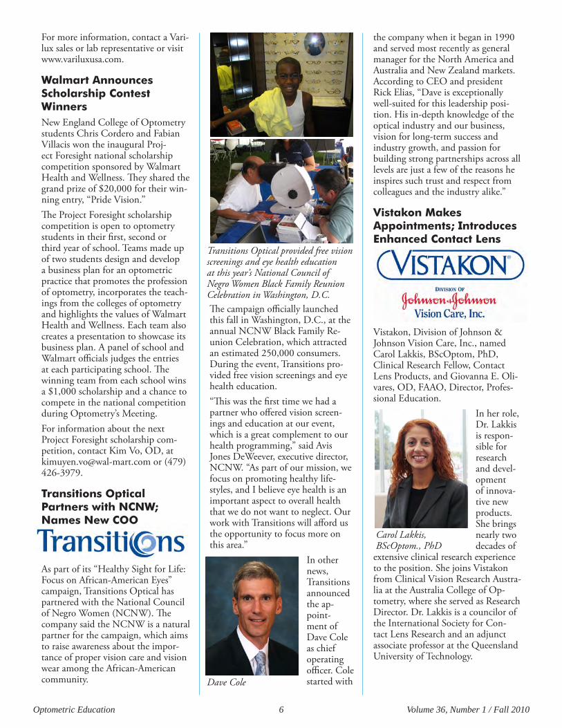
As part of its “Healthy Sight for Life: Focus on African-American Eyes” campaign, Transitions Optical has partnered with the National Council of Negro Women (NCNW). The company said the NCNW is a natural partner for the campaign, which aims to raise awareness about the impor-tance of proper vision care and vision wear among the African-American community.
The campaign officially launched this fall in Washington, D.C., at the annual NCNW Black Family Re-union Celebration, which attracted an estimated 250,000 consumers. During the event, Transitions pro-vided free vision screenings and eye health education.“This was the first time we had a partner who offered vision screen-ings and education at our event, which is a great complement to our health programming,” said Avis Jones DeWeever, executive director, NCNW. “As part of our mission, we focus on promoting healthy life-styles, and I believe eye health is an important aspect to overall health that we do not want to neglect. Our work with Transitions will afford us the opportunity to focus more on this area.”
In other news, Transitions announced the ap-point-ment of Dave Cole as chief operating officer. Cole started with
the company when it began in 1990 and served most recently as general manager for the North America and Australia and New Zealand markets. According to CEO and president Rick Elias, “Dave is exceptionally well-suited for this leadership posi-tion. His in-depth knowledge of the optical industry and our business, vision for long-term success and industry growth, and passion for building strong partnerships across all levels are just a few of the reasons he inspires such trust and respect from colleagues and the industry alike.”
Vistakon Makes Appointments; Introduces Enhanced Contact Lens
Vistakon, Division of Johnson & Johnson Vision Care, Inc., named Carol Lakkis, BScOptom, PhD, Clinical Research Fellow, Contact Lens Products, and Giovanna E. Oli-vares, OD, FAAO, Director, Profes-sional Education.
In her role, Dr. Lakkis is respon-sible for research and devel-opment of innova-tive new products. She brings nearly two decades of
extensive clinical research experience to the position. She joins Vistakon from Clinical Vision Research Austra-lia at the Australia College of Op-tometry, where she served as Research Director. Dr. Lakkis is a councilor of the International Society for Con-tact Lens Research and an adjunct associate professor at the Queensland University of Technology.
Dave Cole
Carol Lakkis, BScOptom., PhD
Transitions Optical provided free vision screenings and eye health education at this year’s National Council of Negro Women Black Family Reunion Celebration in Washington, D.C.
Optometric Education 7 Volume 36, Number 1 / Fall 2010
In her new role, Dr. Olivares is responsible for developing strategies for the implemen-tation of the company’s pro-fessional educa-tional programs across a broad spectrum of groups, includ-ing students, practitioners,
Professional Affairs Consultants and Vistakon Sales and Marketing orga-nizations. She joined the company in 2004 as Manager of the R&D De-sign Clinical Research Group, where she led a multidisciplinary group of optometrists, ophthalmologists, vision scientists and biostatisticians overseeing the clinical development of innovative new contact lens products, including Acuvue Oasys for Astigma-tism. Her team also developed novel methodologies and equipment for testing vision and measuring patients’ experiences with contact lenses.Vistakon also announced the availabili-ty of Acuvue Advance Plus Brand Con-tact Lenses. Acuvue Advance Plus is a redesigned and enhanced successor to the Acuvue Advance lens. Along with Ultra-Clean Technology for deposit resistance and Hydraclear Technology that combines high performance base materials with a moisture-rich wetting agent, the new lens offers improve-ments in initial and overall comfort and visual acuity. In addition, it blocks more than 90% of UV-A rays and 99% of UV-B rays that reach the lens. The company says the Acuvue Advance Plus lens provides patients with the freshness of a two-week modality at a price comparable to a monthly lens. It is available in base curves of 8.3 mm and 8.7 mm at parameters of -0.50D to -6.00D and +0.50D to +6.0D in 0.25D increments and from -6.50D to 12.0D and +6.50D to +8.0D in .50D increments.
Volk Gonio Lens Designed for Better View in Less Time
A new gonioscopy lens from Volk Optical, the G-6, features six closely aligned mirrors to enable a true 360° view during glaucoma screening. The mirrors are equally angled at 63°, eliminating gaps for visualization of the entire anterior segment at 1.0X magnification. During examination, two of the mirrors are aligned in the superior and inferior regions, while the remaining four mirrors provide a continuous view through the nasal and temporal regions. This allows practi-tioners to scan across mirrors quickly, without the confusion of tracking where one view ends and the next begins that can come from rotating the lens. This fast scanning and reduced need to maneuver the slit lamp can reduce examination time.
The G-6 also features a taller, tapered profile, which is easier to hold within the orbit, and a no flange/no fluid design that facilitates use without vis-cous interface solutions. For improved handling flexibility, the lens comes with a ring or a 2-in-1 handle that can be adjusted to create a straight or 45° angled grip.
The Volk G-6 is designed to provide a true 360° view of the anterior segment for glaucoma diagnosis.
Giovanna E. Olivares, OD, FAAO
“A man has madeat least a start on
discovering the meaning ofhuman life when he plantsshade trees under which heknows full well he will
never sit.”- Anonymous
The Partnership Foundation for Optometric Education isplanting, cultivating, and nurturing. Together, this “truepartnership” of state, regional, and national organizationsis making a long-term investment in tomorrow. With theinvestment we make today in optometric education,future generations of practitioners will flourish.
For more information, contact thePartnership Foundation atwww.opted.orgor 301-231-5944, ext 3018.
Have you thought about thefuture of Optometry?
We have!
Optometric Education 8 Volume 36, Number 1 / Fall 2010
Optometric Education 9 Volume 36, Number 1 / Fall 2010
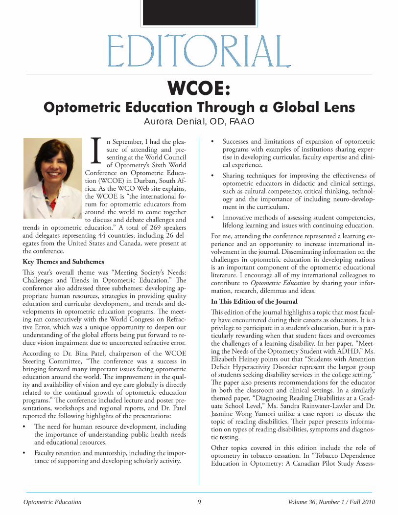
n September, I had the plea-sure of attending and pre-senting at the World Council of Optometry’s Sixth World
Conference on Optometric Educa-tion (WCOE) in Durban, South Af-rica. As the WCO Web site explains, the WCOE is “the international fo-rum for optometric educators from around the world to come together to discuss and debate challenges and
trends in optometric education.” A total of 269 speakers and delegates representing 44 countries, including 26 del-egates from the United States and Canada, were present at the conference.Key Themes and SubthemesThis year’s overall theme was “Meeting Society’s Needs: Challenges and Trends in Optometric Education.” The conference also addressed three subthemes: developing ap-propriate human resources, strategies in providing quality education and curricular development, and trends and de-velopments in optometric education programs. The meet-ing ran consecutively with the World Congress on Refrac-tive Error, which was a unique opportunity to deepen our understanding of the global efforts being put forward to re-duce vision impairment due to uncorrected refractive error. According to Dr. Bina Patel, chairperson of the WCOE Steering Committee, “The conference was a success in bringing forward many important issues facing optometric education around the world. The improvement in the qual-ity and availability of vision and eye care globally is directly related to the continual growth of optometric education programs.” The conference included lecture and poster pre-sentations, workshops and regional reports, and Dr. Patel reported the following highlights of the presentations:• Theneedforhumanresourcedevelopment, including
the importance of understanding public health needs and educational resources.
• Facultyretentionandmentorship,includingtheimpor-tance of supporting and developing scholarly activity.
I
WCOE: Optometric Education Through a Global Lens
Aurora Denial, OD, FAAO
• Successes and limitations of expansion of optometricprograms with examples of institutions sharing exper-tise in developing curricular, faculty expertise and clini-cal experience.
• Sharing techniques for improving the effectiveness ofoptometric educators in didactic and clinical settings, such as cultural competency, critical thinking, technol-ogy and the importance of including neuro-develop-ment in the curriculum.
• Innovativemethodsofassessingstudentcompetencies,lifelong learning and issues with continuing education.
For me, attending the conference represented a learning ex-perience and an opportunity to increase international in-volvement in the journal. Disseminating information on the challenges in optometric education in developing nations is an important component of the optometric educational literature. I encourage all of my international colleagues to contribute to Optometric Education by sharing your infor-mation, research, dilemmas and ideas. In This Edition of the JournalThis edition of the journal highlights a topic that most facul-ty have encountered during their careers as educators. It is a privilege to participate in a student’s education, but it is par-ticularly rewarding when that student faces and overcomes the challenges of a learning disability. In her paper, “Meet-ing the Needs of the Optometry Student with ADHD,” Ms. Elizabeth Heiney points out that “Students with Attention Deficit Hyperactivity Disorder represent the largest group of students seeking disability services in the college setting.” The paper also presents recommendations for the educator in both the classroom and clinical settings. In a similarly themed paper, “Diagnosing Reading Disabilities at a Grad-uate School Level,” Ms. Sandra Rainwater-Lawler and Dr. Jasmine Wong Yumori utilize a case report to discuss the topic of reading disabilities. Their paper presents informa-tion on types of reading disabilities, symptoms and diagnos-tic testing.Other topics covered in this edition include the role of optometry in tobacco cessation. In “Tobacco Dependence Education in Optometry: A Canadian Pilot Study Assess-
Optometric Education Diplomate Program
Now Accepting ApplicationsThe American Academy of Optometry’s Optometric Education Section is now accepting applications for its Optometric Education Diplomate
Program. The Academy Board of Directors approved the program in May.
The granting of diplomate status in Optometric Education is recognition of a focus and expertise in education beyond the level of teaching responsibilities that are commonly held by most faculty. It recognizes advancement in the areas of scholarly activity, educational research,
advanced education and the delivery and transfer of knowledge.
For program criteria and an application visit www.aaopt.org/section/oe/becoming.
Optometric Education 10 Volume 36, Number 1 / Fall 2010
ing Practices and Opportunities,” Dr. Marlee M. Spafford and co-authors state that “tobacco use and dependence is a preventable cause of morbidity and mortality and healthcare providers can be effective facilitators of tobacco cessation among their patients.”This edition also features “Acute Ocular Trauma in a Child: A Teaching Case Report.” Drs. Wendy J. Haaland Stone and Stephanie A. Klemencic present a clinical scenario and edu-cational guidelines to facilitate teaching. This is an impor-tant topic because ocular injuries in children are common and can range in severity. Optometry students and recent graduates must feel confident providing care in these situ-ations.In this edition you will also notice a slight modification in the format of the “Think Tank” feature. The key concept — an opinion-based forum for sharing ideas — remains the same. However, rather than pre-identifying an issue and inviting contributors to submit their comments, we pres-ent “Some Thoughts on Clinical Teaching” from Dr. Mark Vogel. We challenge you, the readers of the journal, to vol-untarily send us your comments on this piece. Do you agree with Dr. Vogel? Have you had the same experiences? Did you come to the same conclusions? Send your comments to me at [email protected], and we will publish them in the next edition of the journal.
Optometric Education 11 Volume 36, Number 1 / Fall 2010
Think Tank ... Some Thoughts on Clinical Teaching
for achieving a goal. They also noted that what was taught to them in their initial clinical education was altered in subsequent courses because of the in-herent inappropriateness of the original material in actual clinical encounters.It should be noted that these were in-telligent and highly competent clini-cal externs, far from the worst I have encountered in more than 30 years of clinical teaching, and that subsequent surveys of other students, residents and graduates of a number of schools have found these impressions to be generally consistent.We Have All Experienced How Not to be TaughtI was reminded of two incidents from my own experience. The first occurred during one of my own externships, at an institution other than the one I at-tended. A friend and I were being su-pervised during patient encounters by a supervisor who had lectured to us about ways to perform certain clinical tests. I felt the methods I had learned previ-ously were better and I performed the tests accordingly. Apparently my class-mate had done the same thing because the supervisor took us into a room and berated us for not performing the tests his way and accused us of not paying at-tention during his lecture. Neither of us offered a defense or explanation, but we left the encounter with little respect for the instructor. Two years later I joined the faculty of that institution and dis-covered that particular individual was one of a few who prided themselves on their ability to “break” students. He continued to teach until his voluntary retirement. The second incident that came to mind occurred a number of years ago while
Mark S. Vogel, OD, FAAO
while ago, I spoke with some fourth-year optometry stu-dents who were completing an externship at our VA hospital.
I wanted to determine what they felt was positive and negative about their experiences in order to perpetuate the strengths of the program and shore up its weaknesses. It was gratifying to learn that they were generally pleased with the externship and compared it favor-ably to a number of others they had previously attended. I also asked them to compare their clinical experience in the current program with what they had encountered in the clinics of their optometry schools. I was startled at the severity of their responses with regard to the attitudes of what they described as a sizeable percentage of their clinical instructors.They described an atmosphere in which they were afraid to express themselves for fear of being told they were either stupid or incompetent. They told of su-pervisors who expected perfection both in knowledge and performance even in the early stages of their clinical training when they had not yet been exposed to certain material either didactically or clinically. They described situations in which a particular instructor insisted that certain formats be followed only to be castigated by a succeeding supervi-sor for doing what they had been previ-ously taught. They noted little consis-tency from one instructor to the next and little tolerance by instructors in their acceptance of alternate methods
A
I was supervising in a first-year clinical methods laboratory. One of my students approached me with a question, which was overheard by another faculty per-son who chastised the student for ask-ing the question. In front of other stu-dents and faculty he loudly proclaimed that the student should not be asking that question because he himself had taught her the material. He informed her that she must be stupid if she did not understand. When he walked away I explained the material to the student who asked the question as well as oth-ers who witnessed the encounter. I told them I would explain it as many times in as many ways as I could to help them understand. Because he was tenured, the instructor taught for many years and was probably the only person on staff who was unaware of his inability to disseminate information effectively.I am sure anyone reading about these experiences would be appalled that such behavior exists in institutions of higher learning. By the same token, I doubt if anyone can think back on his or her own education and deny they encountered such individuals or situa-tions. While I in no way feel that pro-fessional students should be coddled or overly protected, I do feel they should be treated with respect and dignity. I am pleased to say that many of my col-leagues for these many years have not been guilty of such sins. But there have been, and remain, a significant number of individuals, both in institutional and private practice, in and out of optom-etry, who could benefit from a reassess-ment of their positions and responsi-bilities.Concepts Worth ConsideringWith this in mind, I offer the following perspectives on clinical teaching.
Dr. Vogel is an adjunct assistant clinical profes-sor at State University of New York College of Optometry and a staff optometrist at North-port Veterans Affairs Medical Center in North-port, NY.
Optometric educators, we welcome your comments on ...
Optometric Education 12 Volume 36, Number 1 / Fall 2010
1) Teach it correctly the first time. If material changes, change with the times. If new or different techniques or methods are more efficient or appropriate, incorporate them into the syllabus and delete older or less effective material. It is incumbent upon teachers to keep up with new and alternative information and be open and honest enough to change accordingly. It is incorrect to pass outdated or mistaken concepts or methods to another generation. What was new in a text 30 years ago is old today and should be re-evaluated for its effectiveness and timeliness. This is especially true in the education of new practitioners who must learn material one way only to have to alter their methods appropriately in the future. This is unnecessarily confusing and time-consuming in an ever-expanding curriculum. For example, teaching a certain cross-cylinder test to first-year students in a particular man-ner was demonstrated to be of little value more than a decade ago, yet it remains in this form in some cur-ricula. If you are even aware of the Humphriss technique, when was the last time you used it? It, too, remains in some curricula.
When and how should curricula be changed? The first part is easy to an-swer – as often as necessary to stay current in the field and reflect the most efficient model of patient care delivery. The second part is more difficult to answer. Having worked on a curriculum committee at an optometric institution I am keenly aware of how difficult it is to make curriculum changes. The major ob-stacle is obtaining the cooperation of instructors. Each instructor feels that his or her course is infinitely essential to the proper education of students and, therefore, declines to allow even one hour of coursework to be eliminated from its material. This is understandable. Job security is important to everyone, and to admit material is not essential or is outdated is to admit, perhaps, that the individual teaching the class may not be essential.
So, how to proceed? Maybe it is time for our schools to employ a
modality used not only in private healthcare practice but in the busi-ness world in general – outside con-sultancy. The schools should hire people, perhaps successful private practitioners, to dispassionately evaluate what is and is not essen-tial to produce good practitioners. These people should not be gradu-ates of the school being examined. They should not be employees of any school. Our schools already tend to hire their own graduates, which prevents a healthy infusion of new ideas and methods into our training.
2) If you don’t do it, don’t teach it. Many people involved in clinical education are teaching or supervis-ing methods with which they have no immediate experience. A clinical instructor who has no background in the implementation of contact lens care should not be instruct-ing in a clinic where contact lens care is provided. A didactic teacher who teaches clinical methods who does not interact with patients has no frame of reference to know whether what he or she is teach-ing is appropriate in a real clinical encounter. Quite simply, a student should be taught to remove an ap-pendix by someone who actually performs appendectomies rather than by someone who has read or heard about appendectomies.
3) Leave your ego at home. It is easy to know more than your students. Do not make this a source of pride to be dangled before them. A teacher is expected to know more than his students and it is this knowledge that should be passed along in an open, nonthreatening manner.
4) Allow for the fact that there might be alternate approaches to solving a problem. When confronted with alternative methods to handle a particular issue, be open to these concepts and consider allowing another person (student) to imple-ment them as long as no foreseeable harm may arise from such action.
5) Customize your teaching. Explain material at a level that students can understand, not necessarily at the level at which you may con-
verse with your colleagues. Stu-dents do not have the same level of knowledge or experience as do their teachers, and much material is new and potentially confusing. Furthermore, what is obvious to one student may not be obvious to another. Try to communicate with each student at whatever level is necessary to convey information effectively.
6) KISS – Keep It Simple, Stupid. It is easy to confuse and confound. It is not always easy to get to the simplest mode. Take a moment to consider the situation with which you are confronted and try to come up with the simplest explanation, diagnosis, answer or approach.
7) Allow, nay, encourage students to say “I don’t know.” Not to do so engenders fear, deceit, and fabrica-tion. This should be followed by clear explanations and/or assign-ments for students to ferret out the answers to enhance their own edu-cation. Of course, this allowance should not be overly relied upon by students. The continued dem-onstration of ignorance cannot be tolerated.
8) Students can learn as much, or more, from mistakes and poor per-formance as they can from good episodes, if handled properly. Try to make every learning experience a positive learning experience by demonstrating how performance can be improved rather than harp-ing upon how poorly things have already been done.
9) Do not spoon-feed students. They should be encouraged to learn for themselves. Give them the basics then ask and expect them to re-search and think about what they are doing. Students all too often learn things by rote in order to an-swer a question on a test and when asked how that information can be clinically applied have no idea. Try to ask questions that require thought and the assimilation of various pieces of material that must be incorporated into a whole in or-der to arrive at answers. Encourage research. MAKE THEM THINK.
10) Do not teach students merely to en-
Optometric Education 13 Volume 36, Number 1 / Fall 2010
sure they pass boards. While board scores seem important to school ad-ministrators, they are not necessar-ily advantageous to the public. Stu-dents who pass written tests do not necessarily become good clinicians. The current approach to board cer-tification fails in this regard as it does not ensure a doctor can per-form appropriately in the arena of patient contact. All too often stu-dents examine data as if contained in little boxes, ignoring other in-formation in other little boxes that should be combined into a unified whole. Students should be trained to be efficient, effective doctors ca-pable of integrating material from all disciplinary areas into cohesive assessment and treatment plans.
11) It is okay for students to be wrong. They are still learning. In that sense, each of us is still a student. If teach-ers think they have nothing more to learn, they probably should not be teaching. Excessive criticism and overbearing demeanor most likely engender a bad impression of the supervisor and not necessarily en-courage the pursuit of knowledge.
12) Teachers can be wrong. They must be able to admit fallibility when it is appropriate.
13) Teachers can learn from students. It is just as important to listen as it is to talk.
14) Teachers and students do not nec-essarily have to like one another, but teachers must continue to teach and students must continue to learn. Personality conflicts and unhappy interactions are bound to occur at some point. In such situ-ations the teacher must put aside
personal prejudices and handle the educational process professionally. The student must be able to be ma-ture and put personal feelings aside and concentrate on the matters at hand, be they didactic failures or poor clinical performance. The teacher must be able to present the facts of the instance as clearly and unemotionally as possible and not use his or her position of author-ity as the sole basis for demanding obedience.
15) Students should be allowed and encouraged to question everything. They should not be expected to ac-cept a concept simply because the teacher says it is so. It is incumbent upon teachers to provide a rationale and a logical context for anything presented to the student. Teachers constantly challenge students and it is unwise and unfair to disallow challenges from students if present-ed honestly and maturely.
16) Sometimes students deserve to fail. This is included in this treatise because I have seen a number of students passed along because in-structors do not have the heart or fortitude to fail them when it is ap-propriate or because an institution needs to protect its capitation fund-ing. Failure should be accompanied by explanation and encouragement for improvement. However, when students really cannot meet certain professional or personal standards, it is unfair to them and to the pub-lic to allow them to continue in a program.
17) Discuss with, rather than lecture to, students whenever possible. They are people, have brains, and deserve respect.
18) When you become a teacher or su-pervisor, do not forget what it was like to be a student. This will make you a better and more compassion-ate teacher.
Improving, Not Hindering, the Edu-cational ProcessIf all clinical teachers were commit-ted to being thorough and treating their students like people by showing concern for them, listening to them, encouraging communication in two directions and promoting learning, the educational arena would be more ef-fective, efficient, human and humane. Doctor (teacher), heal thyself.
Send Us Your CommentsDo you agree with Dr. Vogel? Have you had similar experiences? Did you come to the same conclusions? Send your comments to Dr. Au-rora Denial at [email protected], and we will publish them in the next edition of the journal.
Optometric Education 14 Volume 36, Number 1 / Fall 2010
MY BEST DAY
Energy Renewal Arrives Twice Yearly
hen asked to select a “best day” in my career in optometric education, I was flooded with won-
derful memories. As I reflected upon each — and tried to rank them by what my colleagues might consider special or worthy of their reading time — I found myself unsettled. What was important to me or the few close colleagues I had consulted would most certainly not be of interest to all. What to do?I decided to try a different approach, realizing that I just might be stretch-ing our editor’s patience and treading on thin ice. I consider myself fortunate to have not one best day, but two, and they occur each and every year. My first best day of the year occurs on new student orientation. After months of overseeing the admissions process, wading through stacks of files and ap-plications and painstakingly working with ICO’s Admissions Committee selecting the best candidates, I see the selected class arrive. I watch their faces and collective energy fill ICO’s Lecture Center.As I stand watching each student pick up a name tag, I reflect upon those I personally encountered during their process of applying. I reflect upon the tremendous effort they all put into demonstrating they were ICO material
W
and worthy of joining our wonderful profession. In each of their eyes, I see many years of effort, and perhaps even dreaming, coming to fruition on this day. I can sense the energy as their en-thusiasm fills the room and for some I can see passion for our wonderful pro-fession already brewing. That is truly a best day for me.My other best day of the year is com-mencement. ICO is fortunate to hold its commencement ceremony in Rock-efeller Chapel on the University of Chicago campus. Rockefeller Chapel is a beautiful, gothic cathedral-inspired, towering stone structure that has served as the home to ICO commencement ceremonies for more than 25 years. In-side the chapel is a beautiful and mas-sive pipe organ, and when I reach the back of the chapel to begin the proces-sional, I still get chills down my spine as I recall my own proud commencement ceremony in that same place.As we walk down the aisle during the processional, we look out at the 1,200 proud faces of the assembled parents, spouses, grandparents, siblings, aunts and uncles and supportive friends. Their rightful pride in their graduate’s accom-plishments is evident. Just past the au-dience seating, the processional passes the graduating class robed in their doc-toral gowns. Three distinct velvet bands on their sleeves signify they have earned a doctoral degree. I see each graduate nearly bursting with pride, some no-ticeably emotional (or is that just me?). They seem to be reflecting upon the hours, days, weeks, months and years of dedicated study and the accomplish-ments that earned them the right to be in this particular grand location on this particular day in May.
During the ceremony, I have the dis-tinct honor of announcing each new doctor’s name as he or she steps forward to receive a diploma and participate in ICO’s solemn hooding ceremony. I can see their pride beaming as they fully re-alize just what they have accomplished. I also see a twinge of anxiety as they contemplate the immense responsibili-ty they are now charged with in execut-ing their duties to their patients. When ICO’s president, Dr. Arol Augsburger, proclaims the doctoral degree upon them, the previously solemn chapel erupts with applause of celebration, and hugs of congratulations are exchanged among the class members. Truly a jubi-lant moment in their lives. During Commencement Day, I find I just can’t hold back my own smile, which emanates in appreciation of these fine young people sharing part of their lives with me along their journey. I consider it a true privilege to be a part of such an incredible undertaking. I feel truly blessed to watch and observe as young college graduates transform themselves into highly educated and knowledgeable doctors of optometry over a span of four years.Following the ceremony, I stroll around to offer my personal congratulations and observe as each new doctor poses for photographs with family members, friends and faculty and attempts to capture the energy and emotion of this wonderful day. My wish is that they will each be able to carry the energy from this day forward as they launch their new careers and face the many challenges that are no doubt ahead. At the end of this glorious day, I find myself retrospective and exhausted. The day is symbolic of the closing of
Mark Colip, OD
Optometric Education 15 Volume 36, Number 1 / Fall 2010
MY BEST DAY
another academic year, and I know that in just three short months we will rev up to begin the journey with another class. I promised myself when I entered optometric education that the day I did not find these two days both exhilarat-ing and fulfilling would be the day I needed to leave optometric education and return to full-time practice.
In closing, I would like to extend that challenge to all of my colleagues in op-tometric education. Students bring our educational institutions to life and they are deserving of every educator’s 100% commitment. Students today require a great deal of energy as we fulfill our re-sponsibilities to help them become the great doctors we know they are capable
of becoming. It is up to each of us to find that energy within ourselves and apply it in our careers as educators on a daily basis. Fortunately for me, that en-ergy renewal source comes twice a year without fail! Make it a great year!
Dr. Colip is Vice President for Student, Alum-ni and College Development at Illinois College of Optometry in Chicago.
(continued)
Invitation to ParticipateUpcoming Theme EditionsImplementing the Teaching of Critical and Clinical Thinking
In optometry, critical thinking related to clinical decisions and patient care is a specific outcome of the educational process. Many optometric institutions have initiated courses dedicated to teaching critical thinking, clinical decision-making and integration of knowledge. Optometric Education is planning a future theme edition that will focus on courses designed to achieve the goals of teaching critical thinking, clinical decision-making and integration of knowledge. We invite all educators involved in these courses to participate in the theme edition. To allow for additional time for outcomes assessment, the deadline to submit articles for this theme edition has been extended to November 30, 2010. Accepted manuscripts will include: innovative teaching meth-odologies, course description and assessment, research on how, when and why students learn about clinical thinking or teaching interventions that increased learning.
Scholarship Scholarly contributions by faculty are a critical component of faculty development, promotion/tenure and delivery of optometric education. Most optometric faculty have minimal formal training in professional writing, research and publication. Scholarly con-tributions move education forward and can significantly impact the profession. Optometric Education is planning a future theme edition that will focus on scholarship. The theme edition is scheduled for publication in 2012. We are sending out this invitation early to allow for adequate time to design appropriate studies. We invite all educators and administrators to participate in this theme edition.
For additional information on the theme editions, contact Dr. Aurora Denial at [email protected].
Optometric Education 16 Volume 36, Number 1 / Fall 2010
ASCOTech
Technology Can Help Students with Learning Disabilities Succeed
ompared with elementary schools or high schools that cater to the masses, there may be a false perception that pro-
fessional school programs don’t need to worry much about students with learn-ing disabilities. After all, we only accept the best and brightest into optometry school, right? In reality, disabilities know no boundaries, and some students with great talent may need extra assis-tance from our academic programs.Although most students with visual, au-ditory, or physical disabilities may not seek out an optometric career path, some students with learning or other more subtle disabilities may very well already be in our classrooms. As those with dis-ability are encouraged to seek out higher educational and career goals, we will be encountering these students with great-er frequency in our classrooms, labora-tories and clinics. This places upon us an important responsibility to provide the necessary tools and resources so that these students can be successful.Accommodations like extended test-ing time or modified student schedules may top the list, but it’s important to remember the ever-growing list of tech-nology solutions that may also lend a helping hand. A literature search pro-vides countless examples of studies that show technology’s benefit to stu-dents with learning disabilities. Such
Geoffrey Goodfellow, OD, FAAODominick M. Maino, OD, MEd, FAAO, FCOVD-A
C
Dr. Goodfellow is assistant dean for Curriculum and Assessment and an associate professor at the Illinois College of Optometry. Dr. Maino is a professor at the Illinois College of Optometry. They invite your feedback about this and all ASCOTech columns and your suggestions for future columns. E-mail them at [email protected] or [email protected]. You can also visit www.MainosMemos.blogspot.com.
assistance also tends to improve self-sufficiency for students who otherwise may be overly dependent on faculty, tu-tors, peers, or parents to help them get through our programs.Here, we have compiled a list of options you and your students may find helpful.
MemoryA variety of tools are available for stu-dents with memory problems. Many software applications allow users to store notes, reminders, pictures and Web pages, as well as email, attach-ments and almost anything else in one
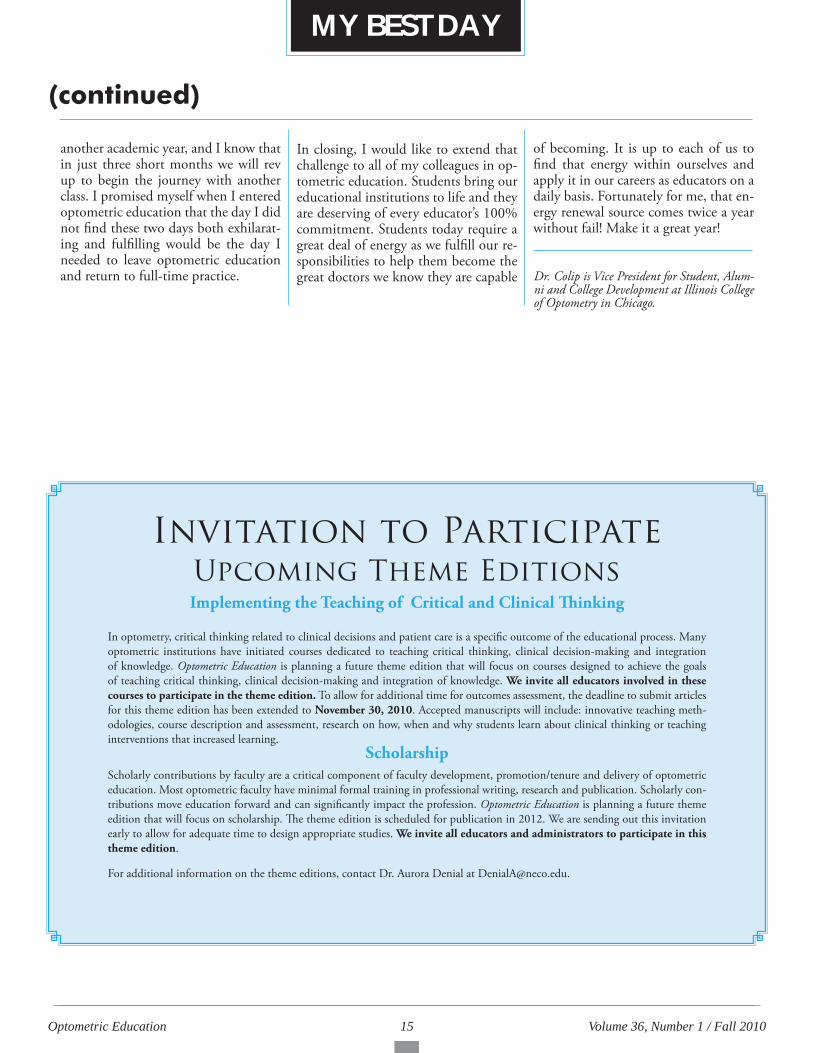
spot. Users can move things around, organize materials in a way that works for them and, best of all, electronically search through their notes by remem-bering only snippets of text. Some of the newer tools are even capable of searching for key words that are part of photos and other scanned materials. Microsoft’s OneNote program is part of the Microsoft Office Suite of software commonly available on most comput-ers and is a great and easily accessible tool for our students to use. (Figure 1) The program Ask Sam (www.asksam.com) is another option.
Figure 1 Microsoft OneNote provides an easy way for students to gather information from many sources and organize it in
a single location.
Optometric Education 17 Volume 36, Number 1 / Fall 2010
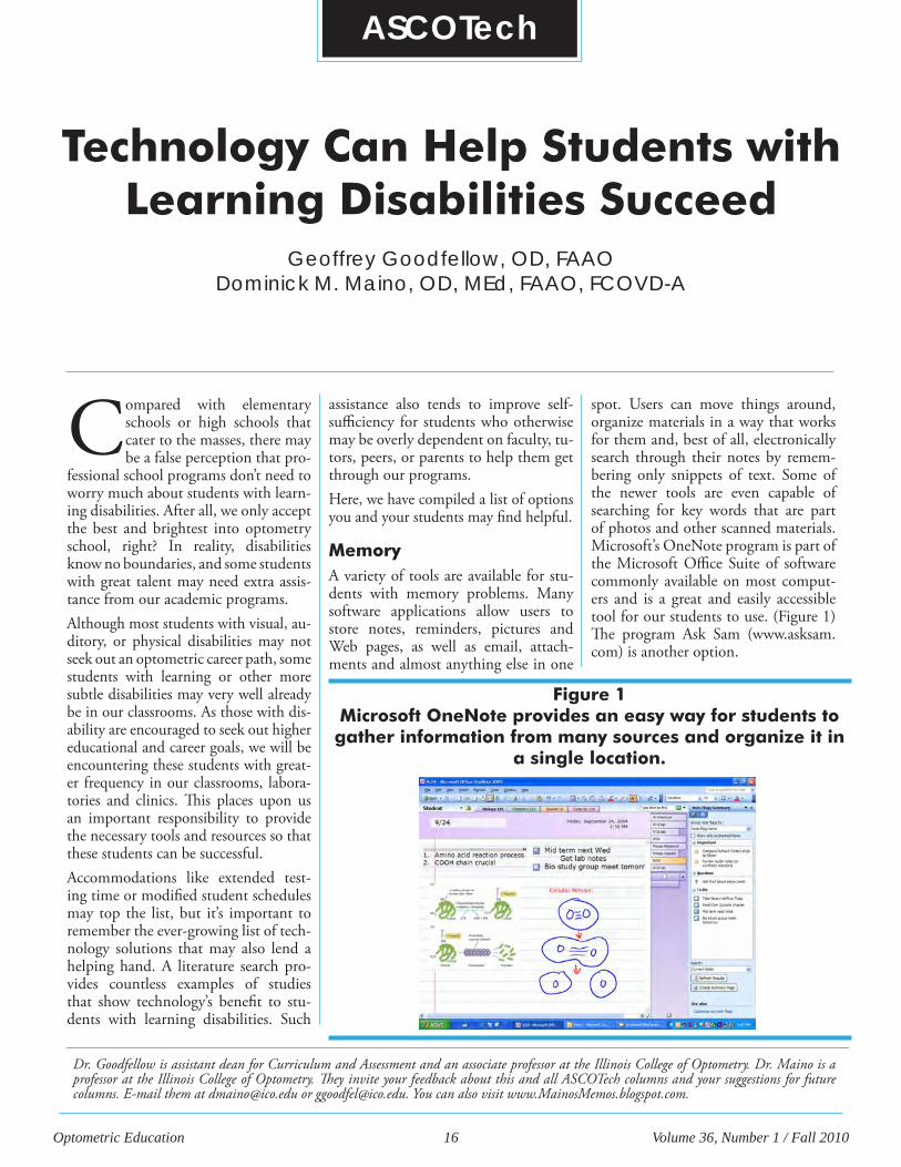
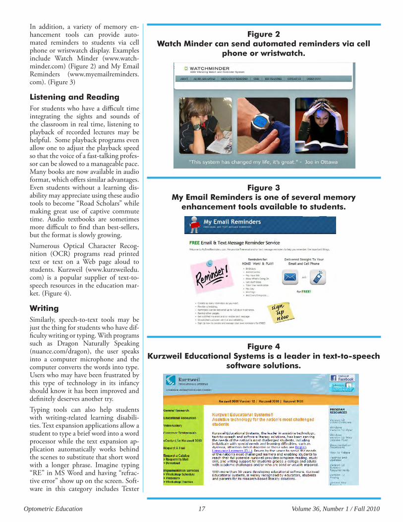
In addition, a variety of memory en-hancement tools can provide auto-mated reminders to students via cell phone or wristwatch display. Examples include Watch Minder (www.watch-minder.com) (Figure 2) and My Email Reminders (www.myemailreminders.com). (Figure 3)
Listening and ReadingFor students who have a difficult time integrating the sights and sounds of the classroom in real time, listening to playback of recorded lectures may be helpful. Some playback programs even allow one to adjust the playback speed so that the voice of a fast-talking profes-sor can be slowed to a manageable pace. Many books are now available in audio format, which offers similar advantages. Even students without a learning dis-ability may appreciate using these audio tools to become “Road Scholars” while making great use of captive commute time. Audio textbooks are sometimes more difficult to find than best-sellers, but the format is slowly growing.Numerous Optical Character Recog-nition (OCR) programs read printed text or text on a Web page aloud to students. Kurzweil (www.kurzweiledu.com) is a popular supplier of text-to-speech resources in the education mar-ket. (Figure 4).
WritingSimilarly, speech-to-text tools may be just the thing for students who have dif-ficulty writing or typing. With programs such as Dragon Naturally Speaking (nuance.com/dragon), the user speaks into a computer microphone and the computer converts the words into type. Users who may have been frustrated by this type of technology in its infancy should know it has been improved and definitely deserves another try.Typing tools can also help students with writing-related learning disabili-ties. Text expansion applications allow a student to type a brief word into a word processor while the text expansion ap-plication automatically works behind the scenes to substitute that short word with a longer phrase. Imagine typing “RE” in MS Word and having “refrac-tive error” show up on the screen. Soft-ware in this category includes Texter
Figure 2 Watch Minder can send automated reminders via cell
phone or wristwatch.
Figure 3 My Email Reminders is one of several memory
enhancement tools available to students.
Figure 4 Kurzweil Educational Systems is a leader in text-to-speech
software solutions.
Optometric Education 18 Volume 36, Number 1 / Fall 2010
(lifehacker.com/software/texter/life-hacker-code-texter-windows-238306.php), Typinator (www.ergonis.com/products/typinator) and Activewords (activewords.com).When spelling is problem, phonetic spelling tools can be used. These al-low the student to type the way a word sounds, and the software produces the correct spelling. However, today’s great word processors that highlight typing/spelling errors in real time may make phonetic spelling software unnecessary.
Number of Resources Continues to GrowAs the rigors of the optometric craft continue to increase, assisting pro-fessional school students who have a learning disability can be challenging. However, a wide variety of technology tools can be very useful, and the list of resources grows daily as software devel-opers work their magic.If you know of other resources that help your students with learning disabilities, let us know about them by emailing us at [email protected] and [email protected].
Optometric Education 19 Volume 36, Number 1 / Fall 2010
Meeting the Needs of the Optometry Student with ADHD
Elizabeth P. Heiney, MS
AbstractThis paper summarizes a presentation given at the 2009 American Academy of Optometry annual conference in Orlando, FL. Students with Attention Deficit Hyperactivity Disorder (ADHD) represent the largest group of students seeking disability services in college. The literature is only beginning to recognize the specific needs of these students in the college setting. While a portion of these stu-dents also pursue graduate degrees, the current literature examining the impact of graduate study on a person with ADHD is extremely limited. This paper ex-pands the knowledge base regarding the challenges faced by the graduate student with ADHD, specifically those in an optometry program. Recommendations for educators that can be applied to the classroom as well as the clinical setting are explored.
Key Words: ADHD, graduate students, optometry students, academic needs, accommodations
Ms. Heiney is a Doctoral Candidate of Clinical Psychology at Spalding University. She is completing her predoctoral internship at Family Service and Guidance Center in Topeka, KS.
Introductionmagine sitting in a classroom, trying to complete an exam or take lecture notes. No matter how diligently you try to focus,
other thoughts continuously interfere. You really want to focus, but your brain is just not cooperating. This is what it is like for students with Attention Deficit Hyperactivity Disorder (ADHD). Once believed to affect only children, ADHD is now understood to be a pervasive dis-ability that continues into adulthood in 55%-75% of cases.1 This translates to ADHD affecting up to 6% of the adult population.2 Although ADHD can be associated with academic underachieve-ment,3 the number of students with ADHD seeking post-secondary educa-tion is increasing due to special educa-tion laws,4 improved diagnostic proce-dures and increased awareness of the impact of this disorder on adults.5 It is estimated that up to 5% of U.S. college students have ADHD.6 A portion of college students with ADHD also seeks graduate education. A literature search using the terms “graduate student” and “ADHD” in the Education Resources Information Center (ERIC) and Education Research Complete databases found no research that cited the prevalence rates of gradu-ate students with ADHD. At this point, the literature regarding this population is limited to acknowledging that gradu-ate students with ADHD continue to face challenges in the education setting and how the Americans with Disabilities Act (ADA) applies to these students.7 Although educational researchers have not yet focused on optometry students, they have identified graduate students in other health professions, including medical students8 and more specifi-cally psychiatry residents with ADHD.7 As these groups represent students in a health profession training program that includes both intense classroom instruc-tion and clinical practice,7 it is likely that their experiences are comparable to those of optometry students with ADHD. Considering the prevalence rates of ADHD in adults as well as the existence of students with ADHD in other health professions, it is expected
I

Optometric Education 20 Volume 36, Number 1 / Fall 2010
that a percentage of optometry students will have ADHD and will subsequently be at risk of having academic difficulty.It is imperative that optometric educa-tors understand how ADHD may af-fect students. Researchers suggest that professors and administrative personnel take a more active role in promoting the academic success of students with ADHD3,9-12 by understanding the chal-lenges that these students face as well as what role they can play in improving their success rate.10 The ADA states that individuals with disabilities, including ADHD, must be afforded reasonable accommodations that allow them to perform at an acceptable level. To maxi-mize outcomes, a collaborative approach between the student, administration and faculty, working within the bound-aries of the ADA, should be sought.7 The purpose of this paper is two-fold: 1) to inform optometric educators how ADHD manifests in students and im-pacts a student’s education and 2) to provide practical recommendations that can be used to help optometry students in their success both in the classroom and in clinical settings.
Attention Deficit Hyperactivity Disorder (ADHD)ADHD is characterized by a pattern of inattention, and/or hyperactive/im-pulsive behavior that manifests across multiple settings.13 It causes significant impairment in the ability to filter out irrelevant information, sustain focus, delay gratification, think before act-ing and problem-solve.10 ADHD cur-rently is divided into three subtypes: 1) Predominantly Inattentive Type; 2) Predominantly Hyperactive/Impulsive Type; and (3) Combined Type. Inat-tentive symptoms include difficulty concentrating, poor attention to de-tail, difficulty completing tasks and forgetfulness. Hyperactive/Impulsive symptoms include significant motor agitation, difficulty awaiting turns and excessive talking.14 While diagnostic criteria emphasize mainly the behavior-al manifestations of ADHD, it should also be conceptualized as a cognitive disorder with implications for academic impairments. Strong evidence exists to support that ADHD affects the neuro-biology of the prefrontal cortex,5 spe-
cifically leading to deficits in executive functioning.5,15 Executive functioning deficits include difficulty with time management, organization and plan-ning.15
ADHD in AdultsWhile ADHD does not have an adult onset, many people are not diagnosed until adulthood. Hyperactive symp-toms typically attenuate, but symptoms of inattention and impulsivity continue into adulthood.1,16 In Adult ADHD, symptoms of inattention include dif-ficulty sustaining attention to reading or paperwork, managing time, com-pleting tasks and focusing and keep-ing track of important items. In adults, symptoms of hyperactivity include an inner sense of restlessness, a sense of overwhelm, excessive talking and fidg-eting while seated. Impulsivity mani-fests as impulsive job changes, speeding while driving, frequent traffic accidents and a quick temper.16 Other typical be-havioral manifestations of ADHD in adults include missing appointments or deadlines, difficulty unwinding and more subtle motor agitation such as pacing or leg shaking,1 cluttered work-spaces, misplaced paperwork and diffi-culty prioritizing. Problems with orga-nization create difficulties completing complex tasks at work.16 Relationship problems and social skill deficits are also common.14
ADHD in the Higher Education EnvironmentRegardless of age, the education set-ting is challenging for individuals with ADHD.11 In addition to the difficul-ties associated with ADHD, additional learning problems are common.3 De-spite these challenges, many seek post-secondary education.17 Research indi-cates that college students with ADHD represent a unique subset of adults with the disorder who are likely to have higher cognitive abilities, a better academic record and more compensa-tory skills than the general population of adults with ADHD.3,4 Nevertheless, these students continue to face signifi-cant difficulties with inattention, which lead to problems with note-taking, out-lining and completing lengthy reading assignments.4 Additionally, they face
executive functioning deficits, includ-ing problems with time management, organization, follow-through, self-monitoring and problem-solving.4,18 While these are not academic skills, per se, they are all necessary skills to be suc-cessful in the academic environment.18 The college environment has minimal structure, requires a large amount of in-dependent learning and gives students a large amount of autonomy.12 This can be difficult to manage for students with ADHD as they must rely heavily on self-discipline to manage their time wisely.6 Academic performance often depends on students’ ability to com-plete long-term projects and educate themselves using multiple sources such as texts, lecture notes and library refer-ence materials.12 They must maintain focus during lengthy lecture classes and make appropriate decisions with mini-mal guidance from professors.6 These same skills are necessary in grad-uate school. While students may have been able to compensate for their defi-cits in an undergraduate program, the increased intensity and volume of grad-uate education may lead to new chal-lenges. Students may find that previous compensatory measures are no longer effective.7 Dr. DeDe Wohlfarth, a clini-cal psychologist and professor in the School of Professional Psychology at Spalding University, has been teaching both undergraduate and graduate stu-dents for 10 years. One of her areas of specialty is ADHD in college students. She maintains that ADHD manifests itself through academic work as well as interpersonal skills.19 When asked about typical issues for graduate professional students with ADHD, she commented, “In graduate school, there is even less structure and even larger projects than an undergraduate program.”Graduate students are not only expected to learn basic concepts and information but also to think critically about this knowledge and apply it to patient care. 19 In addition to the academic side of graduate training, the clinical compo-nent can also be challenging for the stu-dent with ADHD. This includes work-ing within a system of other healthcare professionals, being compassionate and appropriate with patients and commu-nicating effectively with both patients and other professionals.7 In the inter-
Optometric Education 21 Volume 36, Number 1 / Fall 2010
students report that a major difference between undergraduate and graduate training is the need to arrange time on the weekends to practice clinical tech-niques.20 Again, the difficulties in time management that commonly interfere with daily life for a person with ADHD may affect an optometry student’s abil-ity to find time for practicing clinical techniques. Lastly, students will often be in a lecture class for four to five hours at a time.20 The student with ADHD will likely have difficulty focusing for such an extended period of time.As students progress through optometry school, their training becomes more fo-cused on clinical practice.20 Inattention as well as executive functioning deficits (e.g., problem-solving, planning and organization) may cause new challenges in the clinical setting. Critical thinking skills, including the ability to synthesize knowledge about optometric concepts and apply it to assessing patients’ prob-lems, are important for success in an optometry program.21 To apply these skills, the clinician will likely need to ask appropriate questions to extract rel-evant information. The difficulties in-herent to ADHD may cause problems for clinicians with ADHD, as they may become distracted by irrelevant infor-mation and/or may ignore important aspects of the patients’ stories. The as-sociated problems of ADHD thus indi-rectly affect critical thinking. Documentation also requires tedious attention to detail, which can also be a weakness of a person with ADHD. The eye exam itself has the potential to be-come repetitive and understimulating for the clinician with ADHD, which may lead to errors in assessment and/or recording results. Executive functioning deficits also affect time management, a problem cited by many medical profes-sionals.22 It is likely that a student with ADHD will experience difficulties with time management, including problems with ending patient exams on time, completing documentation and stay-ing focused and aware of time spent on activities. Organizational problems may manifest as incomplete or lost pa-perwork. Lastly, interpersonal skills are necessary for working effectively with patients as well as other professionals. This in-cludes appropriately demonstrating
compassion, clearly explaining present-ing problems or effectively gathering in-formation. This also includes effectively navigating the larger healthcare system.7 Persons with ADHD often interrupt others when they are talking, talk exces-sively and miss nonverbal cues.16 This leads to frequent interpersonal relation-ship problems, which may translate into the clinical setting and potentially lead to difficulty building rapport with pa-tients, working effectively with a super-visor and/or communicating effectively with other professionals. Additionally, the associated difficulties of poor time management and poor organization may negatively impact relationships with other professionals.
The Role of Educators and Administrators in Promoting Academic SuccessResearch has identified social support as an important factor in the academic success of the student with ADHD. Within the domain of social support is sensitivity from professors 9,10 as well as various disability support services.4 First, becoming more aware of how ADHD manifests in college students will assist professors in being able to identify these students in their class-rooms. This is not to say that it is the job of the optometric educator to iden-tify students. However, the likelihood of college or graduate school being the first time a student encounters signifi-cant difficulty is high.11,17 Therefore, an educator’s awareness of how ADHD manifests may lead to an early diagnosis and an appropriate referral for evalua-tion. Second, being sensitive to the issues re-lated to the student’s ADHD is help-ful. Research has shown that a professor who is sensitive and understanding to the needs of the student with ADHD positively influences academic suc-cess. Conversely, students often sense a negative attitude from faculty regard-ing their ADHD. This leads to feeling unwanted and unaccepted and creates an additional burden for the student. Conveying empathy as well as encour-aging students to seek disability sup-port services is strongly recommended. The professor’s respect and understand-ing for a student’s need for accommo-
personal domain, Dr. Wohlfarth noted that social skill deficits, which are often disregarded in the undergraduate class-room, become an area for professional development for the graduate student. 19 She explains that blurting out or not focusing can lead to negative reactions from peers. 19 In adherence with the ADA, institutions of higher education must provide a fair and equal education opportunity for students with documented disabilities, including ADHD. This is accomplished by providing students with appropriate accommodations. These accommoda-tions allow students to complete their program requirements adequately but do not alter the requirements or take responsibility away from the student to demonstrate core competencies.7 Ac-cepted classroom accommodations for students with ADHD include extended time for testing, testing in distraction-free environments and note-taking as-sistance.5 Since the graduate school population has not been well-studied, researchers can only speculate about what may be appropriate accommoda-tions for graduate students. Researchers suggest close monitoring of student’s performance, facilitation of learning strategies and providing a structured curriculum. To address possible chal-lenges in the clinical setting, sugges-tions include direct observation, regu-lar feedback, review of documentation and mentoring.7
Optometry Students with ADHDTransitioning from an undergraduate student to an optometric graduate stu-dent can be challenging for many stu-dents because of the added volume and intensity of coursework. Time manage-ment and juggling the increased work-load are two areas that were reported to be the biggest adjustments for first-year optometric students.20 Since time management is a chronic problem for persons with ADHD, it is reasonable to posit that optometry students with ADHD will have difficulty in this area. As in undergraduate programs, these students may struggle with completing lengthy readings and other assignments on time or being able to synthesize a large amount of information in a short period of time. Additionally, optometry
Optometric Education 22 Volume 36, Number 1 / Fall 2010
dations is an important factor in that student’s success.10 Students may have additional ways of compensating other than typical accommodations. For ex-ample, the student may doodle, fidget with a small object or even eat during a lengthy lecture to help maintain focus. The instructor’s flexibility and tolerance of these methods also conveys under-standing to the student. Dr. Wohlfarth strongly recommends that professors be flexible and willing to make accommo-dations in their classrooms.19 “ADHD is a real diagnosis that requires accom-modations to be on a level playing field,” advised Dr. Wohlfarth.19 Due to the lack of available data on op-tometry students with ADHD, no set of accommodations has been defined at this point. However, other medical fields have identified accommodations that can impact the educational expe-rience of students with ADHD. These include closely monitoring a student’s progress through review of written work and direct observation.7 This can be accomplished through mentoring, a positive relationship between the pro-fessor and student specifically geared toward becoming a successful optom-etrist. Within this relationship, the mentor can facilitate the development of learning strategies and provide direct feedback to the student. Determining appropriate accommodations should be a collaborative process specific to the individual student.7 An ideal way to as-sess the individual needs of students is to provide individual meetings where the professor can truly listen to the student. These meetings can serve sev-eral purposes, such as creating built-in timelines and accountability systems for major projects, reviewing the student’s performance and giving specific exam-ples of behaviors that may be problem-atic. Dr. Wohlfarth suggests conduct-ing formative evaluations, done on an ongoing basis instead of summative evaluations.19 “Students need ongoing feedback given in a timely manner that allows students to improve,” said Dr. Wohlfarth. 19 Ongoing and frequent evaluations are also helpful in the clinical setting. As the purpose of clinical training in graduate school is to prepare students for the pro-fessional world, it is reasonable to pro-vide these students with clear feedback
throughout their learning experiences. This will add to the student’s awareness of the problem as well as provide an op-portunity for improving performance. As in the classroom setting, individual meetings in the clinical setting allow for clear expectations to be outlined and detailed feedback to be provided based on short-term performance. For example, the student may be unaware that his/her poor organization is reflect-ing negatively on overall performance. The student will likely need direct and straightforward feedback regarding this issue. The willingness of the supervisor or clinical faculty to help make a plan for remediating this issue can be very effective. One way to possibly minimize the difficulties that will arise in the clin-ical setting is to prepare the student for potential problems prior to beginning a clinical rotation. Talking with a student, walking him/her through a typical day and reviewing responsibilities can make a big difference. Role-playing a patient interview or doing mock examinations is another way to help the student with ADHD get a clear picture of what to expect.The overarching role of the institution is to understand the ADA and to have a clear set of guidelines for handling students with disabilities. Per the ADA, reasonable accommodations provide the student with fair access to exami-nations and courses (when compared to nondisabled students) but do not cause undue burden on the institution or fundamentally alter the academic program.7 As symptoms and severity of ADHD differ from individual to indi-vidual, the needs for specific accommo-dations will vary across individuals.18 Therefore, meeting the specific needs of an individual student with ADHD requires collaboration between the stu-dent, faculty and administration.7 The previously discussed accommodations should be considered a general guide that can be used to determine the needs of students on a case-by-case basis.
ConclusionThe education setting is the environ-ment that prepares students for their future in the professional arena. Stu-dents with ADHD face a variety of difficulties in the academic setting that the average student does not. Although
college students with ADHD are likely to have higher cognitive abilities and compensatory skills than the average adult with ADHD, they have more difficulties than the average student.4 Even students who have learned to be successful in the undergraduate setting may find themselves struggling in the graduate arena.7 Optometry school can be a difficult adjustment. The student with ADHD may face even more chal-lenges and difficulties. Understand-ing and being sensitive to the specific difficulties these students face are key factors in promoting their academic success. Informed educators have the distinct advantage of being able to aid these students in gaining insight into their deficits and developing strategies that improve their performance. This is best achieved through a collaboration between the institution and the student. These efforts can make a significant dif-ference in the academic performance of a student with ADHD and ultimately will positively impact his/her career as an optometrist.
References1. Wasserstein J. Diagnostic issues for
adolescents and adults with ADD. J Clin Psychol. 2005;61(5):535-547.
2. Kessler R, Adler L, Barkley R, Bie-derman J, Conners C, Demler O, et al. The prevalence and correlates of adult ADHD in the United States: Results from the national comorbidity survey replication. Am J Psychiatry. 2006;163:716-723.
3. Frazier T, Youngstrom E, Glutting J, Watkins M. ADHD and achieve-ment: Meta-analysis of the child, adolescent, and adult literatures and the concomitant study with college students. J Learn Disabil. 2007;40(1):49-65.
4. Weyandt L, DuPaul G. ADHD in college students: Develop-mental findings. Developmental Disabilities Research Reviews. 2008:14(4):311-319.
5. Parker D., Benedict K. Assess-ment and intervention: promoting successful transitions for college students with ADHD. Assess-ment for Effective Intervention. 2002;27(3):3-24.

Optometric Education 23 Volume 36, Number 1 / Fall 2010
6. Farrell E. Paying attention to stu-dents who can’t. Chronicle of Higher Education. 2003;50(5):50-52.
7. Elliot H, Arnold E, Brenes G, Sil-via L, Rosenquist P. Attention defi-cit hyperactivity disorder accom-modations for psychiatry residents. Acad Psychiatry. 2007;31(4):290-296.
8. Banks S, Guyer B, Guyer K. A study of medical students and physicians referred for learning disabilities. Ann Dyslexia. 1995;45:233-245.
9. Kaminski P, Turnock P, Rosen L, Laster S. Predictors of academic success among college students with attention disorders. Journal of College Counseling. 2006;9(1):60-71.
10. Perry S, Franklin K. I’m not the gingerbread man! Exploring the experiences of college students di-agnosed with ADHD. Journal of Postsecondary Education and Dis-ability. 2006;19(1):94-109.
11. Heiligenstein E, Keeling R. Presen-tation of unrecognized attention deficit hyperactivity disorder in college students. J Am Coll Health. 1995;43(5):226-229.
12. Brinckerhoff L, Shaw S, McGuire J. Promoting access, accommoda-tions and independence for college students with learning disabilities. J Learn Disabil. 1992;25(7):417-429.
13. American Psychiatric Association. Diagnostic and Statistical Manual of Mental Disorders (4th ed.). Washington, DC: American Psy-chiatric Association, 2000.
14. American Academy of Pediatrics. ADHD: A complete and authori-tative guide. United States: Ameri-can Academy of Pediatrics, 2004.
15. Barkley R. Behavioral inhibition, sustained attention, executive functions: Constructing a unifying theory of ADHD. Psychol Bull. 1997;121:65-94.
16. Biederman J. ADHD in adults: Current concepts and new de-velopments. Can J Psychiatry. 2008;Supplement:1-4.
17. Wierzbicki M, Tyson C. A summa-ry of evaluations for learning and attention problems at a university training clinic. Journal of Postsec-ondary Education and Disability.
2007;20(1):16-27.18. Wolf L. College students with
ADHD and other hidden disabili-ties. Outcomes and interventions. Ann N Y Acad Sci. 2001;931:385-395.
19. Personal correspondence with Dr. DeDe Wohlfarth, Spalding Uni-versity on January 25, 2010.
20. The New England College of Op-tometry Web site. Student Perspec-tives on the First Year Experience in Optometry School. Available at http://www.neco/edu/about-the-college/studentperspectives.html. Accessed March 6, 2010.
21. Denial A, Deng L. Utility of pre-admission criteria in predicting critical thinking skills. Optometric Education. 2008;34(1) 22-26.
22. Skills Cascade Web site. Time Management in the Doctor-Patient Consultation. Available at http://www.skillscascade.com/teaching/time_management.htm. Accessed March 27, 2010.
Optometric Education 24 Volume 36, Number 1 / Fall 2010
Diagnosing Reading Disabilities at a Graduate School Level
Sandra Rainwater-Lawler, MAJasmine Wong Yumori, OD
AbstractReading disability is the most common learning disability. While reading dis-abilities are commonly diagnosed by the fourth grade, symptoms of a well-disguised reading difficulty may manifest during graduate school, when more complex reading and writing skills are required. We report on a medical student reporting increased difficulty with academic tasks and review the process typical-ly used in identifying individuals in graduate school with a reading disability.
Key Words: Reading disability, dyslexia, academic accommodations
Ms. Rainwater-Lawler is Assistant Director, Accommodations and Resource Center (AARC), Western University of Health Sciences, Harris Family Center for Disability and Health Policy.
Dr. Yumori, is an Assistant Professor at Western University of Health Sciences College of Optom-etry.
Backgroundeading disability* is the most common learning disabil-ity. More than 80% of those identified as learning disabled
have a reading disability,1 and the preva-lence of reading problems in the general population ranges from 3%-20%2. It is believed that individuals with read-ing difficulties “possess the intelligence and motivation considered necessary for accurate and fluent reading”3 but have a fundamental deficit in the ability to translate individual letters and letter combinations into sounds; this is known as phonologic awareness4. According to the Diagnostic and Statistical Manual of Mental Disorders (DSM IV), three main criteria are considered in the diag-nosis of a Reading Disorder5:• “Criteria A: The essential feature
of Reading Disorder is reading achievement (i.e., reading accuracy, speed, or comprehension as mea-sured by individually administered standardized tests) that falls sub-stantially below that expected given the individual’s chronological age, measured intelligence, and age-ap-propriate education.
• Criteria B: The disturbance inreading significantly interferes with academic achievement or with ac-tivities of daily living that require reading skills.
• Criteria C: If a sensory deficit ispresent, the reading difficulties are in excess of those usually associated with it.”
An evaluation of a student’s medical, de-velopmental and family history is neces-sary to determine risk factors for a read-ing disability and to rule out secondary forms of reading difficulties. While ge-netic conditions6, traumatic brain inju-ry6, low birth weight7, visual, hearing or motor disorders and mental retardation can lead to learning difficulties (and thus reading difficulties) and need to be iden-tified and addressed, they are not includ-ed in the federal definition of learning disabilities8. Along with personal medi-cal and developmental history, family history is also important to consider. Individuals with a family history of lan-guage, speech or reading difficulties are at a higher risk of developing reading difficulties, with more prevalence among first-degree biological relatives5. There is
R
*The terms “reading disability” and “dyslexia” are often used interchangeably in the literature. 15
Optometric Education 25 Volume 36, Number 1 / Fall 2010
an approximately 60% concordance of phonologic deficiency between identi-cal twins9. Furthermore, 23%-65% of children with a parent who has a read-ing disability will also have a reading disability10. Although a medical examination may reveal signs of neuro-developmental delays, the neurologic examination of an individual with reading disabilities is usually normal11. This may be be-cause other factors can also contribute to reading difficulties. Environmental factors such as poverty, an understimu-lating home environment, low parental education and inadequate instruction play a role in reading and are important to examine. Early identification of and intervention for children with reading disabilities is beneficial, and the prog-nosis is good in a significant percent-age of cases12. Remediation, such as providing highly structured, intense, individualized instruction emphasizing phonologic awareness and phonics in-struction, is crucial in early childhood. While many individuals with reading disabilities are diagnosed by the fourth grade, well-disguised reading difficulty can become more noticeable, severe and debilitating in graduate school. As more complex reading and writing skills are required, and the demands for greater accuracy and speed of decoding in-crease, symptoms of reading disabilities can manifest. For older students, accom-modations such as test accommodations (extra time for reading, reader, scribe, distraction-reduced test-taking environ-ment), tape recorders in the classroom, alternative textbooks and instruction in word processing are helpful. As optometrists and educators, it is important for us to better understand how individuals with reading disabili-ties, particularly those in a graduate level setting, are diagnosed and what resources and accommodations are available to facilitate their success. Fur-thermore, because of the changing dy-namics of our educational system, such as fairer exam provisions and increased academic support, there has been a rise in the number of students with read-ing disabilities in higher education13. In the United Kingdom, the number of students in higher education with dyslexia increased from 0.74% in 1994 to 1.00% in 199614. It is important for optometrists and educators to be
knowledgeable about reading disabili-ties. We report on a case of a student in medical school that was self-referred for an assessment of learning difficulties.Case ReportA self-referred 26-year-old Asian Amer-ican male medical student presented to the Accommodations and Resource Center (AARC) at the Harris Family Center for Disability and Health Policy at Western University of Health Scienc-es requesting an assessment of learning difficulties. He reports that he is an ex-cellent student and denied any special courses or instruction prior to graduate school. However, since his entry into graduate school he has had to request assistance from professors to develop memory strategies and worries that tu-tors have not been able to adequately address his concerns. He also explains that he often goes more in-depth than necessary when studying and feels he needs to know everything to feel con-fident. By report from the individual and his family, his medical history is nonsignificant with no history of in-jury, trauma or atypical illness. He at-tained developmental milestones at the expected stages and he grew up in an intact middle-to-upper-middle class English-speaking community.At the Neuropsychological Assessment, the student completed the Wechsler Adult Intelligence Scale – Fourth Edi-tion (WAIS IV) and Nelson Denny Reading Test (NDRT). The WAIS IV indicated his current level of intellec-tual functioning in the average range. His Verbal Comprehension Index score was recorded and demonstrated perfor-mance in the High Average range. How-ever, on the NDRT, his performance ranged from Borderline to Average. His NDRT Reading Rate and Comprehen-sion were in the Borderline range, more than two standard deviations and more than one-and-one half standard devia-tions, respectively, below his overall in-tellectual functioning, both significant differences. In contrast, his NDRT Vo-cabulary score was in the Average range, somewhat below expectation. Based on this difference between his Av-erage aptitude scores (and High Average score in the Visual Comprehension In-dex) and noncorresponding Below Aver-age achievement scores with a negative medical, developmental and environ-mental history, a diagnosis of reading
disability was given. To accommodate his reading disorder the student was provided with double time for exams, audiotaping of lectures, note-taker or provision of class notes and, whenever possible, early access to course material.
DiscussionReading disability is a complex prob-lem that requires early diagnosis, edu-cational intervention and appropriate accommodations. Educators should be knowledgeable about signs of reading disability in their students. Signs of a reading disability in an adult individual include: • deficitsinreadingcomprehension• lowreadingspeedandaccuracy• difficultywithnote-taking• trouble organizing essays and ex-
pressing ideas in writing• problemswith spelling andgram-
mar.16
Other behavioral signs to look out for include problems with:• shorttermmemory• concentration• distinguishingrightfromleft• self-organization• visualperception.13
These signs may be particularly indica-tive of a reading disability if seen in an individual who excels in other academ-ic areas such as math. Some students with a reading disorder may have been misidentified as lazy when in fact may be working harder than their peers to overcome obstacles and obtain the same grades. If an otherwise normal student is sus-pected of suffering from a reading disor-der, academic support such as tutoring and study skills instruction, including, for example, effective note-taking, essay planning, exam revision, referencing and time management, may be good first steps. At our institution, students suspected of having a learning disability such as a reading disorder may be re-ferred to either AARC or our Learning Enhancement and Academic Develop-ment (LEAD) office. The LEAD Office focuses on providing academic-based counseling, tutoring services and skill development directed at time man-agement, critical thinking, test taking
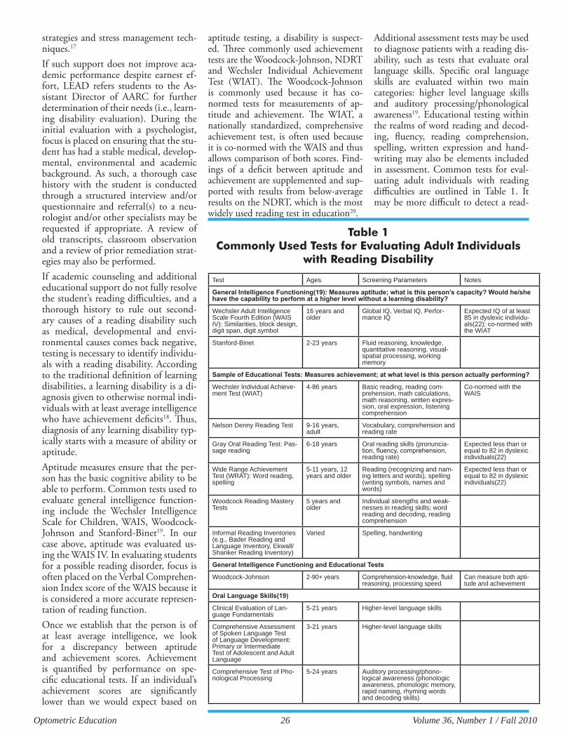
Optometric Education 26 Volume 36, Number 1 / Fall 2010
strategies and stress management tech-niques.17 If such support does not improve aca-demic performance despite earnest ef-fort, LEAD refers students to the As-sistant Director of AARC for further determination of their needs (i.e., learn-ing disability evaluation). During the initial evaluation with a psychologist, focus is placed on ensuring that the stu-dent has had a stable medical, develop-mental, environmental and academic background. As such, a thorough case history with the student is conducted through a structured interview and/or questionnaire and referral(s) to a neu-rologist and/or other specialists may be requested if appropriate. A review of old transcripts, classroom observation and a review of prior remediation strat-egies may also be performed. If academic counseling and additional educational support do not fully resolve the student’s reading difficulties, and a thorough history to rule out second-ary causes of a reading disability such as medical, developmental and envi-ronmental causes comes back negative, testing is necessary to identify individu-als with a reading disability. According to the traditional definition of learning disabilities, a learning disability is a di-agnosis given to otherwise normal indi-viduals with at least average intelligence who have achievement deficits18. Thus, diagnosis of any learning disability typ-ically starts with a measure of ability or aptitude.Aptitude measures ensure that the per-son has the basic cognitive ability to be able to perform. Common tests used to evaluate general intelligence function-ing include the Wechsler Intelligence Scale for Children, WAIS, Woodcock-Johnson and Stanford-Binet19. In our case above, aptitude was evaluated us-ing the WAIS IV. In evaluating students for a possible reading disorder, focus is often placed on the Verbal Comprehen-sion Index score of the WAIS because it is considered a more accurate represen-tation of reading function. Once we establish that the person is of at least average intelligence, we look for a discrepancy between aptitude and achievement scores. Achievement is quantified by performance on spe-cific educational tests. If an individual’s achievement scores are significantly lower than we would expect based on
aptitude testing, a disability is suspect-ed. Three commonly used achievement tests are the Woodcock-Johnson, NDRT and Wechsler Individual Achievement Test (WIAT). The Woodcock-Johnson is commonly used because it has co-normed tests for measurements of ap-titude and achievement. The WIAT, a nationally standardized, comprehensive achievement test, is often used because it is co-normed with the WAIS and thus allows comparison of both scores. Find-ings of a deficit between aptitude and achievement are supplemented and sup-ported with results from below-average results on the NDRT, which is the most widely used reading test in education20.
Additional assessment tests may be used to diagnose patients with a reading dis-ability, such as tests that evaluate oral language skills. Specific oral language skills are evaluated within two main categories: higher level language skills and auditory processing/phonological awareness19. Educational testing within the realms of word reading and decod-ing, fluency, reading comprehension, spelling, written expression and hand-writing may also be elements included in assessment. Common tests for eval-uating adult individuals with reading difficulties are outlined in Table 1. It may be more difficult to detect a read-
Test Ages Screening Parameters Notes
GeneralIntelligenceFunctioning(19):Measuresaptitude;whatisthisperson’scapacity?Wouldhe/shehavethecapabilitytoperformatahigherlevelwithoutalearningdisability?
Wechsler Adult Intelligence Scale Fourth Edition (WAIS IV): Similarities, block design, digit span, digit symbol
16 years and older
Global IQ, Verbal IQ, Perfor-mance IQ
Expected IQ of at least 85 in dyslexic individu-als(22); co-normed with the WIAT
Stanford-Binet 2-23 years Fluid reasoning, knowledge, quantitative reasoning, visual-spatial processing, working memory
SampleofEducationalTests:Measuresachievement;atwhatlevelisthispersonactuallyperforming?
Wechsler Individual Achieve-ment Test (WIAT)
4-86 years Basic reading, reading com-prehension, math calculations, math reasoning, written expres-sion, oral expression, listening comprehension
Co-normed with the WAIS
Nelson Denny Reading Test 9-16 years, adult
Vocabulary, comprehension and reading rate
Gray Oral Reading Test: Pas-sage reading
6-18 years Oral reading skills (pronuncia-tion, fluency, comprehension, reading rate)
Expected less than or equal to 82 in dyslexic individuals(22)
Wide Range Achievement Test (WRAT): Word reading, spelling
5-11 years, 12 years and older
Reading (recognizing and nam-ing letters and words), spelling (writing symbols, names and words)
Expected less than or equal to 82 in dyslexic individuals(22)
Woodcock Reading Mastery Tests
5 years and older
Individual strengths and weak-nesses in reading skills; word reading and decoding, reading comprehension
Informal Reading Inventories (e.g., Bader Reading and Language Inventory, Ekwall/Shanker Reading Inventory)
Varied Spelling, handwriting
GeneralIntelligenceFunctioningandEducationalTests
Woodcock-Johnson 2-90+ years Comprehension-knowledge, fluid reasoning, processing speed
Can measure both apti-tude and achievement
OralLanguageSkills(19)
Clinical Evaluation of Lan-guage Fundamentals
5-21 years Higher-level language skills
Comprehensive Assessment of Spoken Language Test of Language Development: Primary or Intermediate Test of Adolescent and Adult Language
3-21 years Higher-level language skills
Comprehensive Test of Pho-nological Processing
5-24 years Auditory processing/phono-logical awareness (phonologic awareness, phonologic memory, rapid naming, rhyming words and decoding skills)
Table 1 Commonly Used Tests for Evaluating Adult Individuals
with Reading Disability
Optometric Education 27 Volume 36, Number 1 / Fall 2010
ing disability in older students because they may have developed compensatory skills and techniques. Furthermore, test norms may be limited to adolescents.The student described in our case report was diagnosed with a reading disabil-ity because he presents with a negative medical, behavioral and environmental history, has difficulties persisting despite academic-based counseling, tutoring services and skill-building, and dem-onstrates adequate cognitive abilities (as identified through aptitude testing) but significantly lower performance in reading (as identified through achieve-ment testing). A deficit of one-and-a-half standard deviations is considered a meaningful difference21. Notably, it is possible to diagnose a learning disabil-ity in an individual with below-average intelligence, but it is often difficult to find a significant difference between ap-titude and achievement. Academic accommodations allowed this student to better develop strengths to work through limitations from his disability. Besides allowing additional time for exams, audiotaping of lectures, note-taker or provision of class notes and, whenever possible, early access to course material, other resources exist to assist students with reading disabilities. Technologies such as ReadPlease and TextHELP software allow students to simultaneously hear and see text on their computer. More sophisticated software, such as Kurzweil 3000, allows printed material to be scanned and read aloud by the computer. Recordings for the Blind and Dyslexic, which is a li-brary that provides audio recorded text-books, is also a commonly used and of-ten helpful resource. Readers for exams and alternative text are other academic accommodations designed to ensure equal access in the classroom. Special-ized instruction may also be recom-mended to help the student learn skills that tap into natural strengths in order to compensate for areas of weakness. While individual accommodations are important, support from educators is necessary to facilitate optimal student learning. As students with a reading disorder may read slower and with more difficulty than their peers due to difficulties with phonological process-ing, it is important for educators to avoid putting such students in a situ-
ation where this may be highlighted, such as asking the student to read text aloud in class. Other considerations to facilitate academic success for students with a reading disability include pro-viding high-contrast course materials, avoiding putting too much information on one page, writing in shorter, simpler sentences, avoiding fancy, particularly italicized, fonts (12+ sans serif fonts such as Arial are preferred) and consid-ering offering oral assessment opportu-nities13. Multimodal presentation and rehearsal of material to facilitate reten-tion of new information, including use of different or novel presentation for-mats, may also be helpful.While individuals with a reading dis-ability may need more time to process and understand written material, they can still proficiently perform in envi-ronments with high reading and writing demands, such as within the healthcare profession. Recent study results indi-cate that degree classifications achieved by students with reading disorders were not significantly different from those of other students13. Additional workplace support, such as proofreading by col-leagues, typing by administrative staff and using dictation software, may be advisable. Most employers are legally required to make such reasonable ac-commodations.
ConclusionReading disabilities are typically diag-nosed by the fourth grade, but well-disguised reading difficulties may man-ifest in graduate school as reading and writing tasks become more complex. An evaluation to rule out medical, de-velopmental and environmental condi-tions that can lead to secondary forms of reading difficulties is important, and academic-based counseling, tutoring services and academic skill-building need to be considered. If such resources do not fully resolve reading difficulties, testing administered by a trained pro-fessional is necessary to identify indi-viduals with a reading disability.Testing to diagnose a learning disabil-ity, such as a reading disability, typically focuses on ensuring that the otherwise normal individual has at least average intelligence (aptitude) but has achieve-ment scores that are below expected levels. If such a deficit is found within
reading, a reading disability diagnosis is appropriate, and accommodations should be provided to support the stu-dent. Individualized accommodations are essential to the success of the stu-dent’s academic career and ensure stu-dents receive resources specific for their own unique circumstances. Optom-etrists and educators alike need to be aware of the signs of reading disability, how reading disability is diagnosed and what resources and accommodations are available to allow equal access in the classroom and thus facilitate success.
References1. Lerner JW. Educational interven-
tions in learning disabilities. J Am Acad Child Adolesc Psychiatry. 1989;28(3):326-31.
2. Snow C, Burns S, Griffin P. Pre-venting reading difficulties in young children. Washington, DC: National Aacdemy Press; 1998.
3. Shaywitz SE, Shaywitz BA. Dyslex-ia (specific reading disability). Biol Psychiatry. 2005;57(11):1301-9.
4. Hamilton SS, Glascoe FP. Evalu-ation of children with reading difficulties. Am Fam Physician. 2006;74(12):2079-84.
5. Association AP. Diagnostic and Statistical Manual of Mental Dis-orders. Fourth Edition, Text Revi-sion ed. Washington, DC: Ameri-can Psychiatric Association; 2000.
6. Gillberg C, Soderstrom H. Learning disability. Lancet. 2003;362(9386):811-21.
7. Litt J, Taylor HG, Klein N, Hack M. Learning disabilities in children with very low birthweight: preva-lence, neuropsychological corre-lates, and educational interventions. J Learn Disabil. 2005;38(2):130-41.
8. Scheiman MM, Rouse MW. Opto-metric management of learning-re-lated vision problems. Second Edi-tion ed. St. Louis: Mosby Elsevier; 2006.
9. Shaywitz SE. Dyslexia. N Engl J Med. 1998;338(5):307-12.
10. Scarborough HS. Very early lan-guage deficits in dyslexic children. Child Dev. 1990;61(6):1728-43.

Optometric Education 28 Volume 36, Number 1 / Fall 2010
11. McPhillips M, Sheehy N. Preva-lence of persistent primary reflexes and motor problems in children with reading difficulties. Dyslexia. 2004;10(4):316-38.
12. Joint statement-Learning disabili-ties, dyslexia, and vision. Pediat-rics. 2009;124(2):837-44.
13. Lockley S. Dyslexia and higher education: accessibility issues. The Higher Education Academy; 2010. Available at http://www.heacad-emy.ac.uk/assets/York/documents/resources/resourcedatabase/id416_dyslexia_and_higher_education.pdf. Assessed July 12, 2010.
14. Dearing R. Higher Education in the Learning Society. Report of the National Committee of Inquiry into Higher Education. In: Depart-ment for Education and Employ-
ment, editor. London, England; 1997.
15. Vellutino FR, Fletcher JM, Snowl-ing MJ, Scanlon DM. Specific reading disability (dyslexia): what have we learned in the past four de-cades? J Child Psychol Psychiatry. 2004;45(1):2-40.
16. Mortimore T, Crozier WR. Dyslexia and difficulties with study skills in higher education. Studies in High-er Education. 2006;31(2):235-51.
17. Office L. Learning Enhance-ment & Academic Development: LEAD Services. Pomona, CA: Western University of Health Sci-ences; 2009. Available at: http://www.westernu.edu/xp/edu/lead/lead-services.xml. Assessed July 12, 2010.
18. Frankenberger W, Fronzaglio K. A review of states’ criteria and proce-dures for identifying children with
learning disabilities. J Learn Dis-abil. 1991;24(8):495-500.
19. Association TID. Testing for Dys-lexia. 2004. Available at: http://w w w. n y b i d a . o r g / P D F / t e s t -ing2..3.06.pdf. Assessed July 7, 2010.
20. Gordon B, Flippo R. An update on college reading improvement programs in the southeastern United States. Journal of Reading. 1983;27:155-63.
21. Office of Disability Services WOU. Learning Disability. Monmouth, OR: Western Oregon University; 2010. Available at: http://www.wou.edu/student/disability/re-sources/learning.php. Assessed July 12, 2010.
22. Felton RH, Naylor CE, Wood FB. Neuropsychological profile of adult dyslexics. Brain Lang. 1990;39(4):485-97.
Optometric Education 29 Volume 36, Number 1 / Fall 2010
Acute Ocular Trauma in a Child: A Teaching Case ReportWendy J. Haaland Stone, OD, FAAOStephanie A. Klemencic, OD, FAAO
AbstractOcular injuries are common among children, especially boys. Nonpenetrating ocular trauma can cause various types of damage in the eye. The adnexa, an-terior segment and posterior segment can all be affected. This damage can be mild to severe and transient or permanent. This case report of a nonpenetrating ocular injury of a young boy reviews the clinical findings and management.
Key Words: Ocular trauma, closed globe, child, traumatic iritis, commotio retinae
Dr. Stone is an Assistant Professor at the Illinois College of Optometry.Dr. Klemencic is an Assistant Professor and Coordinator of the Primary Care and Ocular Disease Residency Program at the Illinois College of Optometry.
Backgroundrauma to the eye is a common cause of ocular morbidity en-countered by eyecare provid-ers. Nonpenetrating ocular
trauma can manifest in many ways in both the anterior and posterior seg-ments depending on the nature and se-verity of the trauma. The following case report discusses a young boy with ocu-lar trauma and its sequelae. It is most appropriately used as a teaching guide for second-, third- and fourth-year stu-dents, as well as early residents, particu-larly those participating in urgent care clinics. For second-year students, even if they have not begun examining pa-tients, it could be used as a review of ocular anatomy, as all portions of the globe can be affected in blunt trauma. Additionally, the case can be used to re-inforce the uses of ocular health exami-nation skills that second-year students learn (slit lamp, gonioscopy, tonometry and fundus evaluation). For upper-level students and residents examining pa-tients, the potential sequelae of a trauma case will allow for the review of damage to nearly all structures of the eye and the discussion of complex management decisions encountered in these cases.
Student Discussion GuideCase DescriptionPatient LH, a 10-year-old African Amer-ican male, presented to our urgent care clinic with his mother on Saturday, Sept. 20, 2008. He reported having been hit in the right eye two days prior with a plastic toy sword. He was complaining of blurry vision, photophobia, redness and mild pain (2 on a scale of 10). He reported that the pain had improved since the incident, but the blur and pho-tophobia had not. His ocular history was significant for vision therapy in our pediatric clinic for saccadic dysfunction and accommodative insufficiency in 2005. He wore no spectacle correction and had never been prescribed one.Medical history was remarkable for at-tention deficit hyperactivity disorder, which was treated with dexmethylpheni-date (Focalin, Novartis). An allergy to the antibiotic cefuroxime (Ceftin, GlaxoSmithKline) was reported, but no other allergies were known. Family his-tory was positive for hypertension. Ex-amination findings are noted in Table 1.
T
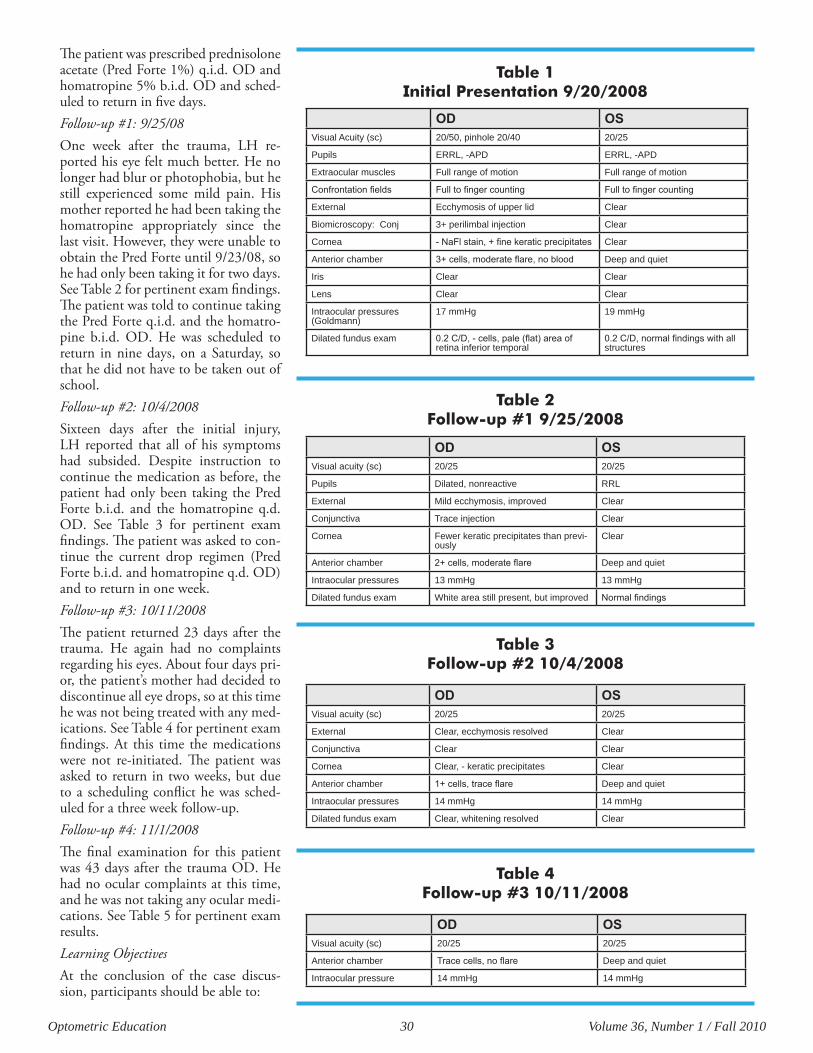
Optometric Education 30 Volume 36, Number 1 / Fall 2010
The patient was prescribed prednisolone acetate (Pred Forte 1%) q.i.d. OD and homatropine 5% b.i.d. OD and sched-uled to return in five days.Follow-up #1: 9/25/08One week after the trauma, LH re-ported his eye felt much better. He no longer had blur or photophobia, but he still experienced some mild pain. His mother reported he had been taking the homatropine appropriately since the last visit. However, they were unable to obtain the Pred Forte until 9/23/08, so he had only been taking it for two days. See Table 2 for pertinent exam findings. The patient was told to continue taking the Pred Forte q.i.d. and the homatro-pine b.i.d. OD. He was scheduled to return in nine days, on a Saturday, so that he did not have to be taken out of school.Follow-up #2: 10/4/2008Sixteen days after the initial injury, LH reported that all of his symptoms had subsided. Despite instruction to continue the medication as before, the patient had only been taking the Pred Forte b.i.d. and the homatropine q.d. OD. See Table 3 for pertinent exam findings. The patient was asked to con-tinue the current drop regimen (Pred Forte b.i.d. and homatropine q.d. OD) and to return in one week.Follow-up #3: 10/11/2008The patient returned 23 days after the trauma. He again had no complaints regarding his eyes. About four days pri-or, the patient’s mother had decided to discontinue all eye drops, so at this time he was not being treated with any med-ications. See Table 4 for pertinent exam findings. At this time the medications were not re-initiated. The patient was asked to return in two weeks, but due to a scheduling conflict he was sched-uled for a three week follow-up. Follow-up #4: 11/1/2008The final examination for this patient was 43 days after the trauma OD. He had no ocular complaints at this time, and he was not taking any ocular medi-cations. See Table 5 for pertinent exam results. Learning ObjectivesAt the conclusion of the case discus-sion, participants should be able to:
OD OSVisual Acuity (sc) 20/50, pinhole 20/40 20/25
Pupils ERRL, -APD ERRL, -APD
Extraocular muscles Full range of motion Full range of motion
Confrontation fields Full to finger counting Full to finger counting
External Ecchymosis of upper lid Clear
Biomicroscopy: Conj 3+ perilimbal injection Clear
Cornea - NaFl stain, + fine keratic precipitates Clear
Anterior chamber 3+ cells, moderate flare, no blood Deep and quiet
Iris Clear Clear
Lens Clear Clear
Intraocular pressures (Goldmann)
17 mmHg 19 mmHg
Dilated fundus exam 0.2 C/D, - cells, pale (flat) area of retina inferior temporal
0.2 C/D, normal findings with all structures
OD OSVisual acuity (sc) 20/25 20/25
Pupils Dilated, nonreactive RRL
External Mild ecchymosis, improved Clear
Conjunctiva Trace injection Clear
Cornea Fewer keratic precipitates than previ-ously
Clear
Anterior chamber 2+ cells, moderate flare Deep and quiet
Intraocular pressures 13 mmHg 13 mmHg
Dilated fundus exam White area still present, but improved Normal findings
OD OSVisual acuity (sc) 20/25 20/25
External Clear, ecchymosis resolved Clear
Conjunctiva Clear Clear
Cornea Clear, - keratic precipitates Clear
Anterior chamber 1+ cells, trace flare Deep and quiet
Intraocular pressures 14 mmHg 14 mmHg
Dilated fundus exam Clear, whitening resolved Clear
OD OSVisual acuity (sc) 20/25 20/25
Anterior chamber Trace cells, no flare Deep and quiet
Intraocular pressure 14 mmHg 14 mmHg
Table 1 Initial Presentation 9/20/2008
Table 2 Follow-up #1 9/25/2008
Table 3 Follow-up #2 10/4/2008
Table 4 Follow-up #3 10/11/2008
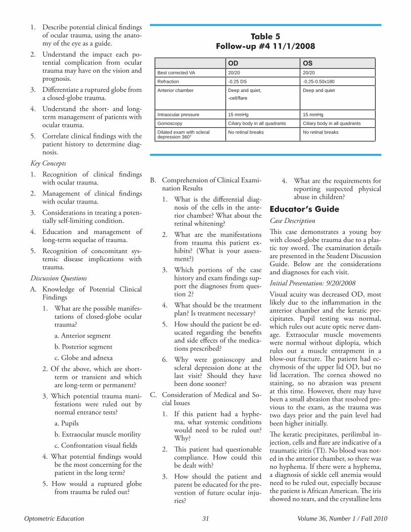
Optometric Education 31 Volume 36, Number 1 / Fall 2010
1. Describe potential clinical findings of ocular trauma, using the anato-my of the eye as a guide.
2. Understand the impact each po-tential complication from ocular trauma may have on the vision and prognosis.
3. Differentiate a ruptured globe from a closed-globe trauma.
4. Understand the short- and long-term management of patients with ocular trauma.
5. Correlate clinical findings with the patient history to determine diag-nosis.
Key Concepts1. Recognition of clinical findings
with ocular trauma.2. Management of clinical findings
with ocular trauma.3. Considerations in treating a poten-
tially self-limiting condition.4. Education and management of
long-term sequelae of trauma.5. Recognition of concomitant sys-
temic disease implications with trauma.
Discussion QuestionsA. Knowledge of Potential Clinical
Findings1. What are the possible manifes-
tations of closed-globe ocular trauma?a. Anterior segmentb. Posterior segmentc. Globe and adnexa
2. Of the above, which are short-term or transient and which are long-term or permanent?
3. Which potential trauma mani-festations were ruled out by normal entrance tests?a. Pupilsb. Extraocular muscle motilityc. Confrontation visual fields
4. What potential findings would be the most concerning for the patient in the long term?
5. How would a ruptured globe from trauma be ruled out?
B. Comprehension of Clinical Exami-nation Results1. What is the differential diag-
nosis of the cells in the ante-rior chamber? What about the retinal whitening?
2. What are the manifestations from trauma this patient ex-hibits? (What is your assess-ment?)
3. Which portions of the case history and exam findings sup-port the diagnoses from ques-tion 2?
4. What should be the treatment plan? Is treatment necessary?
5. How should the patient be ed-ucated regarding the benefits and side effects of the medica-tions prescribed?
6. Why were gonioscopy and scleral depression done at the last visit? Should they have been done sooner?
C. Consideration of Medical and So-cial Issues1. If this patient had a hyphe-
ma, what systemic conditions would need to be ruled out? Why?
2. This patient had questionable compliance. How could this be dealt with?
3. How should the patient and parent be educated for the pre-vention of future ocular inju-ries?
4. What are the requirements for reporting suspected physical abuse in children?
Educator’s GuideCase DescriptionThis case demonstrates a young boy with closed-globe trauma due to a plas-tic toy sword. The examination details are presented in the Student Discussion Guide. Below are the considerations and diagnoses for each visit.Initial Presentation: 9/20/2008Visual acuity was decreased OD, most likely due to the inflammation in the anterior chamber and the keratic pre-cipitates. Pupil testing was normal, which rules out acute optic nerve dam-age. Extraocular muscle movements were normal without diplopia, which rules out a muscle entrapment in a blow-out fracture. The patient had ec-chymosis of the upper lid OD, but no lid laceration. The cornea showed no staining, so no abrasion was present at this time. However, there may have been a small abrasion that resolved pre-vious to the exam, as the trauma was two days prior and the pain level had been higher initially.The keratic precipitates, perilimbal in-jection, cells and flare are indicative of a traumatic iritis (TI). No blood was not-ed in the anterior chamber, so there was no hyphema. If there were a hyphema, a diagnosis of sickle cell anemia would need to be ruled out, especially because the patient is African American. The iris showed no tears, and the crystalline lens
OD OSBest corrected VA 20/20 20/20
Refraction -0.25 DS -0.25-0.50x180
Anterior chamber Deep and quiet, -cell/flare
Deep and quiet
Intraocular pressure 15 mmHg 15 mmHg
Gonioscopy Ciliary body in all quadrants Ciliary body in all quadrants
Dilated exam with scleral depression 360°
No retinal breaks No retinal breaks
Table 5 Follow-up #4 11/1/2008
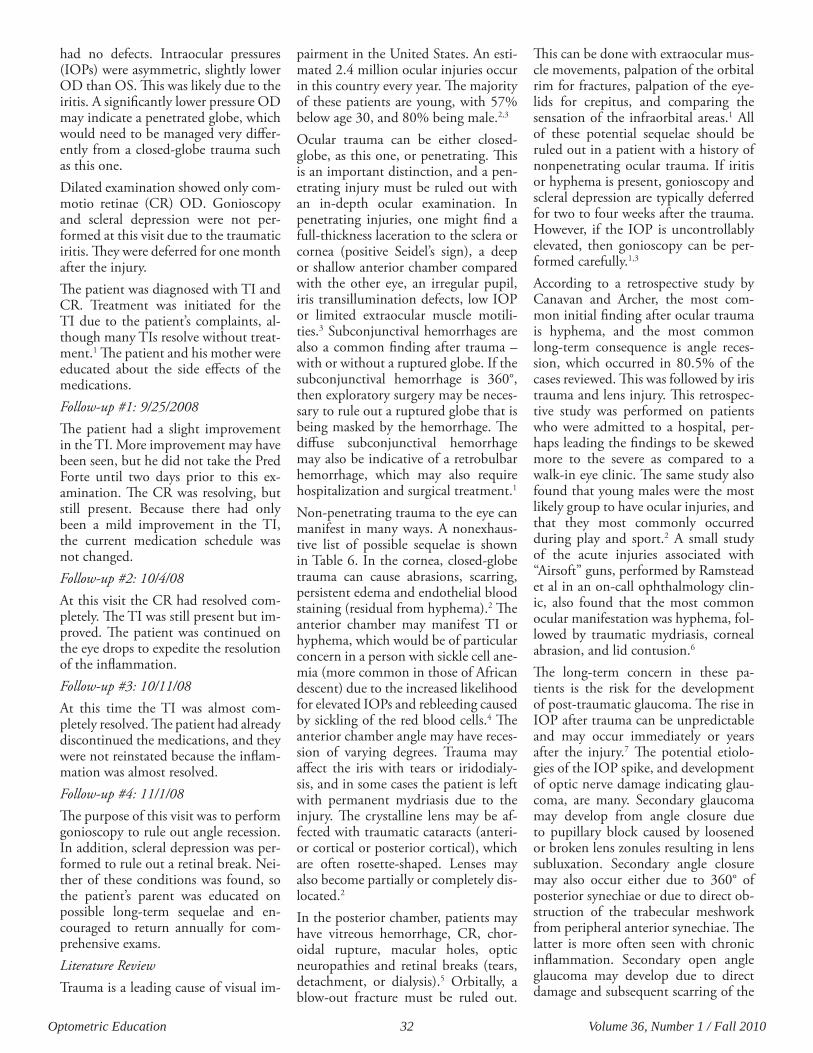
Optometric Education 32 Volume 36, Number 1 / Fall 2010
had no defects. Intraocular pressures (IOPs) were asymmetric, slightly lower OD than OS. This was likely due to the iritis. A significantly lower pressure OD may indicate a penetrated globe, which would need to be managed very differ-ently from a closed-globe trauma such as this one.Dilated examination showed only com-motio retinae (CR) OD. Gonioscopy and scleral depression were not per-formed at this visit due to the traumatic iritis. They were deferred for one month after the injury.The patient was diagnosed with TI and CR. Treatment was initiated for the TI due to the patient’s complaints, al-though many TIs resolve without treat-ment.1 The patient and his mother were educated about the side effects of the medications.Follow-up #1: 9/25/2008The patient had a slight improvement in the TI. More improvement may have been seen, but he did not take the Pred Forte until two days prior to this ex-amination. The CR was resolving, but still present. Because there had only been a mild improvement in the TI, the current medication schedule was not changed.Follow-up #2: 10/4/08At this visit the CR had resolved com-pletely. The TI was still present but im-proved. The patient was continued on the eye drops to expedite the resolution of the inflammation.Follow-up #3: 10/11/08At this time the TI was almost com-pletely resolved. The patient had already discontinued the medications, and they were not reinstated because the inflam-mation was almost resolved.Follow-up #4: 11/1/08The purpose of this visit was to perform gonioscopy to rule out angle recession. In addition, scleral depression was per-formed to rule out a retinal break. Nei-ther of these conditions was found, so the patient’s parent was educated on possible long-term sequelae and en-couraged to return annually for com-prehensive exams.Literature ReviewTrauma is a leading cause of visual im-
pairment in the United States. An esti-mated 2.4 million ocular injuries occur in this country every year. The majority of these patients are young, with 57% below age 30, and 80% being male.2,3 Ocular trauma can be either closed-globe, as this one, or penetrating. This is an important distinction, and a pen-etrating injury must be ruled out with an in-depth ocular examination. In penetrating injuries, one might find a full-thickness laceration to the sclera or cornea (positive Seidel’s sign), a deep or shallow anterior chamber compared with the other eye, an irregular pupil, iris transillumination defects, low IOP or limited extraocular muscle motili-ties.3 Subconjunctival hemorrhages are also a common finding after trauma – with or without a ruptured globe. If the subconjunctival hemorrhage is 360°, then exploratory surgery may be neces-sary to rule out a ruptured globe that is being masked by the hemorrhage. The diffuse subconjunctival hemorrhage may also be indicative of a retrobulbar hemorrhage, which may also require hospitalization and surgical treatment.1
Non-penetrating trauma to the eye can manifest in many ways. A nonexhaus-tive list of possible sequelae is shown in Table 6. In the cornea, closed-globe trauma can cause abrasions, scarring, persistent edema and endothelial blood staining (residual from hyphema).2 The anterior chamber may manifest TI or hyphema, which would be of particular concern in a person with sickle cell ane-mia (more common in those of African descent) due to the increased likelihood for elevated IOPs and rebleeding caused by sickling of the red blood cells.4 The anterior chamber angle may have reces-sion of varying degrees. Trauma may affect the iris with tears or iridodialy-sis, and in some cases the patient is left with permanent mydriasis due to the injury. The crystalline lens may be af-fected with traumatic cataracts (anteri-or cortical or posterior cortical), which are often rosette-shaped. Lenses may also become partially or completely dis-located.2
In the posterior chamber, patients may have vitreous hemorrhage, CR, chor-oidal rupture, macular holes, optic neuropathies and retinal breaks (tears, detachment, or dialysis).5 Orbitally, a blow-out fracture must be ruled out.
This can be done with extraocular mus-cle movements, palpation of the orbital rim for fractures, palpation of the eye-lids for crepitus, and comparing the sensation of the infraorbital areas.1 All of these potential sequelae should be ruled out in a patient with a history of nonpenetrating ocular trauma. If iritis or hyphema is present, gonioscopy and scleral depression are typically deferred for two to four weeks after the trauma. However, if the IOP is uncontrollably elevated, then gonioscopy can be per-formed carefully.1,3 According to a retrospective study by Canavan and Archer, the most com-mon initial finding after ocular trauma is hyphema, and the most common long-term consequence is angle reces-sion, which occurred in 80.5% of the cases reviewed. This was followed by iris trauma and lens injury. This retrospec-tive study was performed on patients who were admitted to a hospital, per-haps leading the findings to be skewed more to the severe as compared to a walk-in eye clinic. The same study also found that young males were the most likely group to have ocular injuries, and that they most commonly occurred during play and sport.2 A small study of the acute injuries associated with “Airsoft” guns, performed by Ramstead et al in an on-call ophthalmology clin-ic, also found that the most common ocular manifestation was hyphema, fol-lowed by traumatic mydriasis, corneal abrasion, and lid contusion.6
The long-term concern in these pa-tients is the risk for the development of post-traumatic glaucoma. The rise in IOP after trauma can be unpredictable and may occur immediately or years after the injury.7 The potential etiolo-gies of the IOP spike, and development of optic nerve damage indicating glau-coma, are many. Secondary glaucoma may develop from angle closure due to pupillary block caused by loosened or broken lens zonules resulting in lens subluxation. Secondary angle closure may also occur either due to 360° of posterior synechiae or due to direct ob-struction of the trabecular meshwork from peripheral anterior synechiae. The latter is more often seen with chronic inflammation. Secondary open angle glaucoma may develop due to direct damage and subsequent scarring of the
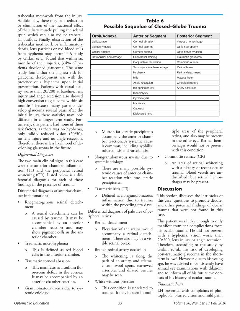
Optometric Education 33 Volume 36, Number 1 / Fall 2010
trabecular meshwork from the injury. Additionally, there may be a reduction or elimination of the tractional effect of the ciliary muscle pulling the scleral spur, which can also reduce trabecu-lar outflow. Finally, obstruction of the trabecular meshwork by inflammatory debris, lens particles or red blood cells from hyphema may occur.7, 8 A study by Girkin et al. found that within six months of their injuries, 3.4% of pa-tients developed glaucoma. The same study found that the highest risk for glaucoma development was with the presence of a hyphema upon initial presentation. Patients with visual acu-ity worse than 20/200 at baseline, lens injury and angle recession also showed high conversion to glaucoma within six months.8 Because many patients de-velop glaucoma several years after the initial injury, these statistics may look different in a longer-term study. For-tunately, this patient had none of these risk factors, as there was no hyphema, only mildly reduced vision (20/50), no lens injury and no angle recession. Therefore, there is less likelihood of de-veloping glaucoma in the future. Differential DiagnosesThe two main clinical signs in this case were the anterior chamber inflamma-tion (TI) and the peripheral retinal whitening (CR). Listed below is a dif-ferential diagnosis for each of these findings in the presence of trauma.Differential diagnosis of anterior cham-ber inflammation:• Rhegmatogenous retinal detach-
ment o A retinal detachment can be
caused by trauma. It may be accompanied by an anterior chamber reaction and may show pigment cells in the an-terior chamber.
• Traumaticmicrohyphemao This is defined as red blood
cells in the anterior chamber.• Traumaticcornealabrasion
o This manifests as a sodium flu-orescein defect in the cornea. It may be accompanied by an anterior chamber reaction.
• Granulomatousuveitisduetosys-temic etiology
o Mutton fat keratic precipitates accompany the anterior cham-ber reaction. A systemic cause is common, including syphilis, tuberculosis and sarcoidosis.
• Nongranulomatous uveitis due tosystemic etiologyo There are many possible sys-
temic causes of anterior cham-ber reaction with fine keratic precipitates.
• Traumaticiritis(TI)o Defined as nongranulomatous
inflammation due to trauma within the preceding few days.
Differential diagnosis of pale area of pe-ripheral retina:• Retinaldetachment
o Elevation of the retina would accompany a retinal detach-ment. There also may be a vis-ible retinal break.
• Branchretinalarteryocclusiono The whitening is along the
path of an artery, and edema, cotton wool spots, narrowed arterioles and dilated venules may be seen.
• Whitewithoutpressureo This condition is unrelated to
trauma. It may be seen in mul-
tiple areas of the peripheral retina, and also may be present in the other eye. Retinal hem-orrhages would not be present with this condition.
• Commotioretinae(CR)o An area of retinal whitening
with a history of recent ocular trauma. Blood vessels are un-disturbed, but retinal hemor-rhages may be present.
DiscussionThis section discusses the intricacies of this case, questions to promote debate, and other potential findings of ocular trauma that were not found in this case.This patient was lucky enough to only manifest transient complications from his ocular trauma. He did not present with a hyphema, vision worse than 20/200, lens injury or angle recession. Therefore, according to the study by Girkin et al., his risk of developing post-traumatic glaucoma in the short-term is low8. However, due to his young age, he was advised to consistently have annual eye examinations with dilation, and to inform all of his future eye doc-tors of his history of ocular trauma.Traumatic IritisLH presented with complaints of pho-tophobia, blurred vision and mild pain.
Orbit/Adnexa AnteriorSegment PosteriorSegmentLid laceration Corneal abrasion Vitreous hemorrhage
Lid ecchymosis Corneal scarring Optic neuropathy
Orbital fracture Corneal edema Optic nerve evulsion
Retrobulbar hemorrhage Endothelial staining Traumatic glaucoma
Conjunctival laceration Commotio retinae
Subconjunctival hemorrhage Retinal break
Hyphema Retinal detachment
Iritis Macular hole
Angle recession Choroidal rupture
Iris sphincter tear Artery occlusion
Iridodialysis
Cyclodialysis
Mydriasis
Cataract
Dislocated lens
Table 6 Possible Sequelae of Closed-Globe Trauma
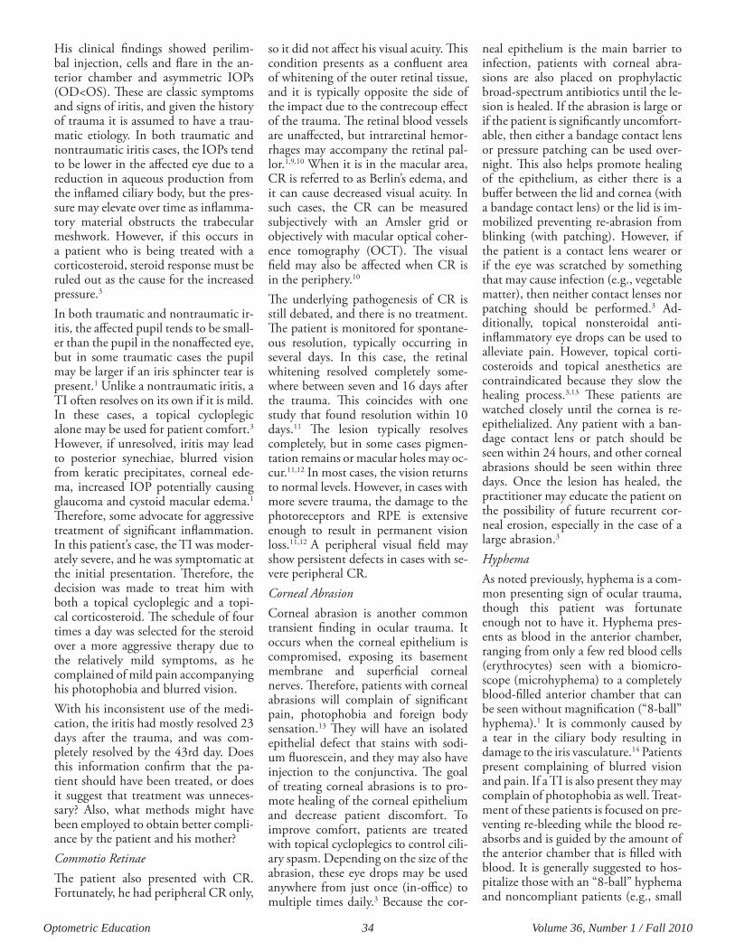
Optometric Education 34 Volume 36, Number 1 / Fall 2010
His clinical findings showed perilim-bal injection, cells and flare in the an-terior chamber and asymmetric IOPs (OD<OS). These are classic symptoms and signs of iritis, and given the history of trauma it is assumed to have a trau-matic etiology. In both traumatic and nontraumatic iritis cases, the IOPs tend to be lower in the affected eye due to a reduction in aqueous production from the inflamed ciliary body, but the pres-sure may elevate over time as inflamma-tory material obstructs the trabecular meshwork. However, if this occurs in a patient who is being treated with a corticosteroid, steroid response must be ruled out as the cause for the increased pressure.3
In both traumatic and nontraumatic ir-itis, the affected pupil tends to be small-er than the pupil in the nonaffected eye, but in some traumatic cases the pupil may be larger if an iris sphincter tear is present.1 Unlike a nontraumatic iritis, a TI often resolves on its own if it is mild. In these cases, a topical cycloplegic alone may be used for patient comfort.3 However, if unresolved, iritis may lead to posterior synechiae, blurred vision from keratic precipitates, corneal ede-ma, increased IOP potentially causing glaucoma and cystoid macular edema.1 Therefore, some advocate for aggressive treatment of significant inflammation. In this patient’s case, the TI was moder-ately severe, and he was symptomatic at the initial presentation. Therefore, the decision was made to treat him with both a topical cycloplegic and a topi-cal corticosteroid. The schedule of four times a day was selected for the steroid over a more aggressive therapy due to the relatively mild symptoms, as he complained of mild pain accompanying his photophobia and blurred vision. With his inconsistent use of the medi-cation, the iritis had mostly resolved 23 days after the trauma, and was com-pletely resolved by the 43rd day. Does this information confirm that the pa-tient should have been treated, or does it suggest that treatment was unneces-sary? Also, what methods might have been employed to obtain better compli-ance by the patient and his mother?Commotio RetinaeThe patient also presented with CR. Fortunately, he had peripheral CR only,
so it did not affect his visual acuity. This condition presents as a confluent area of whitening of the outer retinal tissue, and it is typically opposite the side of the impact due to the contrecoup effect of the trauma. The retinal blood vessels are unaffected, but intraretinal hemor-rhages may accompany the retinal pal-lor.1,9,10 When it is in the macular area, CR is referred to as Berlin’s edema, and it can cause decreased visual acuity. In such cases, the CR can be measured subjectively with an Amsler grid or objectively with macular optical coher-ence tomography (OCT). The visual field may also be affected when CR is in the periphery.10 The underlying pathogenesis of CR is still debated, and there is no treatment. The patient is monitored for spontane-ous resolution, typically occurring in several days. In this case, the retinal whitening resolved completely some-where between seven and 16 days after the trauma. This coincides with one study that found resolution within 10 days.11 The lesion typically resolves completely, but in some cases pigmen-tation remains or macular holes may oc-cur.11,12 In most cases, the vision returns to normal levels. However, in cases with more severe trauma, the damage to the photoreceptors and RPE is extensive enough to result in permanent vision loss.11,12 A peripheral visual field may show persistent defects in cases with se-vere peripheral CR.Corneal AbrasionCorneal abrasion is another common transient finding in ocular trauma. It occurs when the corneal epithelium is compromised, exposing its basement membrane and superficial corneal nerves. Therefore, patients with corneal abrasions will complain of significant pain, photophobia and foreign body sensation.13 They will have an isolated epithelial defect that stains with sodi-um fluorescein, and they may also have injection to the conjunctiva. The goal of treating corneal abrasions is to pro-mote healing of the corneal epithelium and decrease patient discomfort. To improve comfort, patients are treated with topical cycloplegics to control cili-ary spasm. Depending on the size of the abrasion, these eye drops may be used anywhere from just once (in-office) to multiple times daily.3 Because the cor-
neal epithelium is the main barrier to infection, patients with corneal abra-sions are also placed on prophylactic broad-spectrum antibiotics until the le-sion is healed. If the abrasion is large or if the patient is significantly uncomfort-able, then either a bandage contact lens or pressure patching can be used over-night. This also helps promote healing of the epithelium, as either there is a buffer between the lid and cornea (with a bandage contact lens) or the lid is im-mobilized preventing re-abrasion from blinking (with patching). However, if the patient is a contact lens wearer or if the eye was scratched by something that may cause infection (e.g., vegetable matter), then neither contact lenses nor patching should be performed.3 Ad-ditionally, topical nonsteroidal anti-inflammatory eye drops can be used to alleviate pain. However, topical corti-costeroids and topical anesthetics are contraindicated because they slow the healing process.3,13 These patients are watched closely until the cornea is re-epithelialized. Any patient with a ban-dage contact lens or patch should be seen within 24 hours, and other corneal abrasions should be seen within three days. Once the lesion has healed, the practitioner may educate the patient on the possibility of future recurrent cor-neal erosion, especially in the case of a large abrasion.3
HyphemaAs noted previously, hyphema is a com-mon presenting sign of ocular trauma, though this patient was fortunate enough not to have it. Hyphema pres-ents as blood in the anterior chamber, ranging from only a few red blood cells (erythrocytes) seen with a biomicro-scope (microhyphema) to a completely blood-filled anterior chamber that can be seen without magnification (“8-ball” hyphema).1 It is commonly caused by a tear in the ciliary body resulting in damage to the iris vasculature.14 Patients present complaining of blurred vision and pain. If a TI is also present they may complain of photophobia as well. Treat-ment of these patients is focused on pre-venting re-bleeding while the blood re-absorbs and is guided by the amount of the anterior chamber that is filled with blood. It is generally suggested to hos-pitalize those with an “8-ball” hyphema and noncompliant patients (e.g., small
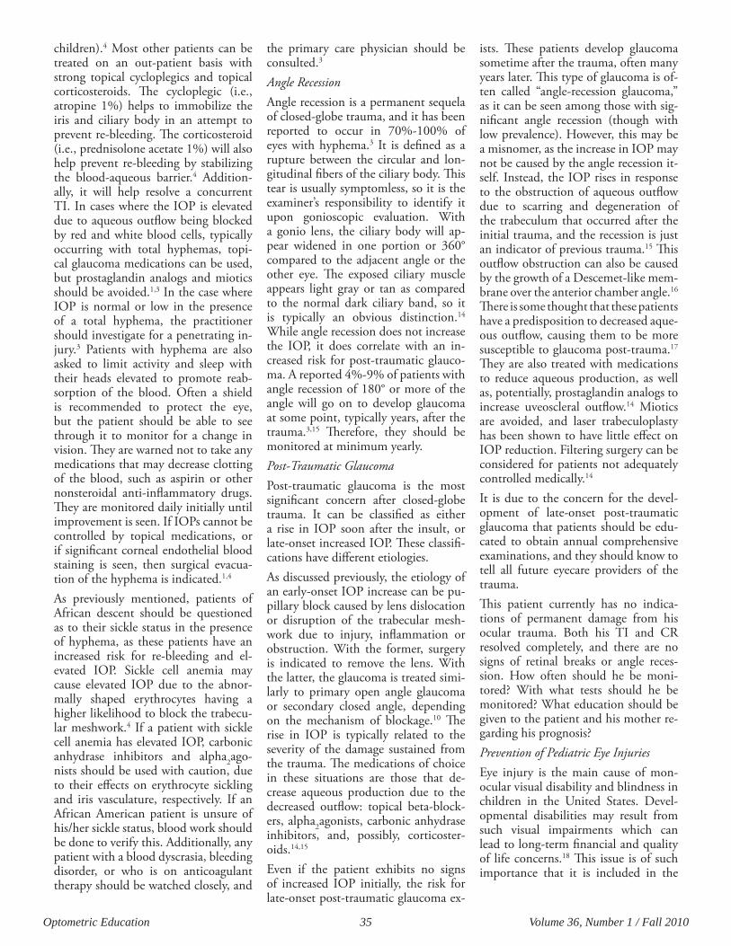
Optometric Education 35 Volume 36, Number 1 / Fall 2010
children).4 Most other patients can be treated on an out-patient basis with strong topical cycloplegics and topical corticosteroids. The cycloplegic (i.e., atropine 1%) helps to immobilize the iris and ciliary body in an attempt to prevent re-bleeding. The corticosteroid (i.e., prednisolone acetate 1%) will also help prevent re-bleeding by stabilizing the blood-aqueous barrier.4 Addition-ally, it will help resolve a concurrent TI. In cases where the IOP is elevated due to aqueous outflow being blocked by red and white blood cells, typically occurring with total hyphemas, topi-cal glaucoma medications can be used, but prostaglandin analogs and miotics should be avoided.1,3 In the case where IOP is normal or low in the presence of a total hyphema, the practitioner should investigate for a penetrating in-jury.3 Patients with hyphema are also asked to limit activity and sleep with their heads elevated to promote reab-sorption of the blood. Often a shield is recommended to protect the eye, but the patient should be able to see through it to monitor for a change in vision. They are warned not to take any medications that may decrease clotting of the blood, such as aspirin or other nonsteroidal anti-inflammatory drugs. They are monitored daily initially until improvement is seen. If IOPs cannot be controlled by topical medications, or if significant corneal endothelial blood staining is seen, then surgical evacua-tion of the hyphema is indicated.1,4
As previously mentioned, patients of African descent should be questioned as to their sickle status in the presence of hyphema, as these patients have an increased risk for re-bleeding and el-evated IOP. Sickle cell anemia may cause elevated IOP due to the abnor-mally shaped erythrocytes having a higher likelihood to block the trabecu-lar meshwork.4 If a patient with sickle cell anemia has elevated IOP, carbonic anhydrase inhibitors and alpha2ago-nists should be used with caution, due to their effects on erythrocyte sickling and iris vasculature, respectively. If an African American patient is unsure of his/her sickle status, blood work should be done to verify this. Additionally, any patient with a blood dyscrasia, bleeding disorder, or who is on anticoagulant therapy should be watched closely, and
the primary care physician should be consulted.3
Angle RecessionAngle recession is a permanent sequela of closed-globe trauma, and it has been reported to occur in 70%-100% of eyes with hyphema.3 It is defined as a rupture between the circular and lon-gitudinal fibers of the ciliary body. This tear is usually symptomless, so it is the examiner’s responsibility to identify it upon gonioscopic evaluation. With a gonio lens, the ciliary body will ap-pear widened in one portion or 360° compared to the adjacent angle or the other eye. The exposed ciliary muscle appears light gray or tan as compared to the normal dark ciliary band, so it is typically an obvious distinction.14 While angle recession does not increase the IOP, it does correlate with an in-creased risk for post-traumatic glauco-ma. A reported 4%-9% of patients with angle recession of 180° or more of the angle will go on to develop glaucoma at some point, typically years, after the trauma.3,15 Therefore, they should be monitored at minimum yearly.Post-Traumatic GlaucomaPost-traumatic glaucoma is the most significant concern after closed-globe trauma. It can be classified as either a rise in IOP soon after the insult, or late-onset increased IOP. These classifi-cations have different etiologies.As discussed previously, the etiology of an early-onset IOP increase can be pu-pillary block caused by lens dislocation or disruption of the trabecular mesh-work due to injury, inflammation or obstruction. With the former, surgery is indicated to remove the lens. With the latter, the glaucoma is treated simi-larly to primary open angle glaucoma or secondary closed angle, depending on the mechanism of blockage.10 The rise in IOP is typically related to the severity of the damage sustained from the trauma. The medications of choice in these situations are those that de-crease aqueous production due to the decreased outflow: topical beta-block-ers, alpha2agonists, carbonic anhydrase inhibitors, and, possibly, corticoster-oids.14,15
Even if the patient exhibits no signs of increased IOP initially, the risk for late-onset post-traumatic glaucoma ex-
ists. These patients develop glaucoma sometime after the trauma, often many years later. This type of glaucoma is of-ten called “angle-recession glaucoma,” as it can be seen among those with sig-nificant angle recession (though with low prevalence). However, this may be a misnomer, as the increase in IOP may not be caused by the angle recession it-self. Instead, the IOP rises in response to the obstruction of aqueous outflow due to scarring and degeneration of the trabeculum that occurred after the initial trauma, and the recession is just an indicator of previous trauma.15 This outflow obstruction can also be caused by the growth of a Descemet-like mem-brane over the anterior chamber angle.16 There is some thought that these patients have a predisposition to decreased aque-ous outflow, causing them to be more susceptible to glaucoma post-trauma.17 They are also treated with medications to reduce aqueous production, as well as, potentially, prostaglandin analogs to increase uveoscleral outflow.14 Miotics are avoided, and laser trabeculoplasty has been shown to have little effect on IOP reduction. Filtering surgery can be considered for patients not adequately controlled medically.14
It is due to the concern for the devel-opment of late-onset post-traumatic glaucoma that patients should be edu-cated to obtain annual comprehensive examinations, and they should know to tell all future eyecare providers of the trauma. This patient currently has no indica-tions of permanent damage from his ocular trauma. Both his TI and CR resolved completely, and there are no signs of retinal breaks or angle reces-sion. How often should he be moni-tored? With what tests should he be monitored? What education should be given to the patient and his mother re-garding his prognosis? Prevention of Pediatric Eye InjuriesEye injury is the main cause of mon-ocular visual disability and blindness in children in the United States. Devel-opmental disabilities may result from such visual impairments which can lead to long-term financial and quality of life concerns.18 This issue is of such importance that it is included in the
Optometric Education 36 Volume 36, Number 1 / Fall 2010
Healthy People 2020 initiatives: “to re-duce visual impairment and blindness in children and adolescents age 17 years and under, and increase the use of pro-tective eyewear in recreational activities and hazardous situations around the home.”19 In one large study, looking at pediatric eye injuries treated in U.S. emergency rooms from 1997-2006, sports was the most common category associated with eye injury, followed by household cleaning chemicals, toys (not including toy guns), furniture and desk supplies.18 Age-related patterns were also found in this study. Eye injuries related to sports, swimming pools/equipment, toy guns and recreational vehicles increased with age. Injuries due to household cleaning chemicals, furniture and baby items de-creased with age.18 It is suggested that pediatric eye trau-ma may be largely prevented by the education of children, use of protec-tive eyewear and increased parental supervision. These tactics may be tai-lored accordingly.18 For example, the American Academy of Pediatrics and the American Academy of Ophthal-mology strongly recommend protective eyewear for all participants in sports in which there is risk of eye injury. Protec-tive eyewear should be mandatory for athletes who are functionally one-eyed and for athletes whose eyecare provid-ers recommend eye protection after eye surgery or trauma.20 What are other examples of patient and guardian education related to ocular in-jury prevention?Concern for Child Abuse in Pediatric Ocular TraumaAs primary care providers, optometrists may need to determine whether an ocular injury is the result of abuse. A misdiagnosis of child abuse can have serious consequences for a child and family. However, a child who is a vic-tim of unreported child abuse can suf-fer further abuse and even death.21 As optometrists, we have an ethical, moral and legal obligation to report suspected abuse in order to protect the child. A detailed history is important in trau-ma cases.21,22 It may be helpful to al-low the parent to lead the history with a narrative of the injury.22 Use direct,
open-ended, non-leading questions when talking with the child.22 Red flags that may alert the provider to possible abuse include a history that is incon-sistent with the injury, no explanation offered for the injury, history that is inconsistent with the child’s develop-mental level, and/or injury blamed on another child or sibling.21 Additionally, conflicting histories given by caretak-ers, delay in seeking medical care, and doctor-shopping may be indicators of physical abuse.22 If abuse is suspected, questioning the child and parent sepa-rately, if possible, may be helpful.21,22 As in any clinical diagnosis, the examina-tion findings should correlate with the history. Healthcare providers are mandated by law to report suspected abuse. Provid-ers are not required to prove the abuse occurred prior to reporting, and, in fact, they are immune from legal and civil liability when reporting concerns for child abuse. Furthermore, provid-ers who fail to report concerns of child abuse to child protective services can be prosecuted.22 Optometrists should be aware of their individual state reporting laws and child protective service con-tacts. In 2008, the U.S. Department of Health & Human Services published a report: Mandatory Reporters of Child Abuse and Neglect: Summary of State Laws. This may be found at: http://www.childwelfare.gov/responding/mandated.cfm. 22 Most states have toll-free numbers for reporting suspected abuse.23 A list of child abuse reporting numbers may be found at: http://www.childwelfare.gov/responding/reporting.cfm. 24
ConclusionThis case is representative of the types of trauma cases optometrists frequently encounter. It is important to be cog-nizant of the possible ocular effects of trauma and the potential long-term effects. Patients with nonpenetrating ocular trauma should be evaluated for the presence of subconjunctival hemor-rhage, corneal abrasion, traumatic iritis, hyphema, choroidal rupture, blow-out fracture, traumatic optic neuropathy, retinal hemorrhages, commotio reti-nae, angle recession and retinal breaks. The patient’s risk for the development
of post-traumatic secondary glaucoma should be assessed, and the patient should be counseled on the risk for its development in the future, as well as future ocular injury prevention. In cas-es of pediatric trauma where physical abuse is suspected, optometrists are re-quired to report the case to appropriate authorities for further investigation.
References1. Ehlers JP, Shah CP, Fenton, GL,
Hoskins EN, Shelsta HN. The Wills Eye manual: office and emer-gency room diagnosis and treat-ment of eye diseases. 5th ed. Phila-delphia: Lippincott Williams & Wilkins; 2008.
2. Canavan YM, Archer DB. Anterior segment consequences of blunt ocular injury. Br J Ophthalmol. 1982;66:549-555.
3. Heiberger MH, Madonna RJ, Ne-hmad L. Emergency care in the optometric setting. New York: McGraw-Hill; 2004.
4. Walton W, Von Hagen S, Grigorian R, Zarbin M. Management of trau-matic hyphema. Surv Ophthalmol. 2002;47(4):297-334.
5. Williams DF, Mieler WF, Williams GA. Posterior segment manifes-tations of ocular trauma. Retina. 1990;10 Suppl 1:S35-44.
6. Ramstead C, Ng M, Rudinsky CJ. Ocular injuries associated with Air-soft guns: a case series. Can J Oph-thalmol. 2008;43(5):584-7.
7. Litwak AB. Secondary open angle glaucoma. Glaucoma handbook. Boston: Butterwork-Heinemann; 2001.
8. Girkin CA, McGwin G, Long C, Morris R, Kuhn F. Glaucoma after ocular contusion: a cohort study of the United States eye injury reg-istry. J Glaucoma. 2005;14:470-473.
9. Majid MA, Hussain HM, Haynes RJ, Dick AD. Buckle, no cryo: scleral buckle with no cryotherapy for retinal detachment secondary to commotio retinae. Br J Oph-thalmol. 2006;90(12):1550-1.
10. Ryan SJ. Retina. 3rd ed. St. Louis: Mosby; 2001.

Optometric Education 37 Volume 36, Number 1 / Fall 2010
11. Sipperley JO, Quigley HA, Gass DM. Traumatic retinopathy in primates: the explanation of com-motio retinae. Arch Ophthalmol. 1978;96(12):2267-73.
12. Alexander LJ. Primary care of the posterior segment. 2nd ed. Nor-walk: Appleton & Lange; 1994.
13. Catalano RA. Ocular emergencies. Philadelphia: Saunders; 1992.
14. Fingeret M, Lewis TL. Primary care of the glaucomas. 2nd ed. New York: McGraw-Hill; 2001.
15. Allingham RR, Damji K, Freed-man S, Moroi SE, Shafranov G, Shields MB. Shields’ textbook of glaucoma. 5th ed. Philadelphia: Lippincott Williams & Wilkins; 2005.
16. Lauring L. Anterior chamber glass membranes. Am J Opthalmol. 1969;68:308.
17. Armaly MF. Effects of corticos-teroids in intraocular pressure and fluid dynamics. II. The effect of dexamethasone in the glau-comatous eye. Arch Ophthalmol. 1963;70:492.
18. Cross JM, Griffen R, Owsley C, McGwin G. Pediatric eye inuries related to consumer products in the United States, 1997-2006. Journal of AAPOS. 2008;12(6):626-28.
19. U.S. Department of Health and Human Services. Healthy Peo-ple 2020 objectives. Available at: http://www.healthypeople.gov/Default.htm. Accessed July 20, 2010.
20. American Academy of Pediatrics, Committee on Sports Medicine and Fitness, American Academy of Ophthalmology, Eye Health and Public Information Task Force. Pro-tective eyewear for young athletes. Ophthalmology. 2004;111:600-603.
21. Harris TS. Bruises in children: nor-mal or child abuse? Journal of Pedi-atric Health Care. 2010;24(4):216-21.
22. Hornor G. Physical abuse: recogni-tion and reporting. Journal of Pe-diatric Health Care. 2005;19(1):4-11.
23. U.S. Department of Health & Hu-man Services. Child Welfare Infor-mation Gateway. Mandatory re-porters of child abuse and neglect: summary of state laws. Available at: http://www.childwelfare.gov/responding/mandated.cfm. Ac-cessed: July 19, 2010.
24. U.S. Department of Health & Hu-man Services. Child Welfare In-formation Gateway. Child abuse reporting numbers. Available at: http://www.childwelfare.gov/re-sponding/reporting.cfm. Accessed: July 19, 2010.
Optometric Education 38 Volume 36, Number 1 / Fall 2010
Tobacco Dependence Education in Optometry: A Canadian Pilot Study Assessing Practices and Opportunities
Marlee M. Spafford, OD, MSc, PhD, FAAOMatthew D. Iley, BSc, ODAnnette S.H. Schultz, RN, PhDRyan D. Kennedy, MAES, PhD
AbstractOptometrists should be part of the healthcare team addressing tobacco prevention and cessation among patients. As part of a pilot Canadian study, we interviewed 18 optometry students and 11 community optometrists to identify the training they have received regarding tobacco use, dependence and cessation. Their train-ing was limited to knowledge about tobacco-related ocular and systemic risks and some skills in asking about smoking behavior in patients older than 15 years. Their training left them lacking necessary: 1) knowledge about tobacco depen-dence and cessation, 2) skills in communicating tobacco advice, and 3) attitudes regarding their potential role in tobacco prevention and cessation.
Key Words: Tobacco use, tobacco cessation, optometrists
Dr. Spafford is a Professor and the Associate Director, Academics & Research, at the University of Waterloo School of Optometry in Waterloo, Ontario, Canada.Dr. Iley is a private practitioner and Adjunct Clinical Lecturer at the University of Waterloo School of Optometry in Waterloo, Ontario, Canada.Dr. Schultz is an Assistant Professor at the University of Manitoba, Faculty of Nursing, in Winnipeg, Manitoba, Canada.Dr. Kennedy is a Scientist at the Propel Centre for Population Health Impact at the University of Waterloo in Waterloo, Ontario, Canada.
Introductionobacco use is a known prevent-able cause of premature death and a less known prevent-able cause of blindness. There
is increasing evidence that healthcare providers can positively affect tobacco cessation among their patients. While optometrists are primary healthcare providers, their voice has been largely silent in health policy documents that guide healthcare providers about tobac-co cessation. In view of these factors, we sought to identify some of the key be-haviors and attitudes among Canadian optometrists and optometry students regarding asking patients about their to-bacco use and making tobacco cessation referrals. This paper reports on the find-ings of a preliminary Canadian study that pertain specifically to the training optometry students and optometrists receive regarding tobacco use, depen-dence and cessation strategies.
Major Health Impact of Tobacco UseTobacco use includes not only smoking cigarettes but also less common tobac-co practices such as chewing tobacco, sniffing tobacco nasally (dry snuff) and inserting tobacco between the lower lip and teeth (moist snuff). The vast ma-jority of the research causally linking tobacco use to morbidity and mortality has focused on smoking cigarettes, in part because of its much higher preva-lence. For example, while 17.9% of Canadians smoke cigarettes, fewer than 1% use smokeless tobacco.1 Research linking tobacco use to eye disease has strictly addressed smoking cigarettes. Smoking is one of the most preventable causes of premature death globally.2 While numerous diseases have been causally linked to smoking, the leading causes of premature death are coronary artery disease leading to myocardial in-farctions and cerebrovascular accidents, lung cancer and chronic obstructive pulmonary disease.2 Public awareness of the negative consequences of smoking is generally high for these diseases. For example, studies in the United King-dom and Singapore reported that most surveyed patients knew that smoking causes lung cancer and heart disease (>85%) as well as stroke (>70%).3,4
While most patients understand that smoking causes premature death, the
T
Optometric Education 39 Volume 36, Number 1 / Fall 2010
proportion who understand smok-ing causes blindness is generally much lower, typically less than 10%3,5,6 with one study4 reporting 31%-37%. This limited awareness is a significant prob-lem because smoking has been shown to cause several eye diseases through ischemic and oxidative mechanisms.7 Smoking has been causally associated with age-related macular degenera-tion,8-12 nuclear and posterior subcap-sular cataract,13-18 thyroid-associated ophthalmopathy,19-21 optic neuropa-thies22-24 and uveitis.25,26
Healthcare Practitioners Addressing Tobacco UseThe role of healthcare practitioners in clinical practice addressing tobacco use is supported by an increasing recogni-tion that tobacco use and dependence is a preventable cause of morbidity and mortality and healthcare providers can be effective facilitators of tobacco cessa-tion among their patients.27-32 The U.S. Surgeon General’s Web site provides a detailed position paper, “Treating Tobacco Use and Dependence: 2008 Update.”33 This document was devel-oped by stakeholders from medicine, nursing, dentistry, pharmacy, epidemi-ology, public health and psychology. Unfortunately, optometry is notably missing as a stakeholder informant or as an identified user of the document. Optometry has also yet to be a stake-holder in the development of Health Canada’s graphic warning labels for tobacco products. (Some of the labels under review are posted at the Tobac-co Labeling Resource Centre, http://www.tobaccolabels.ca/). The exclusion of optometry has occurred despite the current review of a label addressing smoking and blindness. There are two key strategies healthcare practitioners, including optometrists, can use to ef-fectively address tobacco use with their patients: 1) support tobacco cessation and 2) prevent patients from starting to use tobacco.The justification for optometrists pro-viding cessation advice is supported by several studies. First, Gorin and Heck34 found that tobacco quit rates increase when patients receive advice from “any health care professional.” Second, Thornton et al.35 found other health-care practitioners (e.g., pharmacists)
are successfully including the link be-tween smoking and blindness in their cessation advice. And finally, studies of patient perceptions show that quit advice is welcomed and expected.36-38 Our literature search also revealed an encouraging pattern of calls for optom-etry to participate in tobacco cessation strategies.39-44
A number of tobacco cessation coun-seling strategies exist, but perhaps two of the most known are the 5A program promoted by the United States Public Health Service45 and the ABC tool pro-moted by the New Zealand Smoking Cessation Guidelines Group.46 The 5A program encourages practitioners to:1. ask about tobacco use at every visit2. advise those using tobacco to stop3. assess the readiness of those using
tobacco to attempt quitting4. assist those using tobacco with
treatment and/or referral5. arrange follow-up discussions at
subsequent visits.The ABC tool reminds practitioners to:1. ask patients about their tobacco
status2. provide brief advice3. give evidence-based cessation sup-
port to tobacco users wishing to at-tempt quitting.
Healthcare practitioners may also play a role in tobacco use prevention; know-ing the negative consequences of to-bacco may help deter some youth from starting to use it. In fact, a study5 of 260 British youth (16-18 years old) found that in comparison with lung cancer, heart disease and stroke, blindness was the least known but most feared con-sequence of smoking cigarettes. Such findings underscore the perceived im-portance of sight and point to a pos-sible motivator in both tobacco preven-tion and cessation advice.Tobacco use, prevention and cessation curricula within health professional schools and colleges are occurring in medicine,47-54 nursing,55-57 dentistry,58
59 dental hygiene,60, 61 and pharmacy.62,
63 To date, only Hoppe40 has described in the literature a curriculum in a U.S. optometry program, calling for smok-
ing cessation education to become “an integral part of optometric clinical edu-cation.” Hoppe described a two-hour module that followed the 5A program and included an informational handout packet as well as videotaped vignettes illustrating different ways to speak with patients about tobacco use.Based on a review of the literature and anecdotal remarks of optometry stu-dents and optometrists encountered by members of the research team, we sus-pected there were notable training gaps regarding tobacco use and dependency both in the local Doctor of Optom-etry program and optometry continu-ing education courses. Our insufficient knowledge of the training optometry students and optometrists receive re-garding tobacco prompted us to con-duct this preliminary Canadian study and reflect on the resultant barriers and opportunities to optometrists address-ing tobacco use and dependence among their patients.
MethodsSubsequent to institutional ethics clear-ance, focus groups were conducted with optometry students and commu-nity optometrists. The study occurred in the Waterloo Region of southwest-ern Ontario, Canada in the summer of 2009. The only English-speaking Doc-tor of Optometry program in Canada is located in this region.
ParticipantsAt the time of the study, the potential participant pool included 51 practic-ing optometrists and 30 fourth-year optometry students (60 additional fourth-year students were away on ex-ternal clinical placements). Recruit-ment goals were set at involving at least 12 optometry students and 10 optom-etrists. Recruitment methods involved an email, an information meeting and a letter for students. Optometrists re-ceived a phone call and information letter. Twenty interested students were scheduled into three focus groups (S1, S2 and S3) with seven, six, and seven participants, respectively. Two students dropped out of S2, leaving 18 student participants (15 women, 3 men). All students had completed a minimum of three years of university sciences before entering the four-year optometry pro-
Optometric Education 40 Volume 36, Number 1 / Fall 2010
transcripts and then rearranged into thematic charts. Finally, the thematic charts were examined with the goal of finding associations and explanations for the findings, a process referred to by Bryman and Burgess64 as “mapping and interpretation.” This paper exam-ined the four themes as they pertained to training optometrists to facilitate to-bacco cessation referrals.
Results and DiscussionAsking Patients about Tobacco UseThere was widespread agreement among the 18 optometry students that they were trained to ask adult patients about their smoking habits during a complete eye examination. As one student (S1-2) noted, “It’s part of the standard [patient record] form … it should be asked pret-ty much every time they come in.” Typi-cal follow-up questions to patients, who reported smoking, were directed at de-termining how much they smoked daily and how long they had smoked. Several students asked patients if they had ever smoked. For example, one student (S3-1) pointed out, “Even if they say they don’t smoke, I ask them if they ever smoked, like, did they quit? Cause they often answer ‘No, I don’t smoke’ but … they quit like two years ago.”Further focus group discussions revealed limitations to asking patients about their smoking behavior. These limita-tions pertained to the clinic attended and patient age.To date, the clinical training of these optometry students had been limited largely to rotating through a large on-site teaching facility with nine individual clinics. Full and partial eye examinations occurred in the largest (Primary Care) clinic, while most of the other clinics were referral-based, addressing specific areas of eye and vision care (e.g., contact lens, binocular vision, pediatrics, low vision, visual electrodiagnostics). The patient record used in each clinic was formatted to the perceived specific needs of the clinic. In the Primary Care (PC) Clinic, the interview section of the com-plete patient record included a series of ‘yes’ and ‘no’ ocular and medical history tick boxes, including one labeled ‘To-bacco Use’. While the record indicated tobacco use, students asked only about smoking. Neither the PC Clinic par-
tial exam patient record nor the patient records in the other clinics contained a tobacco memory cue. Thus, asking about tobacco use was implicitly com-partmentalized in their training; they were prompted to ask about tobacco use during a complete eye examination but not during partial or other targeted areas of optometry care. For example, tobacco use might not be asked of the patients who were referred into the smaller clin-ics from external sites. The limitation of employing tobacco use questions to pa-tients in the PC Clinic was typified by one student (S2-2), who said, “not for every exam; I think that’s more for just in our primary care setting.”Further focus group discussions re-vealed some debate among the student informants regarding the patient age at which they should routinely start asking about tobacco use. The PC Clinic pa-tients were at least 8-years-old because younger patients were seen in the on-site pediatrics clinic. Of the few students who were willing to specify an age, they had or thought they would routinely start asking patients who were 15- or 16-years-old. One student’s (S2-2) com-ment was typical of other informants: “Yeah, I would feel uncomfortable ask-ing like a 12-year-old … maybe 16.” The Canadian Lung Association65 re-ports that, on average, Canadian youth smoke their first cigarette before they are 13 years old, while Reid & Hammond1 state that almost one in five Canadian youth, between 10 and 14 years, have smoked a cigarette. Thus, waiting to ask about smoking behavior until youth are at least 15 years old may miss identify-ing an important cohort of patients who smoke. In fact, the American Medical Association66 has recommended that pri-mary healthcare providers ask patients as young as 10 years old about their to-bacco use to encourage both prevention and cessation. The optometry students agreed that asking youth about their tobacco use would only occur if their parents were not in the room; they as-sumed smoking questions could prompt tension or a lie if parents were present.Two general approaches were taken by the 11 optometrists in the focus groups. At least half of the practitioners asked their patients about tobacco use only if they could tell their patients smoked (e.g., by smell) or if their patients
gram. Eleven interested optometrists (7 women, 4 men) were scheduled into two focus groups (O1 and O2) with six and five participants, respectively. These optometrists had been practicing for ap-proximately 20 years, on average, with an individual range from five to more than 30 years. All but one had gradu-ated from the local Doctor of Optome-try program. Participant identities were represented by numbers. For example, the seven members of student group S1 were numbered S1-1 to S1-7, and the six members of optometrist group O1 were numbered O1-1 to O1-6.
Focus Group Data CollectionThe multidisciplinary research team, which drew from optometry, nursing and psychology, developed a set of in-terview questions and prompts for each participant group based on a consid-eration of relevant literature as well as clinical and/or research experience. The questions addressed attitudes, practices and training regarding smoking behav-ior and smoking cessation referrals in optometric practice. Focus group facili-tators were experienced in tobacco con-trol research. The student focus groups were facilitated by the study’s principle investigator (RDK), and the optom-etrist focus groups were facilitated by a senior staff member in the University’s Health Psychology Lab (KM). An ad-ditional research assistant (VZ) made field notes during the focus group dis-cussions, which were audio-recorded and later transcribed verbatim.
Data AnalysisWe followed an inductive qualita-tive framework approach64 where data analysis informs theory (as opposed to more quantitative deductive approach-es where data are applied to existing theory). Consistent with a framework approach, reading the focus group transcripts allowed the members of the research team to familiarize themselves with the data. Next, the team identi-fied key issues, themes and concepts by which the data could be further exam-ined. The team identified four broad themes to examine: current practices, rationalizations, barriers and opportu-nities. Data excerpts that fit with the identified themes were indexed in the
Optometric Education 41 Volume 36, Number 1 / Fall 2010
showed signs of eye disease that have been associated with smoking cigarettes. The remaining informants routinely asked first-time patients about their tobacco use, although this information might not be regularly revisited at subse-quent appointments. For example, one optometrist (O2-1) noted, “I would say pretty much any patient usually over the age of 18, you’ll bring it up. … If they’re an existing patient then usually you just backtrack to the first [visit’s record], just to see. You might ask if they’ve changed their habits. I’m not sure if it’s a routine thing, like if we do it routinely or not.”
Supporting Tobacco Cessation among Patients in Optometric PracticeNone of the 29 informants had ever: 1) assessed the interest of their patients in changing their tobacco use, 2) made a referral to a tobacco cessation service, or 3) felt they knew how to make such re-ferrals. As one student (S2-3) admitted, “I wouldn’t even know what I would do if someone asked me.” Most infor-mants routinely provided risk advice for patients who smoked if they demon-strated eye diseases that were known to be causally linked to smoking; this was particularly true of age-related macular degeneration. A few of the students were hesitant to speak about smoking to their patients. There was a concern of going “too far” in discussing tobacco use with patients. Typical of other student infor-mants, one optometry student (S1-6) stated, “I don’t get into a big discussion with them but just as a ‘just to let you know’.” The optometrists were more comfort-able discussing the eye risks of tobacco use with patients but these discussions were triggered typically only by evidence of smoking-associated eye disease being present, and they did not proceed to ad-vising patients to change their smoking behavior. As one practitioner (O1-5) explained, “The topic will come up if the eye health evaluation warrants talk-ing about risk factors. Typically, if there is some macular change and I ask ‘Are you a smoker?’ they’ll say ‘yes’. Then they become very quiet and it becomes a topic they don’t want to engage in. And I don’t take it any further than that … I have no educational materials in my of-fice [about] cessation-related programs because my best guess is that their phy-
sician has already been sitting on them on that topic.”This optometrist’s approach and ratio-nale were echoed by other practitio-ners in the focus groups; there was an assumption that tobacco cessation sup-port and referral was a role best-suited to family physicians. The optometry students also wondered whether cessa-tion support was within their scope of practice but they did appreciate that their responsibilities transcended the visual system. Reflective of this profes-sional identity struggle was this student’s (S1-6) comment about cessation refer-rals: “This sort of does seem to fall in the realm of family physicians and that sort of aspect of their healthcare rather than ours, which might be an issue … so the barrier of taking care of the whole person not simply their eyes.”
Specific Optometric TrainingOverall, the 29 focus group participants felt their training to date had provided useful knowledge about the risks of smoking but no knowledge of tobacco prevention, dependence and cessation strategies and no skill in communicat-ing cessation or prevention advice.The optometry students summarized where in their four-year Doctor of Optometry program they had gained knowledge or skills relevant to tobacco use. Systemic diseases linked to smok-ing were presented in a systemic dis-ease course, while eye diseases linked to smoking were addressed in more than one eye disease course. Thus, the presen-tation of various health effects related to tobacco use was fragmented, potentially affecting student recall of material and shaping risk prevention messages deliv-ered to their patients.Most students specifically recalled learn-ing about the association of smoking to age-related macular degeneration. Some recalled learning about the risk of cataract and the complications associ-ated with diabetes, and no student cited learning any other eye diseases linked to tobacco use. The students also recalled learning prevalence and incidence dis-ease data related to smoking in a public health course but doubted the utility of providing this information to patients. While the students indicated they had learned how to deliver bad news to pa-tients in their communication course,
they had not received training on risk communication related to tobacco use. They also discussed their lack of train-ing concerning tobacco dependence, cessation, referral options and cessation communication. For example, they were unsure how they would assess interest to quit smoking among their patients. The following two comments reflected some of their perceived gaps. (S2-4): “So even having conversations [in class] so that you could understand where those patients … are coming from ... because I have … no concept of what that’s like. So to tell someone just to quit, it’s not that easy just to throw away your pack of smokes … So somehow gaining personal experience so that you could understand would be helpful.” (S3-4): “We don’t really know much about what’s available in the community here or in the communities where we will be practicing and that’s not something we’ve really learned about at all and we probably should.” While the optometry students wanted to receive further training during their pro-gram in these areas, they also pointed to continuing education courses as a use-ful vehicle for training optometrists. For example, one student (S3-2) suggested, “A few hours on smoking and the effects on the eye, and counseling your patient, and information to provide your pa-tients, and programs out there for your patients, I think that would be useful for all optometrists.”The optometrists recalled their train-ing experiences relevant to tobacco use and cessation strategies being limited to school or continuing education lectures about smoking risks to the eye. No train-ing about tobacco dependence, cessation options or related communication strat-egies had occurred. Typical of this stance was one optometrist’s (O2-4) comment, “There hasn’t been any. I haven’t at-tended any lecture where somebody says ‘Okay, you have a smoker, this is the ac-tion plan, this is what you should give.’ It’s just information that you could give the patient and saying it’s a risk factor but as far as any strategies, nothing.”Optometrists in this region were re-quired to complete a minimum of 70 hours of continuing education each three-year period, typically through ap-proved lectures, workshops or electronic courses. While there was a clear emphasis
Optometric Education 42 Volume 36, Number 1 / Fall 2010
on disease management in existing con-tinuing education programs for optom-etrists, no attention had been placed on a focused approach to incorporating the requisite knowledge, skills and attitudes needed to address tobacco dependence, cessation and prevention in optometric practice.While all the focus group participants felt they lacked relevant knowledge and skills relevant to tobacco cessation sup-port for their patients, there was a clear interest in addressing this gap.
ImplicationsThis preliminary study provides an in-dication of the training and perceived competencies of Canadian optometry students and optometrists when ad-dressing tobacco use and dependence among their patients. Overall, the 29 fo-cus group participants felt their training to date had provided useful knowledge about smoking risks but no knowledge of the behaviors associated with tobacco use, dependence, cessation or preven-tion strategies and no skills in commu-nicating advice relevant to tobacco use and dependence.We found that training related to to-bacco use was not addressed compre-hensively. Future optometry students would benefit from learning about all the sequelae of tobacco use in a lecture or lecture series rather than in the cur-rent fragmented presentation across sev-eral compartmentalized courses. While the optometry students were generally more comfortable than the established optometrists regarding asking patients about their smoking behavior in routine eye examinations, their training could be further enhanced. For example, optom-etry students may benefit from learning to broaden their tobacco use history questions to patients who are: 1) using smokeless tobacco (e.g., chew or snuff), 2) attending partial or targeted appoint-ments, 3) younger than 15 years, and 4) accompanied by parents. Organiza-tional changes to the on-site teaching clinic would be needed to support these educational initiatives.Our findings identify the need for addi-tional notable curriculum development in the Doctor of Optometry program we studied. Curriculum revisions could enable optometry students to: 1) gain
knowledge about tobacco prevention, dependence and cessation strategies, 2) develop skills in communicating advice regarding tobacco use and dependence, and 3) acquire attitudes that perceive optometrists as one of the healthcare practitioners that can help their patients attempt cessation strategies. Only one other tobacco-related curriculum in an optometry program has been described in the literature40; it focused on provid-ing tobacco advice strategies in opto-metric practice. The limited attention in the optometric education literature on tobacco dependence and cessation curriculum suggests the potential need for further curricular development and sharing among optometry programs. The limited optometric training found in this study regarding tobacco use and dependence raises the question of whether other addictive drugs and de-pendence are being addressed. While this was not the purview of this study, greater attention in optometric curricula to this broader issue may also be war-ranted.The optometrists in this study also lacked similar tobacco dependence knowledge, skills and attitudes noted among the optometry student partici-pants. Strong support existed among all the informants to develop professional continuing education courses in this area. This study may present the impe-tus for continuing education providers to develop suitable curricula for practic-ing optometrists.Our findings are part of a broader pre-liminary study67 examining the practice patterns, rationalizations, barriers and opportunities to optometrists making tobacco cessation referrals. These collec-tive findings will be helpful in designing a national study of tobacco use and ces-sation practices in Canadian optometry. While this preliminary study points to areas where training needs to improve, we were encouraged by the interest of the optometrists and optometry stu-dents in finding ways to increase their role in tobacco cessation support for their patients.
AcknowledgementsThis study was funded by grants from: 1) CAN-ADAPTT (Canadian Ac-tion Network for Advancement, Dis-semination & Adoption of Practice-
informed Tobacco Treatment), 2) the Ontario Tobacco Research Unit (an Ashley Studentship for Research in Tobacco Control), and 3) the Inter-disciplinary Capacity Enhancement Pan-Canadian Resource Network for Tobacco Control, Policy and Practice (ICE-PRN) Learning Opportunities Program (LOP). Violet Zawada, BSc, BEd, MEd, Katy McEwen, BES, and Carla M. Parkinson, BSc, MSW, were research assistants.
References1. Reid JL, Hammond D. Tobacco
use in Canada: patterns and trends, 2009 Edition (v2). Waterloo, On-tario: Propel Centre for Population Health Impact, University of Wa-terloo, 2009. Accessed April 18, 2010. Available at: http://www.propel.uwaterloo.ca/_global/docu-ments/tobacco_use_in_canada_fi-nalreport_20091030.pdf.
2. McNabola A, Gill LW. The control of environmental tobacco smoke: a policy review, Int J Environ Res Pub Health. 2009;6:741-58.
3. Bidwell G, Sahu A, Edwards R, Harrison RA, Thornton J, Kelly SP. Perceptions of blindness related to smoking: a hospital-based cross-sectional study. Eye. 2005;19:945-8.
4. Ng DHL, Roxburgh STD, San-jay S, Au Eong KG. Awareness of smoking risks and attitudes to-wards graphic health warning labels on cigarette packs: a cross-cultural study of two populations in Singa-pore and Scotland. Eye. 2009;E-pub ahead of print.
5. Moradi P, Thornton J, Edwards R, Harrison RA, Washington S, Kelly SP. Teenagers’ perceptions of blind-ness related to smoking: a novel message to a vulnerable group. Br J Ophthalmol. 2007;91:605-7.
6. Noertjojo K, Maberley D, Bassett K, Courtright P. Awareness of eye diseases and risk factors: identify-ing needs for health education and promotion in Canada. Can J Oph-thalmol. 2006;41:617-23.
7. Solberg Y, Rosner M, Belkin M. The association between cigarette smoking and ocular diseases. Surv Ophthalmol. 1998;42:535-47.
8. Khan JC, Thurlby DA, Shahid H, Clayton DG, Yates JRW, Bradley

Optometric Education 43 Volume 36, Number 1 / Fall 2010
M, Moore AT, Bird AC. Smoking and age related macular degenera-tion: the number of pack years of cigarette smoking is a major deter-minant of risk for both geographic atrophy and choroidal neovas-cularisation. Br J Ophthalmol. 2006;90:75-80.
9. Neuner B, Wellmann J, Dasch B, Behrens T, Claes B, Dietzel M, Pauleikhoff D, Hense HW. Model-ing smoking history: a comparison of different approaches in the MARS Study on age-related maculopathy. Ann Epidem. 2007;17:615-21.
10. Smith,W, Mitchell P, Leeder SR. Smoking and age-related macu-lopathy: The Blue Mountains eye study. Arch Ophthalmol. 1996;114:1518-23.
11. Thornton J, Edwards R, Mitchell P, Harrison RA, Buchan I, Kelly SP. Smoking and age-related macular degeneration: a review of associa-tion. Eye. 2005;19:935-44.
12. Tomany SC, Wang JJ, Van Leeuwen R, Klein R, Mitchell P, Vingerling JR, Klein BEK, Smith W, de Jong PTVM. Risk factors for incident age-related macular degeneration: Pooled findings from 3 continents. Ophthalmol. 2004;111:1280-7.
13. Christen WG, Glynn RJ, Ajani UA, Schaumberg DA, Buring JE, Hennekens CH, Manson JE. Smoking cessation and risk of age-related cataract in men. J Am Med Assoc. 2000;284:713-6.
14. Cumming RG, Mitchell P. Alco-hol, smoking, and cataracts: The Blue Mountains eye study. Arch Ophthalmol. 1997;115(10):1296-303.
15. Kelly SP, Thornton J, Edwards R, Sahu A, Harrison RA. The causal association between tobacco smok-ing and cataract. J Cataract Refract Surg. 2005;31:2395-404.
16. Krishnaiah S, Vilas K, Shamanna BR, Rao GN, Thomas R, Balasu-bramanian D. Smoking and its association with cataract: results of the Andhra Pradesh eye disease study from India. Invest Ophthal-mol. 2005;46:58-65.
17. McCarty CA, Nanjan MB, Taylor HR. Attributable risk estimates for cataract to prioritize medical and public health action. Invest Oph-thal Vis Sci. 2000;41:3720-5.
18. Weintraub JM, Willett WC, Ros-ner B, Colditz GA, Seddon JM, Hankinson SE. Smoking cessa-tion and risk of cataract extraction among US women and men. Am J Epidem. 2002;155:72-9.
19. Cawood TJ, Moriarty P, O’Farrelly C, O’Shea D. Smoking and thy-roid-associated ophthalmopathy: a novel explanation of the biologi-cal link. J Clin Endocrin Metab. 2007;92:59-64.
20. Hegedus L, Brix T, Vestergaard P. Relationship between ciga-rette smoking and Graves’ oph-thalmopathy. J Endocrin Invest. 2004;27:265-71.
21. Thornton J, Kelly SP, Harrison RA, Edwards R. Cigarette smoking and thyroid eye disease: a systematic re-view. Eye. 2007;21:1135-45.
22. Chung SM, Gay CA, McCrary JA. Nonarteritic ischemic optic neu-ropathy. The impact of tobacco use. Ophthalmology. 1994;101:781-3.
23. Rizzo JF, Lessell S. Tobacco am-blyopia. Am J Ophthalmol. 1993;116:84-7.
24. Sadun F, De Negri AM, Carelli V, Salomao SR, Berezovsky A, Andrade R, Moraes M, Passos A, Belfort R, DaRosa AB, Quiros P, Sadun AA. Ophthalmologic find-ings in a large pedigree of 11778/Haplogroup J Leber hereditary op-tic neuropathy. Am J Ophthalmol. 2004;137:271-7.
25. Lin P, Loh AR, Margolis TP, Acha-rya NR. Cigarette smoking as a risk factor for uveitis. Ophthalmology. 2010;117:585-90.
26. Thorne JE, Daniel E, Jabs DA. Kedhar SR, Peters GB, Dunn JP. Smoking as a risk factor for cystoid macular edema complicating inter-mediate uveitis. Am J Ophthalmol. 2008;145:841-6.
27. An LC, Foldes SS, Alesci NL, Bluhm JH, Bland PC, Davern ME, Schillo BA, Ahluwalia JS, Manley MW. The impact of smoking-cessation intervention by multiple health professionals. Am J Prevent Med. 2008;34:54-60.
28. Carr AB, Ebbert JO. Interventions for tobacco cessation in the dental setting. a systematic review. Com-mun Dent Health. 2007;24(2):70-4.
29. Centers for Disease Control and Prevention (CDC). Smoking-
cessation advice from health-care providers--Canada, 2005. Morbid Mortal Weekly Report. 2007;56:708-12.
30. Conroy MB, Majchrzak NE, Regan S, Silverman CB, Schneider LI, Rigotti NA. The association be-tween patient-reported receipt of tobacco intervention at a primary care visit and smokers’ satisfaction with their health care. Nicotine Tob Res. 2005;7(Suppl 1):S29-34.
31. McIntosh S, Ossip-Klein DJ, Spa-da J, Burton K. Recruitment strate-gies and success in a multi-county smoking cessation study. Nicotine Tob Res. 2000;2(3):281-4.
32. Stead LF, Bergson G, Lancaster T. Physician advice for smok-ing cessation. Cochrane Database System Rev (Online). 2008;(2)(2):CD000165.
33. US Surgeon General. Treating to-bacco use and dependence: 2008 update. Cited Dec. 13, 2009. Avail-able at http://www.surgeongeneral.gov/tobacco/.
34. Gorin SS, Heck JE. Meta-analysis of the efficacy of tobacco counsel-ing by health care providers. Can-cer Epidem Biomarkers Prevent. 2004;13:2012-22.
35. Thornton J, Torun P, Edwards R, Harrison A, Kelly SP, Harrison RA, Verma A. Pharmacists could include this information in their advice to clients who smoke, as an additional means to promote smoking cessation. Inter J Pharm Practice. 2008;6:251-6.
36. Campbell HS, Sletten M, Petty T. Patients expect dentists to give them advice to quit tobacco use. J Am Dent Assoc. 1999;130:219-26.
37. Solberg LI, Boyle RG, Davidson G, Mangnan SJ, Carlson CL. Pa-tient satisfaction and discussion of smoking cessation during clini-cal visits. Mayo Clinic Proceed. 2001;76:138-43.
38. Quinn VP, Stevens VJ, Hollis JF, Rigotti NA, Solberg LI, Gordon N, Ritzwoller D, Smith KS, Hu W, Zapka J. Tobacco-cessation servic-es and patient satisfaction in nine nonprofit HMOs. Am J Prevent Med. 2005;29:77-84.
39. Cockburn DM. Is it any of our business if our patients smoke?
Optometric Education 44 Volume 36, Number 1 / Fall 2010
Clin Exper Optom. 2005;88:2-4.40. Hoppe E. A model curriculum for
teaching optometry students about smoking cessation education. Op-tom Educ. 1998;24:12-26.
41. Hoppe E, Frankel R. Optometrists as key providers in the prevention and early detection of malignan-cies. Optometry. 2006;77:397-404.
42. Loo DL, Ng DH, Tang W, Eong KG. Raising awareness of blind-ness as another smoking-related condition: a public health role for optometrists? Clin Exper Optom. 2009;92:42-4.
43. Sheck LH, Field AP, McRobbie H, Wilson GA. Helping patients to quit smoking in the busy optom-etric practice. Clin Exper Optom. 2009;92:75-7.
44. Thompson C, Harrison RA, Wilkinson SC, Scott-Samuel A, Hemmerdinger C, Kelly SP. Atti-tudes of community optometrists to smoking cessation: an untapped opportunity overlooked? Ophthal Physiol Optics. 2007;2:389-93.
45. US Department of Health and Human Services. Healthy people 2010 (2nd Ed). Washington, DC: US Government Printing Office, 2000.
46. McRobbie H, Bullen C, Glover M, Whittaker R, Wallace-Bell M, Fraser T. New Zealand smoking cessation guidelines, N Z Med J. 2007;121(1276):5-8.
47. Geller AC, Brooks DR, Powers CA, Brooks KR, Rigotti NA, Bognar B, McIntosh S, Zapka J. Tobacco cessation and prevention practices reported by second and fourth year students at US medical schools. J Gen Inter Med. 2008;23:1071-6.
48. Geller AC, Zapka J, Brooks KR, Dube C, Powers CA, Rigotti N, O’Donnell J, Ockene J. Tobacco control competencies for US medi-cal students. Am J Pub Health. 2005;95:950-5.
49. Leong SL, Lewis PR, Curry WJ, Gingrich DL. Tobacco world: eval-uation of a tobacco cessation train-ing program for third-year medical students. Acad Med. 2008;83(10 Suppl):S25-8.
50. Mitchell J, Brown JB, Smith C. In-terprofessional education: A nurse practitioner impacts family medi-
cine residents’ smoking cessation counselling experiences. J Interprof Care. 2009;23:401-9.
51. Pederson LL, Blumenthal DS, De-ver A, McGrady G. A web-based smoking cessation and prevention curriculum for medical students: why, how, what, and what next. Drug Alcohol Rev. 2006;25:39-47.
52. Powers CA, Zapka JG, Bognar B, Dube C, Ferry LH, Ferguson KJ, O’Donnell JF, Rigotti N, Thom-son CC, White M, Wilkerson L, Geller AC, McIntosh S. Evaluation of current tobacco curriculum at 12 US medical schools. J Cancer Educ. 2004;19:212-9.
53. Richmond R. The process of introducing a tobacco curricu-lum in medical school. Respirol. 2004;9:165-72.
54. Springer CM, Tannert Niang KM, Matte TD, Miller N, Bassett MT, Frieden TR. Do medical students know enough about smoking to help their future patients? assess-ment of New York city fourth-year medical students’ knowledge of tobacco cessation and treatment for nicotine addiction. Acad Med. 2008;83:982-9.
55. Hornberger CA, Edwards LC. Sur-vey of tobacco cessation curricula in Kansas nursing programs. Nurse Educator. 2004;29:212-6.
56. Lenz BK. Beliefs, knowledge, and self-efficacy of nursing students regarding tobacco cessation. Am J Prevent Med. 2008;35:S494-500.
57. Price JH, Mohamed I, Jeffrey JD. Tobacco intervention train-ing in American college of nurse-midwives accredited education programs. J Midwif Wom Health. 2008;53:68-74.
58. Vanobbergen J, Nuytens P, van Herk M, De Visschere L. Dental students’ attitude towards anti-smoking programmes: a study in Flanders, Belgium. Euro J Dent Educ. 2007;11:177-83.
59. Walsh SE, Singleton JA, Worth CT, Krugler J, Moore R, Wesley GC, Mitchell K. Tobacco cessa-tion counseling training with stan-dardized patients. J Dent Educ. 2007;71:1171-8.
60. Boyd LD, Fun K, Madden TE. Initiating tobacco curricula in
dental hygiene education: a de-scriptive report. Substance Abuse. 2006;27:53-60.
61. Harris JL, Patton LL, Wilder RS, Peterson CA, Curran AE. North Carolina dental hygiene students’ opinions about tobacco cessation education and practices in their pro-grams. J Dent Educ. 2009;73:539-49.
62. Hudmon KS, Bardel K, Kroon LA, Corelli RL, Fenlon CM. To-bacco education in U.S. schools of pharmacy. Nicotine Tob Res. 2005;7:225-32.
63. Lenz TL, Monaghan MS, Hetter-man EA. Therapeutic lifestyle strategies taught in U.S. phar-macy schools. Prev Chronic Dis. 2007;4:1-4.
64. Bryman A, Burgess R. editors. Analysing qualitative data. Lon-don: Routledge, 1993.
65. Canadian Lung Association. Lungs are for life, 2003. Cited Feb 21, 2010. Available from: http://www.lungsareforlife.ca/.
66. American Medical Association. Guidelines for adolescent pre-ventive health services. Chicago: American Medical Association, 1993.
67. Kennedy RD, Spafford MM, Schultz ASH, Iley MD, Zawada V. Smoking cessation referrals in op-tometric practice: a Canadian pilot study. Optom Vis Sci. Revision submitted November 2010.