vwc2010 .com abstract - docvadis · università degli studi di milano - italy prof. silvia moretti...
TRANSCRIPT

WORLD CONGRESS vwc2010.com ABSTRACT BOOK
Milano, September 23-24, 2010

Dear Friends,
it is a great honour and pleasure for me to chair the First Vitiligo World Congress to be held on September 23-24, 2010 at the San Raffaele Scientific Institute in Milano.
Why at the San Raffaele Scientific Institute? Because, using the words of its Founder and President, Don Luigi Maria Verzé, “the San Raffaele complex focuses exclusively on every aspect of man: a blend of soma, intellect and spirit as far as duration and quality are concerned”, as well as because clinical and basic research are core activities of the Institute.
Why a congress dedicated solely to vitiligo? Because in spite of not being a rare disease, of being classified as a disease by the WHO (World Health Organisation) and of being one of the most psychologically devastating chronic skin diseases, with a major impact on both patients and their families, vitiligo is still today underrated and underestimated. Still today there are dermatologists who minimize the impact of the disease, who trivialize the condition or deceive patients’ expectations, leaving them vulnerable to therapies not proven effective, often found within the depths of the Internet.
In view of the fact that these last ten years have witnessed a growing interest for research and an improved understanding of the mechanisms regulating the disease, of its genetic susceptibility and of the role played by autoimmunity, this highly intense conference - the Faculty of which is made up by the most eminent experts in the field - sets itself the goal of becoming an ideal occasion for an innovative and in-depth analysis of vitiligo.
The Congress is going to focus on recent developments in our understanding of the disease, touching on such different research areas as genetics, endocrinology, immunology, photobiology and psychology. Old and new therapeutic approaches for vitiligo are going to be a major issue for debate.
Saturday, September 25, will be dedicated to patients: “Vitiligo: where are we now? Interaction among patients, clinicians and scientists”. Goal of this event is to allow patients to play an active role in the field of research and to provide them with more information on existing therapies.
With the hope that each one of You would make his own contribution to this Congress I send you my best regards,
Prof. Santo Raffaele Mercuri

SCIENTIFIC PROGRAMME 6
ABSTRACT BOOK 13
SESSION I 13
VITILIGO: HISTORY AND CLINICAL ACTUALITY
SESSION II 17
RESEARCH ON VITILIGO: STATE OF PLAY - PART I
SESSION III 25
RESEARCH ON VITILIGO: STATE OF PLAY - PART II
SESSION IV 29
VITILIGO THROUGH THE EYES OF PATIENTS
SESSION V 35
COMPLEMENTARY THERAPIES
SELECTED ABSTRACT 39
SESSION VI 45
MANAGEMENT OF VITILIGO
SESSION VII 51
ULTRAVIOLETS AND PHOTOTHERAPY - PART I
SESSION VIII 59
ULTRAVIOLETS AND PHOTOTHERAPY - PART II
SESSION IX 65
SURGERY AND LONG TERM STABILITY
SELECTED ABSTRACT 69
POSTER PRESENTATION 83
Index

ChairmanProf. Santo Raffaele MercuriIstituto Scientifico Universitario San Raffaele, Milano - Italy
Co-ChairmenProf. Carlo CrostiUniversità degli Studi di Milano - Italy
Prof. Torello LottiUniversità degli Studi di Firenze - Italy
Prof. Gino A. VenaUniversità degli Studi di Bari - Italy
Congress CoordinatorDr. Chiara SoffiantiniIstituto Scientifico Universitario San Raffaele, Milano - Italy
Scientific CommitteeProf. Pier Luca BenciniICLID - Istituto di Chirurgia e Laser - Chirurgia in Dermatologia, Milano - Italy
Prof. Raymond E. BoissyUniversity of Cincinnati College of Medicine, Cincinnati, OH - USA
Prof. Ugo BottoniUniversità degli Studi “Magna Græcia”, Catanzaro - Italy
Dr. Gionata BuggianiUniversità degli Studi di Firenze - Italy
Prof. Piergiacomo Calzavara PintonUniversità degli Studi di Brescia - Italy
Dr. Massimo CantoroIstituto Scientifico Universitario San Raffaele, Milano - Italy
Dr. Santo DattolaOspedale di Melito di Porto Salvo (RC) - Italy
Alida DePaseInternational Liaison, Bergamo - Italy
Prof. Antonino Di PietroOspedale di Inzago, Milano - Italy
Prof. Giuseppe FabriziUniversità degli Studi del Molise, Campobasso - Italy
Dr. Antonia G. GalluccioOspedale “Sacro Cuore di Gesù” Fatebenefratelli, Benevento - Italy
Prof. Carlo GelmettiClinica Dermatologica, Università degli Studi di Milano - Italy
Prof. Alberto GiannettiUniversità degli Studi di Modena - Italy
Dr. Urbà GonzàlezHospital Platò, Barcelona - Spain
Prof. Jana HercogovaBulovka University Hospital, Prague - Czech Republic
4

Dr. Luciano MaviliaIstituto Scientifico Universitario San Raffaele, Milano - Italy
Prof. Silvano MenniUniversità degli Studi di Milano - Italy
Prof. Silvia MorettiUniversità degli Studi di Firenze - Italy
Dr. Luigi NaldiOspedali Riuniti di Bergamo - Italy
Prof. Davinder ParsadPostgraduated Institute of Medical Education & Research, Chandigarh - India
Prof. Andrea PesericoClinica Dermatologica, Università di Padova - Italy
Dr. Mauro PicardoIstituto Dermatologico San Gallicano, Roma - Italy
Prof. Franco RongiolettiSection of Dermatology and Pathology, University of Genova - Italy
Prof. Richard A. SpritzUniversity of Colorado Health Sciences Center, Denver, CO - USA
Prof. Alain TaïebHôpital St. André 1, Bordeaux - France
Prof. Cleto Veller FornasaDermatologia Ospedale Civile, Vicenza - Italy
Prof. Wiete WesterhofNetherlands Institute of Pigmentary Disorders, Amsterdam - The Netherlands
Dr. Giovanni Fabio ZagniDermatologist, Member of SIDeMaST Board, Catania - Italy
Congress venueSan Raffaele Congress CentreVia Olgettina 58 - 20132 Milano (Italy)
Organising SecretariatSan Raffaele Congress CenterVia Olgettina 58 - 20132 Milano (Italy)Ph +39 02 2643 3725 – Fax +39 02 2643 3754www vwc2010 com - e-mail: congress@spr it
CMe accreditationThe First Vitiligo World Congress has received approval from the Italian Continuous Education in Medicine Programme Commission to grant nr 8 credits to specialists in Dermatology and Venereology, Psychiatry, Endocrinology and Paediatrics The First Vitiligo World Congress has been granted for CME accreditation by European Union of Medical Specialists (UEMS) The number of UEMS Credits are not available at printing date They will be communicated online when assigned
Official languageItalian and English Simultaneous translation Italian/English/Italian will be provided
5

Programme Milano, September 23rd - 24th, 2010Thursday, September 23rd
08 45-09 15 Welcome addresses
Prof. Santo Raffaele Mercuri Chairman
sac. prof. Luigi Maria Verzé Presidente
Dott. Mario Cal Vicepresidente
Dott.ssa Gianna Zoppei Sovrintendente Sanitario
Prof. Carlo Crosti Direttore Clinica Dermatologica Università di Milano
Dr. Santo Dattola Presidente Associazione Dermatologi della Magna Grecia
Prof. Torello Lotti Presidente Società Italiana di Dermatologia Medica e Chirurgica e Malattie Sessualmente Trasmesse
Prof. Gino A. Vena Presidente Onorario Associazione Dermatologi della Magna Grecia
Session I Vitiligo: history and clinical actuality Chairpersons: Torello Lotti, Firenze - Italy Richard A. Spritz, Denver CO - USA J.P. Wietze Van der Veen, Amsterdam - The Netherlands Gino A. Vena, Bari - Italy
09 15-09 30 Classifications and clinical variants of vitiligo Jana Hercogova, Prague - Czech Republic
09 30-09 45 Classification of segmental vitiligo involving the face and the neck
Seung-Kyung Hann, Seoul - Korea
09 45-10 00 Vitiligo in pediatric age Giuseppe Fabrizi and Concetto Paolo Agnusdei, Campobasso - Italy
10 00-10 15 differential diagnosis of non segmental Vitiligo (nSV) Alain Taïeb, Bordeaux - France
10 15-10 30 Discussion
10 30-11 00 Coffee break
1st Vitiligo World Congress
6

Programme Milano, September 23rd - 24th, 2010 Session II Research on vitiligo: state of play Part I Chairpersons: Ugo Bottoni, Catanzaro - Italy Luigi Naldi, Bergamo - Italy Andrea Peserico, Padova - Italy Mauro Picardo, Roma - Italy Alain Taïeb, Bordeaux - France
11 00-11 15 The Genetic basis of generalized vitiligo Richard A. Spritz, Denver CO - USA
11 15-11 30 The POMC system in vitiligo Markus Böhm, Münster - Germany
11 30-11 45 PAR-2 involvement in vitiligo pathogenesis Silvia Moretti, Firenze - Italy
11 45-12 00 Inherent cellular and molecular defects in vitiligo melanocytes
Raymond E. Boissy, Cincinnati OH - USA
12 00-12 30 Keynote lecture T lymphocyte-mediated melanocyte destruction and vitiligo in melanoma patients Giorgio Parmiani, Milano - Italy
12 30-12 45 Discussion
12 45-13 15 Keynote lecture Image analysis in vitiligo assessment Luigi Naldi and Simone Cazzaniga, Bergamo - Italy
13 15-14 30 Lunch
Session III Research on vitiligo: state of play Part II Chairpersons: Markus Böhm, Münster - Germany Raymond E. Boissy, Cincinnati OH - USA Antonino Di Pietro, Milano - Italy Silvia Moretti, Firenze - Italy Franco Rongioletti, Genova - Italy
14 30-14 45 Basis and effects of the oxidative stress in Vitiligo pathogenesis Mauro Picardo, Roma - Italy
14 45-15 00 The role of nitric oxide in the pathogenesis of vitiligo: facts and hypothesis
Mario Vaccaro, Messina - Italy
15 00-15 15 The burden of vitiligo J.P. Wietze Van der Veen, Amsterdam - The Netherlands
15 15-15 30 Discussion
1st Vitiligo World Congress
7

Session IV Vitiligo through the eyes of patients Chairpersons: Santo Dattola, Melito di Porto Salvo (RC) - Italy Santo Raffaele Mercuri, Milano - Italy Steven Nisticò, Roma - Italy Davinder Parsad, Chandigarh - India
15 30-15 45 The body on show: between health and well-being. Clinical psychology contribution to the vitiligo treatment Lucio Sarno, Milano - Italy
15 45-16 00 Quality of life in vitiligo Prasad Kumarasinghe, Perth - Australia
16 00-16 40 Alida De Pase introduces: My life with vitiligo: the journey from patient
to researcher Maxine Whitton, Nottingham - United Kingdom Live your best life Lee Thomas, Bloomsfield Hills MI - USA
16 40-16 45 Discussion
16 45-17 00 Coffee break
Session V Complementary therapies Chairpersons: Dario Fai, Parabita (LE) - Italy Seung-Kyung Hann, Seoul - Korea Nanja Van Geel, Ghent - Belgium
Giovanni Fabio Zagni, Catania - Italy 17 00-17 15 Vitiligo: my experience of 24 years and 18 thousand
patients. My protocol and my lotions Antonio Salafia, Mumbai - India
17 15-17 30 When the skin changes the color: “the camouflage” Corinna Rigoni and Alessandra Cantù, Milano - Italy
17 30-17 45 Micropigmentation: another solution in vitiligo camouflage
Milena Lardì, Golden Eye - Italy
17 45-18 00 Discussion and closing remarks
Selected abstract18 00-18 45 Polymorphisms of glutathione S-Transferase M1 and T1:
genetic risk factor for Vitiligo? Serafinella Cannavò, Messina - Italy
Five Cases of vitiligo vulgaris complicated by adult atopic dermatitis Ichiro Katayama, Osaka - Japan
Vitiligo - 8 Years of experience Ahmed Al-Issa, Riyadh - Saudi Arabia
Integrating Indian systems of medicine and modern dermatology in the treatment of Vitiligo Naveen Krishna Tarur
nital-Crystal Clear Maya Tulpule, Pune - India
1st Vitiligo World Congress
8

Friday, September 24th
Session VI Management of vitiligo Chairpersons: Serafinella P. Cannavò, Messina - Italy Silvano Menni, Milano - Italy Davinder Parsad, Chandigarh - India Adrian Tanew, Wien - Austria
09 15-09 30 Management of vitiligo in Czech Republic Jana Hercogova, Prague - Czech Republic
09 30-09 45 Systematic review of treatment for vitiligo. What randomised trials tell us about vitiligo treatment
Urbà Gonzàlez, Barcelona - Spain
09 45-10 00 evidence-based treatments for vitiligo David J. Gawkrodger, Sheffield - United Kingdom
10 00-10 15 evidence-based management of vitiligo. How to define priorities for clinical research
Viktoria Eleftheriadou, Nottingham - United Kingdom
10 15-10 30 Discussion
10 30-11 00 Coffee break
Session VII Ultraviolets and phototherapy - part I Chairpersons: Piergiacomo Calzavara Pinton, Brescia - Italy Viktoria Eleftheriadou, Nottingham - United Kingdom Jana Hercogova, Prague - Czech Republic Luciano Mavilia, Milano - Italy Karin U. Schallreuter, Bradford - United Kingdom
11 00-11 15 Phototherapy of vitiligo Adrian Tanew, Wien - Austria
11 15-11 30 novel approaches in phototherapy of vitiligo Alessia Pacifico, Roma - Italy
11 30-11 45 Microphototherapy Torello Lotti, Firenze - Italy
11 45-12 00 Combined narrow-band UVB therapy and tacrolimus ointment in the treatment of vitiligo in 40 patients
Antonia G. Galluccio, Benevento - Italy
12 00-12 15 Follow-up long-term evaluation of a cohort of vitiligo patients treated with narrow-band UVB phototherapy and tacrolimus ointment
Dario Fai, Parabita (LE) - Italy
12 15-12 30 Discussion
12 30-13 00 Keynote lecture Vitiligo associated organ-specific autoimmune diseases Emanuele Bosi, Milano - Italy
13 00-14 30 Lunch
1st Vitiligo World Congress
9

Session VIII Ultraviolets and Phototherapy - part II Chairpersons: Giuseppe E. Cannata, Imperia - Italy Antonia G. Galluccio, Benevento - Italy Urbà Gonzàlez, Barcelona - Italy Franco Rongioletti, Genova - Italy
14 30-14 45 nB-UVB and topical tacrolimus: our five-years experience in vitiligo treatment
Antonello Baldo, Napoli - Italy
14 45-15 00 Topical steroid combined to excimer laser in the treatment of vitiligo.
Vitiligo and psoriasis in the era of biological drugs Luciano Mavilia, Milano - Italy
15 00-15 15 Polipodium Dario Fai, Parabita (LE) - Italy
15 15-15 30 dead Sea and pseudocatalase combined treatment for vitiligo
Karin U. Schallreuter, Bradford - United Kingdom
15 30-15 45 Vitiligo: effectiveness of UVB “narrow band” plus tacrolimus 0,1% ointment association
Domenico D’Amico and Giancarlo Valenti, Catanzaro - Italy
15 45-16 00 Discussion
Session Ix Surgery and long term stability Chairpersons: Pier Luca Bencini, Milano - Italy David Gawkrodger, Sheffield - United Kingdom Santo Raffaele Mercuri, Milano - Italy Luigi Naldi, Bergamo - Italy Antonio Salafia, Mumbai - India
16 00-16 15 Autologous ORS cell transplantation: adult stem therapy for vitiligo
Thomas Hunziker, Bern - Switzerland
16 15-16 30 Long term results of non cultured epidermal cellular grafting in vitiligo, halo nevi, piebaldism and nevus depigmentosus Nanja Van Geel, Ghent - Belgium
16 30-16 45 Total stability and complete repigmentation in vitiligo: how close are we?
Davinder Parsad, Chandigarh - India
16 45-17 00 Discussion
1st Vitiligo World Congress
10

Selected abstract17 00-18 30 Genetic Variants of the Bche Gene are Associated with Vitiligo in a Brazilian Population Sample Caio Castro, Curitiba - Brasil
Treatment of Vitiligo by means of focused and selective microphoto-therapy using the RATOK®deRM equipment Marina Fantato, Milano - Italy
The clinical safety and efficacy of 308nm excimer light phototherapy for vitiligo patientsSaori Itoi, Suita - Japan
A pilot study of punch grafting followed by excimer laser therapy in stable vitiligoKholoud Qasem, Shuwaik - Kuwait
Is the success of autologous non cultured epidermal suspension transplantation depends on the special medium like melanocyte medium?Ananth Prasad Holla, Mangalore - India
Proinflammatory Cytokines Regulate MITF-related Molecules expression and Melanin Production in vitro. -Possible Pathogenesis of Vitiligo-Yorihisa Kotobuki, Osaka - Japan
Possible Link between Keratinocyte expression of pSTAT3 and Th17 Cell Infiltration to the Lesional Skin in Vitiligo VulgarisAtsushi Tanemura, Osaka - Japan
A Randomised Controlled Trial of Minigrafting Vs ReCell in Stable VitiligoBenjamin Daniel, NSW - Australia
The incidence of leucotrichia in segmental vitiligo: implication of poor response to medical treatmentDong-Youn Lee, Seoul – South Korea
Using research to inform the development of a range of therapeutic interventions for people with vitiligo and other disfiguring conditions.Nichola Rumsey, Bristol - UK
Pathogenesis and treatment of vitiligoRached Smida, Tunis - Tunisia
Classification SMIdA - MOKHTAR of four stages of VitiligoRached Smida, Tunis - Tunisia
18 30 Closing remarks and presentation of the patient day
1st Vitiligo World Congress
11


SESSION IVITILIGO: HISTORY And CLInICAL ACTUALITY
ABSTRACT BOOK

VITILIGO: HISTORY And CLInICAL ACTUALITY
14
Background: Segmental vitiligo (SV) is a distinctive subtype of vitiligo, characterized by unilateral, localized depigmentation of skin mostly delineated along the midline Origins for the distribution of SV has been explained in two broad hypothesis, dermatomal or Blaschkolinear distribution However, SV on the face does not always correspond to either side at an extreme In this work, the author tried to classify the characteristic patterns of SV on the face through long term follow up of patients
Methods: Vitiligo patients who had attended Korean Institute of Vitiligo Research in Drs Woo and Hann’s Skin Center for about nine-year period were enrolled Two hundred eight patients with distinctive segmental vitiligo involving the face among 285 patients with segmental vitiligo were included Photographs of the face from different viewpoints were taken periodically All lesions were categorized into 6 subtypes according to similarities of distribution and morphology
Results: On the face, the most frequent type was type I-a, and followed by type III (22 1%), II (16 3%), IV (13 5%), I-b (11 5%), and type V (9 1%) in descending order of frequency The typical lesions of Type I-a started on one side of middle area of the forehead, crossed the midline of the face around glabella, and extended down and laterally to the eyelid, nose, and cheek Type I-b tends to involve right or left side of forehead In type II, the lesions started on the area between the nose and lip, then arched to the
preauricular area across the cheek or the mandible In type III, the lesion initiated on the lower lip and spread down to the chin and neck Type IV mostly originated on right side of forehead and extend down to the eyelids, nose and cheek areas without crossing the midline In type V, the lesion was confined to the right periorbital area, and can spread to temporal area contiguously There was no significant difference in sex ratio, mean age of onset among the types
Conclusion: The author has revised the classification of SV on the face and neck through long term follow up of patients Majority of subtypes relatively corresponded well to Blaschkolinear distribution However, most frequent type, I-a, shared its distinctive figuration with other pigmentary disorders, such as partial unilateral lentiginosis or nevus spilus, which did not fit with dermatomal or Blaschkolinear patterns The existence of this distinctive figuration suggests there might be local factors which influence on the cutaneous pigmentation in developmental level This classification and figures of several stages of each type can be used to predict the degree and direction of spreading of disease
Limitation: Vague or indistinctive lesions were excluded to avoid from misdiagnosis between focal and early segmental type of vitiligo Also, various stages of each type can not be observed in the same patient without proper treatment due to ethical reason
Classification of segmental vitiligo involving the face and the neckSeung-Kyung HANN
Korea Institute of Vitiligo Research, Drs. Woo and Hann’s Skin Center, Seoul, Korea

VITILIGO: HISTORY And CLInICAL ACTUALITY
15
Vitiligo in pediatric age Giuseppe Fabrizi and Concetto Paolo Agnusdei
University of Molise, Campobasso, Italy
Vitiligo (V) has an estimeed rate of prevalence of about 1% in paedi-atric population, being featured by achromic patches that may appear on any part of the body surface In about the half of cases it starts before the eighteenth year, while, in another 25% of cases it begins before the 8-10 years of age, defin-ing a percentage in prevalence that ranges between the 0,2 and the 0,3 percent in paediatric age
The Authors will discuss on the clinical picture, that is similar to the one of the adult form , with oval or round-shaped lesions, with sharp borders that sometimes may be festooned ,and milk-whitish in col-our Usually V appears before the second year of life, showing a pro-nounced prevalence (from 60 to 70 %) in female babies It’s main clinical features are: Segmental Vitiligo and non Segmental Vitiligo, being the first form more frequent in children Differential diagnosis against other hypopigmentary disorders may be , at times, really difficult So it may be very hard to distinguish a segmental V from an anemic,
achromic or a depigmented naevus Further on, the morpho-clinical details that issue the two different clinical entities will be examined, emphasizing the association of V with other autoimmune diseases like Addison’s, Chron’s disease, thyroiditis, diabetes or the multiendo-crine disease Moreover the relations between V and more rare immunologic syn-dromes , like the Vogt-Koyanagi-Harada, the Castleman disease, the HIV 1 infection and the Down and the Schmidt syndromes will be shown Also the sporadic association of V with more common cutaneous ill-nesses like atopic dermatitis, or psoriasis will be finally examined, taking notes about some ongoing studies on this topic
About the treatment, the Authors will discuss about the various ap-proaches, by topical and general routes, that are avalaible, remarking the safeness, the efficacy and the effect of each one on the quality of life , considering the great psychological impact of this pathology on very young and adolescent patients

VITILIGO: HISTORY And CLInICAL ACTUALITY
16
There is still a lack of consensus among experts about several aspects of this disease though they can identify it without much difficulty in most instances The primary defect(s) underlying common NSV remains unclear, but there is an agreement on NSV being the clinical expression of a progressive loss of melanocytes Vitiligo vulgaris/NSV is an acquired chronic pigmentation disorder characterized by white patches, often symmetrical, which usually increase in size with time, corresponding to a substantial loss of functioning epidermal and sometimes hair follicle melanocytes As such, the definition needs to be completed by a list of disorders which may clinically overlap with NSV (the acquired generalised hypomelanoses), but which are clearly attributable to known etiologic factors Conditions to exclude comprise- the inherited or genetically induced hypomelanoses Usually, contrary
to vitiligo, hypopigmented patches are present at birth, but in patients of low phototype, hypopigmented patches are usually discovered after
the first sun exposure, sometimes in the second or third year of life Piebaldism may be mistaken for vitiligo when the patient without informative family history comes with symmetrical limb patches without midline anomalies
On the other hand, vitiligo universalis is sometimes misdiagnosed as albinism when the history cannot be obtained properly
- vitiligo-like or true vitiligo conditions (“syndromic vitiligo”) are seen in the context of monogenic disorders
- Post-infl ammatory hypomelanoses:- Para-malignant hypomelanoses/mycosis fungoides - Para-malignant hypomelanoses/melanoma-associated
depigmentation - Para-infectious hypopigmentation: - Post traumatic leucoderma- Melasma- Occupational and drug-induced depigmentation
differential diagnosis of non segmental Vitiligo (nSV)Alain Taïeb
Hôpital St. André 1, Bordeaux, France

SESSION IIReSeARCH On VITILIGO: STATe OF PLAY
PART I
ABSTRACT BOOK

ReSeARCH On VITILIGO: STATe OF PLAY - PART I
18
The Genetic basis of generalized vitiligoRichard A Spritz
Human Medical Genetics Program, University of Colorado Denver, Denver, CO, USA
Generalized vitiligo is a multifactorial, polygenic autoimmune disease in which melanocyte loss results in patchy depigmentation of skin and hair, and is associated with elevated risk of other autoimmune diseases To identify generalized vitiligo susceptibility genes, we organized VitGene, a multi-center consortium, and conducted a genomewide association study, an approach that is well-suited to identify common genetic variants that predispose to polygenic disease We genotyped 579,146 single-nucleotide polymorphisms (SNPs) in 1514 Caucasian (CEU) generalized vitiligo cases and compared genotypes of cases with those of 2813 CEU controls We then tested 56 SNPs in two replication sets, one comprising 677 independent CEU cases and 1106 CEU controls, and the other comprising 183 CEU trios and 332 CEU multiplex families We detected significant association of generalized vitiligo with SNPs in at least 13 genes Eight have previously been associated with other autoimmune diseases: HLA class I, HLA class II, PTPN22, LPP, CCR6, IL2RA, UBASH3A,
and C1QTNF6 We also confirmed association of generalized vitiligo with NLRP1, specifically in multiplex families Three other vitiligo loci are novel immune-related genes: RERE, FOXP1, and GZMB An additional major non-immune gene, TYR, encodes tyrosinase, a key melanocyte enzyme and principal vitiligo autoantigen We detected epistasis between HLA-A*02 and the major allele of TYR variant R402Q, suggesting a possible inverse relationship between susceptibility to vitiligo and melanoma, perhaps reflecting genetically-based variation in immune surveillance We also identified an important quantitative locus for vitiligo age-of-onset in the HLA class II region Altogether, these relatively common gene variants account for only ~9% of the total genetic risk for generalized vitiligo, suggesting that additional relatively uncommon variants, in these genes and perhaps others, may account for a significant fraction of vitiligo risk Our findings thus highlight the complex polygenic and autoimmune basis of generalized vitiligo

ReSeARCH On VITILIGO: STATe OF PLAY - PART I
19
The proopiomelanocortin (POMC) system is an important regulatory system of the skin It not only controls the pigmentary response by delivering melanotropic signals via POMC, melanocortin peptides and endogenous opioids towards melanocytes but also by controlling skin inflammatory and immune responses Therefore, disruption of the cutaneous POMC system may play a pathogenetic role in vitiligo Several lines of evidence in fact indicate that distinct components of the POMC system are altered in vitiligo Firstly, circulating levels of alpha-MSH in the peripheral blood of patients with vitiligo appear to be altered although controversial findings have been reported Secondly, mRNA expression levels of MC-1R, MC-4R and POMC were found to differ in lesional versus non-lesional skin of vitiligo patients Interestingly, oxidation of some POMC peptides reduces their immunoreactivity in vitro possibly explaining the reduced epidermal expression of these POMC peptides, e g of alpha-MSH, in lesional skin of patients with vitiligo as shown by immunofluorescence analysis However, the relevance of variant alleles of MC1R and its natural
antagonist, agouti signalling protein (ASIP), is controversial Recent investigations further point to abnormalities in alpha-MSH-mediated signalling in melanocytes from patients with vitiligo Such deviations, e g impaired generation of intracellular cAMP and reduced activation of the transcription factor CREB, would lead to reduced induction of melanogenic enzymes - but perhaps even more relevant - to an increased susceptibility of these cells towards oxidative stressors which normally are counteracted by alpha-MSH via induction of anti-oxidative enzymes, e g Nrf-dependent enzymes and/or catalase Finally, POMC-related peptides and derivatives could be of some value for the therapeutic management of vitiligo Albeit alpha-MSH and a synthetic ACTH peptide have disappointed in the past in the treatment of vitiligo (perhaps due to aberrations of the POMC system in vitiligo!) related novel peptides with anti-inflammatory and anti-oxidative properties may become useful alone in combination with established therapies for vitiligo patients
The POMC system in vitiligoMarkus Böhm
Department of Dermatology, University of Münster, Münster, Germany

20
ReSeARCH On VITILIGO: STATe OF PLAY - PART I
PAR-2 involvement in vitiligo pathogenesisSilvia Moretti
Division of Dermatology, Department of Critical Care medicine and Surgery, University of Florence, Florence, Italy
Protease-activated receptors (PARs) consist of a family of 7 trans-membrane domains, G protein coupled receptors, uniquely activated by serine proteases via the proteolytic cleavage of their extracellular N-terminus The newly exposed, tethered sequence binds and activates the receptor PARs are expressed in several tissues by a large variety of cell types and are implicated in numerous biologic effects PAR-2 is stimulated by several trypsin-like enzymes including trypsin and mast-cell tryptase PAR-2 is expressed by keratinocytes in the epidermis and it appears to be involved in regulation of wound healing, skin inflammation, and epidermal pigmentation via modulation of melanosome uptake in vivo Because vitiligo, a pigmentary skin disorder
characterized by melanocyte impairment, is associated with loss of melanin in keratinocytes, PAR-2 may be involved in this condition In twenty-three vitiligo patients with active disease, PAR-2 expression was demonstrated to be significantly lower in lesional versus perilesional and normal epidermis, by both immunohistochemistry and molecular biology Cultured keratinocytes from depigmented lesions exhibited a lower PAR-2 function compared to normal keratinocytes In addition, in normal cultured keratinocytes the exposure to acrolein or H2O2 induced a decreased expression and function of PAR-2 PAR-2 expression appears to be reduced in vitiligo and its down-regulation seems to be associated with pigment loss and oxidative stress

21
ReSeARCH On VITILIGO: STATe OF PLAY - PART I
In addition to a genetic aberration in the immune system, the etiology of vitiligo appears to also have a genetic defect in the melanocyte itself Prior studies have demonstrated that the melanocytes in the skin of vitiligo patients can exhibit morphologic abnormalities both at the light and electron microscope levels without immunocytes in the vicinity The relative inability to culture melanocytes from pigmented skin of vitiligo patients using routine procedures and the fragility of established cultures of vitiligo melanocytes has been demonstrated Various physiological parameters of melanocytes have been reported to be altered in vitiligo melanocytes that include induction and/or combating of oxidative stress, lipid alterations, etc The most suggestive
evidence that vitiligo melanocytes possess a genetic aberration was demonstrated by a correlation of an isoform of tyrosinase with vitiligo by a genomewide association study Contact/occupational vitiligo is the most obvious form of the disease that correlates a precipitating factor (primarily phenolic and catecholic derivatives) with the onset of melanocyte destruction in some individual exposed to these cytotoxins suggesting an inherent defect in the vitiligo melanocyte The Smyth line chicken, an avian model for Vitiligo, clearly exhibits an inherent melanocyte defect via several parameters It is yet to be determined whether there is a single or several autonomous defective pathways affected in the vitiligo melanocyte
Inherent cellular and molecular defects in vitiligo melanocytesRaymond E Boissy
Department of Dermatology & Cell Biology, University of Cincinnati College of Medicine, Cincinnati, OH, USA

22
ReSeARCH On VITILIGO: STATe OF PLAY - PART I
Keynote lectureT lymphocyte-mediated melanocyte destruction and vitiligo in melanoma patients
Giorgio ParmianiMelanoma Unit, San Raffaele Foundation Scientific and University Institute, Milan, Italy
Vitiligo is an autoimmune disorder characterized by melanocyte loss which results in patchy depigmentatin of skin and/or hairs Patients with melanoma may develop this autoimmune disease as a consequence of different types of therapy and such a phenomenon was found to be significantly associated with a good clinical response, particularly in melanoma patients receiving immunotherapy (e g IL-2, IFN-alfa, vaccines) Though the mechanism of such an association is not completely understood, experimental evidence suggests a limited role of antibody response to TRP-2 and a more relevant role of CD8 cytotoxic T lymphocytes (CTL) directed against TRP-2 or other pigmentation-associated molecules In fact, an activation of the anti-melanocyte CTL occurs during the progression of melanoma and their cross-reaction with the same pigmentation-related proteins express by normal melanocytes may explain the development of vitiligo However, we have shown in untreated melanoma subjects that activation of spontaneous T cell reactions against at least 3 different melanocyte antigens (i e MART1, tyrosinase, gp100) occurs only after the onset of melanoma progression, particularly in advanced metastatic stage though only a minority of these patients also develop vitiligo The reasons for this low frequency of vitiligo in these patients lies in the need the patients have to break the immune tolerance for these normal self proteins and to mount and develop strong and highly frequent melanocyte antigen-specific T cells, a situation that
rarely occurs in absence of immune manipulation However, recent data in animal models and melanoma patients particularly under treatment with the anti-CTLA4 antibody (Ipilimumab or Tremelimumab) suggest that the development of autoimmune vitiligo and of anti-melanoma immune response may require different and alternative cellular and molecular mechanisms The different gene loci variant profile may also be involved in the generation of the two autoimmune-mediated disease as shown from recent large-population-based studies Thus, additional studies are needed to clarify the mechanism of spontaneous generalized vitiligo and of melanoma-associated vitiligo induced by immunological treatment of melanoma patients
ReferencesAnichini A, Maccalli C, Mortarini R, Salvi S, Mazzocchi A, Squarcina P, Herlyn M, Parmiani G Melanoma cells and normal melanocytes share antigens recognized by HLA-A2-restricted cytotoxic T c ell clones from melanoma patients J Exp Med 1993; 177: 989-998
S, Tuting T Peripheral CD8+T cell tolerance against melanocytic self-antigens in the skin is regulated in two Stepps by CD4+T cells and local inflammation: Implications fort he pathophysiology of vitiligo J Invest Dermatol 2005; 124: 144-150
Uchi H, Stan R, Turk MJ, Engelhorn ME, Goldberg SM, Wolchok JD, Houghton AN Unraveling the complex relationship between cancer immunity and autoimmunity: lessons from melanoma and vitiligo Adv Immunol 2006; 90: 215-41

23
ReSeARCH On VITILIGO: STATe OF PLAY - PART I
Keynote lectureImage analysis in vitiligo assessment
Luigi Naldi, Simone CazzanigaCentro Studi GISED, Fondazione per la ricerca Ospedale Maggiore, Bergamo, Italy
Vitiligo causes irregular pale patches of skin due to loss of melanocytes in the affected areas The therapeutic response to treatment is repigmentation of the skin However the repigmentation process is very slow and it is usually observable after several months of treatment There is a need for objective methods to assess therapeutic response in vitiligo Image analysis offers means to analyze in an objective way variations over time in the extension of vitiligo patches We present our experience with the combination of Ultraviolet (UV) fluorescence photography and an automatic image analysis system UV photography
is based on the principle of UV rays being more selectively absorbed by melanin in the epidermis as compared with visible light In UV fluorescence photography a source of UV radiation, filtered with an UV transmission filter, is aimed at the subject in a darkened room The subject reflects the UV and emits visible fluorescence in the region 400-700 nm UV radiation is then blocked by an UV absorbing filter and only fluorescence light is recorded by the camera Our image analysis system automatically detects light areas in image using optimal combination of segmentation and morphological reconstruction algorithms


SESSION IIIReSeARCH On VITILIGO: STATe OF PLAY
PART II
ABSTRACT BOOK
26
ReSeARCH On VITILIGO: STATe OF PLAY - PART II
Basis and effects of the oxidative stress in vitiligo pathogenesisMauro Picardo
Cutaneous Physiopathology San Gallicano Dermatological Institute, Rome, Italy
The oxidative stress has been reported as associated with active vitiligo Excessive hydrogen peroxide level as well as defective activity of different antioxidants characterize the skin and the blood of patients during the active phase of the disease Currently, the true origin of the oxidative stress is still uncertain Previously, we found in cultured vitiligo melanocytes a strict interdependence between lipid composition and the perioxidation of the inner mitochondrial membrane with an abnormal cardiolipin distribution The alteration of membrane organization appears to related to a defect of the intracellular pathway of signal transduction
Moreover an increase in the expression of the rate limiting enzyme of cholesterol biosythesis (HMG-CoA reductase) was also detected Mild oxidative stress was capable to further compromise the functionality of the cellular membrane In vivo treatments with antioxidants have been shown to be capable to decrease the cutaneous and systemic oxidative stress and improve the therapeutical response to phototherapy We suggest that the modification of lipid component in vitiligo cells may be the biochemical basis for mitochondrial alteration and subsequently intracellular ROS generation

27
ReSeARCH On VITILIGO: STATe OF PLAY - PART II
Background: Despite the well-known importance of nitric oxide (NO) in several physiological and pathophysiological conditions, its role in human melanogenesis is still under investigation In normal skin, UVA and UVB induce NO production, particularly by keratinocytes and melanocytes, through the activation of constitutive nitric oxide synthase (c-NOS) leading to an increase in tyrosinase activity and melanin synthesis Moreover, normal human melanocytes in culture can express inducible NOS (i-NOS) when stimulated by LPS/cytokines, suggesting a possible participation of i-NOS in hypopigmentary disorders Aim: We tested the hypothesis that in vitiligo the expression of NOS isoforms is modified compared with normal skin Methods: In twelve patients with active, non segmental vitiligo, biopsies were obtained from inflammatory/lesional and white/lesional skin; site matched biopsies of normal skin from 5 healthy males served as controls We evaluated the expression of c-NOS and i-NOS by means of confocal laser scanning microscopy and Western Blot analysis
Results: In inflammatory/lesional skin a significantly higher expression of i-NOS was detected compared with healthy skin; in white/lesional skin, expression of n-NOS in some melanocytes was also observed Conclusions: We provide evidence that changes in the distribution of NOS isoforms exists and suggest the possible role of this alteration in the pathogenesis of vitiligo Imbalance of epidermal cytokines at sites of lesions, in fact, could cause tetrahydrobiopterin overexpression and i-NOS activation, with NO overproduction, loss and self-destruction of melanocytes Furthermore, large amounts of NO could lead to self-destruction of melanocytes and reduce “de novo” attachment of melanocytes to the extracellular matrix, causing skin depigmentation The finding of melanocytes with n-NOS activity in vitiligo white/lesional skin shows that such cells are not totally lacking and still have some metaboloc functions If vitiligo is really a nitric oxide mediated disease, the use of NOS inhibitors, nitric oxide scavengers or tetrahydrobiopterin inhibitors should be considered in its treatment
The role of nitric oxide in the pathogenesis of vitiligo: facts and hypothesisMario Vaccaro
Institute of Dermatology, University of Messina, Italy

28
ReSeARCH On VITILIGO: STATe OF PLAY - PART II
The burden of vitiligoJ P Wietze van der Veen, dermatologist
Netherlands Institute for Pigment Disorders and Department of Dermatology, Academic Medical Centre, University of Amsterdam, The Netherlands
Vitiligo is a common disease already recognized in early medicine In many cultures the conspicuous features with milky white spots on social visible skin areas, has led and often still leads to social exclusion Half of the patients experience their first symptoms during childhood , and negative experiences like being teased at school, have been associated with low disease-related quality of life (QOL) in adult life (1) Many patients suffer from a low self-esteem leading to ineffective coping and many have psychiatric co-morbidity and a low QOL (2) Cultural aspects and colour of skin further attribute to the burden of disease(3) Some publications from Europe indicate that having vitiligo in the face has less impact on QOL than having lesions on the chest (in women) and on arms, legs or feet (2,3), while the extent of the disease does not necessarily influence QOL (4)
Literature Linthorst Homan M W ,De korte J , Grootenhuis M A et al Impact of childhood vitiligo on 1 adult life Br J Dermatol (2008);159(4):915-20Sampogna F ,Raskovic D ,Guerra L et al Identification of categories at risk for high 2 quality of life impairment in patients with vitiligo Br J Dermatol (2008);159(2):351-9Linthorst Homan M W ,Spuls P I ,De Korte J et al The burden of vitiligo Patient 3 characteristics associated with quality of life J Am Acad Dermatol (2009);61:411-20Linthorst Homan M W , Sprangers M A , De Korte J et al Characteristics of patients with 4 universal vitiligo and health-related quality of life Arch Dermatol (2008);144:1062-4

SESSION IVVITILIGO THROUGH THe eYeS OF PATIenTS
ABSTRACT BOOK

VITILIGO THROUGH THe eYeS OF PATIenTS
30
The body on show: between health and well-being. Clinical psychology contribution to the vitiligo treatmentLucio Sarno
San Raffaele Scientific Institute, Milan, Italy
Full Professor in Clinical Psychology and Psychotherapy, Dean of Psychology School, University Vita-Salute San Raffaele, Milan, ItalyHead of Clinical and Health Psychology Department, San Raffaele Hospital, Milan, ItalyFull member and trainer analyst of International Psychoanalitical Association (IPA)Full member and trainer analyst of the Italian Institute of Group Psychoanalysis (IIPG) and
European Federation for Psychoanalytic Psychotherapy in the Public Sector (EFPP)
The vitiligo highly weighs on patients’ body-psychic identity as the location of its symptoms is the body itself (face, limbs, groin, genitals ) The blemishes due to the modified skin colour lead to a feeling of not being “aesthetically” pleasant; this feeling is worsened by possible social reject, highly characterizing contemporary society with the increasing importance given to the aesthetic self Psychological consequences, even if linked to peoples’ variability, can lead to conditions of increasing severity: starting from embarrassment that produces shame and feelings of inadequacy and/or unacceptability R self- esteem falling or loss R social contact avoidance (anxiety R social phobia) R loneliness R social isolation R depression Social self can equally be interested: social school integration during childhood and adolescence, social experience especially during adolescence and early adulthood, integration and achievement in at work during adulthood It can equally be impaired the full expression of one’s identity in close relationships (romantic and sexual)
Psychological assistance can support an increasing self acceptance, improving both the integration of the body-psychic identity and socio-adaptive defences Psychological help can be carried out both in individual and group settings In this case, group activities should be addressed specifically to the vitiligo affected patients as well as they could be addressed to mixed groups (people differently diagnosed), expressly in order to encourage social integration Anyway, the psychological contribution to the vitiligo treatment can go further; we have to start from considering that stress, together with physical and psychic traumas, are commonly considered as triggering factors for the manifestation of the vitiligo symptoms Again, stress and traumas are also very important factors in the process of emotional regulation So, we can assume that through a psychological support, specifically addressed to improving one’s abilities of regulating emotions (emotions linked to both triggering factors and to the illness condition) we could achieve:
better compliance to treatments;1 relapses prevention, as the psychological support is a continuous 2 treatment, carried on even while patients aren’t medically treated;a facilitating factor to a stable symptom remission 3
VITILIGO THROUGH THe eYeS OF PATIenTS
31
Quality of life in vitiligoPrasad Kumarasinghe, Consultant Dermatologist
Department of Dermatology, Royal Perth Hospital, Perth, Australia
Vitiligo can cause a severe impact on the patients’ lives, particularly in those with darkly pigmented skin It can cause psychological and even psychiatric problems Vitiligo can directly or indirectly impact on job prospects and personal relationships Even in the fair skinned persons inability to tan in these patches can cause psychological effects Feeling uncomfortable in social gatherings, and anxiety are common among vitiligo patients Quality of life can be affected in various ways, from ‘perceived’ ostracization, and stigmatization, to poor self confidence, real issues in relationships, marriage, job prospects and social behaviour The impact is much more when vitiligo occurs in visible areas such as face, forearms and hands Cultural practices and social expectations also have to be considered in evaluating quality of life in vitiligo patients Camouflage also plays an important role in minimizing the negative impact of the visible vitiligo lesions As treatment can be ineffective in extensive and acrofacial vitiligo, counselling patients to cope with vitiligo is also important Patients requiring total depigmentation in near universal
vitiligo also need counselling, as the pigmentation may never come back The patient has to get used to living with the depigmented skin all over Sometimes, even after total depigmentation, breakthrough pigmentation may cause new stress and anxiety Constant protection from sun may become troublesome Children with vitiligo may be emotionally scarred, which can impact in their self-confidence in adult lives They too need attention and counselling Dermatology Life Quality Index(DLQI), although not primarily designed for vitiligo, is a tool that can be used to assess the quality of life in vitiligo patients Vitiligo patient support groups also help individual patients alleviate anxiety about the disease Psychological impact of vitiligo should not be under-estimated; appropriate intervention measures should be offered where necessary Each patient’s expectations should be analysed separately as the psychological impact can vary depending on the person and the social setting rather than the extent of depigmented skin

32
VITILIGO THROUGH THe eYeS OF PATIenTS
My life with vitiligo: the journey from patient to researcherMaxine Whitton, BA (Hons), Hon MSc
Cochrane Skin Group, Nottingham, UK
I have had vitiligo for more than 55 years In common with people who have this disease I have experienced isolation, anger, anxiety, fear of it spreading, lack of confidence and self-esteem, depression and despair because treatments are unsatisfactory and there is no cure In some cultures with a strong stigma associated with vitiligo, sufferers may be ostracised and treated as outcasts In India many people mistakenly believe it is leprosy
The course of my disease has been unpredictable, including spontaneous repigmentation of lesions on my knees By the time I was in my middle years it had spread to such an extent I became very depressed at the prospect of my black skin becoming white and losing my identity Although I used cosmetic camouflage on my face I could not cover up the patches all over my body
Different things have helped me to cope with vitiligo at various stages in my life:
Support of friends and family•Joining the Vitiligo Society Patient support groups play a vital role in •supporting people with vitiligo Psychological counselling based on CBT restored my self esteem •when my vitiligo was at its worst Despite the lack of evidence in the literature I strongly believe that it should be routinely offered to patients who need it
Different therapies including PUVAsol, PUVA, NB- UVB had limited •success Pseudocatalase made a dramatic improvement in my vitiligo lasting for nearly 6 years, but there are now signs of recurrence Recently tacrolimus has improved spots on my nose A strong desire to understand as much as possible about vitiligo has •also helped me to cope As an academic librarian I discovered that there was very little research into vitiligo compared to other common skin diseases Many general practitioners were ignorant of the disease and its effects on those who have it and often trivialise it Some still deny it is a disease
Having vitiligo has been a major trigger in my involvement in research I was a member of the NICE Partners Council, the James Lind Alliance, and a patient representative on the UKDCTN I am a supporting member of the ESPCR, a member of the Cochrane Skin Group and was lead author of the Cochrane systematic review of interventions for vitiligo Fifty seven RCTs have been assessed in this review but the quality of the studies is poor and the evidence for the use of the interventions they describe is not robust My perspective as a patient influenced the choice of outcomes for the review, in particular >75% repigmentation and cessation of the spread of the disease
I am now lead of the Vitiligo Workstream of the SPRUSD project based in Nottingham None of this would have happened if I did not have vitiligo Far from blighting my life, vitiligo has enriched it
33
VITILIGO THROUGH THe eYeS OF PATIenTS
Live your best lifeLee Thomas
Bloomsfield Hills MI - USA
I got my first spot on my scalp and like a bad thought it spread and eventually took over my entire body At first of was distraught I was at a loss why me Why now Then I began to wonder what people saw when they looked at me Did they even see me anymore? Or did they see a monster Would they shake my hand or just move to the other side of the elevator when I get on Should I go and hide? It was tough Then one day where I saw disaster became strength What I thought was weakness become beauty The contrast of color was amazing and a shockingly beautiful Then I knew what people saw when they looked at me they saw a strong, man full of honor integrity and honesty They saw a hard worker and a good citizen, a loving neighbor, brother, and son They saw a good man standing proud I KNOW they saw all of those things because THEY WERE LOOKING AT ME Greetings!! My name is Lee Thomas and my mission here is simple I want
to inspire you to live your best life Because LIFE IS THE GIFT I’ve been going across the country speaking to groups of all sizes, telling my story And it’s much more than the story of a guy who is suffering through a devastating disease We all have things we have to get over My story is an example of living without boundaries We all have something that we believe will keep us from getting there keep us from succeeding Some people are too big or some people too small There are physical challenges of all shapes and sizes Some you can see like me, some remain hidden Whatever the reason you have for not grabbing life by the fun I stand here in front of you, a black man turning white that goes on television in front of a few hundred thousand people every morning, as an example of living your dream I am here to tell you that your best life is possible


SESSION VCOMPLeMenTARY THeRAPIeS
ABSTRACT BOOK

COMPLeMenTARY THeRAPIeS
36
Vitiligo: My experience of 24 years and 18 thousand patients. My protocol and my lotions.Antonio Salafia
Vimala Dermatological Centre, Mumbai, India
In 1984 I joined (Vimala Dermatological Centre)VDC as Dermatologist Here the Medical Officer in charge used oral Dapsone in Vitiligo, after hearing from other nuns that the same drug was used, with success, in their hospital in Andhra Pradesh In 1984 I met Dr C Frati at IDI, Rome, and learned of his experience with Vitamin B6 Back to Bombay, Dapsone and Vitamin B6 became the main treatment for Vitiligo Local application: none of the available creams, lotions were satisfactory, so I started making lotions based on the studies of G Prota and other research workers The lotions gave good results and gradually became refined, improved In the last 24 years I have treated about 20 thousand Vitiligo patients (many records are lost): these lotions appear to be more effective than any other available in the market: this will be demonstrated with a good number of photos My experience tells me:1 Vitiligo is first and foremost a metabolic disorder (proposed by
C Frati), Oxidative stress plays a role, so does Neural Damage in certain cases Antibodies studies in about 390 patients tells me that Autoimmunity and antibodies are a consequence of the disease not its cause
2 Systemic treatment is the correct approach, while lotions and creams do speed-up the re-pigmentation process (important for the patients’ morale)
3 Steroids serve only one purpose: to stop the deleterious actions of the antibodies – whenever present- and they are not curative, neither are they required in all cases
4 Sun-exposure is very important (however too much sun light is deleterious) because of the combined action of UVA+B and Infra red
5 The most effective curative medicines are: Dapsone in dosage 05 mg Kg/ daily, along with Vitamin B6 in dosages 1-4 mgs kg/b/w Ferrous Sulphate, Copper Sulphate and Folic acid are an important part of the treatment
6 The lotions that I have been making for the last 18 years are effective (provided one knows how and which one to use) They have been used by thousands of patients in India and few hundreds of patients in Italy, USA, UK, and all the Arab countries
Conclusion: Vitiligo is most probably a metabolic disorder Most of the patients respond well to systemic therapy and certain creams/lotions

COMPLeMenTARY THeRAPIeS
37
When the skin changes the color: “the camouflage”Corinna Rigoni, Alessandra Cantù
Associazione Donne Dermatologhe d'Italia, Milan, Italy
Vitiligo is a bizarre and enigmatic disorder, characterized by the appearance of white patches on the skin This condition often brings quality of life to a lower level and decreases self-esteem It seems that 75% of vitiligo patients consider it a disfiguring disease, and in many countries it seems to be a cause of social isolation, depression,, discrimination However, a positive and empathetic approach to the vitiligo patients is mandatory, and it is very important to bild up an adequate treatment schedule also based on the consideration of social relationships of the patients
Skin is an organ of communication and is the organ of the touch and vitiligo more or less is influenced by psycological factors and stress events Camouflage is the art of using cosmetics products in order to hide hypomelanosis due to vitiligo Skin-colouring cosmetics give very satisfactory responses The imperfections of the skin can be treated in the face of women, men and children considering the patient’s ethnic, cultural and esthetic background

38
COMPLeMenTARY THeRAPIeS
Micropigmentation: another solution in vitiligo camouflageMilena Lardì
Golden Eye, Italy
Micropigmentation is an aesthetic speciality which includes the introduction of special pigments in the skin , with the purpose to amend, correct, beautify and balance certain facial or body features Specifically, the aesthetic micropigmentation (better known as Permanent Make-up) is used to modify and redefine the shape of eyebrows, eyes and lips; the corrective micropigmentation, instead, is used to correct improper shapes and skin tones; the reconstructive micropigmentation is used to apply a camouflage on scars or vitiligo, to pigment the scalp in case of alopecia or hair loss and to reconstruct the breast areola after surgery In these cases, the Micropigmentist works close to the doctor, who may be a dermatologist, a doctor of aesthetic medicine or a cosmetic surgeon
The Micropigmentation treatment - by definition - is much like a tattoo, but from tattoo Micropigmentation differs in terms of duration, type of pigment used, depth of color implant and type of equipment used
A very important application of Reconstructive Micropigmentation is aimed to solve problems related to vitiligo The result obtained by this method appears to be practical and less restrictive, because it doesn’t fear the sultry weather conditions, it makes possible the use of clear tone clothing and it is resistant to abrasion and water The result of Micropigmentation permits to go in public places in complete serenity, like gyms, swimming pools and sauna The duration of the coverage of the treatment can vary from one to several hours and the number of sessions is closely related to the extension of the area affected by vitiligo The result you get is incredibly covering, and extremely natural

SeLeCTed ABSTRACT
SELECTED ABSTRACT

SeLeCTed ABSTRACT
40
Polymorphisms of glutathione S-Transferase M1 and T1: genetic risk factor for vitiligo?F Guarneri1, A Asmundo2, D Sapienza2, S P Cannavò1
1Section of Dermatology and 2Section of Legal Medicine, Department of Territorial Social Medicine - University of Messina, AOU “G. Martino”, Messina, Italy
Glutathione S-transferases (GSTs) are a family of enzymes that catalyze detoxification of exogenous and endogenous electrophilic compounds through glutathione conjugation Found in several organs, including skin, GSTs are important components of organic defensive mechanisms against oxidative stress A wide variety of polymorphisms of GST genes is present in the general population Based on isoelectric points of the enzymes produced, four main groups of human GST polymorphisms have been identified and labeled as GSTA, GSTM, GSTP and GSTT Recently, particular interest has been focused on GSTM1 and GSTT1, for which a relevant part of the population (with marked inter-ethnic differences) carries a “null” genotype, i e homozygosis for a genetic deletion, resulting in no enzyme production The scientific interest in GSTM1 and GSTT1 is due to the discovery of a statistically significant correlation between the “null” genotype and several diseases where oxidative stress plays a well known causal role: in dermatology, examples are psoriasis, allergic dermatoses, solar keratosis, melanoma and other skin cancers (1) Vitiligo is notoriously a multifactorial disease, for which three pathogenic mechanisms, not mutually exclusive, have been postulated and experimentally confirmed: autoimmune, toxic and neurogenic It is generally accepted that oxidative stress due to increased epidermal levels of H
2O
2 is a leading cytotoxic mechanism of melanocyte loss in
vitiligo; a recent study found significantly suppressed mRNA and protein expression of GST M1 isoform in vitiligo patients, thus highlighting that the redox control provided by this enzyme plays an important role in
melanocyte homeostasis (2) In spite of the above evidence and the epidemiologic data showing the high frequency of familial cases of vitiligo, only two studies have been made on the possible correlation between this disease and the GSTM1/GSTT1 null genotype The study by Uhm et al (3) showed an association between vitiligo and GSTM1 null in Korean patients, while the study by Liu et al (4) showed a higher frequency of vitiligo in patients with a GST null or GSTM1/T1 double-null genotype We present the results of a pilot study made in our Institute on the correlation between the GSTM1/GSTT1 genotype and vitiligo Our study, the first performed on Mediterranean subjects (coming from Sicily and Calabria, areas located in the middle of the Mediterranean sea and well known for being the crossroads of several populations across the centuries) and their parents, prompts interesting considerations about the role and the relative importance, in ethnically different populations, of the antioxidant protection provided by GSTs This gives a possible explanation of the variable (and sometimes apparently contrasting) results obtained, not only for which concerns vitiligo, by different workgroups
ReferencesGuarneri F et al Clin Exp Dermatol. 2009 Oct 23 [Epub ahead of print] Kostyuk VA et al Antioxid Redox Signal. 2010 Apr 20 [Epub ahead of print] Uhm YK et al Life Sci. 2007; 81: 223-7 Liu L et al J Invest Dermatol. 2009; 129: 2646-52

SeLeCTed ABSTRACT
41
Five cases of vitiligo vulgaris complicated by adult atopic dermatitisTomoko YAJIMA MD , Megumi NISHIOKA MD , Yorihisa KOTOBUKI MD , Saori ITOI MD , Atushi TANEMURA MD PhD , Ichiro KATAYAMA MD PhD
Department of Dermatology Integrated Medicine, Graduate School of Medicine, Osaka University, Japan
We report five cases of vitiligo vulgaris (VV) complicated by adult type atopic dermatitis It is well known that VV is occasionally associated with autoimmune thyroid disease, diabetes mellitus or systemic lupus erythematosus Recent reports strongly suggest that genetic variations in NALP1 are closely associated with generalized vitiligo especially complicated with autoimmune or autoinflammatory diseases possibly related to aberrant innate immune response to microorganisms From this point of view, several reports of complication of psoriasis vulgaris and VV are available in the literatures, however, complication of atopic dermatitis with VV has been rarely reported Therefore, we would like to present clinical profiles of the patients with VV complicated by atopic dermatitis We experienced five cases of VV with atopic dermatitis All patients were diagnosed as adult-type atopic dermatitis and had been medicated with
topical glucocorticoid and anti-histamines Average age was 25 6 (18-32) and male/female ratio is 1:4 In all cases, childhood atopic eczema extended to adult and vitiligo had arisen during the course of atopic dermatitis One case was proved to have thyroid goiter with positive anti-thyroid antibody and one case had a son with VV but not complicated by atopic dermatitis All cases showed vulgaris type-vitiligo overlying on the pre-existing eczematous skin lesions except for one case Clinically, these vitiligos were clearly differentiated from post inflammatory depigmented patches, or pityriasis alba because of their well demarcated characteristic features and complete pigmentary loss of the lesions In conclusion, vitiligo might be induced in relation to certain cases of atopic dermatitis after melanocyte injury secondary to eczema NALP1 polymorphism should be analyzed in these cases

42
SeLeCTed ABSTRACT
Vitiligo - 8 Years of experienceAhmed Al-Issa, M D , FRCP(C), Consultant Dermatologist
National Center for Vitiligo, Riyadh, Saudi Arabia
Since the National Center for Vitiligo was established in 2002, we continue to receive a high load of patients with different clinical manifestations We offer different treatment modalities including narrow band UVB, excimer laser, autologous non-culture melanocyte transplantation, and depigmentation
I have been exposed to unusual Vitiligo presentations, and also to unpredictable therapeutic results
I will try to summarize 8 years experience of some clinical and therapeutic tips and I hope it will be very enjoyable and knowledgeable session

43
SeLeCTed ABSTRACT
Integrating Indian systems of medicine and modern dermatology in the treatment of VitiligoNaveen Krishna Tarur, Saravu Narahari, Aggithaya GM, Bose KS, Prasanna KS
IAD Kotekani Rd, Kasaragod, Institute Of Applied Dermatology, Karnataka, India
India is birth place of Ayurveda and Yoga, Ayurveda is renowned nature based traditional system of medicine Reverse pharmacological approach involving Ayurveda can accelerate drug discovery for Vitiligo Integration of modern medicine with traditional systems, especially for resolving enigmatic Skin disorders like Vitiligo can be beneficial Therefore India can be good base for future Vitiligo research at comparatively low cost of research However modern education and economic growth are yet to do good to Vitiligo patients in India which is world’s biggest home for Vitiligo Institute of Applied Dermatology (www indiandermatology org) has done some initial work on which evidence based studies could be carried in future
Systematic Review of Ayurveda Literature for Vitiligo: Although Ayurveda draws an international attention for its ability to treat chronic diseases there is a great need to locate published studies and pool them together IAD has integrated the biomedical knowledge on systematic review without compromising on patient treatment principles in this traditional system of medicine We have developed a comprehensive search strategy to locate scientific publications on Vitiligo by exploring existing databases, containing Ayurveda publicationsi (http://systematicreviewinayurveda org/)
Image acquisition standardization and developing a prognostic tool for vitiligoThe most challenging task in evidence based treatment, for Vitiligo, is to quantitatively assess disease progression and apprise patients Standardizing image capturing and analysis are not done till now So we have been working in collaboration with Indian Institute of Science (IISC www iisc ernet in) and have standardized Image acquisition Now we are in the process of developing software which offers comparative analysis of the images captured, thus quantifying the extent of pigmentation Clinical trials: we are developing integrative treatment protocol by combining the best of Ayurveda, Yoga, Homoeopathy and Biomedicine We have treated 380 patients with mixed successii, Future plansSome of our patients are working closely with clinicians and researchers They are launching a Vitiligo focused organization, VICARE (Vitiligo care and research, an NGO www vicare in ) whose prime objectives are to foster Vitiligo research and improve QOL of Vitiligo patients, especially in India IAD would be willing to partner with likeminded foundations in furtherance of this cause
Referencesi Conducting Literature Searches on Ayurveda in PubMed, Indian and other databases
Journal of Alternative and complementary medicine (in press)ii Integrating modern dermatology and Ayurveda in the treatment of Vitiligo and
Lymphoedema in India; International Journal of Dermatology (in press)

44
SeLeCTed ABSTRACT
nital-crystal clearMaya Tulpule
Shweta Association, Pune, India
Indian association of dermatologist, venerologist and leperologist have decided to focus on vitiligo after eradication of leprosy 19 th may has been celebrated as world vitiligo day all over india since the year 2008
Historical aspects- vitiligo mentioned as ‘kilas’ in ancient text atharva veda- rigveda mentions that a person who steals clothes gets vitiligo in
next life- charak used ‘bawachi’ plant powder as medicine[purified salts are
used in modern medicine]- son of bhagavan shrikrishna-samb had vitiligo- mahakavi kalidas-a great sanskrit poet had vitiligo
Perspective in india- higher incidence than global average- earlier onset at least by 5 years- slightly higher percentage in women- social taboo affecting quality of life- psycho social problems- matrimonial issues- social segregation- inconsistent, haphazard treatment and rampant quackery
Shweta association-first self help support group- 1500 members from various states of india- provides platform to vitiligo people to come to gather, care and share- holistic approach to improve quality of life by
counsellingproviding cosmetic camouflageemployment bureaumarriage bureaupromote social awareness by holding monthly meetings, lectures, public programsprograms in schools and parent counsellingpublishing booklets, fliers, brochures and in house journal ‘colours of mind’organizing essay competitionsstaging road plays, dramasvolunteer training programsproduction of award winning feature film on vitiligo -’nital-crystal clear’ in the year 2006
Research promotion- genetic studies with national institute of immunology by collecting
blood samples- psychological studies and coping up methods with institute of
psychological health,thane- data collection for socio economic and genetic studies,treatment
aspects,food habits etc

SESSION VIMAnAGeMenT OF VITILIGO
ABSTRACT BOOK

MAnAGeMenT OF VITILIGO
46
Systematic review of treatment for vitiligo. What randomised trials tell us about vitiligo treatment.Urbà González
Hospital Plató, Barcelona, Spain
Lots of medical papers are published each year, making it impossible for health care workers to keep up to date on current medical knowledge Reviews are needed to provide manageable information for decisions on health policy and individual treatment but only systematic reviews are objective and rigorous The steps in a systematic review include: developing a research question, conducting a thorough literature search of published and unpublished studies, using relevance and validity tools to assess the studies, synthesizing the findings and writing a report
The Cochrane Collaboration is an international, independent, not-for-profit organization of over 28,000 contributors from more than 100 countries, dedicated to making up-to-date, accurate information about the effects of health care readily available worldwide Their contributors work together to produce systematic reviews of healthcare interventions, known as Cochrane Reviews, which are published online in The Cochrane Library Cochrane Reviews are intended to help providers, practitioners and patients make informed decisions about health care, and are the most comprehensive, reliable and relevant source of evidence on which to base these decisions
An updated Cochrane systematic review based exclusively on randomized clinical trials (RCT) was published at the beginning of 2010 to assess
all kind of interventions used to manage vitiligo This review assessed 57 RCT which covered a wide range of interventions and found some evidence from a number of small individual studies to support existing therapies for vitiligo However, the differences in the design and outcome measures in the RCT meant that the usefulness of these studies was limited Most trials assessed interventions in the area of repigmentation but none for the other possible approaches to the management of vitiligo such as cosmetic camouflage or depigmentation There was only one study of psychological interventions None of the RCT was able to demonstrate long-term benefits Very few studies were conducted on children or included segmental vitiligo
The evidence for this review gives clear priorities for research More robust RCT are needed to fully establish the efficacy and safety of current widely used interventions Patient centred outcomes should be incorporated into the study design There is a need for more high quality clinical research into treatments for vitiligo using standardised measures to address permanence of repigmentation and quality of life Studies on the use of cosmetic camouflage (often recommended instead of treatment) as well as trials of segmental vitiligo and those involving children, who represent a large proportion of people with vitiligo, are also needed

MAnAGeMenT OF VITILIGO
47
evidence-based treatments for vitiligoDavid J Gawkrodger
Department of Dermatology, Royal Hallamshire Hospital, Sheffield S10 2JF, UK
This presentation is based on the British guideline which was devised by a structured process and is intended for use by dermatologists and as a resource for interested parties including patients (Br J Dermatol 2008;159:1051–76) Recommendations and levels of evidence have been graded according to the method developed by the Scottish Inter-Collegiate Guidelines Network Where evidence was lacking, research recommendations were made The types of vitiligo, process of diagnosis in primary and secondary care, and investigation of vitiligo were assessed Vitiligo is a common disease that causes a great degree of psychological distress In its classical forms it is easily recognised and diagnosed Treatments considered include offering no treatment other than camouflage cosmetics and sunscreens, the use of topical potent or highly potent corticosteroids, of vitamin D analogues, and of topical calcineurin inhibitors, and depigmentation with p-(benzyloxy) phenol Treatments for vitiligo are generally unsatisfactory The initial approach to a patient who is thought to have vitiligo is to make a definite diagnosis, offer psychological support and suggest supportive treatments such as the use of camouflage cosmetics and sunscreens, or in some cases after discussion the option of no treatment Active therapies, after an explanation of potential side-effects, include the topical use of potent or highly potent steroids or calcineurin inhibitors
for a defined period of time (usually 2 months), and following which an assessment is made to establish whether or not there has been a response The use of systemic treatment, e g corticosteroids, ciclosporin and other mmunosuppressive agents was analyzed Phototherapy was considered, including narrowband ultraviolet B (UVB), psoralen with ultraviolet A (UVA), and khellin with UVA or UVB, along with combinations of topical preparations and various forms of UV Surgical treatments that were assessed include full-thickness and split skin grafting, mini (punch) grafts, autologous epidermal cell suspensions, and autologous skin equivalents The effectiveness of cognitive therapy and psychological treatments was considered Narrow-band ultraviolet B (NBUVB) or in some cases PUVA,, are the most effective treatment presently available and can be considered for symmetrical types of vitiligo Depigmenting treatments and surgical approaches may be appropriate for vitiligo in selected cases There is no evidence that presently available systemic treatments are helpful and safe in vitiligo Cognitive therapies can help patients cope with the disease Therapeutic algorithms using grades of recommendation and levels of evidence have been produced for children and for adults with vitiligo
48
MAnAGeMenT OF VITILIGO
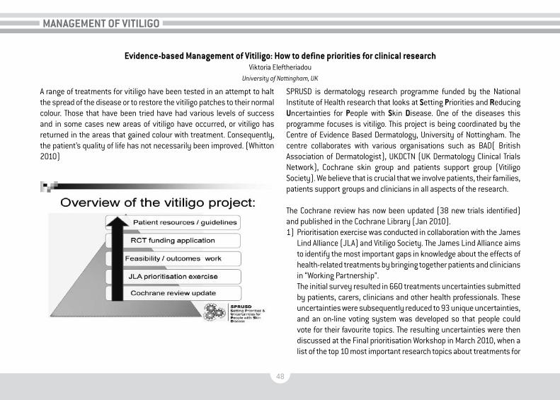
evidence-based Management of Vitiligo: How to define priorities for clinical researchViktoria Eleftheriadou
University of Nottingham, UK
A range of treatments for vitiligo have been tested in an attempt to halt the spread of the disease or to restore the vitiligo patches to their normal colour Those that have been tried have had various levels of success and in some cases new areas of vitiligo have occurred, or vitiligo has returned in the areas that gained colour with treatment Consequently, the patient’s quality of life has not necessarily been improved (Whitton 2010)
SPRUSD is dermatology research programme funded by the National Institute of Health research that looks at Setting Priorities and Reducing Uncertainties for People with Skin disease One of the diseases this programme focuses is vitiligo This project is being coordinated by the Centre of Evidence Based Dermatology, University of Nottingham The centre collaborates with various organisations such as BAD( British Association of Dermatologist), UKDCTN (UK Dermatology Clinical Trials Network), Cochrane skin group and patients support group (Vitiligo Society) We believe that is crucial that we involve patients, their families, patients support groups and clinicians in all aspects of the research
The Cochrane review has now been updated (38 new trials identified) and published in the Cochrane Library (Jan 2010) 1) Prioritisation exercise was conducted in collaboration with the James
Lind Alliance (JLA) and Vitiligo Society The James Lind Alliance aims to identify the most important gaps in knowledge about the effects of health-related treatments by bringing together patients and clinicians in “Working Partnership”
The initial survey resulted in 660 treatments uncertainties submitted by patients, carers, clinicians and other health professionals These uncertainties were subsequently reduced to 93 unique uncertainties, and an on-line voting system was developed so that people could vote for their favourite topics The resulting uncertainties were then discussed at the Final prioritisation Workshop in March 2010, when a list of the top 10 most important research topics about treatments for

49
MAnAGeMenT OF VITILIGO
vitiligo were agreed between patients and healthcare professionals (dermatologists, nurses, GP, psychologists, camouflage practitioners etc) All uncertainties identified during this process have been uploaded onto the DUETs (UK Database of Uncertainties about the Effects of treatments) website for reference and guidance of future research projects
The list of the Top 10 research priorities for the treatment of vitiligo will be presented on the day of the congress
One of the identified research topic will be developed into a feasibility trial, which is a small pilot trial to inform a large randomised multicentre trial
2) A full protocol for a large multicentre trial on treatment of vitiligo will be designed and a funding application will be prepared for submission to the relevant funding body
3) We will also be producing decision aids to help patients and clinicians make informed choices about treatment


SESSION VIIULTRAVIOLeTS And PHOTOTHeRAPY - PART I
ABSTRACT BOOK

ULTRAVIOLeTS And PHOTOTHeRAPY - PART I
52
Phototherapy of vitiligoAdrian Tanew
Department of General Dermatology, Medical University of Vienna, Austria
Topical therapy with corticosteroids and calcineurin inhibitors or, alternatively, surgical procedures can be considered as first choice treatment for localized stable vitiligo In contrast, phototherapies (UVB phototherapy, PUVA) are the mainstay for the management of extensive and active vitiligo In the last decade narrowband UVB, where available, has largely replaced PUVA as the first line phototherapy for vitiligo Narrowband UVB has several advantages over PUVA It does not require the administration of a photosensitizer rendering the treatment safer,
simpler and better tolerated Evidence from recent studies indicate that narrowband UVB provides for a faster and greater response than PUVA In addition, repigmentation induced by narrowband UVB is more even in color Phototherapies can be combined with a variety of topical and systemic treatments with the aim of accelerating or increasing the therapeutic response However, data as to the usefulness of such combination regimens, in particular with regard to the long-term outcome, are mostly scarce and partly conflicting

ULTRAVIOLeTS And PHOTOTHeRAPY - PART I
53
novel approaches in phototherapy of vitiligoAlessia Pacifico
Phototherapy Unit, S. Gallicano Institute, IRCCS, Rome, Italy
Vitiligo is a common skin disease characterized by loss of normal melanin pigments in the skin and its pathogenesis is still unclear Treatment modalities include PUVA, NB UVB phototherapy, now considered as the “gold standard”, topical and systemic steroids, topical calcineurin inhibitors, topical vitamin D analogues in monotherapy or in association with phototherapy, and surgical treatment Calcineurin inhibitors can increase the effectiveness of NB UVB, but the risk of enhancing UV carcinogenesis has to be definitely ruled out with further investigations, so this association should not be routinely recommended Some recent reports have focused on the possible benefits of associating vitamin D analogues with PUVA or NB UVB New ultraviolet sources capable of delivering large fluencies of narrowband ultraviolet B selectively to vitiliginous lesions in a shorter period of time have been introduced: this treatment modality has been defined as “targeted phototherapy” Two systems emitting high energy have been developed: 308 nm excimer laser and a non-laser device (308 nm Monochromatic Excimer Light, MEL) The advantages of MEL over the laser system are lower
operating costs and the fact that also large areas can be treated rapidly In contrast, the 308 nm excimer laser systems produce a small spot size which requires multiple treatments of adjacents areas to cover the lesional skin Adverse events are limited to mild erythema Conventional NB UVB remains the best treatment option for diffuse vitiligo; targeted phototherapy may represent a new therapeutic option for the management of non extensive vitiligo in order to achieve repigmentation in a shorter time and with better patients compliance as compared with other current modalities
References:1) Leone G, Iacovelli P, Paro Vidolin A, Picardo M Monochromatic excimer light 308 nm in
the treatment of vitiligo: a pilot study J Eur Acad Dermatol Venereol 2003; 17: 531-72) Leone G, Pacifico A, Iacovelli P, Paro Vidolin A, Picardo M Tacalcitol and narrow band
phototherapy in patients with vitiligo3) Mehrabi D, Panda AG A randomized, placebo-controlled, double-blind trial comparing
narrowband UV-B Plus 0 1% tacrolimus ointment with narrowband UV-B plus placebo in the treatment of generalized vitiligo Arch Dermatol 2006; 142: 927-9

54
ULTRAVIOLeTS And PHOTOTHeRAPY - PART I
MicrophototherapyTorello Lotti
University of Florence, Italy
Vitiligo is a primitive, chronic, acquired disorder of pigmentation, which represents both a medical and an esthetical challenge Even if some may say it is still an unmanageable disease, many studies on new therapeutical protocols seems to show increasing evidence that an effective treatment is possible However, melanocytes usually react slowly to treatments, so that it could take even several months to reach acceptable results in term of repigmentation, and affected subjects must be aware of it and acquiesce with the possibility of a long-lasting therapy Among all the possible therapeutic approaches, UVB narrowband microphototherapy may be considered a mainstay The efficacy of UV-B light in vitiligo is due both to cutaneous immune-suppression and the increased melanogenesis Evidence-based guidelines indicate that NB-UVB phototherapy is recommended for generalized vitiligo Furthermore, UVB treatments can be safely used in pregnant women and in children, and are related to less erythema, no phototoxic effects and no epidermal thickening after long-term irradiation The microphototherapic approach took place from the consideration
that vitiligo patients undergoing phototherapy were receiving a high cumulative dose of radiation during their lives, thus leading to secondary cutaneous disorders. On the contrary, microphototherapy is based on a photo-exposition limited to well-defined areas, avoiding the collateral effects associated to diffuse phototherapy (photoaging, erythema and burns, excessive tanning of the non-affected skin, among the others). UV-B radiation, with a wavelength peak at 308-311 nm, is selectively delivered to the white patches, thus allowing to obtain the drastic reduction of the total dose of radiation and the possibility to deliver different doses of UV-B radiation in different areas of the body (i.e. the hands need more UV-B than the eyelids to repigment): in other words, it is possible to have a treatment optimized and taylored on each and every subject. Microphototherapy is particularly useful in patients affected by segmental vitiligo and bilateral symmetrical vitiligo, in which the total amount of body surface involved is less than 20% Microphototherapy is not administered to subjects with actinic sensitivity (SLE, Xeroderma pigmentosum, porphyriasis, cutaneous viral infections) and in subjects treated with topical or systemic photosensitizing agents

55
ULTRAVIOLeTS And PHOTOTHeRAPY - PART I
Combined narrow-band UVB therapy and tacrolimus ointment in the treatment of vitiligo in 40 patientsAntonia G Galluccio
U.O. Dermatologia Ospedale “Sacro Cuore di Gesu’” Fatebenefratelli Benevento - Centro di Fototerapia - Centro Psocare, Benevento, Italy
Vitiligo is a chronic disease characterized by white patches of skin caused by lack or total absence of cells responsible for skin pigmentation, called melanocytes Phototherapy with UVB narrow-band was introduced in the late 90 for the treatment of vitiligo, with good results The key feature of the lamps is to emit UVB rays with a peak at 311 nm, a characteristic that led to the designation of “narrow band” as opposed to UVB “broadband” Are then eliminated wavelengths most harmful to the skin causing erythema and burns Tacrolimus ointment is a medication approved for the treatment of atopic dermatitis, with moderate to severe in children (aged 2 years and older) who have not responded to conventional therapy (0 03% ointment) and treatment of atopic dermatitis, moderate to severe in adults who do not respond adequately or who are intolerant of conventional therapies (0 1% ointment)
The skin, tacrolimus acts directly on T helper 2 transcription by inhibiting interleukin 2 and thus reducing the reactivity of lymphocytes T helper 2 with respect to exogenous antigens In recent years, many scientific works have been performed documenting the efficacy of tacrolimus in vitiligo showing how the first positive effects occur after 2-3 months of treatment Of particular interest is’ the article by Do D, Cassano N, Vena GA “Narrow-band UVB phototherapy combined with tacrolimus ointment in vitiligo: a review of 110 patients” that showed the effectiveness of the association between tacrolimus and NB UVB in a substantial number of patients 110 Based on this scientific evidence in the literature the authors evaluated the use of tacrolimus in combination with phototherapy with NB UVB in 40 patients with vitiligo on the face and neck In the same patients other vitiligoidee localized patches on the trunk and limbs were treated only with UVB nb

56
ULTRAVIOLeTS And PHOTOTHeRAPY - PART I
Follow-up long-term evaluation of a cohort of vitiligo patients treated with narrow-band UVB phototherapy and tacrolimus ointment*D Fai,° N Cassano,° G A Vena
*Phototherapy Unit - Dermatology Service, AUSL LECCE, Gagliano del Capo, Salento, Italy - °2nd Dermatology Clinic – University of Bari, Italy
Current treatments for vitiligo generally provide unpredictable and often unsatisfactory results Topical corticosteroids, topical calcineurin inhibitors and phototherapy (narrow-band UVB and psoralen+UVA) are the most prescribed options In severe and refractory cases, the effects of combination therapies can be evaluated, In our experience, for instance, relatively good results can be obtained by combining narrow-band UVB phototherapy and tacrolimus ointment, as demonstrated by a large
experience carried out in 110 patients treated with such a combination treatment for 16 weeks Despite the potential benefit in the short-term, vitiligo lesions can relapse in many cases after a variable period from discontinuation of the combined treatment Nevertheless, the literature contains scarce information about the efficacy of vitiligo treatments in the long term We report the data relative to the follow-up assessments of the original cohort of 110 patients previously mentioned

57
ULTRAVIOLeTS And PHOTOTHeRAPY - PART I
Keynote lectureVitiligo associated organ-specific autoimmune diseases
Emanuele BosiDepartment of Internal Medicine, San Raffaele Scientific Institute and San Raffaele Vita-Salute University, Milan, Italy
Vitiligo is an acquired hypomelanotic disorder characterised by circumscribed depigmented areas in the skin, resulting from the loss of functional melanocytes The frequent association of vitiligo with other autoimmune diseases, together with studies claiming the presence in affected patients of circulating pigment cell-specific autoantibodies and autoreactive T lymphocytes supports the hypothesis of an autoimmune pathogenesis This assumption is reinforced by the association of vitiligo with polymorphisms of genes or gene regions known to be associated with other autoimmune disorders (e g HLA, AIRE, PTPN22) Epidemiological studies showed in unselected patients with vitiligo, and in their first degree relatives, elevated prevalence of a number of
organ-specific autoimmune diseases, including thyroiditis, gastritis, pernicious anaemia, type 1 diabetes, Addison’s disease, and some non-organ-specific autoimmune diseases such as systemic lupus erythematosus Such data indicate a genetic predisposition, common to several autoimmune disorders, most likely with a polygenic basis Unfortunately, the persistence ignorance about the identity of the environmental agents and the mechanisms responsible for their interaction with the genetic background resulting in the initiation and perpetuation of the autoimmune process, hinder the development of efficacious strategies for prevention and therapy


SESSION VIIIULTRAVIOLeTS And PHOTOTHeRAPY - PART II
ABSTRACT BOOK

60
ULTRAVIOLeTS And PHOTOTHeRAPY - PART II
nB-UVB and topical tacrolimus: our five-years experience in vitiligo treatmentAntonello Baldo
University of Naples, Italy
Vitiligo is a common, idiopathic, acquired, depigmenting disease characterized by loss of normal melanin pigments in the skin The current treatment of vitiligo is not satisfactory according to the opinions of both the patient population and the dermatologists In this randomized and comparative study, we investigated the safety and efficacy of narrow band ultraviolet B as monotherapy towards topical tacrolimus therapy We randomized two groups of patients Each group was compound by
100 patients The first one was irradiated with NB-UVB (311 nm) twice a week for 9 months time; the second group applied tacrolimus ointment 0,1% twice a day for the same period Before starting therapy and after 3, 6 and 9 months of therapy, a clinical and photographic evaluation of percentage of repigmentation was performed and DLQI questionnaire was completed In each group, treatments were well-tolerated Results will be presented

61
ULTRAVIOLeTS And PHOTOTHeRAPY - PART II
Topical steroid combined to excimer laser in the treatment of vitiligo. Vitiligo and psoriasis in the era of biological drugsLuciano Mavilia and Santo Raffaele Mercuri
Unit of dermatology and Cosmetology, Ospedale San Raffaele, Milan, Italy
Targeted phototherapy with single-wavelength laser light is a treatment alternative that may prove to be a time-efficient and effective therapeutic option for the management of vitiligo In our report, we use topical steroid before and after the treatment with the 308nm excimer laser for the treatment of vitiligo both to modulate the immune system and to minimize the possible side effects of the therapy using high dosage with weekly sessions The majority of patients with head and neck involvement can expect to achieve complete repigmentation in less than four months
In the second part of the lecture, we report the co-existence of vitiligo and psoriasis in some patients It is not suprising to see association of vitiligo and psoriasis as they affect 1% to 3% of the population normally Many pigment cell biologists and dermatologists have concluded that vitiligo is an autoimmune disease and psoriasis, the T cell mediated skin disease, may be associated We also report as the face of psoriasis is changing in the era of vitiligo and that we cannot say the same thing about vitiligo yet

62
ULTRAVIOLeTS And PHOTOTHeRAPY - PART II
PolipodiumD Fai, I Romano
Parabita, Lecce, Italy
P leucotomos is a tropical fern plant that has long been known by Native Americans who believe it has antitumoral and anti-inflammatory effectsThis plant extract has no reported side-effects and toxicology tests show that P leucotomos is noncarcinogenic and nonteratogenic a has immunomodulatory properties
P leucotomos has also antioxidant properties, quenching of freeradicals, lipid peroxidation and reactive oxygen species such as the hydroxyl radical, singlet oxygen, superoxide anion and hydrogen peroxide These data support the use of polypodium leucotomos as an adjuvant in combination therapy of vitiligo

63
ULTRAVIOLeTS And PHOTOTHeRAPY - PART II
Vitiligo: effectiveness of UVB "narrow band" plus tacrolimus 0,1% ointment associationDomenico D’Amico, Giancarlo Valenti
U.O.C. Dermatologia, Az. Osp. “Pugliese-Ciaccio”, Catanzaro, Italy
Vitiligo is a chronic disease with unknown pathogenesis characterized by the appearance of segmental or not segmental achromic areas vitiligo vulgaris (or not segmental) is the most common feature with a prevalence of approximately 0,5% in Italy and a progressive course of evolution Despite it was considered only an “aesthetic” disease and beyond the possible association with other autoimmune diseases, vitiligo also leads to a serious psychological damage with an important impact on social and intimate relationships, that requires implementation
of all therapeutic measures The limited therapeutic options and the inconstant results frustrate the hopes of patients and the aspirations of physicians The narrow-band UVB are currently considered the gold standard for vitiligo treatment, the results seem to improve if topical therapy is associated with calcineurin inhibitors We present some personal experiences of vitiligo vulgaris, extended to large skin areas where treatment with UVB narrow band associated with the application of tacrolimus 0 1% ointment has been very effective


SESSION IXSURGeRY And LOnG TeRM STABILITY
ABSTRACT BOOK

SURGeRY And LOnG TeRM STABILITY
66
Autologous ORS cell transplantation: adult stem cell therapy for vitiligoThomas Hunziker
Department of Dermatology, University of Bern, Switzerland
The optimal modality of melanocyte transplantation for depigmented skin lesions is still to debate So far, the focus has been on procedures of isolation, application and eventually cultivation using interfollicular epidermal melanocytes harvested by skin biopsy Only 4 publications reported on vitiligo treatment in hairy skin by hair follicle grafting As for keratinocytes, a stem cell pool resides in the outer root sheath (ORS) of hair follicles also for melanocytes, illustrated clinically by the well-known phenomenon of follicular repigmentation in healing vitiligo Melanocytes or stem cells thereof present in the ORS of plucked anagen scalp hair follicles can be used to treat depigmented skin In proof of concept studies, we applied solutions of autologous ORS cells isolated by trypsinisation of plucked anagen scalp hair follicles to stable lesions of vitiligo Transepidermal delivery was achieved either by continuous or fractional laser de-epidermisation or by microneedling
using a dermaroller After reepithelisation, treated areas were irradiated 2-3 times weekly with suberythematogenous doses of 311 nm UVB or Excimer laser Several patients experienced extensive repigmentation, some of them even in sham-treated or non-treated areas In conclusion, melanocytes or stem cells thereof present in the ORS of plucked anagen scalp hair follicles can be used to treat depigmented skin Thereby, harvesting of these autologous cells is non-invasive, which allows easy, immediate and repeated application Using the patient’s own skin as an incubator and thus avoiding cultivation of the cells in vitro is a further practical and also regulatory advantage of this technology, since malignant transformation of melanocytes upon stimulation of proliferation in vitro is still an issue Optimal application modalities and cell numbers as well as concomitant treatments have to be defined

SURGeRY And LOnG TeRM STABILITY
67
Long term results of non cultured epidermal cellular grafting in vitiligo, halo nevi, piebaldism and nevus depigmentosus Nanja van Geel, MD, PhDa, Elien Wallaeysa, Boon Kee Goh, MD, PhDb, Martine De Mila, Jo Lambert, MD, PhDa
aDepartment of Dermatology, Ghent University Hospital, De Pintelaan 185, 9000 Ghent, Belgium -bPigment Clinic, National Skin Center, 1 Mandalay Road, Singapore
Background: Several surgical techniques are available for the treatment of stable leukoderma The use of non cultured epidermal cellular grafting has been introduced in 1992 Data on long term follow-up regarding stability of the repigmented area, time to achieve the final repigmentation, color matching, reaction to sun exposure and patient’s satisfaction of treatment are limited reported Objectives: To evaluate the long term results of the non cultured epidermal cellular grafting technique in patients with different types of leukoderma, including segmental vitiligo (33), generalized vitiligo (33), mixed vitiligo (6), halo nevi (11), piebaldism (3) and nevus depigmentosus (1) Methods: Patients were evaluated by clinical examination and/or a questionnaire in a retrospective setting after transplantation of autologous non cultured cellular grafting Percentage of repigmentation was evaluated in 82 patients using a digital imaging analysis system (mean follow up 15 months) Long term results were evaluated by 54
patients using a questionnaire up to 7 7 years after treatment (mean 4 years) Results: More than 75% repigmentation was achieved in 70 7% of patients Best results were obtained in segmental vitiligo, halo nevi and piebaldism, whereas results in generalized or mixed vitiligo were inferior According to the patients, final repigmentation was achieved after a mean of 10 months post treatment In 80 4% some color mismatch (hyper-and hypo-pigmentation) was reported between the treated area and the surrounding skin, although this was not disturbing for the majority This color mismatch was reported significantly less after sun exposure (p= 0 012) During follow-up 6 7% of patients, all with generalized vitiligo, observed some loss of the achieved repigmentation Conclusion: Autologous epidermal cellular grafting achieves a high percentage of repigmentation, which was maintained during follow-up in the majority of patients Although it improves quality of life, in most patients a perfect color matching was seldom obtained

68
SURGeRY And LOnG TeRM STABILITY
Total stability and complete repigmentation in vitiligo: how close are we?Davinder Parsad
Department of Dermatology, Venereology & Leprosy, Postgraduate Institute of Medical Education and Research, Chandigarh, India
The development of effective treatment for vitiligo depends on understanding the mechanisms of depigmentation and repigmentation The basic pathogenesis of vitiligo in general, or for any of the putative subsets of vitiligo, is not fully known although substantial strides have been made in pathogenesis and the treatment of vitiligo Since the etiopathogenesis of depigmentation in vitiligo is still obscure, the source of pigmentation in the repigmentating lesion and its stability is also not fully known
Here are two main goals of any vitiligo treatment; first is to stop the arrest of further depigmentation and second is to induce repigmentation The first goal can only be archived fully if we could unravel the mechanisms underlying the disappearance of melanocytes in vitiligo If this can be
achieved repigmentation should be rather simple to accomplish with a combination of medical and/or surgical treatment Surgical management has evolved significantly and given a ray of hope in stable vitiligo
However, there is lot of scope for improvement So more research should happen in this field so that our aim will shift from “any repigmentation” to “complete and normal repigmentation” and we can achieve a scar less healing of those “bruised souls” The mechanistic aspects of melanocyte repopulation in vitiligo and of other factors that trigger and influence melanocyte growth, maturation and survival have been little explored A better understanding of vitiligo repigmentation will provide new therapeutic perspectives leading to development of an ideal weapon against vitiligo

SeLeCTed ABSTRACT
SELECTED ABSTRACT

70
SeLeCTed ABSTRACT
Genetic variants of the Bche Gene are associated with vitiligo in a brazilian population sampleL M Nascimento1, C C S de Castro1,2, R I Werneck 1 and M T Mira1
1 Group for Advanced Molecular Investigation, Program of Graduate Studies in Health Sciences, Pontifical Catholic University of Parana, Curitiba, Brazil 2 Department of Dermatology, Santa Casa de Misericordia Hospital, Pontifical Catholic University of Parana, Curitiba, Brazil
Vitiligo is an acquired systemic, chronic disease characterized by macules devoid of melanic pigment and identifiable melanocytes Oxidative stress and the accumulation of free radicals in the epidermal layer of affected skin have been shown to be involved in the pathophysiology of vitiligo Epidermal butyrylcholinesterase (BchE) protein expression and enzyme activitie are altered in vitiligo We hypothesize that genetic variants of the BchE gene are associated with the occurrence of vitiligo To test this hypothesis, we genotyped 10 tag SNPs spanning the entire gene in 600 individuals distributed in 212 trios composed by an affected child and both parents Markers were genotyped in the platform SEQUENOM MassARRAY which uses the iPLEX assay to incorporate mass-modified terminal nucleotides in the SBE step that are then detected by matrix-assisted laser desorption/ionization-time of flight (MALDI-TOF) mass spectrometry Family-based association analysis was performed in the software FBAT
Allele “G” of BchE intragenic tag SNP rs1355538, allele “A” rs4680607, allele “A” rs1803274 were associated with an increased risk for vitiligo in our population (P = 0 004, OR = 1 96, 95% IC = 1 21-3 17; P=0 03, OR = 2 77, 95% IC = 1 03 – 7 43; P= 0 04, OR = 1 38, 95% IC = 1 01 – 1 95 respectively) Best evidence for association in the family-based population was obtained for a haplotype composed by risk alleles of markers rs1355538 and rs4680607 (P = 0 002) In addition, we observed an age-dependent enrichment of rs1803274 “A” allele in young affected individuals using cut-off age of diagnosis of 25 years old In conclusion, we propose the BchE as a new susceptibility gene for vitiligo, possibly implicating in vitiligo pathogenesis

71
SeLeCTed ABSTRACT
Treatment of vitiligo by means of focused and selective microphoto-therapy using the RATOK®deRM equipmentM Fantato
Ratok®derm Dermatological Clinic of Milan, Italy
Vitiligo is a skin disease characterized by the onset of hypopigmented patches in different skin areas, with psychological implications that often affect the social life of patients In the past few years, several therapeutic attempts were made to stop the development of the disease after its onset Photochemotherapy, both topical and systemic, was historically used, however with side effects ranging from the possible development of cataract to the triggering of carcinogenic processes In the past few years, other irradiation sources were thus considered, including UVB-rays, which do not require any combination with psoralens or other molecules In 1986, a new equipment was conceived at the Ratok®derm Centre of Milan, based on the selective and focused output of UVB-rays The benefit of this method is that only the affected areas are treated without any contraindications, not even in child treatment The equipment has a generator capable of producing a mono and
multipunctiform UVB-irradiation with a 200mW/cm2 fluency (at the output of the optical system) and 4-8 sec pulses on the treated patches throughout a 15-30 minute session The therapeutic protocol provides for five daily sessions within 5 days Subsequent cycles provide for one or more treatments performed 7 to 10, maximum 30, days apart The cycle should be repeated until treatment is completed, usually for 6 to 18 months In the last years Ratok®derm methodology has been improved by the introduction of the bath of light (dynamic total body 311 narrow band) Moderate to good results exceeded 75% The author discuss the results obtained on a sample of over 2,000 vitiligo patients treated and followed up and stress the absence of any treatment-related side effects

72
SeLeCTed ABSTRACT
The clinical safety and efficacy of 308nm excimer light phototherapy for vitiligo patientsSaori Itoi, Atsushi Tanemura, Megumi Nishioka, Kazuko Sakimoto, Eriko Iimuro, Ichiro Katayama
Department of Dermatology Integrated Medicine, Osaka University Graduate School of Medicine, Japan
Background: It has been shown that a phototherapy as PUVA and narrowband UVB is one significant treatment modality for vitiligo and that narrowband UVB may be prior to PUVA phototherapy Although the clinical effectiveness of excimer light emitting 308nm of UV is compared to that of other wave length in treatment with vitiligo patients, the clinical safety for vitiligo is not fully elucidated
Aim: The purpose of this study is to assess both of the safety and clinical efficacy of 308 nm excimer light monotherapy in vitiligo patients
Methods: Fourteen Japanese patients suffering from generalized and local vitiligo were enrolled in this study One particular vitiligo lesion was selected and treated with excimer light phototherapy weekly or twice a week for 3 to 5 months (total dose of exposure was ranged from 2 04 to 32 33 J/cm2) The lesion was assessed with the following clinical parameters reflecting a vitiliginous alteration before and after phototherapy; lesional size measured by NIH image and color change of L*a*b* values calculated by a coloriometer The perilesional normal skin was covered by a sunprotective flim and UV cut cream The L*a*b* color space is the most common one to measure object color without subjective contamination The reduction of L* space means decrease in skin whiteness and being close to perilesional skin color Delta L* value
was calculated by the ratio of L* value of lesional to perilesional skin Since a* space represents erythematous change after UV exposure, increase in the score expresses sunburn skin damage To propose a safe increasing dose of UV, 30mJ/cm2 for 4 patients and 45mJ/cm2 for 10 patients were configured If erythematous change and irritation remains at the next visit, the dose was reduced to last but one If vitiligo patients noticed repigmentation, the dose was settled for further treatment
Results: No patients had severe burn injury both with 30 and 45 mJ/ cm2 of increasing doses There is no significant difference of mean initial dose with erythema or irritation between the above two settings Initial repigmentation occurred at 4 to 13 times of treatments and 8 patients noticed repigmentation more than 25 % of lesions area at the end of study Whereas vitiligious lesions on the trunk received 40 8% of repigmentation, those on forehead and elbow had minimal repigmentation with slight erythema irrespective of step increase in dose L* value and delta L* value significantly decreased after a phototherapy There is no significant difference of a* value between before and after treatment
Conclusions: A 308nm excimer light is effective and well-tolerated therapeutic option for Japanese vitiligo patients

73
SeLeCTed ABSTRACT
A pilot study of punch grafting followed by excimer laser therapy in stable vitiligoAl-Harbi R , Tawfik W , Al-Aradi I, Qasem K
Asa’d al-Hamad dermatology centre, Shuwaikh, Kuwait
Background: Vitiligo is a common pigmentary disorder Punch grafting followed by Excimer laser is yet not an established therapy for stable vitiligo The aim of this pilot study was to evaluate the efficacy of the combination treatment of excimer laser in perigraft pigmentation and compare it with perigraft pigmentation without an intervention and pigmentation with the excimer laser, alone in patients with stable vitiligo
Methods: Ten patients with stable vitiligo of various clinical types were subjected to punch grafting The vitiligo patches were divided into 4 groups; Group I received post punch-grafting excimer laser, group II received punch-grafting alone, group III received excimer laser alone and group IV received no intervention to serve as a control During the follow-up period of 2 months, one patient dropped out, nine patients were evaluated for pigment spread and side effects
Results: In group I, the average pigment spread was 51-75%, whereas in group II, it was 0-25%, showing a lower pigment spread, in group III it was 26-50% and in group IV it was 0-25% There was a difference in response to therapy in patients having segmental vitiligo as compared with generalized vitiligo Erythema, blister formation and graft displacement were the important side effects seen in some patients in groups I-III
Conclusion: The study shows that the pigment spread with the combination of punch-grafting and excimer lasaer is higher than that with wither of them, alone However, larger studies with long-term follow-up are required to establish this

74
SeLeCTed ABSTRACT
Is the success of autologous non cultured epidermal suspension transplantation depends on the special medium like melanocyte medium?Holla A P1, Parsad D2
1 Department of Dermatology, Srinivasa Institute of Medical Sciences, Mangalore, India 2 Department of Dermatology, Postgraduate Institute of Medical Education and Research, Chandigarh, India
Objective: To compare the outcome of autologous non cultured epidermal suspension transplantation procedure where suspension was prepared with phosphate buffered saline in one group and suspension prepared using melanocyte culture medium in the other Secondary objective was to prove that there is no need of special medium in this particular procedure if proper selection criteria are followed Methods: 23 stable vitiligo patients (17 females and 6 males; age range 10 – 40 yrs; mean 21 956 ± 7 666 years) were included in the study and total 48 lesions were treated 15 patients were having vitiligo vulgaris, 6 had segmental vitiligo and 2 had focal vitiligo In group A (12 patients, 25 lesions) non cultured autologous epidermal suspension obtained from split thickness skin graft was prepared using phosphate buffered saline (PBS) In group B(11 patients, 23 lesions) melanocyte medium was used to prepare the same In both groups, suspension was transplanted to the dermabraded recipient area with a modified procedure of transplantation Patients were given methylcobalamin and advised daily sun exposure Follow up period was 6 months Results were assessed based on the extent of repigmentation, colour match and adverse events An evaluation was also done based on pre and post treatment DLQI scores and patient satisfaction questionnaire at 6 months
Results: There were no significant statistical differences in the patient and the lesion characteristics between the two study groups Repigmentation was successful (repigmentation > 75%) in 22/25 lesions (88%) in group A and in 20/22 lesions (90 90%) in group B Color match at 6 months was excellent in 23/25 (92%) lesions and 19/22(82 60%) lesions in group A and B respectively Mean percentage of change in pre and post treatment DLQI was 69 66±18 154 in group A and 67 40± 19 135 in group B No significant adverse events were reported in both the groups Patients in both the groups were highly satisfied with outcome Conclusion: Outcome of this comparative study confirms that there is no need of any special medium like melanocyte culture medium in the autologous non cultured epidermal suspension transplantation Phosphate buffered saline achieves similar efficacy if proper selection criteria were followed Moreover it decreases the cost and also it is safe since there is no theoretical risk of carcinogenesis as with melanocyte medium

75
SeLeCTed ABSTRACT
Proinflammatory Cytokines Regulate MITF-related Molecules expression and Melanin Production in vitro. -Possible pathogenesis of vitiligo-
Yorihisa Kotobuki1,2, Atsushi Tanemura1, Satoshi Serada2, Tetsuji Naka2, Ichiro Katayama1
1Department of Dermatology Integrated Medicine, Osaka University Graduate School of Medicine, Japan2Laboratory for Immune Signal, National Institute of Biomedical Innovation, Japan
Background: Alterations in cellular immunity including CD4+T and CD8+T cells have been in the pathogenesis of vitiligo However, some previous reports propose the role for lesional cytokines such as IL-1b, IL-6, and TNF-a in inhibiting melanocyte tyrosinase activity associated with a particular skin depigmentation disorder, vitiligo The cytokine network which is important for disease formation of psoriasis and atopic dermatitis may be also involved in the pathogenesis of vitiligo Aim: The purpose of this study is to identify whether the proinflammatory cytokines which are likely produced by helper T cells regulate melanocyte activity and melanin production directly Method: After human moderately pigmented melanocyte was treated with recombinant IL-1b, IL-6, IL-17A, and IL-22 proteins, the transcription level of MITF was measured by quantitative PCR Moreover, it was examined for the transcription levels of tyrosinase, TYRP-1, and DCT as melanogenic molecules and Bcl-2 and HIF-1a as cell survival ones Melanin contents assay was also performed with the treatment of the above cytokines The concentration of cytokines examined was 10 and 50ng/ml To make sure a direct effect of exogenous cytokines for melanocyte, we examined the phosphorylation of STAT3 by the addition of IL-6 and that of NF-kB by the addition of IL-1b and IL-17A, respectively
Results: All of cytokines phosphorylated either STAT3 or NF-kB in human melanocyte in vitro, suggesting the presence of downstream signaling to proinflammatory cytokines in melanocyte The treatment with IL-1b and IL-6 significantly decreased MITF transcription level as shown in the previous study (p=0 0003, 0 0022, respectively) Interestingly, the transcription level was increased about 1 7-fold in the treatment with IL-22, whereas IL-17A decreased as well as IL-1b and IL-6 (p=0 0217) However, the transcription level of the downstream molecules of MITF and the amounts of melanin production were suppressed in the treatment with not only IL-1b, IL-6, and IL-17A but also IL-22 compared to no stimulation Conclusions: These findings firstly demonstrate the direct effect of proinflammatory cytokines for MITF and its downstream genes expression and melanin production and that not only IL-1b, IL-6, TNF-a but also IL-17A and IL-22 are involved in melanocyte activity Since IL-22 has the opposite effects for between MITF expression and melanin production, there may be auto-regulation factors to inhibit MITF signaling downstream of IL-22 in melanocyte The presence of cytokine network and the impairment of IL-17A - IL-22 balance in Th17 cells may be implicated in a new insight of the pathogenesis of vitiligo

76
SeLeCTed ABSTRACT
Possible Link between Keratinocyte expression of pSTAT3 and Th17 Cell Infiltration to the Lesional Skin in Vitiligo VulgarisAtsushi Tanemura1, Yorihisa Kotobuki1,2, Satoshi Serada2, Tetsuji Naka2, Ichiro Katayama1
1Department of Dermatology Integrated Medicine, Osaka University Graduate School of Medicine, Japan2Laboratory for Immune Signal, National Institute of Biomedical Innovation, Japan
Background: Although hypotheses such as autoimmune reaction, a disease due to abnormalities in the biochemical or metabolic pathways are considered, the exact pathogenesis is still unknown It has been demonstrated that the close interaction between keratinocyte and melanocyte in the epidermis plays a part of role for melanocyte survival and melanogenesis and that dysfunction of the surrounding keratinocytes or production of keratinocyte-derived cytokines as IL-6 can be seen in the vitiligo skin IL-6 is known as one of the proinflammatory cytokines which can activate signal transducer and activator of transcription 3 (STAT3) in inflammatory cells and epidermal keratinocyte Moreover, as well as the cellular immunity including CD4+T and CD8+T cells in autoimmune vitiligo, CD4+IL-17A+ Th17 cells which is important for disease formation of psoriasis may be associated with the pathogenesis of vitiligo Aim: The purpose of this study is to identify whether STAT3 phosphorylation in keratinocyte and Th17 cell infiltration occurs in vilitigo skin and compare those numbers between lesional and perilesional skin and to assess the possibility of functional link between keratinocyte expression of STAT3 and infiltration of Th17 cells in vilitigo skin Method: Eighteen skin biopsy specimens including lesional and non-lesional skin were taken from the same number of vitiligo patients
The patients consisted of 10 female and 8 male between age 27 and 81 years We performed an immunostaining for phosphorylated STAT3 (pSTAT3) in keratinocyte and IL-17A in accordance with CD4 staining using paraffin-embedded archival tissue section Thereafter we counted the number of phosphorylated STAT3 keratinocytes and IL-17A+CD4+ cells in lesional and perilesional skin Three skin samples from non-tanned area were also tested Results: Keratinocyte expression of pSTAT3 occurred in 9 skin specimens (50%) Vitiliginous lesional skin had significantly higher number of pSTAT3 compared to perilesional skin (p=0 0032) Immunofluorescent staining showed that most of CD4+ cells expressed IL-17A, suggesting dense infiltration of Th17 cells in vitiligo skin, whereas a few number of CD8+ cells were detected Vitiligo skin tissues (n=7) with pSTAT3 in keratinocyte were significantly densely infiltrated by Th17 cells compared to those (n=8) with no pSTAT3 (p=0 03) Conclusions: These findings demonstrate the significant phosphorylation of STAT3 in ketratinocyte and recruit of Th17 cells in vitiligo skin Th17 cell infiltration through IL-6-STAT3 signaling linage in lesional keratinocyte may be one of an important phenomenon to induce melanocyte disappearance and/or impairment of melanin synthesis instead of alteration in CD4 and CD8 cells-associated immunity

77
SeLeCTed ABSTRACT
A Randomised Controlled Trial of Minigrafting Vs ReCell in Stable VitiligoB S Daniel1, S S Venugopal1, 3, L K Martin1, R Wittal2, 3, L M Rhodes1, 3, J Le Guay2, D F Murrell1
1 Dermatology department, St George Hospital, Sydney, Australia2 Vitiligo Clinic, Skin and Cancer Foundation, Darlinghurst, Sydney, Australia
3 University of New South Wales, Sydney, Australia
Vitiligo is a common pigmentation disorder associated with an impaired quality of life Treatment of hypopigmented skin has been a therapeutic challenge with many therapies deemed ineffective Surgical treatment of vitiligo is aimed at stable localised areas, resistant to medical therapy Surgical interventions include split thickness grafts, punch grafts and melanocyte transplantation by cultured and non-cultured methods Non-cultured Epidermal Suspension (NCES) involves preparing a split thickness skin graft which is processed into an epidermal cell suspension with the ReCell device and transplanted onto dermabraded skin affected with vitiligo The ReCell device is an effective tool in the management of wounds in patients with burns Its role as a surgical treatment in vitiligo has been published as case reports and this study seeks to determine if repigmentation differs between the non-cultured Epidermal Suspension (NCES) and minigrafting sites Though previous studies using the ReCell device have reported some success in repigmentation, these results were limited by patient numbers, short follow up periods, and the absence of randomisation and a comparison arm We are currently conducting a prospective intra-
patient single centre randomised comparison trial for patients with stable vitiligo A pilot mini-graft is used as part of the inclusion criteria and as a more objective measure of disease stability Those who meet the inclusion criteria have both minigrafting and NCES performed on randomised anatomically paired sites Pre and post-procedure pictures of donor and recipient sites are used to evaluate the percentage of repigmentation which is assessed by an independent blinded investigator Quality of life (QOL) assessments and other subjective data is gathered before the procedure and at every visit The primary outcome of the trial is the percentage repigmentation at 12 months of NCES as compared with the conventional minigrafting Secondary outcomes include percentage repigmentation at 3 and 6 months, cosmetic outcome of recipient and donor sites assessed by investigators, colour matching, time to repigmentation and change in QOL scores during the trial We report out interim results of 15 patients who have been followed up for 12 months Our results indicate a potential beneficial use of the ReCell device in the treatment of stable vitiligo

78
SeLeCTed ABSTRACT
The incidence of leucotrichia in segmental vitiligo: implication of poor response to medical treatmentDong-Youn Lee, Kyu-Dong Jung, Cho-Rok Kim, Ji-Hye Park, Joo-Heung Lee
Department of Dermatology, Samsung Medical Center, Sungkyunkwan University School of Medicine, Seoul, South Korea
In vitiligo, the melanocyte of hair follicle is one of the major sources for repigmentation Segmental vitiligo seems to be often associated with white hairs However, in case of small vellus hairs it is often difficult or impossible to detect hair color Thus, the real incidence of leucotrichia in segmental vitiligo has not been known In this study, we examined about the existence of white hairs in the lesional skin of 82 patients with segmental vitiligo The records and clinical pictures of the patients who visited our vitiligo clinic, department of dermatology, Samsung Medical Center during the last 5 years were reviewed retrospectively When it was difficult to detect hair color with the naked eyes or a magnifier, a digital microscope with 30 magnification was used In some patients, we detected that the many hairs in the lesional skin consist of white color by the digital microscopy Among them
we evaluated the effects of medical treatment such as phototherapy and surgical treatment in the patients who had the majority of white hairs in the lesional skin Interestingly, all 82 patients showed leucotrichia in segmental vitiligo independent of age and disease duration The amount of white hairs was variable However, surrounding normal skin showed black hairs Some patients had white hairs more than 90% in the lesional skin and they showed poor response to medical treatment Based on our results, all patients with segmental vitiligo may be associated with leucotrichia Many white hairs in segmental vitiligo explain poor response to medical treatment The examination of hair color with a digital microscope may be very useful for the prediction of treatment outcome and decision of treatment modalities

79
SeLeCTed ABSTRACT
Using research to inform the development of a range of therapeutic interventions for people with vitiligo and other disfiguring conditionsProf Nichola Rumsey
Centre for Appearance Research, University of the West of England, Bristol, UK
The psychosocial effects of having a condition that is visible to others can be profound A significant proportion of those affected experience negative effects on their self perceptions and social functioning, however others adjust positively This paper reports the results of a series of recent studies involving 1265 participants with disfigurements resulting from a range of conditions, including vitiligo Using both quantitative and qualitative methods and with a clear focus on the translation of results into therapeutic interventions, this programme of research examined the psychological characteristics of those who adjust positively and those who experience high levels of distress
The results of these studies showed that adjustment and distress were not well explained by biomedical factors such as the cause and severity of the condition Neither was time found to be a great healer in the
longitudinal element of the research Instead, adjustment involved a range of psychological factors, particularly biases in cognitions (ways of thinking) and patterns of behaviour The results offered a strong endorsement of the potential utility of therapeutic interventions of varying levels of intensity This research has been used to inform the development of interventions designed to ameliorate distress and promote positive adjustment in those adversely affected by disfiguring conditions such as vitiligo These include a manual to guide face to face interventions, and the development of a range of online interventions including facilitated support groups and self-administered programmes for adults and young people Examples of the ways in which these interventions could be beneficial to people distressed by vitiligo will be given

80
SeLeCTed ABSTRACT
Pathogenesis and treatment of vitiligoProfessor Insaf Mokhtar1, Dr Smida Rached2
1Head of Dermatology Department in the university hospital center ,Habib Thameur, Tunisia and president of the Tunisian Dermatologists Association, Tunis, Tunisia2Researcher in the pathophysiology of vitiligo, Tunis, Tunisia
Vitiligo is due to a high rate of free cortisol, which will lead to rise in phenylalanine hydroxylase The latter will cause an overproduction of H202 and this will degrade the membrane lipids( this phenomenon has been demonstrated on plants by SMIRNOFF IN 1993 when he injected the leaves with Our goal is to demonstrate that a diet rich in linoleic acid (corn oil, sunflower) and low in alphalinolenic acid (blue fish, flaxseeds,wholemeal bread) leads to the rise of free cortisol which will lead to higher rates of most enzymes including phenylalanine hydroxylase which will lead to overproduction of H2O2 creating oxidative stress that will damage the tetrahydrobiopterin (BH4), denature proteins (melanin) and degrade the lipid membrane (a phenomenon that has been proved by SMIRNOFF on plants in 1993 Also,the high rate of free cortisol will increase rates of lymphocytes and monocytes We were able to prove that in mixing 75% of olive oil and 25% of corn oil or sunflower (rich in linoleic acid) and giving ten milliliters of this mixture to patients for six weeks ,will lead to a rise in free cortisol and a decrease the total one It will also increase the level of lymphocytes and monocytes Free cortisol is the antagonist of vitamin D2-D3 (25 hydroxy) We took a sample of 25O vitiligo patients (120 women and 130 men) They made a blood test including vitamin D2-D3 (25 hydroxy), total cortisol and free cortisol We also took another sample of 250 healthy patients We found
low levels of vitamin D3-D2 in 236 patients (94% of total) with an average of 39,8 n mol / l (standard rate between 75 and 150 n mol / l ) against 34% of D2-D3 vitamin level in the group not suffering from vitiligo We found in all men suffering from vitiligo premature ejaculation explained by impairment of BH4, which releases nitric oxide (NO), essential for proper sexual act We found a correlation between very low Vitamin of D2-D3 (lower then 25 n mol / l ) and autoimmune diseases including Thyroid (comes first with 28%), and diabetes type 1( with 8% ) These 250 patients were cured thanks to the oil whose main role is to raise the rate of free cortisol ; and this enhances the activity of anzymes as BH4 and Tyrosine This invention is now the intellectual property of Tunisia
ReSULTSThere was a repigmentation of the affected areas in 100% of the patients and 43% of the patients were completely cured It takes us eight weeks to witness the repigmentation It depends on the rate of vitamin D2-D3 and the length of the infringement The lower vitamin D3-D2 is witnessed,the slower the response to treatment is The genital area is the first to be cured,(nine months), followed by the face (1 year) and hands (2à3 years) Finally, 1OO% of men suffering from premature ejaculation had a significant improvement after 6 months

81
SeLeCTed ABSTRACT
Classification SMIdA - MOKHTAR of four stages of vitiligoProfessor Insaf Mokhtar1, Dr Smida Rached2
1Head of Dermatology Department in the university hospital center ,Habib Thameur, Tunisia and president of the Tunisian Dermatologists Association, Tunis, Tunisia2Researcher in the pathophysiology of vitiligo, Tunis, Tunisia
Vitiligo is due to the rise of free cortisol which will cause the increase of phenylalanine hydroxylase This will lead to an overproduction of H2O2, which will degrade the lipid membranes (This phenomenon has been experimented on plants by SMIRNOFF in 1993 and proved that when injecting H2O2 into the leaves a degradation of membrane lipids and destruction of chlorophyll occur) Depending on the intensity of the overproduction of H2O2 it will have four stages according to the accumulation of lipids in the epidermis, dermis and hypodermis STAGe 1 Degradation of membrane lipids in the epidermis causing Stage 1 with white appearance of the lesion and black hair
STAGe 2 Degradation of membrane lipids in the epidermis and part of the dermis just above the root of the hair follicle causing Stage 2 with mat-white appearance of the lesion and black hair
STAGe 3Degradation of membrane lipids in the epidermis and dermis just below the root of the hair follicle causing Stage three with white appearance of the lesion and white hair STAGe 4 Degradation of membrane lipids in the epidermis, dermis and hypodermis The hair follicles won’t be fed and this causes hair loss


POSTER PRESENTATION
POSTeR PReSenTATIOn

POSTeR PReSenTATIOn
84
Psychosocial effects of vitiligo on sexual lifeSeung-Kyung HANN1, Do-Young KIM2, Sang-Ho OH3, Yee-Jin SHIN4
1Korean Institute of Vitiligo Research, Drs. Woo and Hann’s Skin Center, Seoul, Korea2Branch of Daegu and Gyeongbuk, Korean Hansen Welfare Association, Daegu, Korea
3Department of Dermatology and Cutaneous Biology Research Institute, Yonsei University College of Medicine, Seoul, Korea4Department of Psychiatry, Yonsei University College of Medicine, Seoul, Korea
Background; Depigmentation induced by vitiligo leaves disfigurement, which has a negative impact on quality of life, and even on sexual life This study was conducted to investigate the effect of vitiligo on interference with sexual relationships and to identify clinical factors affect on the sexual dysfunction Methods; Patients were asked during their clinical visit to answer a question, ‘Has vitiligo interfered with sexual life?’ Various clinical factors, such as sex, age, duration, marital status, type of vitiligo, extent of disease, and involvements on genitalia and face were investigated Logistic regression analysis was used to analyze the association between independent variables and the presence of sexual dysfunction Results; Among 148 participants, 26 (17 6%) patients reported that vitiligo affect adversely on their sexual life After multivariate adjustment, patients with generalized type of vitiligo had more affected adversely their sexual life compared to those with localized type (OR: 3 16; 95%
CI: 1 03 to 9 66) Genital involvement did not show the statistically significant influence on sexual life in univariate model However, the involvement of vitiligo on genital area was also a significant variable on combined logistic regression model with generalized type (OR; 7 13; 95% CI 1 75 to 29 05) Other variables, such as as sex, age, duration, marital status, and involvement on the face did not show the significant effects on sexual life Conclusion; Considerable numbers of patients with vitiligo reported problems in their sexual life Dermatologist should pay attention to the sexual well-being of patients, especially when vitiligo involved on widespread area and especially on genital region Limitation; Sexual dysfunction has either psychological or biological origins Detailed studies covering diverse elements of sexual behavior are needed

POSTeR PReSenTATIOn
85
detection of Autoantigen of Melanocyte Reacting with Autoantibodies in Sera of Vitiligo Patients by ProteomicsJi Young Kim1, Jeong Eun Do1, Keun Jae Ahn1, Moon Jung Choi2, Yoon Ho Hwang3, Sang Ho Oh1
1Department of Dermatology and Cutaneous Biology Research Institute, Yonsei University College of Medicine, Seoul, Korea2CNP Skin and laser clinic, Seoul, Korea
3Buyeo Public Health Center, Buyeo-Gun, Korea
Vitiligo is an acquired idiopathic hypomelanotic disorder characterized by circumscribed depigmented macules resulting from the loss of cutaneous melanocytes Although the exact cause of vitiligo remains obscure, autoimmunity may play a role in the development of the disease The present study was undertaken to find out autoantigens in the melanocytes that reacts with autoantibody in the sera of vitiligo patients through proteomics and 2 dimentional -Western blotting We collected sera of 22 active vitiligo patients and 14 normal controls, The existence of IgM autoantibody against proteins of cultured melanocytes was verified through ELISA method After sorting out sera with higher values in ELISA, we performed 2-dimensional immunoblotting assay to detect the site of the protein spot reacting with pooled serum in vitiligo patients against cultured melanocyte protein Subsequently, the protein was identified using matrix-assisted laser desorption/ionization-time of flight mass spectrometry (MALDI-TOF MS) by separating the same site in the electrophoresis gel In the IgM ELISA for melanocyte antigen, the mean(±SD) OD of sera from vitiligo (n=22) was 0 77(±0 85) and normal control subjects (n
= 14) was 0 53(±0 32) When values higher than 1 17(the mean for normal control+2 times the SD) were considered positive, five of 22 vitiligo patients (22 7%) had positive result for IgM Through MALDI-TOF MS analysis, sera from patients with vitiligo yielded multiple spots, and the purified proteins were identified as: heat shock protein 70, crystal structure of phosphorylation-mimicking mutant T356d Of Annexin Vi, aminopeptidase B, glucose-6-phosphate dehydrogenase, fibrin beta, S-adenosylhomocysteine hydrolase, actin-related protein 1, NADP-dependent isocitrate dehydrogenase, elongation factor Tu, enoyl coA hydratase, and zinc finger protein 623 isoform 1 From this preliminary data, we will perform ELISA and Western blotting using recombination of identified autoantigens to evaluate the rate of existence of antigen-specific antibody in sera of vitiligo patients The detection of specific autoantigen reacting with autoantibody in vitiligo will be helpful for understanding autoimmune pathogenesis of vitiligo and offer a powerful tool for identifying novel serum markers that may display clinical usefulness in the progression of vitiligo

86
POSTeR PReSenTATIOn
2,4,6-octatrienoic acid promotes melanogenesis in normal human melanocytesE Flori1, A Mastrofrancesco1, V Maresca1, B Bellei1, G Giuliani2, S Briganti1, Mauro Picardo1
1Laboratory of Cutaneous Physiopathology and Integrated Center of Metabolomics Research, San Gallicano Dermatologic Institute (IRCCS ) Rome, Italy; 2 Giuliani SpA, Milan, Italy
Melanogenesis is a biosynthetic way controlled by several mediators affecting intracellular signal transduction pathways Peroxisome-activated receptors (PPARs) are ligand-activated transcription factors that include receptors for retinoids and modulate important functions of skin physiology In melanocytes PPARγ is expressed at detectable levels and promotes melanogenesis and differentiation in response to pharmacological agonists We have observed that Parrodienes, a class of compounds sharing some structural features with carotenoids and retinoids, promote cell antioxidant defence and counteract senescence-like phenotype in fibroblasts Furthermore we found that the same compounds are able to stimulate PPARγ activation Parrodienes possess a retinoic-like structure, and nuclear receptors such as retinoic acid receptor and PPARs are involved in the control of pigmentation On
these basis we selected 2,4,6-octatrienoic acid (Octa), a derivative of parrodienes, to investigate its capacity to stimulate melanogenesis in normal human melanocytes (NHM) Exposure of NHM to Octa promoted a significant up-regulation of tyrosinase expression and activity The melanin content was also significantly increased by Octa treatment Moreover we evaluated the expression level of PPARγ and microphthalmia-associated transcription factor (MITF), the key transcription factor of melanocyte differentiation, proliferation and survival The observed increase in pigmentation was associated with a stimulation of both PPARγ and MITF expression These data suggest a regulatory role of Octa in melanogenesis of NHM, offering future perspectives of its employment to enhance skin photoprotection and possibly for the treatment of pigmentary disorders


WORLD CONGRESSvwc2010.com
vwc2010.com
PartnersSponsors
Savoma Medicinali S.p.A.
Under the Auspices of
Associazione Dermatologidella Magna Grecia
D.D.I.Donne Dermatologhe Italiaamicheperlapelle
S O I N S CO S M É T I Q U E SDE HAUTE -PRÉC I S ION