wastewater treatment alters microbial colonization of
TRANSCRIPT

RESEARCH ARTICLE
Wastewater treatment alters microbial
colonization of microplastics
John J. KellyID1*, Maxwell G. London1, Amanda R. McCormick1, Miguel Rojas1, John
W. Scott2, Timothy J. Hoellein1
1 Department of Biology, Loyola University Chicago, Chicago, Illinois, United States of America, 2 Illinois
Sustainable Technology Center, Prairie Research Institute, Champaign, Illinois, United States of America
Abstract
Microplastics are ubiquitous contaminants in aquatic habitats globally, and wastewater
treatment plants (WWTPs) are point sources of microplastics. Within aquatic habitats micro-
plastics are colonized by microbial biofilms, which can include pathogenic taxa and taxa
associated with plastic breakdown. Microplastics enter WWTPs in sewage and exit in sludge
or effluent, but the role that WWTPs play in establishing or modifying microplastic bacterial
assemblages is unknown. We analyzed microplastics and associated biofilms in raw sew-
age, effluent water, and sludge from two WWTPs. Both plants retained >99% of influent
microplastics in sludge, and sludge microplastics showed higher bacterial species richness
and higher abundance of taxa associated with bioflocculation (e.g. Xanthomonas) than influ-
ent microplastics, suggesting that colonization of microplastics within the WWTP may play a
role in retention. Microplastics in WWTP effluent included significantly lower abundances of
some potentially pathogenic bacterial taxa (e.g. Campylobacteraceae) compared to influent
microplastics; however, other potentially pathogenic taxa (e.g. Acinetobacter) remained
abundant on effluent microplastics, and several taxa linked to plastic breakdown (e.g. Kleb-
siella, Pseudomonas, and Sphingomonas) were significantly more abundant on effluent
compared to influent microplastics. These results indicate that diverse bacterial assem-
blages colonize microplastics within sewage and that WWTPs can play a significant role in
modifying the microplastic-associated assemblages, which may affect the fate of microplas-
tics within the WWTPs and the environment.
Introduction
Microplastic particles are contaminants found in aquatic habitats throughout the world,
including marine [1–4] and freshwater ecosystems [5–9]. Aquatic organisms ranging from
invertebrates to fish can ingest microplastics [10–12], which can negatively affect their diges-
tive systems and cause exposure to toxic chemicals [13–20]. Consumer products, including
personal care products (e.g., soaps, lotions, and cleansers that contain microplastic abrasives)
and synthetic textiles (fabrics composed of polymers such as acrylic and polyester), are sources
of microplastics to the environment [21–23]. Microplastics from consumer products enter
PLOS ONE
PLOS ONE | https://doi.org/10.1371/journal.pone.0244443 January 6, 2021 1 / 19
a1111111111
a1111111111
a1111111111
a1111111111
a1111111111
OPEN ACCESS
Citation: Kelly JJ, London MG, McCormick AR,
Rojas M, Scott JW, Hoellein TJ (2021) Wastewater
treatment alters microbial colonization of
microplastics. PLoS ONE 16(1): e0244443. https://
doi.org/10.1371/journal.pone.0244443
Editor: Robert Nerenberg, University of Notre
Dame, UNITED STATES
Received: June 17, 2020
Accepted: December 9, 2020
Published: January 6, 2021
Copyright: © 2021 Kelly et al. This is an open
access article distributed under the terms of the
Creative Commons Attribution License, which
permits unrestricted use, distribution, and
reproduction in any medium, provided the original
author and source are credited.
Data Availability Statement: Raw sequence data
from this study can be downloaded from National
Center for Biotechnology Information (NCBI)
Sequence Read Archive (SRA) with accession
number PRJNA638613.
Funding: This research was funded by the Illinois
Water Resource Center, Graduate School of Loyola
University Chicago, and a grant from the National
Science Foundation to TJH (CAREER Award
1552825).
Competing interests: The authors have declared
that no competing interests exist.

domestic wastewater (sewage) through normal use of these products (e.g. washing with soaps
and laundering of textiles) and can enter the environment directly if untreated sewage is
released through combined sewer overflows or leaking sewage infrastructure. Municipal
wastewater treatment plants (WWTPs) remove the majority of microplastics from sewage
[24–27], but microplastics are still present in WWTP effluent and the high flow rates of many
WWTPs can release large amounts of microplastics [28, 29]. Therefore WWTPs are point
sources of microplastics to aquatic environments [6, 8, 30, 31].
Microplastics in both freshwater [6, 8, 32] and marine environments [33–37] are colonized
by microbial biofilms, which are diverse assemblages of microorganisms attached to a surface
[38]. Many bacterial taxa are capable of biofilm formation, which is controlled by multiple
genetic pathways and often involves the expression of type IV pili and the production of extra-
cellular polysaccharides [39]. Biofilms offer microbes protection from a variety of stressors
[40] including antimicrobials [41] and can function as reservoirs of antibiotic resistance in the
environment [42]. The formation of biofilms on microplastic can influence particle buoyancy
[43–45] and transport [46], and may contribute to microplastic breakdown [33, 36]. Microbial
communities on microplastics can include potentially pathogenic bacterial taxa, including Vib-rio, Campylobacter, and Arcobacter [6, 33, 47, 48], which may colonize microplastics during its
transport in sewers and in the wastewater treatment process. WWTPs are designed to remove
pathogens from sewage, but microbial communities on microplastics in rivers downstream of
WWTPs included bacterial taxa associated with human gastrointestinal infections [6, 8], sug-
gesting that microplastics may play a role in transporting pathogens through WWTPs by pro-
viding a buoyant surface for attachment. In addition, microplastics can enhance transport of
wastewater-associated taxa within rivers, with potentially negative implications for organisms
and ecosystem processes downstream [32]. Currently the composition of microbial assem-
blages colonizing microplastics within sewage and the potential effects of wastewater treatment
on pathogenic microbes associated with microplastics are unknown. Microplastic bacterial
assemblages can also include taxa linked to plastic decomposition [6, 8, 49], which could influ-
ence the persistence of microplastics in the environment. WWTPs are known to contain
microbes responsible for breakdown of a variety of anthropogenic organic compounds [50]
including plasticizers [51], but the role that WWTPs play in establishing or altering microplas-
tic bacterial assemblages is unknown. These knowledge gaps limit our ability to manage
WWTPs to limit the release of microplastics and microplastic-associated pathogens.
The goal of the current study was to quantify and characterize microplastics in three critical
stages of wastewater treatment, and to provide the first analysis of bacterial assemblages
attached to microplastics within WWTPs to determine if the composition of these assemblages
changes during transport through domestic WWTPs. We sampled two activated sludge
WWTPs in Illinois that are point sources of microplastics to their receiving streams [8]. For
each plant we characterized microplastics and associated bacterial assemblages in raw sewage,
effluent water, and sludge.
Materials and methods
Field sites
Samples were collected from two activated sludge WWTPs in DuPage County, IL, that treat
primarily domestic wastewater, the Greene Valley Wastewater Facility in Woodridge, IL and
the Bartlett Wastewater Treatment Plant in Bartlett, IL. The Greene Valley Wastewater Facility
and the Wastewater Treatment Plant of Bartlett both provided access to their facilities for sam-
ple collection. At the time of sampling the Woodridge facility filtered its effluent through a 76
cm sand filter while the Bartlett facility did not use a sand filter, and neither plant disinfected
PLOS ONE Wastewater treatment alters microbial colonization of microplastics
PLOS ONE | https://doi.org/10.1371/journal.pone.0244443 January 6, 2021 2 / 19
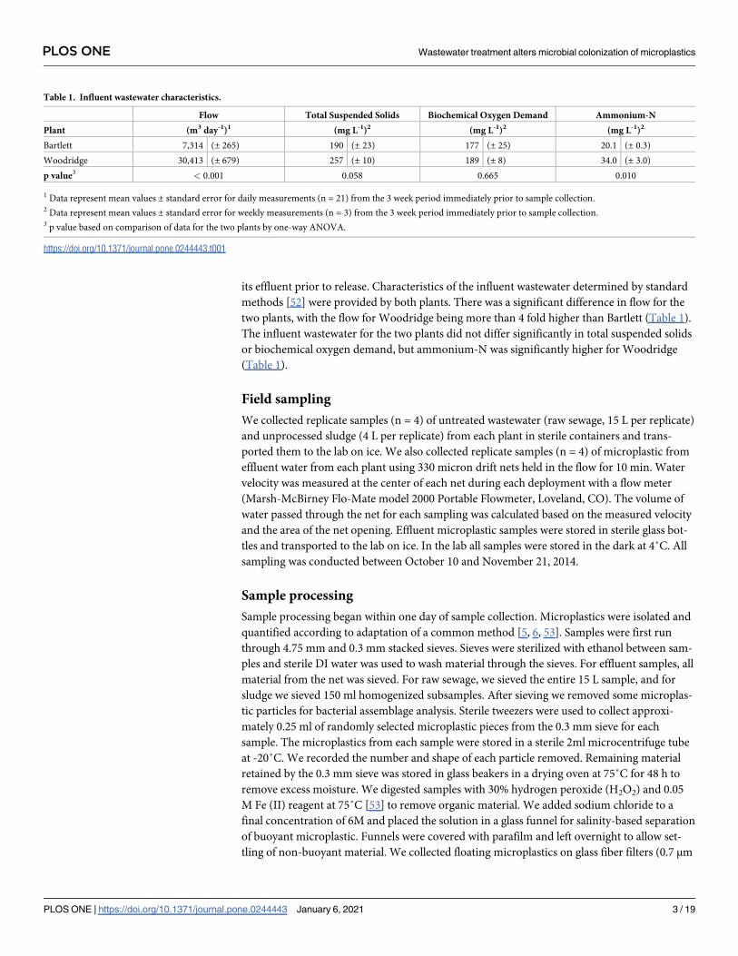
its effluent prior to release. Characteristics of the influent wastewater determined by standard
methods [52] were provided by both plants. There was a significant difference in flow for the
two plants, with the flow for Woodridge being more than 4 fold higher than Bartlett (Table 1).
The influent wastewater for the two plants did not differ significantly in total suspended solids
or biochemical oxygen demand, but ammonium-N was significantly higher for Woodridge
(Table 1).
Field sampling
We collected replicate samples (n = 4) of untreated wastewater (raw sewage, 15 L per replicate)
and unprocessed sludge (4 L per replicate) from each plant in sterile containers and trans-
ported them to the lab on ice. We also collected replicate samples (n = 4) of microplastic from
effluent water from each plant using 330 micron drift nets held in the flow for 10 min. Water
velocity was measured at the center of each net during each deployment with a flow meter
(Marsh-McBirney Flo-Mate model 2000 Portable Flowmeter, Loveland, CO). The volume of
water passed through the net for each sampling was calculated based on the measured velocity
and the area of the net opening. Effluent microplastic samples were stored in sterile glass bot-
tles and transported to the lab on ice. In the lab all samples were stored in the dark at 4˚C. All
sampling was conducted between October 10 and November 21, 2014.
Sample processing
Sample processing began within one day of sample collection. Microplastics were isolated and
quantified according to adaptation of a common method [5, 6, 53]. Samples were first run
through 4.75 mm and 0.3 mm stacked sieves. Sieves were sterilized with ethanol between sam-
ples and sterile DI water was used to wash material through the sieves. For effluent samples, all
material from the net was sieved. For raw sewage, we sieved the entire 15 L sample, and for
sludge we sieved 150 ml homogenized subsamples. After sieving we removed some microplas-
tic particles for bacterial assemblage analysis. Sterile tweezers were used to collect approxi-
mately 0.25 ml of randomly selected microplastic pieces from the 0.3 mm sieve for each
sample. The microplastics from each sample were stored in a sterile 2ml microcentrifuge tube
at -20˚C. We recorded the number and shape of each particle removed. Remaining material
retained by the 0.3 mm sieve was stored in glass beakers in a drying oven at 75˚C for 48 h to
remove excess moisture. We digested samples with 30% hydrogen peroxide (H2O2) and 0.05
M Fe (II) reagent at 75˚C [53] to remove organic material. We added sodium chloride to a
final concentration of 6M and placed the solution in a glass funnel for salinity-based separation
of buoyant microplastic. Funnels were covered with parafilm and left overnight to allow set-
tling of non-buoyant material. We collected floating microplastics on glass fiber filters (0.7 μm
Table 1. Influent wastewater characteristics.
Flow Total Suspended Solids Biochemical Oxygen Demand Ammonium-N
Plant (m3 day-1)1 (mg L-1)2 (mg L-1)2 (mg L-1)2
Bartlett 7,314 (± 265) 190 (± 23) 177 (± 25) 20.1 (± 0.3)
Woodridge 30,413 (± 679) 257 (± 10) 189 (± 8) 34.0 (± 3.0)
p value3 < 0.001 0.058 0.665 0.010
1 Data represent mean values ± standard error for daily measurements (n = 21) from the 3 week period immediately prior to sample collection.2 Data represent mean values ± standard error for weekly measurements (n = 3) from the 3 week period immediately prior to sample collection.3 p value based on comparison of data for the two plants by one-way ANOVA.
https://doi.org/10.1371/journal.pone.0244443.t001
PLOS ONE Wastewater treatment alters microbial colonization of microplastics
PLOS ONE | https://doi.org/10.1371/journal.pone.0244443 January 6, 2021 3 / 19
nominal pore size, Whatman, Inc. Piscataway, NJ, USA). Filters were placed in aluminum
pans, covered with aluminum foil, and dried at 60˚C.
Microplastic quantification
For each filter every piece of plastic was counted manually using a dissecting microscope. Each
of the microplastic particles was categorized as fragment, pellet, foam, film, or fiber. Microplas-
tics that were removed for bacterial community analysis prior to digestion were included in
counts. Our analyses also included controls (n = 5) to account for procedural and reagent con-
tamination, which consisted of deionized water placed in sample containers and digested in
parallel with our samples. Contamination in controls was low (an average of 4.67 fibers per
sample, and no contamination by fragments, foam, pellets, or film) and was accounted by sub-
tracting this value from all samples [6, 8].
Polymer analysis
Representative samples of each microplastic type from each treatment plant were analyzed by
pyrolysis gas chromatography mass spectrometry (py-GCMS; CDS Analytical 5200 pyroprobe
and Varian 3800 gas chromatograph). A sample was inserted into a quartz capillary tube with
quartz wool plugs, then loaded into the pyroprobe and heated to 750˚C for 90 s. GC injection
port and transfer line were constant at 325˚C (split ratio of 10:1). Restek Rtx-5MS capillary col-
umn (30 m x 0.25 mm x 0.25 μm df) with carrier gas helium (flow rate of 2.0 mL min-1) was
used for separation. The oven increased from 40˚C to 325˚C (heating rate of 10˚C min-1) and
was held for 20 min at 325˚C. GC was connected to Saturn 2000 ion trap mass spectrometer,
with heated transfer line (325˚C) and ion trap (220˚C), which collected all mass to charge ions
(m/z) from 35–550. We analyzed blanks between each sample to check for carry-over and
none occurred. Pyrograms were generated by averaging the mass spectra over the entire chro-
matogram, and then we searched the CDS Analytical 2013 pyrolysis library for the best match.
DNA extraction and sequencing
DNA was extracted from microplastic samples collected from raw sewage, sludge, and effluent
using MoBio Power Soil DNA kit. Partial 16S rRNA genes were amplified from DNA samples
using primers 515F and 806R, which amplify the V4 hypervariable region [54], and amplifica-
tion was confirmed by agarose gel electrophoresis. DNA extractions were also run without
samples as controls for kit contamination, and no amplification was observed for these kit con-
trols. Equimolar amounts of amplicons from each sample were sequenced in a 2 x 250 bp
paired-end format using an Illumina MiSeq [55] by DNA Services Facility, University of Illi-
nois at Chicago. Raw sequence data from this study can be downloaded from National Center
for Biotechnology Information (NCBI) Sequence Read Archive (SRA) with accession number
PRJNA638613.
Analysis of DNA sequence data
Sequences were processed using mothur v.1.42.3 [56] following the MiSeq Standard Operating
Procedure [57]. Briefly, paired reads were assembled and demultiplexed, and any sequences
with ambiguities or homopolymers > 8 bases were removed. Sequences were aligned using the
SILVA-compatible alignment database available within mothur. Chimeric sequences were
removed using VSEARCH [58]. Sequences were classified using the mothur-formatted version
of the RDP training set (v.9) and any unknown (i.e. not identified as bacterial), chloroplast,
mitochondrial, archaeal, and eukaryotic sequences were removed. Sequences were clustered
PLOS ONE Wastewater treatment alters microbial colonization of microplastics
PLOS ONE | https://doi.org/10.1371/journal.pone.0244443 January 6, 2021 4 / 19
into operational taxonomic units (OTUs) based on 97% sequence identity and were also
grouped into amplicon sequence variants (ASVs). OTUs were assigned to families and genera
by comparison to the RDP training set. In order to avoid biases associated with uneven num-
bers of sequences across samples, the entire dataset was randomly subsampled to 10,226
sequences per sample. For unidentified OTUs we selected a representative sequence, defined
as the sequence with minimum distance to other sequences within the OTU, and compared
these representative sequences to the NCBI 16S rRNA database using Megablast.
Statistical analyses
Microplastic concentrations and relative abundances of microplastic categories (fragment, pel-
let, foam, film, or fiber) were not normally distributed based on the Shapiro-Wilk test
(p<0.001). Therefore the effects of treatment plant (Bartlett and Woodridge) and sample type
(sewage, effluent, and sludge) on these data were analyzed by the non-parametric Kruskal-
Wallis Test followed by the Dwass-Steel-Chritchlow-Fligner Test for all pairwise comparisons
using Systat v13. For bacterial assemblage data, we quantified diversity for each sample based
on taxonomic richness (i.e. total number of OTUs observed) and the Shannon index [59].
Richness and diversity data were normally distributed based on the Shapiro-Wilk test
(p>0.05) so the effects of treatment plant and sample type on richness and diversity were ana-
lyzed by two-way ANOVA and Tukey’s HSD Test using Systat v13. Bacterial assemblages were
further compared by calculating dissimilarities for each pair of samples based on theta index
[60] for both OTUs and ASVs and visualizing the resulting dissimilarity matrices using non-
metric multidimensional scaling (nMDS). Statistical significance of differences in assemblages
between sample types based on theta index for both OTUs and ASVs was assessed by analysis
of molecular variance (AMOVA) [61], a nonparametric analog of traditional analysis of vari-
ance. Effect of sample type on relative abundance of the 25 most abundant bacterial families
was assessed by one-way ANOVA with Benjamini-Hochberg correction for false discovery
rate [62]. Metastats analysis [63] was used to identify OTUs that were differentially abundant
between sewage and effluent samples and between sewage and sludge samples, and ANOVA
with Benjamini-Hochberg correction for false discovery rate was used to assess significance of
differences in relative abundances of these OTUs between the sample types.
Results
Microplastics were present in all sample types (sewage, effluent, and sludge) from both plants
(Bartlett and Woodridge) (Table 2). The microplastics included fragments, pellets, foam, film,
and fibers (Fig 1), and were composed of several different polymers, including polyethylene,
polypropylene, and polystyrene (S1 Table). There was no significant effect of plant on micro-
plastic concentrations (p = 0.204) but there was a significant effect of sample type (p<0.001)
and there were significant differences in microplastic concentrations between each of the sam-
ple types (p<0.005 for all pairwise comparisons). When each sample type was compared indi-
vidually across plants, Bartlett had significantly higher microplastic concentrations than
Woodridge in sewage (p = 0.020) and effluent (p = 0.021), but the concentrations were not sig-
nificantly different between plants for sludge (p = 0.083; Table 2).
Both WWTPs had significantly higher microplastics in the incoming sewage relative to the
effluent (p = 0.002), corresponding to a reduction in microplastic concentration of more than
99% for both WWTPs (Table 2). High microplastic retention rates for both WWTPs resulted
in an average sludge microplastic concentration > 140,000 pieces/m3 (Table 2). While reten-
tion of microplastics was high, effluent microplastic concentrations corresponded to releases
PLOS ONE Wastewater treatment alters microbial colonization of microplastics
PLOS ONE | https://doi.org/10.1371/journal.pone.0244443 January 6, 2021 5 / 19

of approximately 9,470 microplastic pieces per day for Bartlett, and 273 microplastic pieces per
day for Woodridge (Table 2).
There were some differences in microplastic particle types among the sample types within
the WWTPs. Fibers were the most common microplastic type in incoming sewage for both
Woodridge and Bartlett, accounting for> 50% of sewage microplastic for both plants (Fig 1).
Relative abundance of fibers showed no difference between plants (p = 0.954), but was differ-
ent among sample types (p<0.006), with effluent having significantly fewer fibers than sewage
and sludge (p = 0.024 and 0.013, respectively). These data indicate that WWTPs selectively
retained microplastic fibers in sludge. Pellets showed the opposite trend, with no significant
differences between WWTPs (p = 0.246), but the relative abundance of pellets was different
among sample types (p = 0.014): higher in effluent (>20%) than in sewage (<10%) and sludge
(< 5%), indicating that pellets were not retained by WWTPs as effectively as fibers. There were
Table 2. Microplastic concentrations by plant and sample type.
Plant Sewage1 Effluent1 Sludge1 Retention Rate (%)2 Particles Released (#/day)3
Bartlett 1,987 (± 68) 1.295 (± 0.214) 335,533 (± 69,761) 99.93 9,471
Woodridge 1,161 (± 178) 0.009 (± 0.001) 140,533 (± 17,294) 99.99 273
1 Data represent mean values (n = 4) ± standard error in units of No. m-3.2 Retention rate equals the percentage of particles in sewage that were retained by the plant.3 The number of particles released per day was estimated based on the concentration of microplastics in the effluent and the average daily flow of the treatment plant.
https://doi.org/10.1371/journal.pone.0244443.t002
Fig 1. Relative abundance of microplastic particle types within each sample type (sewage, effluent, and sludge) from each of
two WWTPs (Bartlett and Woodridge). Each data point represents the mean value (n = 4).
https://doi.org/10.1371/journal.pone.0244443.g001
PLOS ONE Wastewater treatment alters microbial colonization of microplastics
PLOS ONE | https://doi.org/10.1371/journal.pone.0244443 January 6, 2021 6 / 19

no significant differences between WWTPs or sample types in relative abundance of other
microplastic types (fragments, foam, or film).
Bacterial assemblages attached to microplastic samples were analyzed by high-throughput
amplicon sequencing of 16 rRNA genes. Bacterial assemblage taxonomic richness (i.e., the
number of OTUs observed), was significantly different among sample types (sewage, effluent,
and sludge) (2-way ANOVA, p = 0.001) but there was no difference between WWTPs
(p = 0.547) and no significant interaction (p = 0.933) (Fig 2). Microplastics in sludge had sig-
nificantly higher bacterial taxonomic richness than microplastics in sewage or effluent, which
were not different from one another. In contrast to taxonomic richness, Shannon diversity of
bacterial assemblages attached to microplastics showed no significant differences among sam-
ple types (2-way ANOVA, p = 0.641) or between WWTPs (p = 0.456), and no significant inter-
action (p = 0.701) (S2 Table).
Multivariate statistical analyses (nMDS ordination and AMOVA) based on OTUs (97%
sequence identity) indicated no significant difference in composition of microplastic-attached
bacterial assemblages from the two WWTPs (p = 0.183), but there were significant differences
among the 3 sample types (p<0.001). Each sample type had a microplastic-attached bacterial
assemblage distinct from the others (sewage vs. effluent p = 0.003, sewage vs. sludge p = 0.007,
and effluent vs. sludge p = 0.011) (Fig 3). Pairwise comparisons of microplastic-attached bacte-
rial assemblages from each of the sample types (sewage, effluent, and sludge) across plants
showed a significant difference in bacterial assemblage between Bartlett effluent and Wood-
ridge effluent (p = 0.027) but no significant differences in bacterial assemblage by plant for
sewage (p = 0.189) or sludge (0.094). As expected, grouping of 16S rRNA gene sequences into
ASVs identified a higher total number of ASVs (30,946) compared to OTUs based on 97%
sequence identity (11,307), but beta diversity analyses based on ASVs produced results that
were equivalent to the analyses based on OTUs. Specifically, AMOVA of the ASV data showed
no significant difference in composition of microplastic-attached bacterial assemblages from
the two WWTPs (p = 0.183), but there were significant differences among the 3 sample types
(p<0.001) and significant pairwise differences between the sample types (sewage vs. effluent p
=<0.001, sewage vs. sludge p = 0.012, and effluent vs. sludge p = 0.009). In addition, nMDS
ordination based on the ASV data (S1 Fig) showed a highly similar pattern to that based on
OTUs (Fig 3).
Microplastic-attached bacterial assemblages included several families that contain patho-
genic bacterial taxa or taxa associated with the human gut microbiome (Fig 4), and sequences
from some of these families varied in relative abundance in different stages of wastewater treat-
ment. For example, Campylobacteraceae sequence abundance varied significantly with sample
type (p = 0.001), representing ~11% of bacterial sequences on sewage microplastic, but less
than 1% on effluent microplastic (p = 0.002) and approximately 1% on sludge microplastic
(p = 0.004). Within the family Campylobacteraceae 94% of the sequences were assigned to the
genus Arcobacter. Sequences from several other families, including Bacteroidaceae, Aeromona-daceae, and Lachnospiraceae, showed similar trends, being most abundant on sewage micro-
plastics and less abundant on effluent and sludge microplastics, but the effects of sample type
on relative abundances of these families were not significant (p = 0.188, 0.150, 0.079, respec-
tively). Abundance of Moraxellaceae sequences did not differ between sewage and effluent
microplastics (~12% and 11%, respectively), but was much lower on sludge microplastic
(<0.5%), although this difference was not statistically significant (p = 0.143). The genus Acine-tobacter accounted for 96% of sequences identified to the Moraxellaceae family, and Acineto-bacter sequences were similarly abundant on both sewage and effluent microplastics
(p = 0.909) but significantly less abundant on sludge microplastics (p = 0.009). Finally, the rela-
tive abundance of the family Sphingomonadaceae varied significantly with sample type
PLOS ONE Wastewater treatment alters microbial colonization of microplastics
PLOS ONE | https://doi.org/10.1371/journal.pone.0244443 January 6, 2021 7 / 19

(p<0.001) and was ~10 fold more abundant on effluent microplastics than sewage or sludge
microplastics. Within the family Sphingomonadaceae 69% of the sequences were assigned to
the genus Sphingomonas.Several bacterial OTUs had significant differences in relative abundance between sewage
and effluent microplastics (Table 3). Relative abundance of an unclassified Enterobacteriaceaegenus accounted for more than 11% of bacterial sequences on effluent microplastics but only
~1% on sewage microplastics. BLAST analysis indicated that the representative sequence from
this unclassified genus showed highest percent identity (>98%) to several species within the
genus Klebsiella, including Klebsiella pneumoniae and Klebsiella aerogenes. Similarly,
sequences from the genus Sphingomonas were greater than 10-fold more abundant and
sequences from the genus Pseudomonas were twice as abundant on effluent microplastics com-
pared to sewage microplastic. In contrast, several OTUs were significantly less abundant on
effluent microplastics, including Arcobacter, which decreased by a factor of 20, and an unclas-
sified Gammaproteobacteria genus, which decreased by a factor of 10. The representative
sequence from this unclassified Gammaproteobacteria genus fell within the family Aeromona-daceae and had a percent identity >96% for multiple species within the genus Aeromonas.
Several bacterial OTUs also had significant differences in relative abundance between sew-
age and sludge microplastics samples (Table 4). There were significantly lower relative abun-
dances of Acinetobacter (36-fold), Arcobacter (17-fold), Aeromonas (27-fold), an unclassified
Comamonadaceae genus (37-fold), and an unclassified Gammaproteobacteria genus (9-fold)
on sludge microplastics compared to sewage microplastics. The representative sequence from
the unclassified Comamonadaceae genus had a percent identity >98% for multiple species
within the genus Acidovorax, and the unclassified Gammaproteobacteria genus was the same
OTU discussed above that matched with the genus Aeromonas. Sludge microplastics also had
significantly higher relative abundances of another unclassified Comamonadaceae genus
Fig 2. Number of operational taxonomic units (based on 97% identity in 16S rRNA amplicons) observed within bacterial
communities attached to microplastic particles collected from three sample types (sewage, effluent, and sludge) from two
WWTPs (Bartlett and Woodridge). Each data point represents the mean value (n = 4) with error bars representing standard error.
https://doi.org/10.1371/journal.pone.0244443.g002
PLOS ONE Wastewater treatment alters microbial colonization of microplastics
PLOS ONE | https://doi.org/10.1371/journal.pone.0244443 January 6, 2021 8 / 19

(30-fold) and an unclassified Xanthomonadaceae genus (100-fold). The representative
sequence from this unclassified Comamonadaceae genus had a 99.6% identity to a strain from
the genus Ideonella.
Discussion
Microplastics were identified in untreated sewage, effluent water, and sludge from two
WWTPs treating primarily domestic wastewater. Patterns of particle abundance throughout
the WWTPs were consistent with previous assessments. Microplastic concentrations in
untreated sewage ranged from 800–2,000 particles/m3, which is comparable to published data
which ranged from 1,000 to> 50,000 particles/m3 [24–26]. Both WWTPs in this study
retained > 99% of influent microplastic, which is similar to [24, 25, 27] or higher than [26, 64]
values reported in prior studies, which ranged from 72% to 99%. The slight difference in reten-
tion between the two WWTPs suggests that sand filtration of effluent enhanced microplastic
retention, which has been reported previously [8, 65]. Effluent microplastic concentrations�1
particle/m3, which is on the low end of published values (for review see [66]), support the con-
clusion that both WWTPs were effective at removal of microplastics. Despite high retention
both WWTPs released microplastic in their effluent on the order of hundreds to thousands of
pieces per day. This finding agrees with a previous study which reported that both of these
WWTPs were point sources of microplastics to their receiving streams [8] and with studies
Fig 3. nMDS ordination of bacterial assemblage composition for biofilms attached to microplastic particles
collected from three sample types (sewage, effluent, and sludge) from two WWTPs (Bartlett and Woodridge).
Each point represents the bacterial assemblage from one individual sample. Bacterial assemblage analysis was based on
high-throughput amplicon sequencing of partial 16 rRNA genes, clustering sequences into OTUs (97% sequence
identity) and comparison of assemblages based on the theta index. Stress value of ordination = 0.2997.
https://doi.org/10.1371/journal.pone.0244443.g003
PLOS ONE Wastewater treatment alters microbial colonization of microplastics
PLOS ONE | https://doi.org/10.1371/journal.pone.0244443 January 6, 2021 9 / 19

that have detected microplastics in effluent from a range of WWTPs [28, 29]. However, the
very low concentration of microplastic in the effluent from both plants suggests the possibility
that daily microplastic discharge could be highly variable, so future studies should examine
temporal variability in effluent microplastic concentrations.
Changes in the relative abundance of microplastic particle types between sewage and efflu-
ent suggest selective retention. Microfibers were the most common type of microplastics in
influent sewage (>50%) and showed higher relative abundance in the sludge relative to the
effluent, suggesting net fiber retention. While similar selective retention of fibers was reported
in previous studies [25, 67], fibers are one of the most common forms of microplastics detected
in aquatic habitats [8, 23, 31]. One possible explanation for this discrepancy is that fibers may
be entering aquatic ecosystems through additional routes, including surface runoff, airborne
deposition, or direct release of untreated wastewater via combined sewer overflows or leaking
sewage infrastructure [31, 68]. In contrast to fibers, pellets were less well retained by WWTPs
and increased in relative abundance in effluent water. Previous studies have also reported that
Fig 4. Average relative abundance of the 25 most abundant bacterial families within microplastic-attached bacterial
assemblages from three sample types (sewage, effluent, and sludge). Each bar represents the mean (n = 8). Bacterial
families were identified based on high-throughput amplicon sequencing of partial 16 rRNA genes.
https://doi.org/10.1371/journal.pone.0244443.g004
PLOS ONE Wastewater treatment alters microbial colonization of microplastics
PLOS ONE | https://doi.org/10.1371/journal.pone.0244443 January 6, 2021 10 / 19

pellets are less well retained by WWTPs compared to other microplastic types [26]. The con-
trasting retention of fibers and pellets may reflect differences in their physical properties, such
as density, shape, buoyancy, or hydrophobicity, that might influence the tendency of these par-
ticles to settle or become trapped within flocs. Determination of properties that influence
microplastic retention in WWTPs would be a good topic for future controlled, manipulative
experiments.
Table 3. Relative abundances of bacterial OTUs with the largest differences in relative abundance between sewage microplastic and effluent microplastic.
Genus Sewage1 Effluent1 p-value2
Enterobacteriaceae_unclassified 1.15 (± 0.33)% 11.03 (± 4.54)% 0.007 �
Arcobacter 7.38 (± 2.05) 0.34 (± 0.16) 0.000 �
Pseudomonas 3.72 (± 1.67) 7.68 (± 1.42) 0.022 �
Gammaproteobacteria_unclassified 2.35 (± 0.61) 0.29 (± 0.11) 0.000 �
Uliginosibacterium 0.84 (± 0.46) 0.00 (± 0.00) 0.021 �
Aurantimonas 0.10 (± 0.04) 0.92 (± 0.53) 0.046
Propionivibrio 1.02 (± 0.23) 0.21 (± 0.12) 0.000 �
Sphingomonas 0.06 (± 0.04) 0.86 (± 0.30) 0.001 �
Pantoea 0.17 (± 0.08) 0.96 (± 0.29) 0.001 �
Zoogloea 0.94 (± 0.27) 0.16 (± 0.07) 0.001 �
Rhizobium 0.05 (± 0.02) 0.81 (± 0.34) 0.006 �
Faecalibacterium 0.67 (± 0.30) 0.02 (± 0.01) 0.007 �
Roseburia 0.70 (± 0.29) 0.08 (± 0.04) 0.008 �
Formivibrio 0.65 (± 0.37) 0.07 (± 0.05) 0.047
1 Data represent mean values (n = 8) ± standard error.2 Asterisks (�) indicate significant effects of sample type (sewage v. effluent) based on Benjamini-Hochberg correction.
https://doi.org/10.1371/journal.pone.0244443.t003
Table 4. Relative abundances of bacterial OTUs with the largest differences in relative abundance between sewage microplastic and sludge microplastic.
Genus Sewage1 Sludge1 p-value2
Acinetobacter 9.94 (± 4.45)% 0.28 (± 0.23)% 0.009 �
Arcobacter 7.38 (± 2.05) 0.43 (± 0.40) 0.000 �
Comamonadaceae_unclassified 0.15 (± 0.04) 5.44 (± 2.94) 0.030 �
Aeromonas 5.02 (± 2.45) 0.18 (± 0.11) 0.017 �
Comamonadaceae_unclassified 3.21 (± 1.66) 0.35 (± 0.09) 0.037 �
Gammaproteobacteria_unclassified 2.35 (± 0.61) 0.26 (± 0.25) 0.001 �
Flavobacterium 1.72 (± 0.88) 0.14 (± 0.13) 0.033 �
Arcobacter 1.94 (± 0.40) 0.57 (± 0.41) 0.004 �
Xanthomonadaceae_unclassified 0.01 (± 0.01) 1.00 (± 0.49) 0.015 �
Zoogloea 0.94 (± 0.27) 0.14 (± 0.10) 0.002 �
Propionivibrio 1.02 (± 0.23) 0.28 (± 0.24) 0.008 �
Trichococcus 0.64 (± 0.29) 0.05 (± 0.03) 0.015 �
Cloacibacterium 0.67 (± 0.16) 0.13 (± 0.08) 0.001 �
Bacteroides 0.52 (± 0.24) 0.06 (± 0.05) 0.021 �
Sulfurospirillum 0.57 (± 0.19) 0.10 (± 0.06) 0.005 �
1 Data represent mean values (n = 8) ± standard error.2 Asterisks (�) indicate significant effects of sample type (sewage v. sludge) based on Benjamini-Hochberg correction.
https://doi.org/10.1371/journal.pone.0244443.t004
PLOS ONE Wastewater treatment alters microbial colonization of microplastics
PLOS ONE | https://doi.org/10.1371/journal.pone.0244443 January 6, 2021 11 / 19

High retention of microplastic particles by both WWTPs resulted in high concentrations of
microplastics in sludge, similar to previous reports [67]. Concentrations measured in this
study for untreated sludge were on the order of hundreds of thousands of particles/m3, which
is approximately ten times lower than reported in a previous study of digested sludge [25],
with this discrepancy likely due to the reduction in sludge volume resulting from digestion
which would lead to increased microplastic concentration. Sewage sludge is commonly applied
to land in the U.S. [69], and there is no regulatory framework to assess or limit sludge micro-
plastics [70]. Thus, microplastics in sludge may be a concern for soil ecosystems [71]. In addi-
tion, surface runoff from applied biosolids could transport land-applied microplastic to
surface waters (i.e., agricultural streams), but this has not yet been assessed.
Microplastics in the environment encompass a wide diversity of sizes and materials [72],
some of which were not captured with our methods. Microplastics smaller than 300 microns
(including particles in the nanometer size range) were not included in the sampling techniques
used in our study, suggesting that our numbers may be an underestimate of the total micro-
plastics in wastewater. Furthermore, microplastics with a density > 1.3 g cm-3 (e.g. polyvinyl
chloride) were less likely to be captured with the salinity-based separation we used. This would
be especially relevant for the sludge, since high density particles would be most likely to have
settled into the sludge. Additional work is needed to consolidate an array of sampling methods
that could capture a broader range of the total microplastic particle assemblage in wastewater.
Microplastic particles from all samples were colonized by diverse microbial assemblages,
which were attached to the plastic surfaces as they were not washed off during microplastic
collection. Several of the most abundant taxa within these microplastic-attached bacterial
assemblages are known biofilm formers, including Pseudomonadaceae [73], Moraxellaceae[74], Enterobacteriaceae [75], and Comamonadaceae [76]. Our study represents the first analy-
sis of microplastic-attached bacterial assemblages within WWTPs and our results suggest that
the wastewater treatment process has strong effects on these assemblages. Species richness and
taxonomic composition differed across stages of wastewater treatment, and trends were con-
sistent for the two WWTPs included in this study. Specifically, there was no difference in spe-
cies richness of bacterial assemblages attached to microplastic in influent sewage versus
effluent water, but there was a significant difference in taxonomic composition of these
assemblages.
Several potentially pathogenic bacterial taxa were lower in abundance on effluent micro-
plastics than on influent sewage microplastics, suggesting that these taxa were negatively
affected by passage of microplastic through WWTPs. For example, sequences identified to the
genus Arcobacter and to its family Campylobacteraceae were significantly more abundant on
microplastics in influent sewage and less on effluent and sludge microplastic. The family Cam-pylobacteraceae and the genus Arcobacter include multiple taxa associated with human gastro-
intestinal infections such as gastroenteritis [77, 78]. The same pattern was observed for the
genus Aeromonas, which includes multiple species associated with human disease [79]. The
decreases in relative abundances of Campylobacteraceae, Arcobacter, and Aeromonas on efflu-
ent microplastics suggests that wastewater treatment could help to limit release of these poten-
tially pathogenic taxa to the environment. However, previous studies have identified these taxa
on microplastics in rivers receiving wastewater inputs [6, 32], including the rivers receiving
effluent from these specific WWTPs [8], so wastewater treatment may not completely remove
these taxa from microplastics.
Other bacterial taxa associated with human infections were abundant on microplastics in
both influent sewage and effluent water, suggesting that abundance of these taxa was not
altered by wastewater treatment. For example, the genus Acinetobacter and its family Moraxel-laceae were abundant on both sewage and effluent microplastics. Members of the genus
PLOS ONE Wastewater treatment alters microbial colonization of microplastics
PLOS ONE | https://doi.org/10.1371/journal.pone.0244443 January 6, 2021 12 / 19

Acinetobacter are involved in a wide range of human infectious diseases and are a common
cause of nosocomial infections [80]. Failure of wastewater treatment to reduce the abundance
of these taxa on microplastics could result in their release to the environment, and indeed
these taxa were identified previously on microplastics in rivers receiving wastewater inputs [6,
32], including the rivers receiving effluent from the specific WWTPs analyzed in this study [8].
Moreover, prior work demonstrated that Moraxellaceae sequences were still detected on
microplastics up to 2km downstream of a WWTP [32], suggesting that microplastics could be
a vector for transport of this potentially pathogenic taxon within rivers.
Finally, sequences from several bacterial genera were significantly more abundant on efflu-
ent microplastics compared to sewage microplastic, suggesting that these taxa increased in
abundance during wastewater treatment. Sequences from the family Enterobacteriaceae that
had a high percentage identity to Klebsiella pneumoniae and Klebsiella aerogenes increased by
a factor of 10. Both of these Klebsiella species are part of the normal human microbiome, but
they are also common causes of opportunistic and nosocomial infections [81]. Klebsiella pneu-moniae has also been shown to be capable of biodegrading polyethylene [82]. We are not
aware of previous studies reporting the presence of Klebsiella on microplastics, but it’s high
abundance on effluent microplastics is noteworthy due to its connections to humans and to
plastic breakdown. Sequences from the Pseudomonas genus were also significantly more abun-
dant on effluent microplastics compared to sewage microplastics. Species from the genus Pseu-domonas are common biofilm formers [83] and have been linked to breakdown of a wide
range of plastic polymers [84–89] as well as production of enzymes that contribute to plastic
biodegradation [90]. The abundance of Pseudomonas sequences on microplastics within
WWTP effluent agrees with previous detection of this genus on microplastic collected from
urban rivers, including the rivers receiving effluent from the WWTPs analyzed in this study
[6, 8, 32]. Moreover, prior work demonstrated that Pseudomonas increased in abundance on
microplastics in an urban river with distance from a WWTP [32]. Finally, sequences from the
genus Sphingomonas and its family Sphingomonadaceae were 10-fold more abundant on efflu-
ent microplastic compared to sewage microplastic. Bacteria from the genus Sphingomonas are
able to degrade various complex organic compounds, including polycyclic aromatic hydrocar-
bons [91], the plasticizer bisphenol A [92] and plastic monomers [93, 94], and they produce a
polysaccharide that enhances biofilm formation on plastic surfaces [95]. The observed
increases in Klebsiella, Pseudomonas, and Sphingomonas on microplastics in effluent suggests
some specific selection for these organisms on plastic within WWTPs, perhaps related to the
surface as a growth substrate and their capacity to break down plastic polymers.
The observed shifts in the composition of microplastic bacterial assemblages during passage
through the WWTPs could be driven by several factors, including interactions with microbes
within the plants and changes in environmental conditions at different stages of treatment.
For example, oxygen concentration can affect biofilm development [96] and the active pump-
ing of air into the aeration tank increases oxygen availability. Experimental investigations of
the effects of oxygen on microplastic biofilm development could provide insights into the
mechanisms underlying the observed shifts in microplastic bacterial assemblages during
wastewater treatment.
Bacterial assemblages attached to microplastics in sludge were also different than influent
sewage, suggesting that microplastic-attached bacterial assemblages changed during transfer of
microplastics from sewage to sludge. Bacterial assemblages on sludge microplastics showed
higher species richness, indicating that additional bacterial taxa colonized microplastics during
wastewater treatment. This colonization may enhance plastic retention, perhaps by increasing
sedimentation rates within the settling tank. Previous work has demonstrated that microbial
colonization of microplastics can increase settling of microplastic particles [43, 44] and
PLOS ONE Wastewater treatment alters microbial colonization of microplastics
PLOS ONE | https://doi.org/10.1371/journal.pone.0244443 January 6, 2021 13 / 19

microplastic retention in streams [46]. Bacterial colonization of microplastics during wastewa-
ter treatment could also enhance retention by incorporating microplastics into flocs. In our
study, sequences from the family Xanthomonadaceae and one unclassified Xanthomonadaceaegenus were dramatically more abundant on sludge microplastics than sewage microplastics. A
Xanthomonas strain was previously isolated from sewage sludge and was shown to have high
surface hydrophobicity and ability to co-aggregate with other wastewater bacteria, suggesting
that it might play a role in bioflocculation within WWTPs [97]. Microplastic surfaces are also
hydrophobic, which might enhance interaction with Xanthomonas and contribute to incorpo-
ration of microplastics into flocs. Sequences from an unclassified Comamonadaceae genus that
matched to the genus Ideonella were also dramatically more abundant on sludge microplastic
than sewage microplastic. An Ideonella species was recently reported to be capable of degrad-
ing and assimilating carbon from polyethylene [98], so the increase in this taxa in sludge may
be linked to plastic breakdown. The potential for WWTP bacteria to biodegrade microplastics
would be a good topic for future controlled, manipulative experiments. In addition, the com-
position of microbial communities within WWTPs can vary seasonally [99, 100] and by geo-
graphic location [101, 102], so analyzing microplastic microbiomes within WWTPs across
seasons and in more spatially separated locations would be valuable.
Supporting information
S1 Fig. nMDS ordination of bacterial assemblage composition for biofilms attached to
microplastic particles collected from three sample types (sewage, effluent, and sludge)
from two WWTPs (Bartlett and Woodridge). Each point represents the bacterial assemblage
from one individual sample. Bacterial assemblage analysis was based on high-throughput
amplicon sequencing of partial 16 rRNA genes, grouping sequences into ASVs, and compari-
son of assemblages based on the theta index. Stress value of ordination = 0.2997.
(PDF)
S1 Table. Identification of microplastic polymer types by PyGCMS.
(PDF)
S2 Table. Shannon diversity of microplastic attached bacterial assemblages.
(PDF)
Acknowledgments
The authors thank Greene Valley Wastewater Facility and Wastewater Treatment Plant of
Bartlett for providing access to their facilities and sharing data. We thank Lisa Kim for labora-
tory support.
Author Contributions
Conceptualization: John J. Kelly, Timothy J. Hoellein.
Formal analysis: John J. Kelly, Maxwell G. London, Miguel Rojas.
Funding acquisition: John J. Kelly, Timothy J. Hoellein.
Investigation: Maxwell G. London, Amanda R. McCormick, John W. Scott.
Methodology: Maxwell G. London, Amanda R. McCormick, Miguel Rojas, John W. Scott.
Project administration: John J. Kelly.
Supervision: John J. Kelly, Timothy J. Hoellein.
PLOS ONE Wastewater treatment alters microbial colonization of microplastics
PLOS ONE | https://doi.org/10.1371/journal.pone.0244443 January 6, 2021 14 / 19

Writing – original draft: John J. Kelly.
Writing – review & editing: Maxwell G. London, Amanda R. McCormick, Miguel Rojas, John
W. Scott, Timothy J. Hoellein.
References1. Moore CJ, Moore SL, Leecaster MK, Weisberg SB. A comparison of plastic and plankton in the North
Pacific central gyre. Mar Pollut Bull. 2001; 42: 1297–1300. https://doi.org/10.1016/s0025-326x(01)
00114-x PMID: 11827116
2. Moore CJ, Moore SL, Weisberg SB, Lattin GL, Zellers AF. A comparison of neustonic plastic and zoo-
plankton abundance in southern California’s coastal waters. Mar Pollut Bull. 2002; 44: 1035–1038.
https://doi.org/10.1016/s0025-326x(02)00150-9 PMID: 12474963
3. Law KL, Moret-Ferguson S, Maximenko NA, Proskurowski G, Peacock EE, Hafner J, et al. Plastic
accumulation in the North Atlantic subtropical gyre. Science. 2010; 329: 1185–1188. https://doi.org/
10.1126/science.1192321 PMID: 20724586
4. Doyle MJ, Watson W, Bowlin NM, Sheavly SB. Plastic particles in coastal pelagic ecosystems of the
Northeast Pacific ocean. Mar Environ Res. 2011; 71: 41–52. https://doi.org/10.1016/j.marenvres.
2010.10.001 PMID: 21093039
5. Eriksen M, Mason S, Wilson S, Box C, Zellers A, Edwards W, et al. Microplastic pollution in the surface
waters of the Laurentian Great Lakes. Mar Pollut Bull. 2013; 77: 177–182. https://doi.org/10.1016/j.
marpolbul.2013.10.007 PMID: 24449922
6. McCormick AR, Hoellein TJ, Mason SA, Schluep J, Kelly JJ. Microplastic is an abundant and distinct
microbial habitat in an urban river. Environ Sci Technol. 2014; 48: 11863–11871. https://doi.org/10.
1021/es503610r PMID: 25230146
7. Castañeda RA, Avlijas S, Simard MA, Ricciardi A. Microplastic pollution in St. Lawrence River sedi-
ments. Can J Fish Aquat Sci. 2014; 71: 1767–1771. https://doi.org/10.1139/cjfas-2014-0281
8. McCormick AR, Hoellein TJ, London MG, Hittie J, Scott JW, Kelly JJ. Microplastic in surface waters of
urban rivers: concentration, sources, and associated bacterial assemblages. Ecosphere. 2016; 7:
e01556.
9. Baldwin AK, Corsi SR, Mason SA. Plastic debris in 29 Great Lakes tributaries: relations to watershed
attributes and hydrology. Environ Sci Technol. 2016; 50: 10377–10385. https://doi.org/10.1021/acs.
est.6b02917 PMID: 27627676
10. Murray F, Cowie PR. Plastic contamination in the decapod crustacean Nephrops norvegicus (Lin-
naeus, 1758). Mar Pollut Bull. 2011; 62: 1207–1217. https://doi.org/10.1016/j.marpolbul.2011.03.032
PMID: 21497854
11. McNeish RE, Kim LH, Barrett HA, Mason SA, Kelly JJ, Hoellein TJ. Microplastic in riverine fish is con-
nected to species traits. Scientific reports. 2018; 8: 1–12.
12. Foley CJ, Feiner ZS, Malinich TD, Hook TO. A meta-analysis of the effects of exposure to microplas-
tics on fish and aquatic invertebrates. Sci Total Environ. 2018; 631: 550–559. https://doi.org/10.1016/j.
scitotenv.2018.03.046 PMID: 29529442
13. Browne MA, Dissanayake A, Galloway TS, Lowe DM, Thompson RC. Ingested microscopic plastic
translocates to the circulatory system of the mussel, Mytilus edulis (L.). Environ Sci Technol. 2008; 42:
5026–5031. https://doi.org/10.1021/es800249a PMID: 18678044
14. Rios LM, Jones PR, Moore C, Narayan UV. Quantitation of persistent organic pollutants adsorbed on
plastic debris from the Northern Pacific Gyre’s “eastern garbage patch”. Journal of Environmental
Monitoring. 2010; 12: 2226–2236. https://doi.org/10.1039/c0em00239a PMID: 21042605
15. Wright SL, Rowe D, Thompson RC, Galloway TS. Microplastic ingestion decreases energy reserves
in marine worms. Current Biology. 2013; 23: R1031–R1033. https://doi.org/10.1016/j.cub.2013.10.
068 PMID: 24309274
16. Wright SL, Thompson RC, Galloway TS. The physical impacts of microplastics on marine organisms:
a review. Environmental pollution. 2013; 178: 483–492. https://doi.org/10.1016/j.envpol.2013.02.031
PMID: 23545014
17. Rochman CM, Hoh E, Kurobe T, Teh SJ. Ingested plastic transfers hazardous chemicals to fish and
induces hepatic stress. Scientific reports. 2013; 3: 3263. https://doi.org/10.1038/srep03263 PMID:
24263561
18. Teuten EL, Saquing JM, Knappe DR, Barlaz MA, Jonsson S, Bjorn A, et al. Transport and release of
chemicals from plastics to the environment and to wildlife. Philos Trans R Soc Lond B Biol Sci. 2009;
364: 2027–2045. https://doi.org/10.1098/rstb.2008.0284 PMID: 19528054
PLOS ONE Wastewater treatment alters microbial colonization of microplastics
PLOS ONE | https://doi.org/10.1371/journal.pone.0244443 January 6, 2021 15 / 19

19. Watts AJ, Urbina MA, Corr S, Lewis C, Galloway TS. Ingestion of plastic microfibers by the crab Carci-
nus maenas and its effect on food consumption and energy balance. Environ Sci Technol. 2015; 49:
14597–14604. https://doi.org/10.1021/acs.est.5b04026 PMID: 26529464
20. Barboza LGA, Vieira LR, Branco V, Carvalho C, Guilhermino L. Microplastics increase mercury bio-
concentration in gills and bioaccumulation in the liver, and cause oxidative stress and damage in
Dicentrarchus labrax juveniles. Scientific reports. 2018; 8: 1–9.
21. Gregory MR. Plastic ‘scrubbers’ in hand cleansers: a further (and minor) source for marine pollution
identified. Mar Pollut Bull. 1996; 32: 867–871.
22. Fendall LS, Sewell MA. Contributing to marine pollution by washing your face: Microplastics in facial
cleansers. Mar Pollut Bull. 2009; 58: 1225–1228. https://doi.org/10.1016/j.marpolbul.2009.04.025
PMID: 19481226
23. Browne MA, Crump P, Niven SJ, Teuten E, Tonkin A, Galloway T, et al. Accumulation of microplastic
on shorelines woldwide: sources and sinks. Environ Sci Technol. 2011; 45: 9175–9179. https://doi.
org/10.1021/es201811s PMID: 21894925
24. Carr SA, Liu J, Tesoro AG. Transport and fate of microplastic particles in wastewater treatment plants.
Water Res. 2016; 91: 174–182. https://doi.org/10.1016/j.watres.2016.01.002 PMID: 26795302
25. Lares M, Ncibi MC, Sillanpaa M, Sillanpaa M. Occurrence, identification and removal of microplastic
particles and fibers in conventional activated sludge process and advanced MBR technology. Water
Res. 2018; 133: 236–246. https://doi.org/10.1016/j.watres.2018.01.049 PMID: 29407704
26. Bayo J, Olmos S, Lopez-Castellanos J. Microplastics in an urban wastewater treatment plant: The
influence of physicochemical parameters and environmental factors. Chemosphere. 2020; 238:
124593. https://doi.org/10.1016/j.chemosphere.2019.124593 PMID: 31446275
27. Murphy F, Ewins C, Carbonnier F, Quinn B. Wastewater treatment works (WwTW) as a source of
microplastics in the aquatic environment. Environ Sci Technol. 2016; 50: 5800–5808. https://doi.org/
10.1021/acs.est.5b05416 PMID: 27191224
28. Mason SA, Garneau D, Sutton R, Chu Y, Ehmann K, Barnes J, et al. Microplastic pollution is widely
detected in US municipal wastewater treatment plant effluent. Environmental Pollution. 2016; 218:
1045–1054. https://doi.org/10.1016/j.envpol.2016.08.056 PMID: 27574803
29. Mintenig SM, Int-Veen I, Loder MG, Primpke S, Gerdts G. Identification of microplastic in effluents of
waste water treatment plants using focal plane array-based micro-Fourier-transform infrared imaging.
Water Res. 2017; 108: 365–372. https://doi.org/10.1016/j.watres.2016.11.015 PMID: 27838027
30. Estahbanati S, Fahrenfeld NL. Influence of wastewater treatment plant discharges on microplastic
concentrations in surface water. Chemosphere. 2016; 162: 277–284. https://doi.org/10.1016/j.
chemosphere.2016.07.083 PMID: 27508863
31. Kay P, Hiscoe R, Moberley I, Bajic L, McKenna N. Wastewater treatment plants as a source of micro-
plastics in river catchments. Environmental Science and Pollution Research. 2018; 25: 20264–20267.
https://doi.org/10.1007/s11356-018-2070-7 PMID: 29881968
32. Hoellein TJ, McCormick AR, Hittie J, London MG, Scott JW, Kelly JJ. Longitudinal patterns of micro-
plastic concentration and bacterial assemblages in surface and benthic habitats of an urban river.
Freshwater Science. 2017; 36: 491–507.
33. Zettler ER, Mincer TJ, Amaral-Zettler LA. Life in the “plastisphere”: microbial communities on plastic
marine debris. Environ Sci Technol. 2013; 47: 7137–7146. https://doi.org/10.1021/es401288x PMID:
23745679
34. Carson HS, Nerheim MS, Carroll KA, Eriksen M. The plastic-associated microorganisms of the North
Pacific Gyre. Mar Pollut Bull. 2013; 75: 126–132. https://doi.org/10.1016/j.marpolbul.2013.07.054
PMID: 23993070
35. Harrison JP, Sapp M, Schratzberger M, Osborn AM. Interactions between microorganisms and
marine microplastics: a call for research. Mar Technol Soc J. 2011; 45: 12–20.
36. Reisser J, Shaw J, Hallegraeff G, Proietti M, Barnes DK, Thums M, et al. Millimeter-sized marine plas-
tics: a new pelagic habitat for microorganisms and invertebrates. PloS one. 2014; 9. https://doi.org/10.
1371/journal.pone.0100289 PMID: 24941218
37. Frère L, Maignien L, Chalopin M, Huvet A, Rinnert E, Morrison H, et al. Microplastic bacterial commu-
nities in the Bay of Brest: Influence of polymer type and size. Environmental Pollution. 2018; 242:
614–625. https://doi.org/10.1016/j.envpol.2018.07.023 PMID: 30014939
38. Davey ME, O’toole GA. Microbial biofilms: from ecology to molecular genetics. Microbiology and
molecular biology reviews. 2000; 64: 847–867. https://doi.org/10.1128/mmbr.64.4.847-867.2000
PMID: 11104821
39. O’Toole G, Kaplan HB, Kolter R. Biofilm formation as microbial development. Annual Reviews in
Microbiology. 2000; 54: 49–79. https://doi.org/10.1146/annurev.micro.54.1.49 PMID: 11018124
PLOS ONE Wastewater treatment alters microbial colonization of microplastics
PLOS ONE | https://doi.org/10.1371/journal.pone.0244443 January 6, 2021 16 / 19

40. Flemming H. Biofilms and environmental protection. Water Science and Technology. 1993; 27: 1–10.
41. Gilbert P, Das J, Foley I. Biofilm susceptibility to antimicrobials. Adv Dent Res. 1997; 11: 160–167.
https://doi.org/10.1177/08959374970110010701 PMID: 9524452
42. Balcazar JL, Subirats J, Borrego CM. The role of biofilms as environmental reservoirs of antibiotic
resistance. Frontiers in microbiology. 2015; 6: 1216. https://doi.org/10.3389/fmicb.2015.01216 PMID:
26583011
43. Fazey FM, Ryan PG. Biofouling on buoyant marine plastics: An experimental study into the effect of
size on surface longevity. Environmental Pollution. 2016; 210: 354–360. https://doi.org/10.1016/j.
envpol.2016.01.026 PMID: 26803792
44. Lagarde F, Olivier O, Zanella M, Daniel P, Hiard S, Caruso A. Microplastic interactions with freshwater
microalgae: hetero-aggregation and changes in plastic density appear strongly dependent on polymer
type. Environmental pollution. 2016; 215: 331–339. https://doi.org/10.1016/j.envpol.2016.05.006
PMID: 27236494
45. Kooi M, Nes EHv, Scheffer M, Koelmans AA. Ups and downs in the ocean: effects of biofouling on ver-
tical transport of microplastics. Environ Sci Technol. 2017; 51: 7963–7971. https://doi.org/10.1021/
acs.est.6b04702 PMID: 28613852
46. Hoellein TJ, Shogren AJ, Tank JL, Risteca P, Kelly JJ. Microplastic deposition velocity in streams fol-
lows patterns for naturally occurring allochthonous particles. Scientific Reports. 2019; 9: 1–11.
47. Kirstein IV, Kirmizi S, Wichels A, Garin-Fernandez A, Erler R, Martin L, et al. Dangerous hitchhikers?
Evidence for potentially pathogenic Vibrio spp. on microplastic particles. Mar Environ Res. 2016; 120:
1–8. https://doi.org/10.1016/j.marenvres.2016.07.004 PMID: 27411093
48. Harrison JP, Hoellein TJ, Sapp M, Tagg AS, Ju-Nam Y, Ojeda JJ. Microplastic-associated biofilms: a
comparison of freshwater and marine environments. In: Wagner M, Lambert S, editors. Freshwater
microplastics. Cham, Switzerland: Springer; 2018. pp. 181–201.
49. Delacuvellerie A, Cyriaque V, Gobert S, Benali S, Wattiez R. The plastisphere in marine ecosystem
hosts potential specific microbial degraders including Alcanivorax borkumensis as a key player for the
low-density polyethylene degradation. J Hazard Mater. 2019; 380: 120899. https://doi.org/10.1016/j.
jhazmat.2019.120899 PMID: 31326835
50. Fang H, Cai L, Yu Y, Zhang T. Metagenomic analysis reveals the prevalence of biodegradation genes
for organic pollutants in activated sludge. Bioresour Technol. 2013; 129: 209–218. https://doi.org/10.
1016/j.biortech.2012.11.054 PMID: 23247148
51. O’Grady DP, Howard PH, Werner AF. Activated sludge biodegradation of 12 commercial phthalate
esters. Appl.Environ.Microbiol. 1985; 49: 443–445. https://doi.org/10.1128/AEM.49.2.443-445.1985
PMID: 16346734
52. American Public Health Association. Standard methods for the examination of water and wastewater.
American Public Health Association (APHA): Washington, DC, USA. 2005.
53. Masura J, Baker J, Foster G, Arthur C. Laboratory methods for the analysis of microplastics in the
marine environment: recommendations for quantifying synthetic particles in waters and sediments.
2015: 1–39.
54. Caporaso JG, Lauber CL, Walters WA, Berg-Lyons D, Lozupone CA, Turnbaugh PJ, et al. Global pat-
terns of 16S rRNA diversity at a depth of millions of sequences per sample. Proceedings of the
national academy of sciences. 2011; 108: 4516–4522.
55. Caporaso JG, Lauber CL, Walters WA, Berg-Lyons D, Huntley J, Fierer N, et al. Ultra-high-throughput
microbial community analysis on the Illumina HiSeq and MiSeq platforms. ISME J. 2012; 6: 1621–
1624. https://doi.org/10.1038/ismej.2012.8 PMID: 22402401
56. Schloss PD, Westcott SL, Ryabin T, Hall JR, Hartmann M, Hollister EB, et al. Introducing mothur:
Open-Source, Platform-Independent, Community-Supported Software for Describing and Comparing
Microbial Communities. Appl Environ Microbiol. 2009; 75: 7537–7541. https://doi.org/10.1128/AEM.
01541-09 PMID: 19801464
57. Kozich JJ, Westcott SL, Baxter NT, Highlander SK, Schloss PD. Development of a dual-index
sequencing strategy and curation pipeline for analyzing amplicon sequence data on the MiSeq Illu-
mina sequencing platform. Appl Environ Microbiol. 2013; 79: 5112–5120. https://doi.org/10.1128/
AEM.01043-13 PMID: 23793624
58. Rognes T, Flouri T, Nichols B, Quince C, Mahe F. VSEARCH: a versatile open source tool for metage-
nomics. PeerJ. 2016; 4: e2584. https://doi.org/10.7717/peerj.2584 PMID: 27781170
59. Shannon CE. A mathematical theory of communication. ACM SIGMOBILE Mobile Computing and
Communications Review. 2001; 5: 3–55.
60. Yue JC, Clayton MK. A similarity measure based on species proportions. Communications in Statis-
tics-Theory and Methods. 2005; 34: 2123–2131.
PLOS ONE Wastewater treatment alters microbial colonization of microplastics
PLOS ONE | https://doi.org/10.1371/journal.pone.0244443 January 6, 2021 17 / 19
61. Excoffier L, Smouse PE, Quattro JM. Analysis of molecular variance inferred from metric distances
among DNA haplotypes: application to human mitochondrial DNA restriction data. Genetics. 1992;
131: 479–491. PMID: 1644282
62. Benjamini Y, Hochberg Y. Controlling the false discovery rate: a practical and powerful approach to
multiple testing. Journal of the Royal statistical society: series B (Methodological). 1995; 57: 289–300.
63. White JR, Nagarajan N, Pop M. Statistical methods for detecting differentially abundant features in
clinical metagenomic samples. PLoS computational biology. 2009; 5: e1000352. https://doi.org/10.
1371/journal.pcbi.1000352 PMID: 19360128
64. Leslie HA, Brandsma SH, Van Velzen M, Vethaak AD. Microplastics en route: Field measurements in
the Dutch river delta and Amsterdam canals, wastewater treatment plants, North Sea sediments and
biota. Environ Int. 2017; 101: 133–142. https://doi.org/10.1016/j.envint.2017.01.018 PMID: 28143645
65. Michielssen MR, Michielssen ER, Ni J, Duhaime MB. Fate of microplastics and other small anthropo-
genic litter (SAL) in wastewater treatment plants depends on unit processes employed. Environmental
Science: Water Research & Technology. 2016; 2: 1064–1073.
66. Habib RZ, Thiemann T, Al Kendi R. Microplastics and Wastewater Treatment Plants—A Review. Jour-
nal of Water Resource and Protection. 2020; 12: 1.
67. Mahon AM, O’Connell B, Healy MG, O’Connor I, Officer R, Nash R, et al. Microplastics in sewage
sludge: effects of treatment. Environ Sci Technol. 2017; 51: 810–818. https://doi.org/10.1021/acs.est.
6b04048 PMID: 27936648
68. Zhang Y, Kang S, Allen S, Allen D, Gao T, Sillanpaa M. Atmospheric microplastics: A review on cur-
rent status and perspectives. Earth-Sci Rev. 2020: 103118.
69. Lu Q, He ZL, Stoffella PJ. Land application of biosolids in the USA: A review. Applied and Environmen-
tal Soil Science. 2012; 2012.
70. Harrison EZ, McBride MB, Bouldin D. Land application of sewage sludges: an appraisal of the US reg-
ulations. International Journal of Environment and Pollution. 1999; 11: 1–36.
71. Nizzetto L, Futter M, Langaas S. Are agricultural soils dumps for microplastics of urban origin? Envi-
ronmental Science & Technology. 2016; 50: 10777–10779. https://doi.org/10.1021/acs.est.6b04140
PMID: 27682621
72. Barrows AP, Neumann CA, Berger ML, Shaw SD. Grab vs. neuston tow net: a microplastic sampling
performance comparison and possible advances in the field. Analytical Methods. 2017; 9: 1446–1453.
73. Ghafoor A, Hay ID, Rehm BH. Role of exopolysaccharides in Pseudomonas aeruginosa biofilm forma-
tion and architecture. Appl Environ Microbiol. 2011; 77: 5238–5246. https://doi.org/10.1128/AEM.
00637-11 PMID: 21666010
74. Moen B, Røssvoll E, Måge I, Møretrø T, Langsrud S. Microbiota formed on attached stainless steel
coupons correlates with the natural biofilm of the sink surface in domestic kitchens. Can J Microbiol.
2016; 62: 148–160. https://doi.org/10.1139/cjm-2015-0562 PMID: 26758935
75. Burmølle M, Bahl MI, Jensen LB, Sørensen SJ, Hansen LH. Type 3 fimbriae, encoded by the conjuga-
tive plasmid pOLA52, enhance biofilm formation and transfer frequencies in Enterobacteriaceae
strains. Microbiology. 2008; 154: 187–195. https://doi.org/10.1099/mic.0.2007/010454-0 PMID:
18174137
76. Li L, Jeon Y, Lee S, Ryu H, Santo Domingo JW, Seo Y. Dynamics of the physiochemical and commu-
nity structures of biofilms under the influence of algal organic matter and humic substances. Water
Res. 2019; 158: 136–145. https://doi.org/10.1016/j.watres.2019.04.014 PMID: 31026675
77. Engberg J, On SL, Harrington CS, Gerner-Smidt P. Prevalence of Campylobacter, Arcobacter, Helico-
bacter, Andsutterella spp. in human fecal samples as estimated by a reevaluation of isolation methods
for campylobacters. J Clin Microbiol. 2000; 38: 286–291. PMID: 10618103
78. On SL. Taxonomy of Campylobacter, Arcobacter, Helicobacter and related bacteria: current status,
future prospects and immediate concerns. J Appl Microbiol. 2001; 90: 1S–15S. https://doi.org/10.
1046/j.1365-2672.2001.01349.x PMID: 11422556
79. Janda JM, Abbott SL. The genus Aeromonas: taxonomy, pathogenicity, and infection. Clin Microbiol
Rev. 2010; 23: 35–73. https://doi.org/10.1128/CMR.00039-09 PMID: 20065325
80. Joly-Guillou M. Clinical impact and pathogenicity of Acinetobacter. Clinical microbiology and infection.
2005; 11: 868–873. https://doi.org/10.1111/j.1469-0691.2005.01227.x PMID: 16216100
81. Podschun R, Ullmann U. Klebsiella spp. as nosocomial pathogens: epidemiology, taxonomy, typing
methods, and pathogenicity factors. Clin Microbiol Rev. 1998; 11: 589–603. PMID: 9767057
82. Awasthi S, Srivastava P, Singh P, Tiwary D, Mishra PK. Biodegradation of thermally treated high-den-
sity polyethylene (HDPE) by Klebsiella pneumoniae CH001. 3 Biotech. 2017; 7: 332. https://doi.org/
10.1007/s13205-017-0959-3 PMID: 28955629
PLOS ONE Wastewater treatment alters microbial colonization of microplastics
PLOS ONE | https://doi.org/10.1371/journal.pone.0244443 January 6, 2021 18 / 19

83. Mann EE, Wozniak DJ. Pseudomonas biofilm matrix composition and niche biology. FEMS Microbiol
Rev. 2012; 36: 893–916. https://doi.org/10.1111/j.1574-6976.2011.00322.x PMID: 22212072
84. Balasubramanian V, Natarajan K, Hemambika B, Ramesh N, Sumathi CS, Kottaimuthu R, et al. High-
density polyethylene (HDPE)-degrading potential bacteria from marine ecosystem of Gulf of Mannar,
India. Lett Appl Microbiol. 2010; 51: 205–211. https://doi.org/10.1111/j.1472-765X.2010.02883.x
PMID: 20586938
85. Tribedi P, Gupta AD, Sil AK. Adaptation of Pseudomonas sp. AKS2 in biofilm on low-density polyethyl-
ene surface: an effective strategy for efficient survival and polymer degradation. Bioresources and Bio-
processing. 2015; 2: 14.
86. Kathiresan K. Polythene and plastics-degrading microbes from the mangrove soil. Rev Biol Trop.
2003; 51: 629–633. PMID: 15162769
87. Cacciari I, Quatrini P, Zirletta G, Mincione E, Vinciguerra V, Lupattelli P, et al. Isotactic polypropylene
biodegradation by a microbial community: physicochemical characterization of metabolites produced.
Appl.Environ.Microbiol. 1993; 59: 3695–3700. https://doi.org/10.1128/AEM.59.11.3695-3700.1993
PMID: 8285678
88. Arkatkar A, Juwarkar AA, Bhaduri S, Uppara PV, Doble M. Growth of Pseudomonas and Bacillus bio-
films on pretreated polypropylene surface. Int Biodeterior Biodegrad. 2010; 64: 530–536.
89. Shimao M. Biodegradation of plastics. Curr Opin Biotechnol. 2001; 12: 242–247. https://doi.org/10.
1016/s0958-1669(00)00206-8 PMID: 11404101
90. Bhardwaj H, Gupta R, Tiwari A. Communities of microbial enzymes associated with biodegradation of
plastics. Journal of Polymers and the Environment. 2013; 21: 575–579.
91. Bastiaens L, Springael D, Wattiau P, Harms H, deWachter R, Verachtert H, et al. Isolation of adherent
polycyclic aromatic hydrocarbon (PAH)-degrading bacteria using PAH-sorbing carriers. Appl Environ
Microbiol. 2000; 66: 1834–1843. https://doi.org/10.1128/aem.66.5.1834-1843.2000 PMID: 10788347
92. Oshiman K, Tsutsumi Y, Nishida T, Matsumura Y. Isolation and characterization of a novel bacterium,
Sphingomonas bisphenolicum strain AO1, that degrades bisphenol A. Biodegradation. 2007; 18: 247–
255. https://doi.org/10.1007/s10532-006-9059-5 PMID: 16821103
93. Arnold M, Reittu A, von Wright A, Martikainen PJ, Suihko M. Bacterial degradation of styrene in waste
gases using a peat filter. Appl Microbiol Biotechnol. 1997; 48: 738–744. https://doi.org/10.1007/
s002530051126 PMID: 9457801
94. Li J, Gu J, Yao J. Degradation of dimethyl terephthalate by Pasteurella multocida Sa and Sphingomo-
nas paucimobilis Sy isolated from mangrove sediment. Int Biodeterior Biodegrad. 2005; 56: 158–165.
95. Czieborowski M, Hubenthal A, Poehlein A, Vogt I, Philipp B. Genetic and physiological analysis of bio-
film formation on different plastic surfaces by Sphingomonas sp. strain S2M10 reveals an essential
function of sphingan biosynthesis. Microbiology. 2020: micro000961. https://doi.org/10.1099/mic.0.
000961 PMID: 32762802
96. Goller CC, Romeo T. Environmental influences on biofilm development. In: Anonymous Bacterial Bio-
films.: Springer; 2008. pp. 37–66.
97. Malik A, Kakii K. Pair-dependent co-aggregation behavior of non-flocculating sludge bacteria. Biotech-
nol Lett. 2003; 25: 981–986. https://doi.org/10.1023/a:1024009511113 PMID: 12889835
98. Yoshida S, Hiraga K, Takehana T, Taniguchi I, Yamaji H, Maeda Y, et al. A bacterium that degrades
and assimilates poly (ethylene terephthalate). Science. 2016; 351: 1196–1199. https://doi.org/10.
1126/science.aad6359 PMID: 26965627
99. Ju F, Guo F, Ye L, Xia Y, Zhang T. Metagenomic analysis on seasonal microbial variations of activated
sludge from a full-scale wastewater treatment plant over 4 years. Environmental microbiology reports.
2014; 6: 80–89. https://doi.org/10.1111/1758-2229.12110 PMID: 24596265
100. Zhang B, Yu Q, Yan G, Zhu H, Yang XuX, Zhu L. Seasonal bacterial community succession in four
typical wastewater treatment plants: correlations between core microbes and process performance.
Scientific reports. 2018; 8: 1–11.
101. Wu L, Ning D, Zhang B, Li Y, Zhang P, Shan X, et al. Global diversity and biogeography of bacterial
communities in wastewater treatment plants. Nature microbiology. 2019; 4: 1183–1195. https://doi.
org/10.1038/s41564-019-0426-5 PMID: 31086312
102. Zhang B, Ning D, Van Nostrand JD, Sun C, Yang Y, Zhou J, et al. Biogeography and Assembly of
Microbial Communities in Wastewater Treatment Plants in China. Environ Sci Technol. 2020; 54:
5884–5892. https://doi.org/10.1021/acs.est.9b07950 PMID: 32259441
PLOS ONE Wastewater treatment alters microbial colonization of microplastics
PLOS ONE | https://doi.org/10.1371/journal.pone.0244443 January 6, 2021 19 / 19