water as an active constituent in cell biology - college of computer
TRANSCRIPT

Water as an Active Constituent in Cell Biology
Philip Ball*
Nature, 4-6 Crinan Street, London N1 9XW, U.K.
Received December 28, 2006
Contents1. Introduction 12. Water as a Liquid and Solvent 2
2.1. Water Structure 22.2. Small-Molecule Solutes: Hydrophiles 42.3. Small-Molecule Solutes: Hydrophobes 52.4. Large Hydrophobic Solutes and Surfaces 72.5. The Influence of Ions: Structure-Making and
Structure-Breaking9
2.6. Long-Range Hydrophobic Interactions and theRole of Bubbles
11
2.7. Hydrophilic Surfaces 133. The Aqueous Environment of the Cell 144. Protein Hydration: Nonspecific Effects 15
4.1. The Hydration Shell 154.2. Dynamics, Cooperativity, and the Glass
Transition17
5. Protein Hydration: Specific Roles of Water inStructure and Function
19
5.1. Secondary Structure 195.2. Protein−Protein Interactions 195.3. Mediation of Ligand Binding 205.4. Functional Tuning 225.5. Allostery 235.6. Hydrophobic Cavities 235.7. Electron Transfer 235.8. Involvement of Bound Water in Catalytic
Action24
5.9. Proton Wires 245.10. Function of Protein Channels 26
6. Water and Nucleic Acids 277. Conclusions 308. Acknowledgments 309. Note Added in Proof 30
10. Note Added after ASAP Publication 3011. References 30
1. IntroductionWhen Szent-Gyo¨rgyi called water the “matrix of life”,1
he was echoing an old sentiment. Paracelsus in the 16thcentury said that “water was the matrix of the world and ofall its creatures.”2 But Paracelsus’s notion of a matrixsanactive substance imbued with fecund, life-giving propertiesswas quite different from the picture that, until very recently,molecular biologists have tended to hold of water’s role inthe chemistry of life. Although acknowledging that liquid
water has some unusual and important physical and chemicalpropertiessits potency as a solvent, its ability to formhydrogen bonds, its amphoteric naturesbiologists have re-garded it essentially as the backdrop on which life’s molec-ular components are arrayed. It used to be common practice,for example, to perform computer simulations of biomol-ecules in a vacuum. Partly this was because the computa-tional intensity of simulating a polypeptide chain waschallenging even without accounting for solvent moleculestoo, but it also reflected the prevailing notion that water doeslittle more than temper or moderate the basic physicochem-ical interactions responsible for molecular biology. WhatGerstein and Levitt said 9 years ago remains true today:“When scientists publish models of biological molecules injournals, they usually draw their models in bright colors andplace them against a plain, black background”.3
Curiously, this neglect of water as an active componentof the cell went hand in hand with the assumption that lifecould not exist without it. That was basically an empiricalconclusion derived from our experience of life on Earth:environments without liquid water cannot sustain life, andspecial strategies are needed to cope with situations in which,because of extremes of either heat or cold, the liquid isscarce.4-6 The recent confirmation that there is at least oneworld rich in organic molecules on which rivers and perhapsshallow seas or bogs are filled with nonaqueous fluidstheliquid hydrocarbons of Titan7smight now bring some focus,even urgency, to the question of whether water is indeed a* E-mail: [email protected].
Philip Ball is a science writer and a consultant editor for Nature, wherehe worked as an editor for physical sciences for more than 10 years. Heholds a Ph.D. in physics from the University of Bristol, where he workedon the statistical mechanics of phase transitions in the liquid state. Hisbook H2O: A Biography of Water (Weidenfeld & Nicolson, 1999) was asurvey of the current state of knowledge about the behavior of water insituations ranging from planetary geomorphology to cell biology. Hefrequently writes about aspects of water science for both the popular andthe technical media.
10.1021/cr068037a CCC: $71.00 © xxxx American Chemical SocietyPAGE EST: 34.6Published on Web 12/21/2007

unique and universal matrix of life, or whether on the con-trary it is just the one that happens to pertain on our planet.
Fundamental to that question is the role that water playsin sustaining the biochemistry of the cell. It has becomeincreasingly clear over the past 2 decades or so that water isnot simply “life’s solvent” but is indeed a matrix more akinto the one Paracelsus envisaged: a substance that activelyengages and interacts with biomolecules in complex, subtle,and essential ways. There is now good reason to regard the“active volume” of molecules such as proteins as extendingbeyond their formal boundary (the van der Waals surface,say), by virtue of the way they shape and manipulate theshell of water that surrounds them. Moreover, the structureand dynamics of this hydration shell seem to feed back ontothose aspects of the proteins themselves so that biologicalfunction depends on a delicate interplay between what wehave previously regarded as distinct entities: the moleculeand its environment. Many proteins make use of bound watermolecules as functional units, like snap-on tools, to mediateinteractions with other proteins or with substrate moleculesor to transport protons rapidly to locations buried inside theprotein.
Here I review the case for considering water to be aversatile, adaptive component of the cell that engages in awide range of biomolecular interactions. In order to providesome basis for assessing water’s often-alleged “uniqueness”to life, however, I shall try to highlight throughout this paperthe distinctions between generic and specific behaviors ofbiological water. That is to say, some of its roles andproperties may be expected from any small-molecule liquidsolvent. Others depend on water’s hydrogen-bonding capac-ity, but not in a way that could not obviously be fulfilledalso by other hydrogen-bonded liquids. But some of water’sbiochemical functions do indeed seem to be quite unique tothe H2O molecule. From an astrobiological perspective, thequestion is then whether we can regard these latter roles asoptional or essentialfor any form of life to be tenable.
2. Water as a Liquid and Solvent
2.1. Water StructureWater is not like other liquids, but neither is it wholly
different. The structure of a so-called simple liquidsone inwhich the molecules can be represented as particles thatinteract via some spherically symmetric potential functionscan be depicted in terms of a radial distribution function (rdf)g(r), which is related to the liquid densityF(r) around aparticle atr ) 0: F(r) ) Fbg(r), whereFb is the bulk density.The rdf for a simple liquid interacting through the Lennard-Jones potential (V ∼ σ/r12 - σ/r6, whereσ is the moleculardiameter) is not very different from that of a “hard-sphere”fluid, in which the particles experience no intermolecularforce until they touch, whereupon they act as infinitely hardspheres (V ) 0 (r > σ); V ) ∞ (r e σ)). In both cases, therdf is oscillatory, with a prominent first peak aroundr ) σand smaller subsequent peaks at separations close tor ) nσthat decay rapidly until the density reaches the bulk averagevalue (Figure 1). This implies that the oscillatory densityprofile is dominated by the steep repulsive core of theparticles and is related to the geometric aspects of a dense,random packing of particles. These repulsive interactionscreate short-ranged ordering in the liquid that the inclusionof an attractive potential (without which there is no liquid-gas transition) modifies only slightly.
Liquid water also has an oscillatory rdf. In this case asingle functiong(r) will not suffice to fully characterize theliquid structure, because there are two types of atom in themolecules: H and O. So one must define a series of partialrdfs gXY(r), which denote the probability of finding an atomY a distancer from the center of atomX. For example,gOO(r) indicates the radial distribution of oxygen atoms inother H2O molecules around any given molecule. The partialrdfs for water, determined by neutron scattering,8 are shownin Figure 2.
The structure is more complex than the oscillatory profilewith decaying amplitude found for simple fluids such as hard-sphere or Lennard-Jones systems or for real liquid argon;but the basic features look qualitatively similar. This localstructuring of liquid water has, however, a quite differentorigin. Whereas hard-sphere repulsion controls the short-range order of simple liquids, the structure seen in Figure 2is primarily due to the attractive interactions between watermolecules: the hydrogen bonds. These generate a peak ingOO(r) at a separation considerably greater than the mean“molecular diameter” (radius of gyration)σsthe peak is atabout 1.4σ. In other words, the molecules do not, on average,sit as “close” as do the particles of a simple liquid. They areheld apart by the hydrogen bonding, which imposes geo-metric constraints on the molecular positions: the hydrogenatom in an O‚‚‚H-O union sits, on average, roughly alongthe axis between the two oxygen atoms. In other words, thehydrogen bond is linear. If it is “bent”, the orbital overlap ispoorer and the bond is weaker. Thus, one might say thathydrogen bonding keeps the H2O molecules at “arm’slength”, preventing them from packing as closely as theywould in a simple liquid. The hydrogen bonds are direc-tional: they bind the water molecules into particular spatialorientations.
Figure 1. Typical radial distribution functions (rdfs) of simplefluids: (a) hard spheres; (b) Lennard-Jones potential.
Figure 2. Partial radial distribution functions (rdfs) for water at298 K. The first (off the scale) peak ingOH(r) is the intramolecularpeak. Data courtesy of Alan Soper, ISIS.
B Chemical Reviews Ball
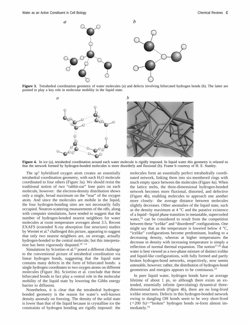
The sp3 hybridized oxygen atom creates an essentiallytetrahedral coordination geometry, with each H2O moleculecoordinated to four others (Figure 3a). We should resist thetraditional notion of two “rabbit-ear” lone pairs on eachmolecule, however: the electron-density distribution showsonly a single, broad maximum on the “rear” of the oxygenatom. And since the molecules are mobile in the liquid,the four hydrogen-bonding sites are not necessarily fullyoccupied. Neutron-scattering measurements of the rdfs, alongwith computer simulations, have tended to suggest that thenumber of hydrogen-bonded nearest neighbors for watermolecules at room temperature averages about 3.5. RecentEXAFS (extended X-ray absorption fine structure) studiesby Wernet et al.9 challenged this picture, appearing to suggestthat only two nearest neighbors are, on average, actuallyhydrogen-bonded to the central molecule; but this interpreta-tion has been vigorously disputed.10-14
Simulations by Sciortino et al.15 posed a different challengeto the conventional picture of tetrahedral coordination vialinear hydrogen bonds, suggesting that the liquid statecontains many defects in the form of bifurcated bonds: asingle hydrogen coordinates to two oxygen atoms on differentmolecules (Figure 3b). Sciortino et al. conclude that thesebifurcated bonds in fact play a central role in the molecularmobility of the liquid state by lowering the Gibbs energybarrier to diffusion.
Nonetheless, it is clear that the tetrahedral hydrogen-bonded geometry is the reason for water’s well-knowndensity anomaly on freezing. The density of the solid stateis lower than that of the liquid because in crystalline ice theconstraints of hydrogen bonding are rigidly imposed: the
molecules form an essentially perfect tetrahedrally coordi-nated network, linking them into six-membered rings withmuch empty space between the molecules (Figure 4a). Whenthe lattice melts, the three-dimensional hydrogen-bondednetwork becomes more fluxional, distorted, and defective(Figure 4b), enabling molecules to approach one anothermore closely: the average distance between moleculesslightly decreases. Other anomalies of the liquid state, suchas the density maximum at 4°C and the putative existenceof a liquid-liquid phase transition in metastable, supercooledwater,16 can be considered to result from the competitionbetween these “icelike” and “disordered” configurations. Onemight say that as the temperature is lowered below 4°C,“icelike” configurations become predominant, leading to adecreasing density, whereas at higher temperatures thedecrease in density with increasing temperature is simply areflection of normal thermal expansion. The notion17,18 thatwater is best viewed as a two-phase mixture of distinct icelikeand liquid-like configurations, with fully formed and partlybroken hydrogen-bond networks, respectively, now seemsuntenable, however; rather, the distribution of hydrogen-bondgeometries and energies appears to be continuous.14
In pure liquid water, hydrogen bonds have an averagelifetime of about 1 ps, so although there exists an ex-tended, essentially infinite (percolating) dynamical three-dimensional network (Figure 4b), there are no long-livedicelike structures. Defects in this hydrogen-bonded networkowing to dangling OH bonds seem to be very short-lived(<200 fs)s“broken” hydrogen bonds re-form almost im-mediately.19
Figure 3. Tetrahedral coordination geometry of water molecules (a) and defects involving bifurcated hydrogen bonds (b). The latter areposited to play a key role in molecular mobility in the liquid state.
Figure 4. In ice (a), tetrahedral coordination around each water molecule is rigidly imposed. In liquid water this geometry is relaxed sothat the network formed by hydrogen-bonded molecules is more disorderly and fluxional (b). Frame b courtesy of H. E. Stanley.
Water as an Active Constituent in Cell Biology Chemical Reviews C

For water’s role as a solvent, particularly in the cell, oneof the key questions is how this network structure is affectedby the introduction of a solute molecule or proximity to asurface. In each case, the structural response of liquid waterdepends on whether the foreign body is hydrophobic orhydrophilic. But in more or less all cases the issue has beencontentious and is still not well resolved.
2.2. Small-Molecule Solutes: Hydrophiles
Water is an extremely good solvent for ions. In part,this is a result of water’s high dielectric constant, whichscreens the ions’ Coulombic potential effectively and pre-vents the aggregation and crystallization of ions of oppo-site charge. By the same token, water is an efficient sol-vent for biomolecular polyelectrolytes such as DNA andproteins, shielding nearby charges on the backbone from oneanother.
As a polar species, the water molecule can engage infavorable Coulombic interactions with ions and other polarsolutes. According to the traditional picture, water moleculeswill solvate cations by orienting their oxygen atoms towardthe ion, whereas they will adopt the opposite configurationfor anions (Figure 5a). Such reorientation, however, perturbsthe hydrogen-bonded network, and the question is howreadily this disruption can be accommodated.
The key technique for investigating this issue is neutronscattering, particularly because of the large scattering crosssection of hydrogen and because of the facility for alteringthe various cross sections by isotopic substitution, enablingthe several water-water and ion-water partial rdfs to bedisentangled. Pioneering work by Enderby, Neilson, Soperand their co-workers20 has confirmed the expected picturein which anions such as chloride are coordinated to waterin the hydration shell via hydrogen atoms, such that theH-O‚‚‚Cl- bond is almost linear, whereas for cations likenickel the water molecules are oriented with the oxygenatoms facing toward the ions (Figure 5b). Recent XAS(X-ray absorption spectroscopy) studies of cation hydrationhave been interpreted as implying little or no geometricdistortion of “water structure” over long ranges,21,22althoughdivalent cations do seem to induce a redistribution of chargeamong water molecules in the solvation shell, leading tochanges in the X-ray absorption spectra.22 This very localview of the effects of ions on water structure is supportedby first-principles MD (molecular dynamics) simulations,which reveal only some degree of preferential orientation inthe first hydration shell for both monovalent and divalentions,23 and by femtosecond pump-probe spectroscopy,
which shows that ions have essentially no influence on therotational dynamics of water molecules beyond the firsthydration shell.24 It is important to note, moreover, thatneutron-scattering studies caution against too static a visionof hydration: there is clearly a lot of dynamic variation inthese structures.
While, as we shall see later, the hydration of hydrophilicregions of proteins has been investigated extensively, therehas been surprisingly little work to date on small, biologicallyrelevant hydrophiles. As a result, the “ground rules” forinterpreting the hydration of larger biomolecules have notreally been established. What little we do know about suchsimple “model” species, however, suggests that it may notbe possible either to generalize or to reduce the structuraland dynamic aspects of hydration to simplistic rules ofthumb. A neutron-scattering study of the hydration ofL-glutamic acid in alkaline solution25 suggested a significantdisruption of the water structure, with the average numberof water-water hydrogen bonds being reduced from 1.8 to1.4. But this was at high concentrations of both glutamicacid and NaOH (each 2 M), so it is not clear how the resultsmight relate to a physiological context. In contrast,L-prolineengages in strong hydrogen bonding with water but in amanner that seems barely to perturb the hydrogen-bondednetwork at all.26 Proline is an osmoprotectant, preventingprotein denaturation in the face of water stress. One possiblemechanism for this invokes the formation of a looselyassociated sheath of proline molecules around a protein to“chaperone” itssomething that appears feasible without anysignificant disruption of the protein’s hydration.
One of the best studied small hydrophiles is urea, whichcan be considered a mimic of a hydrophilic residue but isalso known for its tendency to denature proteins. A neutron-scattering study shows that the urea molecule can “substitute”quite readily for water in the hydrogen-bonded network: therdf of urea around water in a 1:4 solution looks remarkablylike that of water around water.27 Although urea has nearly3 times the molecular volume of water, the structure of liquidwater is sufficiently “open” that a urea molecule appears todisplace just two waters, offering up to eight hydrogen bondsin place of the displaced pair. The idea that urea “fits” rathereasily into the hydrogen-bonded structure of water has beensupported recently by dielectric spectroscopy.28 Yet despitethis apparently “easy” substitution, incorporating urea intothe network appears to disrupt it, creating a local compressionof the second hydration shell around water molecules in amanner similar to the effect of high pressure on the liquid.All the same, the orientational dynamics of the watermolecules seem largely unaffected even at urea concentra-tions high enough for all the water to be part of hydrationshells.29 Only one water molecule per urea, on average, hasa significantly slower reorientational time constant (about 6times that of bulk water), which can be rationalized accordingto a hydration structure in which one water is complexed tothe urea molecule via two hydrogen bonds.
These findings shed light on the action of urea as a proteindenaturant. Proposed reasons for this behavior have invokeddirect interactions of urea with the protein backbone, forexample, via hydrogen bonds30,31or electrostatic32 or hydro-phobic contacts,33 which act to swell the protein as aprecursor to denaturation, and indirect effects due to thedisruption of “water structure” by urea, making the hydro-phobic groups more readily solvated.34 The experimentalfindings for the effect of urea on water do not seem to support
Figure 5. Solvation of cations and anions. (a) The conventionalview. (b) Water orientations revealed by neutron scattering.
D Chemical Reviews Ball
the idea of significant disruption of the hydrogen-bondednetwork (although one might argue that the mimicking ofhigh pressure in the second hydration shell might encouragesomething akin to pressure-induced denaturation). Rather,it seems best to consider the nature of specific interactionsbetween the various solutes, instead of regarding the proteinas being solvated by “modified water”. Indeed, experimentsand thermodynamic analysis imply that in general a cosolventsuch as urea will be inhomogeneously partitioned around abiological macromoleculesurea tends to expel water froma protein surface, whereas sugars are preferentially excludedfrom the hydration sphere.35
We will see below that the same principlesconsideringlocal, direct interactions rather than global “ water structure”modificationssholds for hydrophobes too. In any event, onecan see that the overall picture of the effect of hydrophileson water structure is rather complex even for this relativelysimple species, demanding a detailed consideration of thestructure and dynamics of the hydration shell(s), and so itseems unwise to fall back on popular but simplistic notionsabout whether solutes such as urea “break” the structure ofthe hydrogen-bonded network36,37 (see below).
2.3. Small-Molecule Solutes: HydrophobesHydrophobic solutes in water experience a force that
causes them to aggregate. This hydrophobic interaction isresponsible for several important biological processes:38 forthe aggregation of amphiphilic lipids into bilayers, with theirhydrophobic tails “hidden” from water and their hydrophilicheads at the surface, for the burial of hydrophobic residuesin polypeptide chains that helps proteins to fold and to retaintheir compact forms, and for the aggregation of proteinsubunits into multi-subunit quaternary structures. There is atendency for even rather small hydrophobic moieties toclusterssomething of the sort seems to happen for methanoldissolved in water, leading to dynamic heterogeneity of themixture.39 The question is whether a single mechanism forthe hydrophobic interaction can account for all thesebehaviors, and if so, what is it?
We do not yet know how to answer either point. Part ofthe reason for that perhaps surprising ignorance may be thatthere has for some time existed an apparent explanation that
made such seemingly good sense that it has been hard todislodge and is still routinely cited in biochemistry textbooks.This is Kauzmann’s “entropic” origin of the hydrophobicattraction,40 which draws on the picture of hydrophobichydration posited in 1945 by Frank and Evans.41
The conventional story is as follows. In order to accom-modate a hydrophobic species, the hydrogen-bonded networkmust be disrupted to create a void. But this can be done insuch a way as to avoid the enthalpic penalty of losinghydrogen bonds, if the water molecules arrange themselvesaround the hydrophobe in a relatively ordered fashion (Figure6a). In the words of Frank and Evans, “when a rare gas atomor nonpolar molecule dissolves in water at room temperatureit modifies the water structure in the direction of greatercrystallinitysthe water, so to speak, builds a microscopiccage around it”.41
This is the “iceberg” model: the hydrophobe is encasedin an icelike shell of water. It has been often suggested42,43
that, based on a description of water structure due toPauling,44 a better model for this pseudocrystalline cage isthat found in gas hydrates, where small hydrophobic speciessuch as methane are enclosed in cavities made frompentagonal rings45 rather than the hexagonal rings of ice.(Hydration structures around the hydrophobic portions ofproteins are now commonly described in terms of pentagonalrings.) Kauzmann pointed out that the price of creating anyrelatively ordered structure of this sort to preserve theintegrity of the hydrogen-bonded network is that the rota-tional and translational freedom of the molecules in the cagewall are compromised: there is an entropy decrease. But iftwo “caged” hydrophobes were to come together, the“structured” water in the region between them is returnedto the bulk, leading to an entropy increase (Figure 6b). Thus,there is an entropically based force of attraction betweenthese solute particles.
The argument seems sound in principle, but the questionis whether hydrophobic hydration really has the “semicrys-talline” character proposed by Frank and Evans. To date,there is no good reason to suppose that it does, and someevidence indicating that it does not. Reviewing the existingdata on hydrophobic hydrationsboth structural and thermo-dynamic, and based on both experiment and computer
Figure 6. (a) “Iceberg” model of hydrophobic hydration. (b) Kauzmann’s explanation of the hydrophobic attraction.
Water as an Active Constituent in Cell Biology Chemical Reviews E

simulationsBlokzijl and Engberts46 concluded that there wasno reason to suppose that hydrogen bonding is enhanced inthe first hydration shell or that the favorable enthalpy ofhydrophobic hydration need be attributed to anything morethan normal van der Waals interactions between water anda nonpolar solute. Moreover, they found no evidence thathydration involved any enhancement in the ordering of watermolecules around the solute; rather, the hydrogen-bondednetwork seemed simply tomaintainits structure, in particularby orienting water molecules such that the O-H bonds aretangential to the solute surface. There is no inconsistencybetween the occurrence of such orientational effects and thelack of any enhancement in structure, since we must bear inmind that the hydrogen-bonded network introduces direc-tional preferences in local water orientation even in the bulk.The importance of attractive solute-water van der Waalsinteractions, relative to considerations of “water structure”and water-water interactions, was emphasized by Ashbaughand Paulaitis,47 whose Monte Carlo simulations show thatalthough water densities in the first hydration shells ofclusters of methane molecules are greater than those of thebulk, the corresponding density for hydrated hard spheres(lacking any attractive interactions) of the same size as themethane clusters decreases as the sphere’s radius increasesand ultimately falls below that of the bulk.
Blokzijl and Engberts suggested that aggregation ofhydrophobes results not from Kauzmann’s entropic mech-anism but from the increasing difficulty in accommodatinghydrophobes within the hydrogen-bonded network as theirconcentration in solution increases: “hydrophobic interac-tions”, they say, “are not so much a result of a structuralproperty of the hydrophobic hydration shell of apolarcompounds but rather reflect the limited capacity of liquidwater to accommodate the apolar solute and maintain itsoriginal network of hydrogen bonds.”46 From the perspectiveof whether there is anything “special” about water thatintroduces a hydrophobic interaction, therefore, the messageis mixed. Yes, the hydrogen-bonded network seems to beimportant, but not because it becomes more highly structuredby hydrophobes; rather, it is because this network is disruptedby too great an accumulation of cavities.
On the other hand, Lucas48 and Lee49,50have proposed thatthis disruption is not a function of the hydrogen-bondednetwork at all but stems merely from the small size of thewater molecules, which creates a high free-energy cost toopening up a cavity to accommodate a hydrophobesanargument based on scaled-particle theory,51 which Stillinger52
first adapted to the case of hydration. Hummer, Pratt, andtheir co-workers53-58 have argued that models based on thespontaneous formation of cavities through density fluctua-tions in liquid water can account quantitatively for thethermodynamics of small-hydrophobe solvationsa claimsupported by MD simulations.59 Yet Southall et al.60 assertthat neither water “structure” nor the small-size effect canby themselves fully explain several of the characteristics ofthe hydrophobic effect, such as its dependence on temper-ature and on solute shape.
While all of these studies help to decouple the real puzzlesof the hydrophobic effect from comfortable but outdatedexplanations, there is a tendency in discussions of thehydrophobic interaction to regard water asthe liquid ratherthana liquid: little comparison has been made with othersmall-molecule liquids, both associated and simple, perhapsbecause of the notion that the hydrophobic effect is clearly
biologically relevant, whereas such solvents are not. As aresult, we lack much basis for comparison in deciding howmuch of water’s behavior as a solvent for solvophobicparticles is unique (and thus perhaps due to its three-dimensional hydrogen-bonded network) and how much isgeneric to other, related liquids. Only when such questionsare answered can we expect to say much about water’ssupposed centrality to life.
Evidently, however, there is nothing unique to water aboutsolvophobic aggregation in general: the existence of reversemicelles in nonpolar solvents indicate as much. Morestrikingly, Huang et al.61 have shown that non-naturalpeptide-like molecules with N-linked rather than C-linkedside chains (N-substituted oligoglycines or “peptoids”62) willfold into well-defined secondary structures in acetonitrile inwhich the polar units (the carbonyl groups) are buried inthe interior (Figure 7): one apparently does not need a strongdegree of solvent structure in order for such packing to occur.It will be very interesting to explore the potential complexityof structure and function available to this and other non-aqueous pseudoprotein chemistry.
All the same, one can imagine there being scope for addedsubtlety in the interactions of a solvophobe and a structuredsolvent like water. Indeed, Hummer and co-workers53,54arguethat if cavity-opening fluctuations are the dominant factorin hydrophobic hydration, water’s unusual equation of statedistinguishes the nature of these effects from those that mightbe anticipated in other liquidssthere is then something“special” about water.
But how, if at all, does water structure manifest itself inthe hydration environment of hydrophobes? Obviously thebest way to deduce the effect of a hydrophobic moleculeplaced within water’s network is to measure it directly; andagain neutron scattering is the ideal probe. But that is nosimple matter, because archetypal small hydrophobes suchas methane and krypton are insufficiently soluble in waterto permit an easy detection of the hydration shells abovethe bulk water signal.
Nonetheless, de Jong et al.63 used neutron scattering todeduce that methane molecules in water are surrounded byabout 19 solvent molecules and that those in the firsthydration shell are tangentially oriented. The structure of ahydrophobic hydration shell has been most aggressivelypursued by Finney, Soper, and their co-workers. Filipponiet al.64 used EXAFS to show that krypton is hydrated by
Figure 7. Backbone folded structure of an N-substituted glycinenonamer peptoid in acetonitrile. The dashed green lines indicatehydrogen bonds. Reprinted with permission from ref 61. Copyright2006 American Chemical Society.
F Chemical Reviews Ball
around 20 water molecules. This hydration structure wasbarely altered by applying pressures of up to 1 kbar.65 Thisnot only throws into question some proposed explanationsfor pressure-induced protein denaturation in terms of hydra-tion changes but also cautions against too heavy a relianceon computer simulations for evaluating hydration structure,some of which66 predict significant pressure-induced changesin the hydration shell of methane.
Molecular dynamics simulations of krypton in aqueoussolution67,68show no sign of the clathrate-like hydration shellsinvoked in the classic iceberg model. That is borne outexperimentally by Bowron et al.,69 who used EXAFS tohighlight the differences between the hydration shell ofkrypton in solution and the clathrate cage of krypton in thecrystalline clathrate hydrate. The former is unambiguouslymore disordered, and the tangential orientation of watermolecules found in the hydrate is not rigorously main-tained in the liquid. Indeed, Finney70 points out that thefirst-neighbor water structure in the hydration shell is allbut unchanged from that in bulk water: “water in thisenvironment still thinks it is liquid water”. And ab initioMD simulations of the diffusion of small hydrophobes inwaterswhich is found experimentally to be anomalouslyfast71sindicate that, far from being icelike, the hydrationshell is unusually dynamic and loose.72 Even if theFrank-Evans iceberg model is no longer interpreted tooliterally in any case, these results give direct cause to doubtany such picture that places emphasis on enhanced “waterstructuring” around hydrophobes.
Another model small hydrophobe is benzene, sincearomatic groups are common in biological molecules. Butwhile phenyl groups are generally regarded as hydrophobic,it has been known for some time that theπ-electron systemcan act as a weak hydrogen-bond acceptor,73 creating strongorientational preferences in the bound water molecules.74 Theconverse is also possible: the lone pairs on the oxygen mayalso interact favorably withπ electrons. This balance appearsto be rather subtle, making it hard to generalize about howaromatic groups are likely to be hydrated. Allesch et al.75
find in first-principles MD simulations of benzene andhexafluorobenzene in water that both molecules act ashydrophobes equatorially but that the axial hydration struc-ture is very different: water molecules typically point towardthe ring hydrogen-first for benzene, but lone-pair-first forhexafluorobenzene.
A particularly attractive minimal model for investigatingthe role of hydration structure on hydrophobic interactionsin biological systems is provided by simple alcohols suchas methanol. Not only are they amphiphiles that mimic“shrunken lipids”, but the juxtaposition of nonpolar groupsand polar, hydrogen-bonding groups shares to that extent thecharacter of a typical portion of a protein’s peptide chain.Moreover, they provide an opportunity to study hydrophobichydration without the problems of low solubility. Dixit etal.76 find that a concentrated solution of methanol (7:3methanol/water ratio) is imperfectly mixed and highlystructured: water clusters bridge hydroxyl groups on thealcohol to form hydrogen-bonded chains that thread througha “fluid” of methyl groups. Most of the water molecules,however, cluster into groups of 2-20, in which their localstructure is surprisingly bulklike, again challenging any ideaof iceberg-like ordering. The segregation of methanol andwater even persists at high dilution of the alcohol:77 in a1:19 mixture of water and methanol, more than 80% of the
alcohol molecules are clustered into groups of 3-8. Again,here neutron scattering shows no evidence for structureenhancement in the hydration shells of the nonpolar portionsof the methanol molecules.78
What, then,is the origin of this clustering effect? Dixit etal.77 propose that it might indeed have an entropic origin,but subtly so. They find that the hydration shells of the watermolecules themselves are slightly altered by the presence ofmethanol (at a 1:19 concentration, most of the watermolecules lie within the hydration shell of at least onemethanol): the second hydration shell becomes slightlycompressed, and the correlations between second-neighborwaters slightly sharpened, leading to a small reduction inentropy. This small decrease in freedom of the watermolecules could promote aggregation of the methanols.“Perhaps it is here”, Finney et al.79 conclude, “in thewater’ssecondshell rather than thealcohol’s first hydration shell,that the entropic driving force for the hydrophobic interactionis to be found”. If so, this is a small effect, and by no meansobvious, and of unproven generalitysso it might be arguedthat explanations based on general factors such as cavityformation53-58 have a stronger appeal, not least because theyare more intuitive. But in any event, these direct structuralprobes seem to have diminished any argument for the classic“enhanced structuring” model of the hydrophobic interactionalong the lines defined by Kauzmann.
2.4. Large Hydrophobic Solutes and SurfacesAlthough small hydrophobic species can be accommodated
in the hydrogen-bonded network of liquid water withoutmuch perturbation of the network, large hydrophobes, forexample the hydrophobic surfaces of proteins, are anothermatter. An attractive interaction evidently exists betweensuch species too, being responsible for protein foldingand aggregation. Does this have the same origin as theforce that causes small hydrophobes to cluster? That is notobvious.
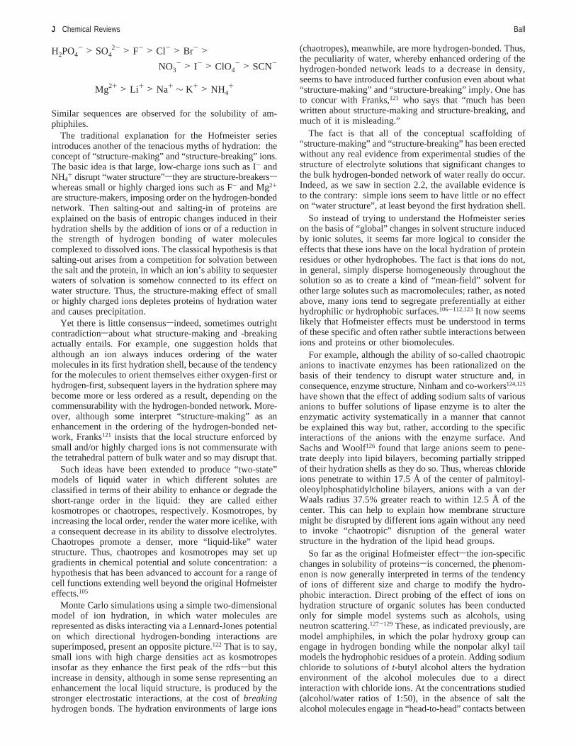
Close to an extended hydrophobic surface, it is geo-metrically impossible for the network to maintain its integrity.It has been proposed that this can even lead to “drying” ofthe interface43sthe formation of a very thin layer of vaporseparating the liquid from the surface. In a highly stimulatingcontribution, Lum, Chandler, and Weeks (LCW)80 argue thatthis difference between small and large hydrophobes shouldlead to qualitatively different behavior, with a crossoverlength scale somewhere in the region of 1 nmsabout thevan der Waals diameter of anR-helix. This idea derives fromthe notion, proposed by Wallqvist and Berne,81 that whentwo hydrophobic surfaces come in close proximity, watercan withdraw from between them (Figure 8) and that the
Figure 8. Hydrophobic attraction in the model of Lum et al.80
The hydrophobic surfaces are surrounded by a thin layer of vapor.At some critical separationDc, there is a collective drying transitionin the space between the surfaces.
Water as an Active Constituent in Cell Biology Chemical Reviews G

resulting imbalance in pressure would cause the two surfacesto attract. In effect, the confined water undergoes “capillaryevaporation”. This kind of drying transition has been seenin simulations of hydrophobic plates in water when theseparation between them falls to just a few molecularlayers.81-83
Simulations of hard-sphere solvation by Rajamani et al.59
support the LCW picture, indicating that the thermodynamicsof hydration is entropically controlled for small hydrophobesbut enthalpic for larger ones. This is precisely what onewould expect if the former case depends on density fluctua-tions and cavity formation while the latter involves theformation of a liquid-vapor interface and thus introducesthe surface tension into the hydration Gibbs energy. Thiscrossover of hydration mechanisms is accompanied by achange in the variation of hydration Gibbs energy∆G withsolute size: ∆G is a linear function of solute volume forsmall sizes but becomes closer to being proportional to solutesurface area for larger sizes. Rajamani et al. find that thecrossover occurs for hard-sphere solute radii of the order ofa few angstroms under ambient conditions but that this sizescale can be “tuned” either by altering thermodynamicparameters (for example, placing the water under negativepressure) or by adding other solutes. Ethanol, for example,decreases the crossover length scale to molecular dimensions.Ashbaugh and Pratt58 show that this picture of a crossoverfrom entropically to enthalpically dominated hydrophobichydration as a function of particle size can be rationalizedby applying scaled-particle theory52 to a thermodynamicanalysis of cavity formation.
But does hydrophobic collapse induced by cooperativedewetting really play any role in the association of hydro-phobic macromolecules, for example, in protein folding andaggregation? Here the picture remains unclear, although itseems fair to say that the LCW model looks increasinglyunlikely to provide a general description of macromolecularhydrophobic interactions. Simulations by ten Wolde andChandler84 suggest that a hydrophobic polymer acquires acompact conformation in water via a process resembling afirst-order phase transition in which the rate-limiting step isthe nucleation of a sufficiently large vapor bubblestheclassical mechanism of heterogeneous nucleation. But simu-lations of protein folding show a more complex situation.Berne’s group found that collapse of the two-domain enzymeBphC, which breaks down polychlorinated biphenyls, showedno sign of a sharp dewetting transition as the domains cametogether.85 Here the drying seen between hydrophobic plates82
seems to be suppressed by attractive interactions betweenthe protein and water: dewetting was recovered when theelectrostatic protein-water forces were turned off and wasstronger still in the absence of attractive van der Waals forces.Thus, it seems that the inevitable presence of such interac-tions in proteins complicates the simple picture obtained fromhydrophobic surfaces. Consistent with this view, MacCullumet al. found that simulations of the dimeric association ofboth polyalanine and polyleucine (A20 and L20) R-helicesshowed no dewetting between the chains until it was inducedsterically by a mere insufficiency of space for a watermonolayer.86
On the other hand, Berne’s group found a first-order-likedewetting transition in simulations of the association of themelittin tetramer, a small polypeptide found in honeybeevenom.87 But single mutations of three hydrophobic isoleu-cine residues to less hydrophobic ones were sufficient to
suppress the dewetting. Is melittin a rarity, even a uniquecase, or might other proteins also exhibit dewetting? Berne’sgroup has performed a survey of the protein data bank tosearch for other structures that might show similar behavior.88
They find that dewetting is indeed rather rare but does happenin several other cases: they identify two two-domainproteins, six dimers, and three tetramers that behave this way.It seems that any significant number of polar residues in thehydrophobic core (which is common) is generally enoughto suppress dewetting. Using the same tools, Berne’s groupfinds preliminary evidence that dewetting may also some-times play a role in ligand binding.
These results suggest that even if the LCW mechanismcan operate in the collapse of some proteins, nonetheless itis extremely sensitive both to the precise chemical nature ofthe protein domains involved and perhaps to the geometryof association: melittin subunit association forms a tubelikeenclosed space, whereas that for BphC is slablike.
Choudhury and Pettitt have attempted to clarify theseissues by returning to the case of two planar, nanoscopichydrophobic plates.89,90 They find89 that the existence orabsence of a wetting layer between the plates at separationsof less than about 1 nm depends on a fine balance betweenthe plate-water interaction energy, the hydrogen-bondingenergy, and the plate size. For example, graphite-like platesmeasuring 11× 12 Å2 undergo a steric dewetting transitionfor separation below about 6.8 Å for a Lennard-Jonesinteraction potential, but dewetting occurs at about 10 Å fora purely repulsive plate-water interaction. This LCW-likebehavior vanishes, however, if the plates are smaller.
This finding seems consistent with the view provided bydensity functional calculations on confined simple liquids,91
which show that, although unfavorable liquid-surfaceintermolecular interactions (relative to liquid-liquid interac-tions) can counteract the usual enhancement of liquid densityclose to a wall owing to packing effects, and can even leadto a depletion in average density here, it takes a ratherextreme set of interaction parameters to induce capillaryevaporation in the manner of the LCW model. In otherwords, the bulk liquid-solid contact angle has to be verylow, and it is far from clear that water in contact with atypical hydrophobic surface (an alkyl-covered surface, say,let alone the hydrophobic surface of a protein) representssuch an extreme case. Choudhury and Pettitt concur90 thatin general, “chemically reasonable” estimates of the plate-water interaction strength lead to a microscopically wet stateand not to plate association triggered by a dewettingtransition. That conclusion is supported by MD simulationsof Bresme and Wynveen, who have studied the effect oninteractions between two hydrophobic solutes of varying theirpolarizability.92 The solute polarizability has a strong influ-ence on the water contact angle, and a drying transitionoccurs only for rather extreme conditions (outside the rangeof permittivities typical for proteins) in which the contactangle is close to 180°. Otherwise a fluid layer remainsbetween the solute surfaces, albeit with a density significantlylower than that of the bulk liquid. This situation is neverthe-less associated with strong hydrophobic forces, showing thatcomplete drying is not essential to promote an attractiveinteraction.
This perspective leads to the more general question of howwater behaves and is structured close to asinglehydrophobicsurface. Although, as Choudhury and Pettitt point out,90
drying in the interplate region may be a cooperative
H Chemical Reviews Ball

phenomenon and thus does not necessarily demand dryingof the isolated surfaces themselves, nonetheless the natureof the interface between water and a hydrophobic surface isthe precondition, one might say, for any discussion ofpotential dewetting by confinement. Moreover, this questionis central to the more general and much-disputed issue ofthe hydrophobic hydration of proteins.
Experiments have tended to give a rather confused andcontradictory picture of this situation,93 although a consensusnow seems to be emerging. X-ray reflectivity measure-ments94-96 suggest that, although there is a depletion in liquiddensity adjacent to alkane monolayers, it is far less pro-nounced than what would be observed for complete dryingand happens only within a few molecular diameters of thesurface. Measurements of water density adjacent to acrystalline paraffin monolayer floating on the surface of watersuggest that the depletion region extends about 1.5 nm intothe liquid phase and that it corresponds to a deficit of aboutone water molecule for every 25-30 Å2 of the paraffinsurface.94 On the other hand, MD simulations in the samestudy indicated the formation of a very thin (about 1 Å) layerof “vacuum” between the water phase and the surfacessomething beyond the resolution of the experiment itself.This latter result might be considered broadly consistent withthe simulations of Pertsin et al.,97 which indicated only avery small reduction in water density close to an alkylatedsurface. Other MD simulations suggest that, contrary to whatwould be expected in the presence of a vapor-like film, thereis significant penetration of water molecules into a layer oftetheredn-C18 chains.98
A more recent high-resolution X-ray reflectivity study95
corroborates the existence of a “hydrophobic gap” for amonolayer ofn-C18 chains on silica but suggests that itextends no further than 1-6 Å from the surface and that itcorresponds to an integrated density deficit of 1.1 Å g cm-3.Similarly, Poynor et al.96 find a density deficit of more than40% extending about 2-4 Å from the surface.
In contrast, some neutron reflectivity studies of the waterdensity adjacent to a self-assembled monolayer of alkyl-thiols99,100 have apparently indicated a density depletionextending for several nanometers. But recent results makethis now seem unlikely. Doshi et al.101 found a reduceddensity extending only 1 nm or less from the surface, thedistance depending on the amount and chemical nature ofdissolved gases (see below; at this stage the influence ofdissolved gases is by no means clearsthe smaller hydro-phobic gap reported by Mezger et al.95 was unaffected by awide range of such gases). And Maccarini et al. report adepletion layer of no more than about 2 Å.102
From a theoretical perspective, even if hydrophobicsurfaces do not induce anything like complete drying, theexistence of a depletion layer extending over distances of2-5 nm would be very perplexing, since there is no obviousphysical interaction in the system that could introduce sucha length scale. That recent results seem to be converging ona depletion layer with a thickness of the same order of thewater molecule itself is therefore reassuring.
It is important to know whether similar effects are seenfor other liquids so that one might elucidate the role (if any)of water’s hydrogen-bonded network. Maccarini et al.102 dofind that depletion layers of a similar order, that is, just afew angstroms thick, appear to be present at the interface ofhydrophilic surfaces and nonpolar liquids, showing that we
should be wary here of attributing anything “special” towater.
How water dynamics might be affected within a hydro-phobic depletion layer is another matter, which has receivedlittle attention. Dokter et al.103 found that nanodroplets inreverse micelles, where the interface is not hydrophobic butis thought nonetheless to have decreased hydrogen bonding,have slow orientational dynamics and relatively immobilewater molecules in the interfacial layer. Jensen et al. alsofound retarded dynamics in simulations of water next to ahydrophobic surface.104 As we will see below, a significantchange in rotational dynamics in this region could haveimplications for the hydrophobic interaction itself.
Any change in the nature of “water structure” close to ahydrophobic surface can be expected to alter its solvatingcharacteristics, leading to the possibility of segregation ofsmall solutes such as ions at or away from such interfaces.105
But it seems likely that this view is putting the cart beforethe horse. There is now strong evidence that some ions doindeed segregate preferentially at, or away from, the air-water interface.106-112 Yet this is not because the water is“different” there, but because there is an intrinsic thermo-dynamic driving force for this segregation, and if anythingwe might expect an excess of ions to alter the properties ofthe solvent rather than vice versa. Since one might expectthe interface of water with a hydrophobic surface to mimicin many respects that with air, this inhomogeneity of solutesat a surface could have significant implications for thesolvation of proteins, as we will see below.
The same applies to the finding that hydronium ions seemto have a preference for the water surface.113-118 This result,which was predicted theoretically113,114,118and confirmed bythermodynamic analysis,111 by surface spectroscopy,114,116,117
and by deuterium exchange at the surface of ice nanocrys-tals,118 apparently has a rather different origin from thesurface segregation of other ions. H3O+ may form three donorhydrogen bonds to neighboring water molecules, but becausemost of the positive charge resides on the oxygen atom, itcan no longer act as a good hydrogen-bond acceptor. Indeed,this makes the oxygen somewhat hydrophobic, so thathydronium acts as an amphiphile.113,115 Both that and thereduced hydrogen-bond capacity encourage the surfaceaccumulation of hydronium, oriented with the oxygen atomoutermost. It has been estimated113,118that this effect shiftsthe surface pH of pure water to around 4.8 or less (althoughthe applicability of this bulk parameter on a localized scaleis not entirely clear). As much the same behavior might beexpected at hydrophobic surfaces, this finding could havesignificant implications for biomolecular hydration that haveyet to be investigated; for example, one might expect to seea shift in the dissociation of protonatable residues close tohydrophobic patches and perhaps even a stabilization ofhydrophobic species by a kind of surfactant behavior ofhydronium.
2.5. The Influence of Ions: Structure-Making andStructure-Breaking
The coexistence of ions and hydrophobes in aqueoussolution has some puzzling consequences. Hofmeister notedin 1888 that some salts tend to precipitate albumin fromsolution (salting-out), whereas others enhance its solubility(salting-in).119,120The Hofmeister series ranks ions in orderof their “salting-out” tendency for proteins:
Water as an Active Constituent in Cell Biology Chemical Reviews I
Similar sequences are observed for the solubility of am-phiphiles.
The traditional explanation for the Hofmeister seriesintroduces another of the tenacious myths of hydration: theconcept of “structure-making” and “structure-breaking” ions.The basic idea is that large, low-charge ions such as I- andNH4
+ disrupt “water structure”sthey are structure-breakersswhereas small or highly charged ions such as F- and Mg2+
are structure-makers, imposing order on the hydrogen-bondednetwork. Then salting-out and salting-in of proteins areexplained on the basis of entropic changes induced in theirhydration shells by the addition of ions or of a reduction inthe strength of hydrogen bonding of water moleculescomplexed to dissolved ions. The classical hypothesis is thatsalting-out arises from a competition for solvation betweenthe salt and the protein, in which an ion’s ability to sequesterwaters of solvation is somehow connected to its effect onwater structure. Thus, the structure-making effect of smallor highly charged ions depletes proteins of hydration waterand causes precipitation.
Yet there is little consensussindeed, sometimes outrightcontradictionsabout what structure-making and -breakingactually entails. For example, one suggestion holds thatalthough an ion always induces ordering of the watermolecules in its first hydration shell, because of the tendencyfor the molecules to orient themselves either oxygen-first orhydrogen-first, subsequent layers in the hydration sphere maybecome more or less ordered as a result, depending on thecommensurability with the hydrogen-bonded network. More-over, although some interpret “structure-making” as anenhancement in the ordering of the hydrogen-bonded net-work, Franks121 insists that the local structure enforced bysmall and/or highly charged ions is not commensurate withthe tetrahedral pattern of bulk water and so may disrupt that.
Such ideas have been extended to produce “two-state”models of liquid water in which different solutes areclassified in terms of their ability to enhance or degrade theshort-range order in the liquid: they are called eitherkosmotropes or chaotropes, respectively. Kosmotropes, byincreasing the local order, render the water more icelike, witha consequent decrease in its ability to dissolve electrolytes.Chaotropes promote a denser, more “liquid-like” waterstructure. Thus, chaotropes and kosmotropes may set upgradients in chemical potential and solute concentration: ahypothesis that has been advanced to account for a range ofcell functions extending well beyond the original Hofmeistereffects.105
Monte Carlo simulations using a simple two-dimensionalmodel of ion hydration, in which water molecules arerepresented as disks interacting via a Lennard-Jones potentialon which directional hydrogen-bonding interactions aresuperimposed, present an opposite picture.122 That is to say,small ions with high charge densities act as kosmotropesinsofar as they enhance the first peak of the rdfssbut thisincrease in density, although in some sense representing anenhancement the local liquid structure, is produced by thestronger electrostatic interactions, at the cost ofbreakinghydrogen bonds. The hydration environments of large ions
(chaotropes), meanwhile, are more hydrogen-bonded. Thus,the peculiarity of water, whereby enhanced ordering of thehydrogen-bonded network leads to a decrease in density,seems to have introduced further confusion even about what“structure-making” and “structure-breaking” imply. One hasto concur with Franks,121 who says that “much has beenwritten about structure-making and structure-breaking, andmuch of it is misleading.”
The fact is that all of the conceptual scaffolding of“structure-making” and “structure-breaking” has been erectedwithout any real evidence from experimental studies of thestructure of electrolyte solutions that significant changes tothe bulk hydrogen-bonded network of water really do occur.Indeed, as we saw in section 2.2, the available evidence isto the contrary: simple ions seem to have little or no effecton “water structure”, at least beyond the first hydration shell.
So instead of trying to understand the Hofmeister serieson the basis of “global” changes in solvent structure inducedby ionic solutes, it seems far more logical to consider theeffects that these ions have on the local hydration of proteinresidues or other hydrophobes. The fact is that ions do not,in general, simply disperse homogeneously throughout thesolution so as to create a kind of “mean-field” solvent forother large solutes such as macromolecules; rather, as notedabove, many ions tend to segregate preferentially at eitherhydrophilic or hydrophobic surfaces.106-112,123It now seemslikely that Hofmeister effects must be understood in termsof these specific and often rather subtle interactions betweenions and proteins or other biomolecules.
For example, although the ability of so-called chaotropicanions to inactivate enzymes has been rationalized on thebasis of their tendency to disrupt water structure and, inconsequence, enzyme structure, Ninham and co-workers124,125
have shown that the effect of adding sodium salts of variousanions to buffer solutions of lipase enzyme is to alter theenzymatic activity systematically in a manner that cannotbe explained this way but, rather, according to the specificinteractions of the anions with the enzyme surface. AndSachs and Woolf126 found that large anions seem to pene-trate deeply into lipid bilayers, becoming partially strippedof their hydration shells as they do so. Thus, whereas chlorideions penetrate to within 17.5 Å of the center of palmitoyl-oleoylphosphatidylcholine bilayers, anions with a van derWaals radius 37.5% greater reach to within 12.5 Å of thecenter. This can help to explain how membrane structuremight be disrupted by different ions again without any needto invoke “chaotropic” disruption of the general waterstructure in the hydration of the lipid head groups.
So far as the original Hofmeister effectsthe ion-specificchanges in solubility of proteinssis concerned, the phenom-enon is now generally interpreted in terms of the tendencyof ions of different size and charge to modify the hydro-phobic interaction. Direct probing of the effect of ions onhydration structure of organic solutes has been conductedonly for simple model systems such as alcohols, usingneutron scattering.127-129 These, as indicated previously, aremodel amphiphiles, in which the polar hydroxy group canengage in hydrogen bonding while the nonpolar alkyl tailmodels the hydrophobic residues of a protein. Adding sodiumchloride to solutions oft-butyl alcohol alters the hydrationenvironment of the alcohol molecules due to a directinteraction with chloride ions. At the concentrations studied(alcohol/water ratios of 1:50), in the absence of salt thealcohol molecules engage in “head-to-head” contacts between
H2PO4- > SO4
2- > F- > Cl- > Br- >
NO3- > I- > ClO4
- > SCN-
Mg2+ > Li+ > Na+ ∼ K+ > NH4+
J Chemical Reviews Ball
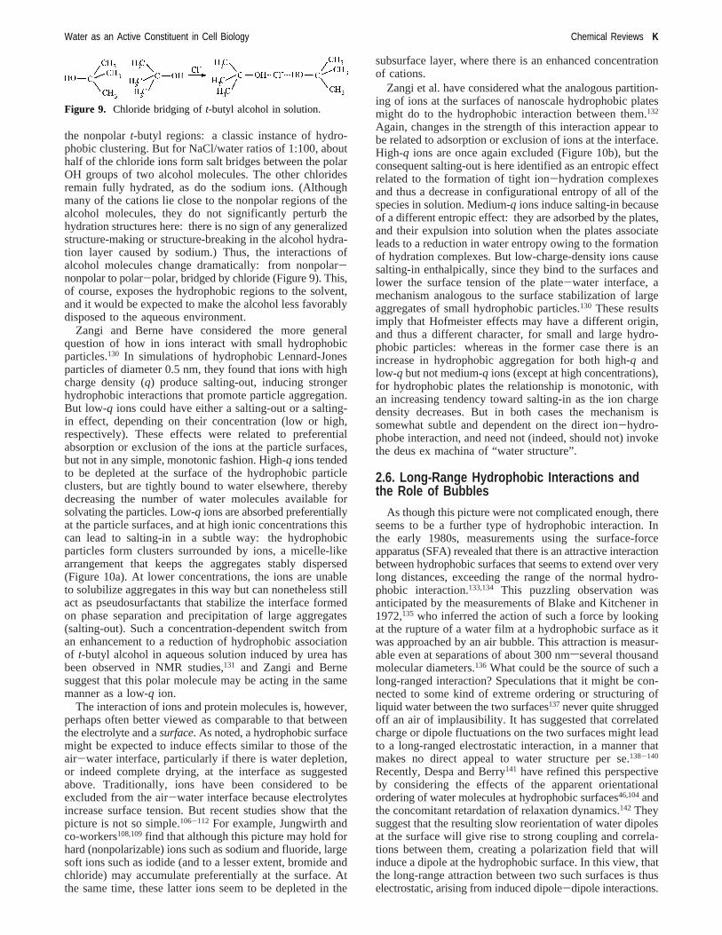
the nonpolart-butyl regions: a classic instance of hydro-phobic clustering. But for NaCl/water ratios of 1:100, abouthalf of the chloride ions form salt bridges between the polarOH groups of two alcohol molecules. The other chloridesremain fully hydrated, as do the sodium ions. (Althoughmany of the cations lie close to the nonpolar regions of thealcohol molecules, they do not significantly perturb thehydration structures here: there is no sign of any generalizedstructure-making or structure-breaking in the alcohol hydra-tion layer caused by sodium.) Thus, the interactions ofalcohol molecules change dramatically: from nonpolar-nonpolar to polar-polar, bridged by chloride (Figure 9). This,of course, exposes the hydrophobic regions to the solvent,and it would be expected to make the alcohol less favorablydisposed to the aqueous environment.
Zangi and Berne have considered the more generalquestion of how in ions interact with small hydrophobicparticles.130 In simulations of hydrophobic Lennard-Jonesparticles of diameter 0.5 nm, they found that ions with highcharge density (q) produce salting-out, inducing strongerhydrophobic interactions that promote particle aggregation.But low-q ions could have either a salting-out or a salting-in effect, depending on their concentration (low or high,respectively). These effects were related to preferentialabsorption or exclusion of the ions at the particle surfaces,but not in any simple, monotonic fashion. High-q ions tendedto be depleted at the surface of the hydrophobic particleclusters, but are tightly bound to water elsewhere, therebydecreasing the number of water molecules available forsolvating the particles. Low-q ions are absorbed preferentiallyat the particle surfaces, and at high ionic concentrations thiscan lead to salting-in in a subtle way: the hydrophobicparticles form clusters surrounded by ions, a micelle-likearrangement that keeps the aggregates stably dispersed(Figure 10a). At lower concentrations, the ions are unableto solubilize aggregates in this way but can nonetheless stillact as pseudosurfactants that stabilize the interface formedon phase separation and precipitation of large aggregates(salting-out). Such a concentration-dependent switch froman enhancement to a reduction of hydrophobic associationof t-butyl alcohol in aqueous solution induced by urea hasbeen observed in NMR studies,131 and Zangi and Bernesuggest that this polar molecule may be acting in the samemanner as a low-q ion.
The interaction of ions and protein molecules is, however,perhaps often better viewed as comparable to that betweenthe electrolyte and asurface. As noted, a hydrophobic surfacemight be expected to induce effects similar to those of theair-water interface, particularly if there is water depletion,or indeed complete drying, at the interface as suggestedabove. Traditionally, ions have been considered to beexcluded from the air-water interface because electrolytesincrease surface tension. But recent studies show that thepicture is not so simple.106-112 For example, Jungwirth andco-workers108,109find that although this picture may hold forhard (nonpolarizable) ions such as sodium and fluoride, largesoft ions such as iodide (and to a lesser extent, bromide andchloride) may accumulate preferentially at the surface. Atthe same time, these latter ions seem to be depleted in the
subsurface layer, where there is an enhanced concentrationof cations.
Zangi et al. have considered what the analogous partition-ing of ions at the surfaces of nanoscale hydrophobic platesmight do to the hydrophobic interaction between them.132
Again, changes in the strength of this interaction appear tobe related to adsorption or exclusion of ions at the interface.High-q ions are once again excluded (Figure 10b), but theconsequent salting-out is here identified as an entropic effectrelated to the formation of tight ion-hydration complexesand thus a decrease in configurational entropy of all of thespecies in solution. Medium-q ions induce salting-in becauseof a different entropic effect: they are adsorbed by the plates,and their expulsion into solution when the plates associateleads to a reduction in water entropy owing to the formationof hydration complexes. But low-charge-density ions causesalting-in enthalpically, since they bind to the surfaces andlower the surface tension of the plate-water interface, amechanism analogous to the surface stabilization of largeaggregates of small hydrophobic particles.130 These resultsimply that Hofmeister effects may have a different origin,and thus a different character, for small and large hydro-phobic particles: whereas in the former case there is anincrease in hydrophobic aggregation for both high-q andlow-q but not medium-q ions (except at high concentrations),for hydrophobic plates the relationship is monotonic, withan increasing tendency toward salting-in as the ion chargedensity decreases. But in both cases the mechanism issomewhat subtle and dependent on the direct ion-hydro-phobe interaction, and need not (indeed, should not) invokethe deus ex machina of “water structure”.
2.6. Long-Range Hydrophobic Interactions andthe Role of Bubbles
As though this picture were not complicated enough, thereseems to be a further type of hydrophobic interaction. Inthe early 1980s, measurements using the surface-forceapparatus (SFA) revealed that there is an attractive interactionbetween hydrophobic surfaces that seems to extend over verylong distances, exceeding the range of the normal hydro-phobic interaction.133,134 This puzzling observation wasanticipated by the measurements of Blake and Kitchener in1972,135 who inferred the action of such a force by lookingat the rupture of a water film at a hydrophobic surface as itwas approached by an air bubble. This attraction is measur-able even at separations of about 300 nmsseveral thousandmolecular diameters.136 What could be the source of such along-ranged interaction? Speculations that it might be con-nected to some kind of extreme ordering or structuring ofliquid water between the two surfaces137 never quite shruggedoff an air of implausibility. It has suggested that correlatedcharge or dipole fluctuations on the two surfaces might leadto a long-ranged electrostatic interaction, in a manner thatmakes no direct appeal to water structure per se.138-140
Recently, Despa and Berry141 have refined this perspectiveby considering the effects of the apparent orientationalordering of water molecules at hydrophobic surfaces46,104andthe concomitant retardation of relaxation dynamics.142 Theysuggest that the resulting slow reorientation of water dipolesat the surface will give rise to strong coupling and correla-tions between them, creating a polarization field that willinduce a dipole at the hydrophobic surface. In this view, thatthe long-range attraction between two such surfaces is thuselectrostatic, arising from induced dipole-dipole interactions.
Figure 9. Chloride bridging oft-butyl alcohol in solution.
Water as an Active Constituent in Cell Biology Chemical Reviews K

Interestingly, Despa and Berry suggest that the orientationalordering of water at a hydrophobic surface provides avindication of Frank and Evans’ “iceberg” model41sacontrast with what Blokzijl and Engberts46 concluded fromthe same basic observation, indicating how little consensusthere is about what precisely is implied by “water structure”in this context.
An alternative explanation for the long-range hydrophobicattraction invokes the formation of submicroscopic bubblesbetween the surfaces, whereupon the meniscus pulls themtogether.136,143Such bubbles are hard to visualize directlysthey would be too small to be seen in optical microscopysand moreover it was not clear how the highly curvedinterfaces could be viable, since they would generate a highinternal gas pressure (via the Laplace equation) that shouldlead to bubble dissolution.
Nonetheless, there is now some evidence that such bubblesmay be formed. Using high-resolution optical microscopy,Carambassis et al.144 saw bubbles about 1µm in diameter inwater in contact with a glass surface coated with fluorinatedalkylsilanes. They observed jumps to contact between thesurface and a similarly coated glass microsphere as it wasbrought toward the surface on the tip of an atomic forcemicroscope (AFM). These jumps occurred at differentseparationsstypically 20-200 nmsin different experimentalruns, suggesting the abrupt appearance of bubbles of varioussizes. Tyrrell and Attard145,146have also imaged submicro-scopic bubbles, about 100 nm in radius and flattened againstthe surface, in AFM studies of hydrophobic surfaces in water.This flattening might explain why the bubbles are not
ruptured by the high pressures that would be inferred if theywere assumed to be spherical with a radius equal to the jump-to-contact distance as the surface is approached by anotherhydrophobic object. More recently, such bubbles were seenalso by Simonsen et al.147 and Zhang et al.148 using the AFM.The latter report flat gas bubbles about 5-80 nm thick and4 µm across that remain stable at such a hydrophobicinterface for over 1 h. But the bubbles form only when aparticular protocol is followed for introducing the gas layer(carbon dioxide): in other words, the presence of the gasphase depends on the previous history of the interface.
A possible objection remains, however, that the bubblesimaged this way might be nucleated by the AFM probe tipitself, rather than pre-existing. Doshi et al.101 argue that adynamically fluctuating water density depletion owing to (orat any rate enhanced by) the adsorption of dissolved gasesat a hydrophobic surface149 could act to help nucleate bubblesheterogeneously when two such surfaces are brought to-gether, as suggested in ref 99, rather than there being anystable bubbles already present at such surfaces.
Thus, it has been proposed that there may be distinctregimes for the interaction between hydrophobic surfaces:a long-ranged attraction created by bridging bubbles (eitherpre-existing or nucleated as the surfaces come together) anda medium-ranged interaction felt at separations of less than20 nm or so where the attraction is of the same type as thatinvolved in protein aggregation and foldingswhatever thatmight entail. If the latter is due (at least for nanometer lengthscales) to the capillary-evaporation mechanism of Lum etal.,80 any such distinction is at risk of becoming blurred: the
Figure 10. (a) Distribution of ions around hydrophobic (Lennard-Jones) particles in water. The hydrophobes are yellow, positive ions arered, and negative ions are blue. Low-q ions (left) are adsorbed preferentially at the particle surfaces, leading to micelle-like clusters ofhydrophobic particles surrounded by ions, which prevents further aggregation and precipitation. High-q ions (right) tend to be depleted atthe particle surfaces, which again leads to the formation of clusters. In the intermediate-q case (center) there is neither adsorption nordepletion, and the hydrophobes remain individually dispersed. Reprinted with permission from ref 130. Copyright 2006 American ChemicalSociety. (b) The distribution of high-q (|q| ) 1.00 e) ions around hydrophobic (LJ) plates at varying plate separationsd. The ions arepreferentially excluded at the surfaces and in the intervening water film, which retreats in a drying transition at aroundd ) 0.96 nm.Reprinted with permission from ref 132. Copyright 2007 American Chemical Society.
L Chemical Reviews Ball

force is then “bubble-driven” in both cases. But it isimportant to maintain the distinction between “bubbleformation” induced by the solvent’s liquid-gas equilibrium(cavitation/drying/capillary evaporation) and that induced bydissolved gas. It is strictly the latter that has been proposedas a mechanism for the long-ranged hydrophobic interaction.
If bridging bubbles are truly responsible for the long-ranged interaction, one would then expect the removal ofdissolved gas from the liquid to influence the effect.Degassing does indeed seem to decrease the range andmagnitude of the attraction150-152 and, consistent with thateffect, to increase nanoparticle adsorption on surfaces153 andto enhance the stability of colloids154,155salthough it is hardto differentiate between bulk and surface effects here.150
Doshi et al.101 found that removal of dissolved gasesdecreased the width of the depletion layer observed byneutron reflectivity, which could make the nucleation ofbubbles less likely. None of this is inconsistent with theobservation of Meyer et al.156 that, although deaeration alteredthe force curves between two hydrophobic surfaces in theSFA for separations greater than about 25 nm, the short-ranged “jump-in” behavior was essentially identical foraerated and partially deaerated solutionsssupporting the ideathat there are indeed two distinct attractive hydrophobicmechanisms involved. It is only fair to conclude, however,that we are still not sure how either of them operates.
Indeed, Meyer et al.157,158have proposed that the principalsource of the long-ranged hydrophobic interaction may haveyet another origin. This force has generally been observedto operate between two surfaces rendered hydrophobic bymonolayer coatings of surfactants: in the SFA, these filmsare typically adsorbed onto sheets of mica. AFM images ofa mica surface coated with the cationic surfactant dimeth-yldioctadecylammonium bromide (DODAB), however, showthat once immersed in water, the monolayer becomes patchyon a scale of about 100 nm. The film delaminates and formsbilayer patches separated by bare mica (Figure 11). Meyeret al. argue that as the two surfaces are brought together,bilayer patches will migrate by a rolling mechanism to bringthem opposite bare patches on the opposing surface. Thisplaces regions of opposite charge facing one anothersthe
cationic head groups of DODAB against the negativelycharged mica surfacesgiving rise to an electrostatic attrac-tion. In this picture, then, there is no real “hydrophobicinteraction” at all: the long range of the attraction is duesimply to the Coulombic force.
Bubble coalescence should be influenced by a long-rangedinteraction between hydrophobic surfaces. It appears to besuppressed by ions,159but in a selective manner: certain com-binations of anions and cations have this effect, whereasothers do not.160 This is deeply perplexing, and there is noknown explanation for it. Since salts in general decrease thesurface tension of water, they would be expected to reducecoalescence; indeed, this has been proposed as the explana-tion for the foaminess of seawater relative to pure water.But the fact that some electrolytes apparently do not havethis effect is truly strange. Craig et al.160 suggested thatcoalescence might be somehow mediated by the long-rangedhydrophobic attraction, which salts might modify in an ion-specific way related to Hofmeister effects. But if this attrac-tion is itself to arise from bubble formation or coalescence,then the argument becomes circular, and one might insteadelect to invert the argument and explain the reduction of thelong-ranged attraction in the presence of salts such as KBrand MgSO4 by the salt effect on bubble coalescence.161,162
The phenomenon provides another reminder of how poorlyunderstood the influence of salts is on water structure andbehavior. Nonetheless, Craig et al.160 propose that thissuppression of bubble coalescence might be physiologicallyuseful, in that the coincidence of the salt concentration formaximum suppression and the concentration in blood sug-gests a role in the avoidance of decompression sickness.
2.7. Hydrophilic SurfacesWe must note with some resignation that the interactions
between two hydrophilic surfaces are equally mired inuncertainties and controversy. Measurements with the SFAhave suggested that there is a monotonically repulsiveinteractions between such surfaces.163-165 But van der Waalsinteraction between surfaces would be attractive, and so onceagain “structuring effects” unique to water are among theexplanations proposed to account for the difference.166-168
Israelachvili and Wennerstro¨m dispute that idea,169 arguingthat in fact the hydration force between two hydrophilicsurfaces is indeed either attractive or, because of the layeringeffects experienced by any liquid close to a sufficientlysmooth solid surface, oscillatory. They suggest that the steeprepulsion often measured between hydrophilic particles andsurfaces at small separations is instead due to the charac-teristics of the surfaces themselves, for example, an entropiceffect caused by increasing confinement of mobile surfacegroups such as silicic acid protrusions on the surface of silica,or the constraints imposed on the fluctuations of bilayermembranes. “As a suspending medium”, they argue, “watershould be seen as an ordinary liquid”.169
In the cell, this situation is commonly encountered whentwo bilayer membranes come into close proximity, sand-wiching a layer of water between the sheets of hydrophilichead groups. A repulsive force is indeed experienced by thebilayers when they are 1-3 nm apart.170,171 Whatever itsorigin, this force is clearly of fundamental importance to themembrane dynamics. Simulations suggest that water mol-ecules within 1 nm of the bilayer surface might haveenhanced orientational order,172,173and Cheng et al.174 haveconfirmed this picture experimentally by using coherent anti-
Figure 11. Patchy structure of surfactant films on mica, as revealedby AFM (a), has been explained in terms of the delamination andfolding over of the monolayer to form bilayers separated by baremica (b). (a) Reprinted with permission from ref 157. Copyright2005 National Academy of Sciences.
Water as an Active Constituent in Cell Biology Chemical Reviews M

Stokes Raman scattering (CARS) microscopy to study waterat lipid bilayer surfaces. They found that these superficialwater molecules are preferentially oriented with their dipolesopposed to those of the lipids (Figure 12) and that they aremore weakly hydrogen-bonded than in the bulk. It is temptingto conclude that in this instance water is not such an“ordinary” solvent after all. But one should not leap too farwith that inference. No one should expect water adjacent toa surface to be bulklike: even simple liquids are layered inthat circumstance by packing effects, and indeed models thatinclude laterally ordered surfaces predict such lateral orderingof the liquid too.175 Both densification induced by molecularpacking and the lateral ordering due to surface structuresuniversal effects expected for any liquidswould be expectedto disrupt the hydrogen bonding in the water layer. So hereas elsewhere, before concluding that a change in “waterstructure” is a consequence of its unique hydrogen-bondednetwork, we must remember to ask not just whether thatstructure is different from that in the bulk but whether thedifferences go beyond those we might expect from the theoryof simpler fluids.
3. The Aqueous Environment of the CellIn that same spirit, whatever else we do and do not know
about water structure and hydration, experience with simpleliquids shows that we cannot expect much if any of the waterin the cell to behave as it does in the bulk. The cytoplasmtypically contains up to 400 g L-1 of macromolecules, whichmay occupy 5-40% of the total volume of the cell,176 andas a result the cell is extremely crowded (Figure 13):macromolecules are typically separated by only 1-2 nm.Such narrow confinement would be expected to alter thestructure of any liquid; for water in hydrophilic pores (Vycorglass, where the surface is covered with OH groups that mayengage in hydrogen bonding), the hydrogen-bonded networkappears to be significantly perturbed at these scales,177 andthe average coordination number is reduced from about 3.6in the bulk to about 2.2. If such a structural change iswidespread in the cytoplasm, the implications for hydrationand hydrophobic association (for example, in terms of the
free-energy costs of displacing bound water) could beprofound, indeed rather fearsomely so.
Moreover, molecular crowding means that diffusion ratesare considerably lowerstypically by a factor of 3-8 in themitochondrion and endoplasmic reticulumsthan in the bulk.NMR studies suggest that in fact confinement of water evenwithin spaces several hundreds of nanometers wide can lowerthe molecular mobility of water significantly below that ofthe bulk,178whereas force-microscopy experiments reveal thatwater confined between two hydrophilic surfaces less thanabout 2 nm apart has a viscosity several orders of magnitudegreater than that of the bulk, apparently owing to greatertetrahedral “ordering” of the liquid and (in this case) a largenumber of hydrogen bonds to the surfaces.179,180 There isevidence181 that crowding in the cytoplasm may make watermolecular motions subdiffusive: the typical timet for amolecule to travel a distancel scales not as the Browniant∼ l2 but as t ∼ l2/R, where R ) 0.74. This means thatmolecules take longer to reach their “target” but then stayin its vicinity for longer.
It seems nature may put this crowding to good effect:since processes that reduce the crush are entropicallyfavorable, both the native stability and the refolding rate ofglobular proteins seem to be enhanced in a crowdedenvironment relative to dilute solutions.182 In addition, someprotein functions may be optimized to the dynamics of thecrowded environment in which they will operate.183
As well as being crowded, it seems likely that thecytoplasm is inhomogeneous: there is increasing evidencethat even soluble proteins in such concentrated solution formrelatively long-lived clusters. Small-angle X-ray and neutronscattering shows that lysozyme forms clusters of about 3-10molecules at volume fractions of between 0.05 and 0.2,184
whereas light scattering from concentrated solution ofbacterial lumazine synthase reveals metastable clusters withlifetimes of around 10 s and a mean radius of about 350 nm(the individual molecules are about 15.6 nm in diameter).185
If water in the cell is evidentlynot like bulk water, thequestion that remains unanswered is whether those differ-ences are sufficiently pronounced to matter. Pretty much allextremes of opinion, and everything in between, can be foundrepresented in the literature. Despite the existence of a well-developed thermodynamic theory of small chemical sys-tems,186 biochemistry texts have tended to employ bulkdescriptions of the thermodynamics and kinetics of thecellular environmentsand generally to surprisingly goodeffect. On the other hand, Pollack has argued that thecytoplasm is like a gel (without the macromolecular networkthat makes ordinary gels cohere), which maintains itsintegrity if relatively large sections of the cell membraneare removed.187,188It has been claimed that the proton NMRrelaxation times of water in cells differ from that in the bulkand, moreover, that the relaxation becomes more “bulklike”in diseased, such as cancerous, cells.189,190 (The recentobservation that most of the cell water in the halophilic DeadSea extremophileHaloarcula marismortuihas a translationaldiffusion coefficient more than 2 orders of magnitude lowerthan that of bulk water191 seems to be exceptional andsomehow due to the high salt concentration, although thisremains as yet unexplained.) The notion often advanced hereis that the cell somehow “tames” bulk water and therebyrenders it “biophilic”. This superficially appealing idea tendsto blur together the many different things that can happento water close to ions and surfaces and in confined spaces
Figure 12. Orientational ordering of water molecules between lipidbilayers, as proposed in ref 174.
N Chemical Reviews Ball

within a cellssome of them perhaps specific to water, somegeneric to any associated liquid, and others to any liquid. Ihave discussed some of the general phenomena that mightbe involved in this putative “taming” (one should really saymodification) of water. Now I shall look at some of thespecific roles that solvation (in general) and hydration (inparticular) play in the structure and behavior of biologicalmacromolecules.
4. Protein Hydration: Nonspecific Effects
4.1. The Hydration ShellThe pioneering early studies of protein secondary structure
by Pauling192,193 have, perhaps understandably, somethingof the in vacuo mentality about them insofar as they stressintra- and intermolecular hydrogen bonding in the polypep-tide chains without much consideration of the role of thesolvent. Thus Pauling’s iconicR-helices andâ-sheets areheld together by hydrogen bonds between polar residues, andthe apparent implication is that there is an enthalpic penaltyto breaking up these structures, without consideration of thehydrogen bonds that can then form between the peptideresidues and water. That picture would actually argue foran enhanced stability of these secondary structural motifs innonpolar solvents, where there would be no such competitionfor hydrogen bonding from the solvent molecules. Indeed,completely unsolvated polyalanine oligomers in the gas phaseremain helical up to at least 450°C, where the peptide isalmost entirely dissociated: the noncovalent interactionsexceed the strength of a covalent bond.194 The role of water,then, appears to be not to stabilize theR-helix but to loosenit and allow flexibility of the peptide chain.
What Pauling’s model lacked, of course, is the existenceof hydrophobic interactions between the nonpolar protein
residuessa feature proposed by Langmuir195 and promotedby Bernal196 in the late 1930s. Crudely speaking, this notioninvokes the burial of hydrophobic residues in the proteininterior. It is now generally accepted that protein folding isdriven primarily by a balance between these two factorssintramolecular hydrogen bonding and hydrophobic inter-actionssalthough there is no consensus on precisely wherethe balance lies. Certainly it is an oversimplification to pres-ent globular proteins as a kind of “polymer micelle” that iswholly hydrophobic inside and wholly hydrophilic on thesurface: the two types of residue are in any case not suffi-ciently segregated along the chain to make such a separationpossible. It is true that on average 83% of nonpolar sidechains are buried in the interior of folded proteinssbut soare 63% of polar side chains and 54% of charged sidechains.197 And as we shall see in section 5.6, it seems unwiseto assume that the hydrophobic interior of a folded proteinis “dry”.
We have seen that the precise mechanism of the attractionbetween hydrophobic groups in water remains imperfectlyunderstood. And it is as well to point out now, beforeexpanding on it below, that any attempt to understand proteinfunction cannot rely on statics (structure) alone but mustconsider also the dynamical behavior. With those provisos,what can be said about the role of water in producing thecharacteristic folded structures of proteins in general? Andhow does water hydrate and decorate those three-dimensionalforms?
A key point is that it does not seem sufficient toincorporate water into protein structure prediction merely viasome heuristic potential that acknowledges the existence ofhydrophobic interactions. This approach has been commonin attempts to simulate protein folding, because an explicit,atomistic representation of the solvent in MD simulations
Figure 13. Cytoplasm is a crowded environment, as revealed in this scale drawing. For clarity, the small molecules are drawn only in theupper right corner. Reprinted with permission from D. Goodsell,The Machinery of Life. Copyright 1993 Springer-Verlag.
Water as an Active Constituent in Cell Biology Chemical Reviews O

remains computationally very expensive, restricting thetimescales and the conformational space that can be accessed.But atomistic simulations of the folding of the SH3 proteindomain have revealed that the process may depend on arather gradual, molecule-by-molecule expulsion of waterfrom the collapsed interior.198-200 These studies characterizefolding as a two-stage process: first, collapse to a near-nativestructure that retains a partially hydrated hydrophobic core,followed by slower expulsion of the residual water. Thiswater might play the part of a lubricant to enable thehydrophobic core to find its optimally packed state. More-over, some water molecules typically remain in the core,hydrogen-bonded to the peptide backbone. NMR evidencesuggests that the SH3 domain of the drk protein ofDroso-phila indeed may adopt a collapsed but loosely structuredconformation.201,202This challenges the picture presented byLum et al.80 and ten Wolde and Chandler84 of an abrupt,collective drying transition as the water is confined betweenbasically hydrophobic surfaces.
Papoian and co-workers203,204 have modified a typicalHamiltonian used for protein structure prediction to incor-porate the possibility of water-mediated interactions betweenresidues. This allows for the formation of relatively long-ranged (6.5-9.5 Å) connections between hydrophilic partsof the folding chain through bridging water moleculesssomething that is not permitted by the “dry” Hamiltonian.Such water bridges can be “squeezed dry” in the later stagesof the folding process: the water acts as a temporary, looseglue that holds the folded chain together until it is ready forfinal compactionsin effect constraining the conformationalfreedom, reducing potential topological frustration, andgenerally “smoothing” the funnel in the folding energysurface.204 There is a substantial improvement in the simula-tion structure predictions for several proteins when thesewater-mediated contacts are included.203 Thus, whereas theclassical Kauzmann model of interactions between hydro-phobic species postulates an attraction resulting from theliberation of “bound” or “ordered” hydration water, thecontrasting picture that emerges here in the interactionsbetweenhydrophilicresidues is one in which the attractionsare promoted enthalpically by water molecules that areconstrained at the macromolecular surface, despite theentropic cost.
Harano and Kinoshita argue that excluded-volume effectscan play a significant role in offsetting the entropic cost ofprotein compaction.205,206This idea, familiar from the theoryof colloidal interactions, explains how large particles (proteinchains and side chains) and small particles (water molecules)tend to phase separate to minimize the volume of spacesurrounding each of the large particles that is inaccessibleto the small particles. This exclusion results in a reductionof translational entropy of the small particles. Harano andKinoshita use simple Lennard-Jones potentials to estimatethat, for a peptide of the order of 50 residues or more, thegain in translational entropy can equal or exceed the loss ofconfigurational entropy of the compact chain. Here, the factthat water is a small solvent molecule with a relatively denseliquid state under ambient conditions plays an important rolein the relatively large gain in translational entropy.
Studies like those above raise the issue of how highlyresolved a model of protein hydration needs to be in orderto capture its important features. The classical picture favoredin biochemistry posits two distinct classes of water moleculein the hydration shell.207 “Bound” waters are hydrogen-
bonded to the protein at specific locations and typicallyremain in position even in an anhydrous environment(nonaqueous solvent, vacuum, or “dry” powder). They canthus be identified crystallographically and are regarded asin some sense an intrinsic part of the protein structure, to allintents “frozen” in place and not liquid-like at all. Typically10% of the “dry mass” of proteins consists of such boundwater. “Free” water, meanwhile, is deemed to remain notonly mobile but essentially bulklike even in the immediatehydration layer of the protein. There is no doubt now thatthis is a highly oversimplified picture. Certainly, some watermolecules in the hydration shell have dynamics very differentfrom those in the bulk, with residence times of up to severalhundred picoseconds.208,209 (Water molecules in proteininteriors can be even slower to exchange with the bulk, withresidence times measured in milliseconds.210,211) But thereis a continuum between these “bound” molecules and thosethat behave dynamically as though they are indeed bulklike.“Bound” and “free” are categories that are so ill-defined asto be positively misleading.
The existence of discrete water-binding sites on a proteinsurface may not necessarily imply that they are occupied bywater molecules. Makarov et al.212 found that, even thoughseveral hundred such sites could be identified in simulationsof myoglobin, only about half of them were typicallyoccupied at any time. This led them to propose that anatomistic representation of tightly bound waters on a proteinsurface may not in general be appropriate and that instead acontinuous density distribution model of the hydration watersis more suitable. Moreover, the residence times of watermolecules in specific hydration sites does not seem to dependin any simple way on the nature of the residue to whichthey bindsits polarity or hydrogen-bonding ability, forexample.212,213 In the simulations of Makarov et al., theseresidence times seemed to depend almost exclusively on thegeometry of the protein surface: hydration waters with longresidence times were those within clefts and cavities,implying that steric hindrance to diffusion is the key factor.And yet it appears, in simulations at least, that the diffusionratenormal rather than parallel to the protein surface is ingeneral the most strongly perturbed component.214 As thesestudies indicate, part of the problem of describing thehydration structure of proteins is that there is no agreed orobvious framework to use: continuum or discrete, static ordynamic, scalar (density) or vector (orientation).
There seems to be considerable variation even in crudemeasures of hydration structure such as the average waterdensity in the first hydration shell. Smolin and Winter215 seean enhancement of only 0.3-0.6% relative to the bulkdensity for simulations of staphylococcal nuclease (SNase),whereas a simulation of hydrated lysozyme216 yields ahydration layer 15% denser than the bulk. X-ray and neutron-scattering studies indicate that a density enhancement of thislatter magnitude might be quite typical for proteins.217 Smolinand Winter describe the hydration layer of SNase in terms215
of water rings, which form structures somewhat reminiscentof clathrate cages around convex nonpolar residues. As seenabove, however, this kind of “crystallographic” picture ofhydration may not be the most appropriate way of thinkingabout the way the hydrogen-bonded network accommodateshydrophobes. The upshot, according to Makarov et al.,214 isthat “in general, the current hydration description does notprovide us sufficient information about the solvent structurearound the protein in its natural aqueous environment. Hence,
P Chemical Reviews Ball
such descriptions can rarely be relied upon in accurate studiesof molecular docking and ligand design and folding”.
Rand218 has suggested that binding and release of watermolecules may play a quite general role in the energetics ofprotein function and that in this context water should bethought of as a ligand as well as a solvent. For example, thethermodynamic measurements of Colombo et al.219 indicatethat about 60 extra water molecules bind to hemoglobinduring the transition from the fully deoxygenated to the fullyoxygenated state, whereas Kornblatt and Hui Bon Hoa220
show that the addition of an electron to cytochromea boundto cytochrome oxidase is accompanied by the binding of 10water molecules to the oxidase. This solvation-energeticcontribution to protein function should apply regardless ofthe precise nature of the “bound” water, but as we can see,the structural details may in fact make a considerabledifference to the quantitative effects of such processes.
4.2. Dynamics, Cooperativity, and the GlassTransition
Any notion that proteins “only work in water” has nowbeen thoroughly dispelled by evidence that enzymes canretain some functionality both in nonaqueous solvents andin a vacuum.221,222But neither of these environments is trulynonaqueous in the sense of being devoid of water: in bothcases, some water molecules remain tightly bound to theprotein. Although this “bound water” may not be enough tofully cover the protein surface with a monolayer “aqueoussheath”, nonetheless proteins seem to require about 0.4 g ofwater/g of protein to achieve their normal functionality.223
This is a rather nonspecific effect, suggesting that there issome general property of this hydration water that activatesthe protein. As indicated earlier, it seems likely that waterlubricates the protein dynamics, giving the peptide chain themobility it needs to conform to and bind its substrate. Butprecisely how this occurs is a complex matter, in which itseems insufficient to regard hydration water as a kind ofall-purpose plasticizer. Broadly speaking, one can considerthe problem from three aspects: how does the protein modifywater dynamics, how does the water modify protein dynam-ics, and how are they coupled?
It seems clear that one cannot generalize about thedynamical behavior of water in the hydration layers: as wehave seen already, there is a very wide range of residencetimes at different sites on the protein surface, and equallythere are big variations in the rotational and translationalrelaxation times. Modig et al.224 find from magnetic relax-ation dispersion measurements of hydrated bovine pancreatictrypsin inhibitor that on average 95% of the water moleculesin the hydration layer have rotational and translationaldynamics retarded by only about a factor of 2. So thedynamics of the hydration shell may be dominated by just asmall number of hydration sites in deep surface pockets andclefts, where the motions of water are much more stronglyperturbed.212 That picture is supported by ultrafast spectro-scopic studies of bovine pancreaticR-chymotrypsin,225 whichshow that 90% of the hydration shell has more or lessbulklike relaxation, while the remainder is appreciably slower(tens of picoseconds). In both experiments and simulations,Li et al. find slow relaxation on timescales of 5-87 ps inthe water dynamics around a tryptophan group Trp7 ofapomyoglobin.226 Bhattacharyya et al. have suggested thatsuch slow dynamics are due to the effect of the protein’spotential field on the hydration water,227 but Li et al. argue
that the protein and water motions are strongly coupled: ifeither the water or the protein is frozen in the simulations,the slow component disappears. Russo et al.228 suggest thatthe dynamics of hydration water are most strongly perturbedat the interfaces between hydrophilic and hydrophobicregions on a protein surface, as a result of the frustrationthat may arise between the differing hydration structuresinvolved.
How does hydration affect protein dynamics? It is gener-ally considered that a protein needs to maintain a delicatebalance between rigidity and flexibility of structure: thespecificity of the folded shape is clearly central to anenzyme’s substrate selectivity, but it must also remain ableto adapt its shape by accessing a range of conformationswithout getting stuck in local energy minima.229 In water,hydrogen bonds that might otherwise form between donorand acceptor groups in the protein side chains, rigidifyingthe structure, may instead be transferred onto solventmolecules, providing greater mobility of the backbone;230 inother words, the kind of intramolecular hydrogen bondingthat is often assumed to be central to protein structure canpotentially inhibit protein function.
For example, Olano and Rick231 find that for both bovinepancreatic trypsin inhibitor (BPTI) and barnase, which havepolar and hydrophobic cavities, respectively, the addition ofa water molecule into the cavity makes the proteins moreflexible by weakening intramolecular hydrogen bonds. Dwyeret al.232 find that water molecules buried in the hydrophobicinterior of mutant staphococcal nuclease, in which a hydro-phobic valine residue is replaced by a glutamic acid residue,increase the dielectric constant in the interior markedly. Suchsolvent penetration would shield charge-charge interactionsin the protein chain, increasing its flexibility as well assignificantly affecting pKa values of ionizable residues.
But as well as merely loosening structure by breakinghydrogen bonds, the solvent might also play a more activerole in protein dynamics. It is been suggested that this roleis to “inject” fluctuations into the protein to boost itsconformational flexibilitysan effect that would involvecooperative aspects of the molecular motions. Simulationsof scytalone dehydratase indicate that water molecules inthe protein’s binding pocket seem to play a part in theconformational flexibility that is necessary for binding ofthe substrate and that there is cooperativity between themotions of the “bound” water molecules and the ligand-freeprotein.233 These cooperative motions assist in the bindingevent by arranging for water molecules to be expelledthrough a “rear gate” as the ligand enters through the bindingsite’s “front gate”.
If indeed one of the roles of the hydration water is toactively excite protein dynamics via liquid-state fluctuations,we might ask whether there is anything special about waterthat enables thissafter all, fluctuations occur in any liquid.That is to say, are there features of water that make thedynamics of the hydration shell qualitatively different fromthose of a solvation layer in a simple liquid, for examplebecause of cooperative effects stemming from the hydrogen-bonded network? Are these fluctuations any different fromthose that take place in the bulk liquid? And do hydrogenbonding or other aspects of water structure promote couplingto the protein?
Both simulations234 and experiments228,235show that waterdynamics in the hydration layer of a peptide are anomalouswith respect to the bulk. Quasi-elastic neutron scattering
Water as an Active Constituent in Cell Biology Chemical Reviews Q

(QENS)228,235 suggests that the translational dynamics ofhydration water for model peptides at room and physiologicaltemperatures are non-Arrhenius-like, with slow and nonex-ponential relaxation dynamics reminiscent of those seen insupercooled water below-20°C. The hydration water seemsto adopt a state akin (albeit not necessarily equivalent) tothat of a glass, with a very rough potential-energy landscapeand slow hopping between local potential minima. Thus, thewater molecules no longer diffuse independently: theirmotion is dependent on that of their near neighbors. It istempting to regard this as a result of the interconnected natureof the hydrogen-bonded network, which is highly constrainedclose to the protein surface and so might develop an enhanceddegree of cooperativity relative to the bulk. That is indeedsuggested by QENS studies228 of solutions of the modelhydrophilic peptideN-acetyl glycine-methylamide: at con-centrations corresponding to a single hydration layer sharedbetween solute molecules, the water dynamics seem to bestrongly coupled to those of the peptides, whereas at lowerconcentrations equivalent to 2-3 hydration layers per solute,there appears to be dynamical coupling between inner andouter hydration layers.
By impressing a pseudoglassy character on its hydrationsphere, the protein may suppress crystallization at lowtemperatures, which might afford freeze-tolerance. After all,a common physiological response of freeze-tolerant organ-isms is to manufacture glass-forming compounds such asglycerol and trehalose, which might further suppress crystal-lization of the solvent when the intrinsic “glassiness” ofhydration water is not sufficient.
But if the protein induces pseudoglassy dynamics in itshydration layer, how then might the hydration dynamics feedback on the behavior of the protein? It seems that this kindof anomalous dynamics is just what a protein needs to attainthe kind of conformational flexibility that is intrinsic to itsfunction. One is tempted to suggest that, if the protein needsto “feed off” the dynamics of its solvation layer, then wateris the ideal solvent because its hydrogen-bonded networkmakes it ideally suited to being “molded” by the protein intoa pseudoglassy state.
There is some evidence to support the idea that thedynamics of a protein can be “slaved” to those of thesolventsthat the large-scale fluctuations of the protein chainreflect those of the solvent.236 Below about 200-220 K,proteins seem to “freeze” into a kinetically arrested state thathas genuine analogies with a glass:237-240 the protein atomsundergo harmonic vibrations in local energy minima but nodiffusive motion. Both experiment241 and simulations242,243
imply that this glasslike transition of a protein coincides withdynamical changes characteristic of a true glass transitionin the solvent. It is suggested244,245that the solvent and proteinmotions are intimately coupled so that as a protein is warmedthrough its glass transition temperature the dynamics of thehydration shell “awaken” motions in the protein. Althoughsimulation of proteins in vacuo show evidence of low-temperature dynamical changes,246 there is no sharp glassliketransition around 200 K unless the protein is hydrated.Bizzarri and Cannistraro speculate that the dynamics of theprotein and solvent are so strongly coupled that they “shouldbe conceived as a single entity with a unique rough energylandscape.” In other words, the protein motions are notsimply “slaved” to those of the solvent, but “the verytopological structure of the protein energy landscape couldbe deeply altered by the spatial organization, as well as by
the dynamical behavior of the hydration water”.234 In supportof that idea, Tarek and Tobias247,248use MD simulations toargue that relaxation in the coupled protein-water hydrogen-bonded network close to the glass transition is governed bytranslational diffusion of water molecules. On the other hand,the dynamical transition at 200-220 K does not appear toaffect the rate-limiting step in enzyme catalysis,249,250 andFenimore et al.251 have suggested that protein function canbe divided into processes that are and are not “slaved” tothe solvent dynamics.
Chen et al. have proposed, however, that the dynamicalchange around 200 K is not a true glass transition at all buta crossover from “fragile” to “strong” behavior in thehydration water.252 “Strong” liquids follow the Arrhenius lawin the variation of viscosity with temperature as the liquidis cooled toward its glass transition temperature, whereasfragile liquids (which is most of them) show significantlyfaster increases in viscosity. A strong-to-fragile crossoverhas been proposed for bulk water near 228 K253 and wasreported at around 225 K for water confined in nano-pores.254,255This behavior is expected to be characteristic ofstrongly associated “tetrahedral” liquids such as water andsilica. Meanwhile, Kumar et al.256 have linked the dynamicaltransition to the first-order phase transition between high-and low-density liquid states of water that is predicted ataround 200 K at high pressure.16 These interpretations, whichare not in fact inconsistent, hinge on aspects of water’s low-temperature behavior that are highly unusual, making at leastthis aspect of biomolecular solvation unlikely to be observedin another solvent system. That the dynamical transition isintimately linked to the intrinsic properties of supercooledwater is further implied by the observation of a similarcrossover between strong and fragile dynamics in thehydration shell of DNA257 and RNA258 at around 220 K.
Something analogous to the effect of temperature onsolvation dynamics might occur as the degree of hydrationis altered.247,248,259 Pizzitutti and Bruni259 find that thedielectric relaxation time for rapid proton motions alongchains of water molecules at the surface of lysozymesameasure of the collective dynamics of the water networksdiverges at the same hydration level as that required forlysozyme to become functional (about 130 water moleculesper protein molecule). Further support for this picture comesfrom MD and neutron-scattering studies of water dynamicsas a function of protein concentration,260 which suggest thatthe water behavior changes qualitatively for low degrees ofhydration: the translational and rotational dynamics aremarkedly slower below the “critical” level of hydrationneeded for proteins to be fully functional. Only when asecondary hydration layer is present might molecular diffu-sion between the inner and outer hydration shells promote“plasticity” of the hydration network and thus catalyze themotions of protein side chains.
Oleinikova and co-workers261,262suggest that this behaviordepends on the formation, at a critical “water coverage” onthe protein surface, of a fully connected hydrogen-bondednetwork of water molecules. In other words, the collectivedynamics become “activated” in a two-dimensional percola-tion transition. This threshold for a single lysozyme moleculeappears to require about 50% of the protein surface to becovered with water, which would correspond to about 66%coverage of the purely hydrophilic regions. This is essentiallyidentical to the percolation threshold for clusters formed ontwo-dimensional square and honeycomb lattices. The typical
R Chemical Reviews Ball

lifetimes of percolating hydration networks are comparablewith the (picosecond) lifetimes of a single water-waterhydrogen bond, and protein side-chain dynamics can becomeslaved to these network dynamics if their characteristictimescales coincide.262
The idea of a “critical hydration threshold” below whichproteins cease to function has, however, recently beenquestioned by Kurkal et al.,263 who find using incoherentneutron scattering that anharmonic, diffusive motions of theprotein chainsgenerally taken to be a signature of “glassy”dynamicssin pig liver esterase are present even for strictlyzero hydration at room temperature. Thus, even thoughenzyme activity does appear to increase with increasinghydration, it is not clear that the “loosening up” generallythought to be induced by water is always essential forfunction.
These considerations prompt the pragmatic question ofhow much water needs to be included explicitly in ahydration shell in order to simulate a protein realistically.Since this is computationally expensive, one might like toget away with including as little water as possible. Hamanehand Buck264 find that a shell just two or three layers thick(using the CHARMM22/CAMP potential function) willsuffice, which implies, perhaps, that the cooperativity ofwater dynamics in the hydration shell does not extend veryfar, at least for the case of lysozyme considered in that study.
The heterogeneity of a protein surface, which is typically50-60% hydrophobic, means that there are significant lateralvariations in water dynamics in the hydration layer: Russoet al. find that water motions are about an order of magnitudefaster near hydrophobic side chains than near hydrophilicresidues.235 They hypothesize that these variations in watermobility might serve a functional role by producing fast-moving “slip streams” on the protein surface, in a manneranalogous to a “nanofluidic” technology, that direct watermolecules to active sites where they mediate recognitionevents260 (see below).
A further, subtle feedback between solvation dynamics andprotein structure and function and has been proposed byDespa et al.142 They suggest that if hydrophobic units on apeptide chain elicit a locally “structured” hydration shell,this slows the dynamics, and thus the dipole reorientationand the dielectric susceptibility, of the water molecules inthat shell. This in turn increases the Coulombic interactionsbetween nearby polar groups. Thus, they conclude, “hydro-phobic residues play an active role in mediating intramo-lecular interactions between the polar side-chain residues ofa protein.” Despa et al. propose that slaving of proteindynamics by solvent motions may in fact be mediatedprimarily by this effect, which can lead to strong dipolefluctuations in some parts of the hydration layer.
Even while the details of the dynamical coupling betweenprotein and hydration water remain to be clarifiedsand thatmay depend on, among other things, an ability to movebeyond a reliance on simulations in probing local dynamicson a heterogeneous protein surface and a better understandingof the issue of “water structure” around polar and nonpolarresiduessit seems clear that there are strong arguments forregarding proteins as fuzzy-edged entities that not onlyinfluence their solvation environments but are in turn “fine-tuned” and modified by these environments in ways thataffect their biological function and their behavior at extremesof temperature and dehydration, and quite possibly atelevated pressure too.
Thus, we can see already that there are apparently variousways in which proteins might make functional use of localvariations in hydration structure.265 It is important torecognize, particularly in the context of the astrobiologicaldebate over whether “nonaqueous life” is feasible, that thissort of utility goes beyond merely exploiting the character-istics that a particular molecule happens to have in aparticular solvent, such as the clustering tendency of am-phiphiles. Rather, it amounts to a functionally motivatedreconstruction of the environmentssomething that may beespecially (if not uniquely) feasible in aqueous solution.
5. Protein Hydration: Specific Roles of Water inStructure and Function
5.1. Secondary StructureGiven that water molecules may evidently mediate the
folding of proteins,203,204it is scarcely surprising to find thatthey may sometimes apparently get “frozen” in place in thefolded structuresnot as a kind of lubrication that has beenimperfectly expelled but as an element of the secondarystructure in their own right. This seems to be the case ininternalin B (Inl B), a bacterial surface protein found inListeria monocytogenesthat helps to activate the bacterium’sphagocytotic defense against the mammalian immune system.The leucine-rich repeat motif of Inl B, which is common toall proteins of the internalin family, contains a series ofstacked loops that are held together by water moleculesbridging the peptide chains.266 These waters are organizedinto three distinct “spines” through the stack and are anintegral part of the secondary structure (Figure 14).
5.2. Protein −Protein InteractionsInteractions between proteins and aggregation of their
subunits are commonly discussed in terms of the sameguiding forces that govern the folding of the primary proteinchain: hydrogen bonding, polar interactions, and hydropho-bic interactions. Just as water-mediated intramolecular
Figure 14. Water “spines” in the secondary structure of the LRRdomain of Inl B. The water molecules are the dark gray spheres,and the lower frame shows the typical hydrogen-bonding pattern.Reprinted with permission from ref 266. Copyright 1999 Elsevier.
Water as an Active Constituent in Cell Biology Chemical Reviews S

contacts may assist in protein folding,203,204so these contactscan serve to facilitate selective recognition in protein-proteininteractions.267 In other words, it is not simply the case thatwater molecules can bridge two proteins: such contacts canbe imbued with significant information content that allowsthe interactions to be discriminating. Thus, protein surfacesin a sense extend the range of their influence via theirhydration shells. These water-mediated interactions may beoptimal when they involve two oppositely charged groups,such as an acid-base pair, where the apparently favorableCoulombic interaction of a direct contact is offset by a largeGibbs energy penalty to the complete desolvation of thecharges that would be required to make such a contact.
Fernandez and co-workers268,269 have proposed a furtherway in which hydrogen-bonding groups on the backbone canmediate the intra- and intermolecular contacts involved inthese processes. They postulate the existence of units calleddehydrons, which have the key property that it is energeti-cally favorable to remove water from their vicinity, makingthem “adhesive” sites for hydrophobic regions. In contrastto the conventional adhesion of two hydrophobic groups viathe hydrophobic interaction, dehydrons are (paired)polargroups that engage in hydrogen bonding. Any hydrogen bondbetween peptide chains (inter- or intramolecular) is stabilizedby removal of nearby water, which decreases the screeningand increases the Coulombic interaction between the polarmoieties. That is why most backbone hydrogen bonds inproteins are in fact dehydrated by being “wrapped” insurrounding hydrophobic groups, which “dry” the hydrogen-bonded region. Dehydrons are hydrogen bonds that are ratherpoorly wrapped in this way, making removal of water, forexample, by the formation of a new peptide-peptide contact,energetically favorable. Ferna´ndez has used the atomic forcemicroscope to measure directly the attractive “dehydronicforce” exerted by a “dehydron” monolayer (a hydroxylatedalklythiol) on an AFM tip carrying a hydrophobic patch ofalkylthiols.270
Many different proteins possess dehydron units268shumanmyoglobin has 16, for instance, and human ubiquitin has12. These units appear to be concentrated at sites that engagein complexation with other proteins, and may play animportant role in protein-protein interactions such as theassociation of capsid assemblies in viruses.
Fernandez et al.271 point out that underdehydrated hydro-gen bonds seem to be a common feature of proteins with apropensity to form amyloid aggregates, which are associatedwith neurodegenerative diseases such as Alzheimer’s, Par-kinson’s, and Huntington’s diseases. Such proteins appearto undergo a conformational change from a soluble, globularform to an insoluble form that aggregates viaâ-sheetformation into fibrils. Prion diseases such as scrapie andCreutzfeld-Jakob disease may be caused by such a confor-mational change in the prion protein PrP from its solublecellular form PrPC to the insoluble PrPSc. Fernandez et al.propose that destabilization of the globular fold, and con-sequent amyloidogenic capacity, is related to the tendencyof dehydron units to promoteâ-sheet aggregation. MDsimulations of the hydration of native human and sheep PrPconfirm the presence of underwrapped hydrogen bonds272
but show that the hydration structure here is surprisinglyfluid, without well-localized water molecules.
Studies of protein-protein interfaces have shown that mostof the binding free energy comes from just a few residues,called hot spots.273-275 Typically, these hot-spot residues are
defined as those for which the binding free-energy differenceon alanine mutation is at least 4.0 kcal/mol, whereas “warmspots” have a binding free-energy difference of 2.0-4.0 kcal/mol. Hot spots are often clustered together and surroundedby a ring (a so-called O-ring) of residues that do not seemto have a role in binding but shelter the hot-spot residuesfrom water.274 MD simulations of hot and warm spots in thebinding of hen egg lysozyme to antibody FVD1.3 seem toconfirm this O-ring hypothesis, showing that these residuesare significantly less accessible to water molecules.276
Moreover, the water molecules that do penetrate to the hotand warm spot sites have unusually long residence times andseem to play essential roles in binding via the formation ofwater bridges.
5.3. Mediation of Ligand BindingThe binding of small molecules to protein receptors is
generally discussed in terms of the replacement of water inthe binding site by ligand groups that are cognate to theadjacent protein surface, through either the juxtaposition ofhydrophobic regions or the formation of hydrogen-bondedcontacts. In either case, the notion is that there may be bothentropic and enthalpic gains that contribute to the ligandbinding affinity.
Although this picture is probably correct in broad outline,it ignores many subtleties. For one thing, some watermolecules are often retained in the binding site. Renzoni etal.277 point out that hydration water can potentially servedistinct and in fact divergent purposes in the mediation ofligand binding by proteins. On the one hand, it can makethe binding surface highly adaptable and thus somewhatpromiscuous; on the other hand, there is evidence that watermolecules occupying crystallographically defined sites in aprotein structure through hydrogen bonding to polar residuescan act as removable “tools” or extensions to the peptidechain for assisting in the specificity of substrate binding.
The former role is illustrated in the mechanism by whicholigopeptide binding protein OppA binds very small (2-5residue) peptides with more or less any amino acid sequence.This lack of specificity is made possible by the fact that allinteractions between the protein and the peptide side chainsare mediated by water: hydration of the voluminous bindingsite creates a highly malleable receptor matrix.278,279Ratherthan acting as a plastic medium that can be arbitrarilymanipulated to accommodate a substrate, this water seemsto constitute a well-defined, “bricklike” filler: some of thewater molecules adopt the same positions for differentsubstrates (Figure 15). In this sense, they are not so much a“filler” at all, but rather an extension of the protein surfacethat bears much the same information content, making highlyspecific hydrogen-bonding interactions with the cavity walls.
In contrast, biology also uses water to achieve selectivity.Chung et al.280 found that a network of water molecules inthe peptide-binding site of the SH2 domain of tyrosine kinaseSrc, which plays an important general role in cell biologyby mediating protein interactions in tyrosine kinase signaling,dictates the specificity of these interactions. Water moleculesplay a similar role in the binding of some protease inhibitorsto their target enzymes,281 and in the binding mechanism ofsome antibodies,282,283 indicating that nature has masteredthe “rules” of incorporating water into the binding sitesufficiently to use them for essentially ad hoc challenges ofmolecular recognition.
A combination of these seemingly contradictory roles ofwatersspecificity and plasticityswas revealed in a crystal-
T Chemical Reviews Ball

lographic study of the binding specificity of the bacterialL-arabinose binding protein (ABP) for various relatedsugars.284 ABP also bindsD-fucose andD-galactose, and inall cases the sugars sit in identical positions and form ninehydrogen bonds with the residues in the binding site. ButwhereasD-galactose has a binding constant comparable tothat of ABP’s “natural” substrateL-arabinose, the bindingconstant ofD-fucose is an order of magnitude smaller. Thetight binding ofL-arabinose can be attributed to the presenceof two water molecules in the binding site that “fill in” apotential void, bridging between the protein and the ligand(Figure 16). These favorable interactions are not availableto D-fucose. ForD-galactose, one of the water molecules isdisplaced by a-CH2OH group on the sugar, and some ofthe “freed” hydrogen-bonding capacity is taken up by thisgroup while some of the “lost” hydrogen-bonding interactionsare redirected toward the other water molecule. By liberatingone of the bound waters, binding ofD-galactose also incursan entropic benefit. Thus, the water molecules here serve asflexible adhesive filling that contributes a degree of selectiv-ity of binding while also allowing the ADP binding pocketto adapt to a different substrate.
Rearrangement of bound water molecules has beenimplicated as a significant factor in protein-carbohydrateinteractions,285,286but has generally been discussed in termsof the displacement of loosely bound, disordered water fromthe protein surface, again providing both enthalpic andentropic benefits in returning these molecules to the liquidphase. Clarke et al.,287 however, found that displacement ofordered water from the binding site can have subtle effectson substrate selectivity in such interactions that depend ona delicate balance between entropic and enthalpic effects.
They studied the affinity of the legume lectin proteinconcanavalin A (Con A) for the two trimannosides1 and2(Figure 17). A water molecule helps to anchor1 to the proteinvia hydrogen bonds. This ordered water molecule is displacedby the -CH2OH group in 2, but this substituent can inprinciple form the same hydrogen bonds with the protein asthose mediated by water in the complex with1. Nonethelessthe enthalpy of binding for2 is 2.3 kcal mol-1 lower thanfor 1, which Clarke et al. explain by a detailed considerationof the number of hydrogen bonds formed by the complexedwater relative to the typical hydrogen-bond occupancy inthe liquid. This enthalpic factor offsets the favorable entropyterm for binding of2 relative to1.
Talhout et al.,288 meanwhile, have shown that the bindingaffinity of several synthetic inhibitors of the serine proteinasetrypsin can depend not just on the factors commonlyenumerated in drug design, such as hydrophobic interactionsand steric hindrance involved in ligand binding, but also onmore subtle considerations such as the free-energetic costof dehydration of the active site.
What these examples illustrate is that, despite the entropicadvantage of expelling bound water from a binding cleft,one cannot generalize about the consequent Gibbs energychange and, thus, about the role of water in protein-substrateinteractions and specificity.265 As a general rule, the questionof whether or not it is advantageous to incorporate a watermolecule at the binding interface hinges on a delicate balance.Confining a water molecule clearly has an entropic penalty,but this might be repaid by the enthalpic gains of hydrogen-bond formationsan issue that must itself be weighed againstthe average number of hydrogen bonds that a bulk watermolecule engages in. Dunitz289,290estimates that transferringa water molecule from an ordered binding site where it isbound by an “average” hydrogen bond to the bulk involvesan overall Gibbs energy change that is close to zero. So it isnot obvious which way the scales will tip in any instance.
This message is illustrated in the binding of variousinhibitors of HIV-1 protease, one of the key targets in AIDStherapies. Crystal structures show that some of these, suchas KNI-272, bind to the enzyme via a bridging watermolecule.291-294 Other inhibitors, such as DMP450,295 havebeen designed specifically to exclude this water molecule,while mimicking its hydrogen-bonding capacity, and have
Figure 15. Schematic picture of water “bricks” in the binding site of OppA.
Figure 16. Active site of arabinose binding protein. Red and greenshow the first and second binding-site shells.
Figure 17. Concanavalin A binds these two sugars with differentspecificity.
Water as an Active Constituent in Cell Biology Chemical Reviews U

found to bind more strongly. Li and Lazaridis296 havecalculated that displacement of the bound water by DMP450is in itself unfavorable relative to KNI-272 but that this costis outweighed by the lower cost of desolvating DMP450 toform the bound complex. So the consequences of eliminatingthe water molecule are both highly specific and not obvious.
The roles of water in protein-substrate binding are furthercomplicated by the fact that the solvent molecules are notalways sufficiently localized to be evident in structuralstudies. Thermodynamic measurements, however, can revealthem. For example, changes in hydrogen bonding and in localpacking of water molecules can give rise to changes in thethermally accessible “soft” vibrational and rotational statesthat will be manifested in changes in heat capacity. Bergqvistet al.297 have used measurements of heat capacities to studythe binding of the TATA-box binding protein of thethermophilePyrococcus woeseito its cognate DNA. Bindingproduces a large release of hydration water, as well as a smalluptake of ions, and changes in the heat capacity for mutationsof the protein can be rationalized in terms of changes in thehydration environment of the bound complex.
Despite such evidence of water’s role as a moderator andmediator in protein-substrate interactions in a manner thatcan increase binding affinity and selectivity, the difficultyof predicting and interpreting this role means that there hasbeen little effort to date to make use of the versatility ofhydration water in drug design.277,286,298The strategy fordesigning target binding sites is thus generally that ofeliminating hydration water and replacing the correspondinghydrogen bonds with protein-ligand interactions. Indeed,Renzoni et al.277 concluded that “it may be that the veryversatility that enables water to bind between interactingmolecules makes the design of purpose-built water bindingsites an impossibly complex problem”. Nonetheless, theypropose a strategy for the rational inclusion of watermolecules at the binding interface. One can imagine severalgeneral situations: the water molecule could be boundprimarily to the ligand, to the protein, or approximatelyequally to both; or it could be situated at the periphery ofthe binding site299 (Figure 18). Barillari et al. have soughtto facilitate the judicious use of water molecules in drugbinding sites by classifying them according to how easilydisplaced they are by ligands.300 By studying the thermo-dynamics of six proteins complexed with a variety of ligands,they say that the water molecules can be apportioned intotwo classes: those that are readily displaced (by at least someligands) and those that never are. Although they admit that
no linear correlation exists between the binding free energiesof waters and the change in binding affinity of ligands thatdisplace them, they conclude that the latter class of “con-served” water molecules may be usefully used in the designof drug docking: in effect, they serve as “part of the protein”,available for hydrogen bonding to the ligand.
More generally, the release of bound water on ligandbinding is often assessed via scoring functions that aim toevaluate the effect of particular recognition motifs on thebinding affinity. Friesner and co-workers have pointed out,however, that such scoring procedures seem inadequate fordescribing the contributions of two motifs in particular:strongly hydrophobic cavities enclosing water clusters andthe formation of hydrogen bonds at sites enclosed by hydro-phobic groups.301 Young et al. have shown that the hydrationof the binding cavities is particularly perturbed in such cases,imposing unusually large entropic and enthalpic penaltiesand thereby stabilizing the protein-ligand complex.302 Thisaccounts, for example, for the large binding affinity of thestreptavidin-biotin complex. Young et al. propose that thesemotifs are attractive targets for drug design.
5.4. Functional TuningAn indication of the active role of hydration in determining
protein function was provided by Ohno et al.,303 who haveused quantum-chemical methods to calculate how thereactivity of ribonuclease T1 is influenced by the solvent.They find that the electronic state of the enzyme in vacuo isquite different from that in solution and that, in particular,hydration shifts the spatial distribution of the frontier orbitalsof the protein into its active site. Thus, hydration not onlyhelps to maintain the native structure but also “tunes” thecatalytic behavior.
Further evidence of the “tuning” of protein function bycrystallographic water is provided by the light-sensitivetransmembrane protein bovine rhodopsin. The central chro-mophore of rhodopsin, retinal, is tuned to different wave-lengths in the red, green, and blue cone cells of the retina.This wavelength selectivity is achieved by small alterationsof the retinal conformation owing to its protein environment.Okada et al.304 showed that two water molecules seem toplay a central part in this spectral tuning by participating ina hydrogen-bonded network stretching between polar resi-dues in the retinal site (Figure 19).
Figure 18. Schematic representations of the ways in which watermolecules could be used for designed protein-substrate binding.The water molecule could be bound largely to the protein (a), largelyto the ligand (b), or approximately equally to both, either in thebinding site itself (c), or at its periphery (d).
Figure 19. Hydrogen-bond network around the Schiff base ofretinal (shown in purple) in rhodopsin. Two water molecules, shownas light blue spheres, play a crucial role in this network. Reprintedwith permission from ref 304. Copyright 2002 National Academyof Sciences.
V Chemical Reviews Ball

5.5. Allostery
Protein-protein contacts mediated by water molecules cannot only serve to assist in recognition and docking but mayalso play a mechanistic role in function. Autenrieth et al.305
have studied the changes in structured water molecules atthe interface between the cytochromec2 redox protein (cytc2) of Rhodobacter sphaeroides, which facilitates photosyn-thetic electron transfer, and the photosynthetic reaction center(RC), as cytc2 switches between its two redox states. Thedocking of cytc2 to RC is finely tuned to be a relativelyweak interaction, since the association needs to be tran-sient: the reduced form of cytc2 docks to the RC, releasesan electron, and detaches. MD simulations of the crystalstructure of the complex shows that the primary hydrophobicdocking domain contains water molecules bridging contactsbetween small polar residues in this region. In switching fromthe reduced to the oxidized form of cytc2, the bindinginterface undergoes only very slight reorganization limitedprimarily to a change in the interfacial water from a fairlystructured arrangement to a less tightly bound, more fluctuat-ing structure. This change could facilitate the undocking ofthe cytochrome once it is oxidized: in this sense, theinterfacial water acts as a kind of latch.
Something similar is observed in the allosteric regulationof oxygen binding to hemoglobin. The hemoglobin of themolluskScapharca inaequiValVis is dimeric, and the interfaceof the subunits contains a cluster of 17 well-ordered watermolecules. In contrast to mammalian hemoglobin, where thecooperativity of oxygen binding is due to marked changesin the quaternary structure of the hemoglobin complex, inthe hemoglobin ofScapharcathis cooperativity seems tostem from more subtle structural changes. In particular,oxygenation is accompanied by loss of six of the orderedinterfacial water molecules. Royer et al.306 found that thesewaters have a central role in cooperative oxygen binding,enabling allosteric interactions between the subunits by actingas a kind of transmission unit. The water cluster helps tostabilize the low-affinity form of the protein, whereas amutant form that lacks two of the hydrogen bonds from thiscluster tends to adopt the high-affinity conformation instead.Thus, loss of interfacial water occasioned by oxygen bindingto one of the wild-type subunits helps to promote thetransition to the high-affinity conformation of the othersubunit. In human hemoglobin, as we noted above, solvationalso seems to play a role in allostery, but perhaps more interms of the general energetics of hydration changes than inspecific behavior of ordered, “bound” water molecules.218
5.6. Hydrophobic Cavities
Many proteins contain hydrophobic cavities in their cores.But these are not necessarily dry, or even water-repelling.In a high-pressure crystallographic study of a mutant of T4lysozyme containing a highly hydrophobic cavity, Collinset al.307 find that only modest pressure will enable four watermolecules to enter this space and that the free-energy penaltyof filling such cavities can be small or even zero. Theysuggest that this might help to account for the pressure-induced denaturation of most proteins.
The X-ray structure of the tetrabrachion protein of thehyperthermophileStaphylothermus marinusreveals severalhydrophobic cavities in the 70 nm long “stalk” segment thatare all filled with water at 100 K.308 Simulations of thisstructure by Yin et al.309 suggest that the two largest cavities,
containing 7-9 and 5 water molecules at room temperature,are close to switching to a dry state at the organism’s optimalgrowth temperature of 365 K, which may offer a dockingmechanism for the binding of the two proteases present inthe active form of the protein: emptying of the cavities, asnonpolar anchors on the proteases plug in, would provide athermodynamic driving force for binding.
As indicated in section 2.3, conventionally “hydrophobic”groups can even engage in hydrogen bonding. Ordered,hydrogen-bonded clusters have been reported in hydrophobiccavities of small supramolecular assemblies.310,311Yoshizawaet al.311 found a cluster of 10 water molecules encapsulatedin a self-assembled hydrophobic cavity with tetrahedralsymmetry, in which the water molecules form an icelikeadamantoid cluster that they call “molecular ice”, with theoxygen atoms oriented toward the aromatic groups of thecage. Here, it seems, the constraints of geometry andintermolecular interactions have conspired to produce agenuine case of what is literally a sort of inverse Frank-Evans “iceberg”. Although the high symmetry and good fitof the cavity here is likely to represent a rather special case,such an extreme degree of water ordering would be expectedto provide a large entropic driving force for the displacementof the water cluster by a suitably sized guest molecule.
Open-ended hydrophobic pores are “cavities” of a quitedifferent nature, potentially allowing throughflow of water,which are discussed in section 5.10.
5.7. Electron Transfer
Electron transfer between proteins and other biomoleculesplays a central role in several important biological processes,including photosynthesis and respiration. It was once con-sidered sufficient to regard water as an essentially homoge-neous dielectric medium that might intervene between theelectron donor and acceptor species and thereby lead to anexponential decay of the transfer rate with increasingseparation. But in recent years it has become clear that thestory is not so simple. For example, two ordered watermolecules bound at the interface between the redox centersof cross-linked azurin proteins appeared to facilitate electrontransfer considerably.312Similarly, the rate of electron transferbetween molecules in crystalline tuna cytochromec wasincreased by three bridging water molecules,313 and waterbetween the two copper centers of peptidylglycineR-ami-dating monooxygenase appears to facilitate electron transferover distances of 10-11 Å.314 Lin et al.315 have proposedthat there are two distinct ways in which water can mediatethe coupling of redox groups in proteins (in addition to thesituation in which these groups are close enough to be indirect van der Waals contact without intervening solvent).At separations of around 10 Å, structured water at theinterface can establish facile electron-transfer pathways thatincrease the rate and reduce the decay constant. At largerseparations (more than about 12 Å) the intervening solventis bulklike, and the coupling is relatively weak, as seen inearlier work. One might expect the intermediate regime,involving “structured” water, to be highly sensitive to thedynamics of both the protein chain and the hydration watersthe issue of the coupling between them, discussed above,then becomes critical. Such a dependence on conformationalfluctuations is expected316,317 but remains to be studiedexperimentally.
Water as an Active Constituent in Cell Biology Chemical Reviews W

5.8. Involvement of Bound Water in CatalyticAction
Water in the active site of a protein can play more than apurely structural role: as a nucleophile and proton donor, itcan be a reagent in biochemical processes. A few examplesmay suffice to illustrate this very general role.
A water molecule in the bacterial enzyme zinc lactamase,which is involved in resistance to lactam antibiotics, appar-ently acts as a nucleophile to initiate splitting of the lactamring.318 Hydrogen bonding between this water molecule anda zinc-bound aspartate group increases its polarity andnucleophilicity, while the carboxylate group of the aspartatepotentially provides a source for the proton that reacts withthe cleaved ring.
Erhardt et al.319 find that the protein-degradation enzymebovine lens leucine aminopeptidase (blLAP) seems tofunction by “drip-feeding” water molecules to the active site,where they engage in nucleophilic hydrolysis of peptidebonds. Here too the active site contains two zinc ions (Zn1and Zn2), coordinated to a glutamate residue; they sit nextto a “water channel” that penetrates the protein. Watermolecules are delivered in a coordinated fashion to Zn2,where they can be converted to nucleophilic OH by a simpleproton transfer to an oxygen atom coordinated to Zn1. Theactive site looks rather like an automated mechanism inwhich moving parts (due to molecular rotations) transfer the“sticky” reagent H2O in a sequential fashion through suc-cessive hydrogen-bond making and breaking.
In an ab initio molecular mechanics study of the mecha-nism of DNA polymerase IV in the thermophilic archaeonSulfolobus solfataricus, Wang et al. found that watermolecules in the coordination sphere of the catalytic Mg2+
ion appear to play two important roles.320 The enzyme addsa nucleotide to a growing DNA chain by catalyzing thereaction of the terminal 3′-OH group with theR-phosphateof the new deoxyribonucleoside triphosphate, eliminatingpyrophosphate. The initial, and rate-limiting, step is protontransfer from 3′-OH to phosphate, which happens via abridging water molecule. And the cleaving of pyrophosphatefollowing linkage of the polynucleotide chain and thedeoxyribonucleoside involves another water-mediated protonrelay that protonates theγ-phosphate and partly neutralizesits negative charge.
Proton transfer facilitated by a bridging water moleculealso seems to occur in horseradish peroxidase, where itenables the transfer of a proton from iron-coordinated H2O2
to a His residue in the active site321sthe first step in cleavageof the O-O bond. Ab initio simulations without this bridgingwater arrive at an energy barrier considerably greater thanthat found experimentally, because of the large separationof the proton source and sink.
5.9. Proton WiresOne of the most striking consequences of the extended
hydrogen-bonded structure of liquid water is the rapiddiffusion rate of protons, which is considerably higher than
that of other monovalent cations. The traditional explanation,the so-called Grotthuss mechanism,322,323invokes the fact thatprotons moving through the network do not, like othercations, have to “drag” a solvent shell with them. Rather,the water molecules solvating a hydronium (H3O+) ion canactually facilitate proton transport by shuttling it to anothermolecule (Figure 20). In this way, a specific proton doesnot itself diffuse through the medium; rather, there is acooperative transfer of protons between successive molecules.
The real picture seems not to be quite so simple, however,because the hydronium ion appears to have a significantlydifferent hydration state to that of a water molecule: MDsimulations indicate an average hydration number of about3, rather than about 4. So intermolecular transfer of the protonentails a significant rearrangement of the hydrogen-bondnetwork. Agmon323 has proposed that this reorganizationprecedesproton transfer, leading him to call it the “Mosesmechanism” by analogy with the parting of the Red Seabefore the Israelites could cross it. Day et al.324 reportedsupport for this mechanism from MD simulations using theempirical valence bond (EVB) methodology. Kornyshev etal.325 argue from a comparison between MD simulations andexperimental data that motion of the proton may involve notonly transfer between a hydronium ion and a neighboringwater molecule, accompanied by the necessary rearrange-ments of hydrogen bonds, but also what they call “structuraldiffusion” of the more complex protonated species H5O2
+,the so-called Zundel ion, which involves the concerteddisplacement of two or three protons:
In fact, the rearrangements of the hydrogen-bond networkthat accompany proton transfer appear to be even morecomplex than this.326Using a multistate EVB approach, Lapidet al. find that although the Zundel ion is indeed theintermediate in proton-hopping between adjacent watermolecules, this process is coupled to hydrogen-bond reor-ganization over at least three hydration shells, with bondmaking and breaking in the second hydration shell possiblyrepresenting the rate-determining step.327 Within this picture,there are two classes of hydrogen bond that contribute tothe process: those emanating from the protonated moleculestabilize it, whereas those pointing toward this center aredestabilizing. The “Red Sea” then has two complexions: the“red” (destabilizing) bonds do part in front of the protonand close up behind it, but the “blue” (stabilizing) bonds dothe opposite. The cooperativity involved is thus bothextensive and complex.
Mezer et al.328 suggest that proton-hopping involvingH5O2
+ and other protonated water clusters can transferprotons between nearby residues on the surface of proteins,via the network of “rigidified” water molecules in thehydration shell. Deprotonation of an acidic surface residuecreates a Coulomb cage which hinders the diffusion of theproton into the bulk for a sufficiently long time that it canbecome bound by another acceptor site on the surface. Such
Figure 20. Grotthuss mechanism.
(H2O‚‚‚H+‚‚‚H2O)‚‚‚H2O f H2O‚‚‚(H2O‚‚‚H+‚‚‚H2O)
X Chemical Reviews Ball

a mechanism is supported by observations of proton transferbetween two sites a few angstroms apart on the fluoresceinmolecule.328 Suzuki and Sota329 find distinct loops ofhydrogen-bonded water molecules between hydroxyl groupsat the surface of the sugarâ-ribofuranose, using ab initioMD simulations. Such “circular hydrogen-bond networks”,first identified in crystal structures by Saenger,330 persistingat least for several picoseconds in solution, may increasethe dipole moments of the water molecules concerned, andcould also act as proton-conducting pathways.
Proton-hopping in a hydrogen-bonded chain, along thelines of the original Grotthuss mechanism, remains a viableprocess for water molecules in confined geometries in whichthe formation of a bulklike, three-dimensional network isnot possible. The existence of such “proton wires” has beenpostulated in a variety of proteins, where they provide proton-conduction channels connecting the interior and exterior ofthe molecules. For example, there is a 23 Å water wire inthe photosynthetic reaction center ofR. sphaeroides,331
disruption of which by site-directed mutagenesis disturbs thefunctioning of the protein complex.332Water chains also seemto play an important proton-conduction role in the cata-lytic activity of carbonic anhydrase326,333,334and of somecytochromes,326,335-340and in proton motion through the pore-forming membrane peptide gramicidin A. A chain of threewater molecules has been posited to exist in the ammoniachannel AmtB ofE. coli, although its role here remainsunclear.341 Pomes and Roux342 argue that even for one-dimensional proton-conducting water wires there is someelement of a “Moses mechanism”sa degree of reorganizationof the chain is needed for proton translocation, and thisreorientation of water dipoles might constitute the rate-limiting step of the process.
These one-dimensional proton wires have been modeledby considering the water-filled hydrophobic channels ofcarbon nanotubes.343-347 Dellago et al.344 calculate using abinitio MD that the proton mobility along one-dimensionalwater chains inside a nanotube can be enhanced by theordering imposed by confinement and may be about 40 timesgreater than that in bulk water, if no hydrogen-bondingdefects are present in the chain. This conclusion wasanticipated in calculations by Brewer et al., who foundenhanced proton diffusion within narrow, smooth, cylindricalhydrophobic pores.348 Increasing confinement here promotesmobility by organizing the water molecules into a one-dimensional proton wire and by preventing stabilization ofthe protonated center through hydration.
Simulations and experiment suggest that water in largernanotubes may adopt relatively ordered structures not foundin the bulk: Byl et al.347 found stacks of rings in which theintra-ring hydrogen bonds are comparable to those in thebulk, whereas the inter-ring bonds are weaker. Mashl et al.346
found a state with structural and dynamical characteristicsof both the liquid state and of hexagonal ice. The protonconductivity of these water “tubes” depends sensitively ontheir structures, making it possible that the proton mobilitycould be switched “on” or “off” by small and subtleperturbations of the confining environment.
A water wire in the transmembrane proton pump bacte-riorhodopsin (bR) has been studied extensively. Crystal-lographic studies349,350and MD simulations351,352have shownthat there are several ordered water molecules within theinternal cavity of the extracellular half of bR. Following lightabsorption and photoisomerization of the chromophore
retinal, bR undergoes a transition between so-called L andM conformations, in which a proton is transferred from aSchiff base on the chromophore to an aspartate residue(Asp85), accompanied by the release of a proton to theextracellular surface. Before this event, the latter proton isstored on some group X that has not yet been definitivelyidentified. FTIR spectra353,354are consistent with the idea thatthe proton from the Schiff base moves, via bound waters, toAsp85 (Figure 21), protonation of which induces a movementon Arg82 that triggers proton release from X.
Several studies have pointed toward a hydrogen-bondednetwork of internal water molecules as the most likelycandidate for X. Such a one-dimensional network spans thedistance from the Schiff base on the buried chromophore tothe external surface of the protein. Two glutamate groupsnear the extracellular surface (Glu204 and Glu194) have alsobeen proposed as candidates for X, and indeed at face valueit seems unlikely that a proton would reside in the hydrogen-bonded chain rather than in the carboxylate groups of theglutamate residues. But it seems that an H5O2
+ clusterstheZundel cationsmay indeed be the preferred proton storagesite.355,356The H5O2
+ group is stabilized by the delocalizationof charge across both water molecules and by favorableinteractions with the two glutamate residues.355 Quantum-chemical simulations354 show that the H5O2
+ group may havea symmetric hydrogen-bonding arrangement, with the protonshared equally between the two water molecules. Thissuggests the possibility that proton migration involvestransformations between a hydrated Zundel cation H5O2
+‚(H2O)4 and the “Eigen” complex H3O+‚(H2O)3, where theproton is asymmetrically hydrogen-bonded.357 Garczarek andGerwert354 suggest that the movement of Arg82 triggeredby protonation of Asp85 destabilizes the H5O2
+ complex insuch a way as to create a connection between this water wireand the protonated Asp85. This enables the water cluster tobe reprotonated following release of a proton at the extra-
Figure 21. “Proton wire” in bacteriorhodopsin connects thechromophore to the outer surface of the membrane, providing aproton-conducting channel. The water molecules are shown as grayspheres in the left-hand frame. Courtesy of K. Gerwert.
Water as an Active Constituent in Cell Biology Chemical Reviews Y

cellular surface. Mathias and Marx358have refined this picturestill further using hybrid density functional and force-fieldsimulations to calculate the IR spectra of the proton-releasecomplex, which they compare with experiment.359 Theysuggest that the water cluster that facilitates proton storageand release is in fact best not viewed in terms of either ofthe classical Zundel or Eigen complexes but involves a waterwire, stabilized by Glu194 and Glu204, in which the protonicdefect is highly delocalized. They propose that such one-dimensional water chains might serve quite generally as“proton sponges” in proteins.
Lee and Krauss360 suggest that a water wire is alsoinvolved in proton transportto the (initially deprotonated)Schiff base from the cytoplasmic side of bR, which occursduring the M-N transition of the photocycle. Here the protonmotion is initiated at the Asp96 residue in the cytoplasmicregion, which is connected to the Schiff base in the M statevia a narrow channel. Part of this channel is hydrophobic,but water molecules have been identified in this region, nearthe Schiff base, in an X-ray structure of the N state.361 Leeand Krauss360 show that a water wire threading linking Asp96to the nitrogen of the Schiff base can effect extremely fastproton transportswithin about 0.05 ps if the Asp85 residuehydrogen-bonded to the Schiff base is deprotonated. Thus,they postulate that the rate-limiting step of this part of thephotocycle is likely to be the formation of the water wireitself.
The participation of water wires in the function of bR andrelated proton pumps is consistent with the finding ofFurutani et al.362 that only rhodopsins that contain stronglyhydrogen-bonded water molecules are able to act as protonpumps. Moreover, adding such molecules to a mutant of thechloride pump halorhodopsin converted it to a protonpump.363
5.10. Function of Protein ChannelsThe water-conducting protein channel aquaporin provides
an interesting contrast to these proton wires. The functionof aquaporin proteins is to mediate water transport,364 which
it appears to do via a chain of nine hydrogen-bondedmolecules. But if this chain were to permit rapid transmem-brane proton motion, that would disturb the delicate chargebalance across the membrane. So aquaporin must somehowdisrupt the potential proton wire that threads through it. Infact, aquaporin proteins achieve water conductance rates ofaround 109 molecules s-1 while preventing transmission ofall ions, protons included.
It has been proposed that this is achieved by the introduc-tion of a defect into the hydrogen-bonded chain.365-367
Simulations by de Groot and Grubmu¨ller366 of water per-meation through human aquaporin-1 and the closely relatedbacterial glycerol factor GlpF suggest that the “protonbarrier” occurs at a conserved arginine residue in bothchannels, whereas another conserved region, the asparagine-proline-alanine (NPA) motif, provides a size-selective filterthat prevents passage of other small molecules. Tajkhorshidet al.,365 on the other hand, suggested that, in GlpF at least,it is the interaction of one of the water molecules withsurrounding asparagine residues in the NPA region thatintroduces the defect responsible for proton-blocking. Theseinteractions enforce opposite orientations of water moleculesin the two halves of the chain to either side, so that protontransfer from either end becomes arrested in the middle,where a water molecule acts as a hydrogen-bond donor toboth of its neighbors (Figure 22). This defect in a water wire,called an L defect, was also found to disrupt proton transportalong one-dimensional water chains in carbon nanotubes.344
But the proton impermeability of aquaporin may be insteaddue to electrostatic rather than structural barriers, for examplebeing induced by oppositely orientedR-helical domains thatconverge in the NPA region where, in addition to arrangingthe proton-shuttling paths in opposite directions as describedabove, they produce a bipolar, positively charged electrostaticfield.368,369 Voth and co-workers have now shown thatelectrostatics, rather than water-wire defects, seem to pre-dominate.326,370,371Point mutations in the seemingly crucial,narrow NPA region of the aquaporin channel372 have helpedto resolve the matter here: Beitz et al. found that a double
Figure 22. One possible mechanism for the proton impermeability of aquaporin invokes a defect in the chain of water molecules threadingthrough the protein pore (left), such that Grotthuss-like proton transport can only take place toward the defect from either direction (right).Reprinted with permission fromScience(http://www.sciencemag.org), ref 365. Copyright 2002 American Association for the Advancementof Science.
Z Chemical Reviews Ball
mutation of aquaporin-1 with the Arg-195 residue removedhas an enhanced ability to conduct cations.373 Chen et al.370
show that this can be explained by invoking three contribu-tions to the free-energy barrier for proton transport: thebipolar field,372 electrostatic repulsion by Arg-195, and thedehydration penalty imposed by the narrow neck of the pore.The first of these contributes about half of the Gibbs-energy barrier to proton permeation, and this barrier doesnot seem to be significantly lowered for protons, relative toother cations, by their unique ability to delocalize their chargein Zundel-type configurations.371 This dominant influenceof electrostatics on gating is supported by simulations ofwater permeation through a carbon nanotube.374 When apositive charge is introduced at the midpoint of the nanotubeand just outside of the tube wall, it may induce reorientationof water molecules in a single-file chain threading throughthe tube so as to produce an L defect. Dellago and Hummerhave found that proton desolvation in a single-file water chainwithin a carbon nanotube is by itself sufficient to suppressproton transport significantly, despite the high proton mobil-ity along such water wires.375
Voth and co-workers326,376-378 have proposed that rathersimilar effects may operate in the M2 proton channel of theinfluenza A virus, where again a constriction blocks thepassage of non-proton ions because of the desolvation thiswould entail. In principle, a continuous chain of hydrogen-bonded water molecules could thread the pore to produce aproton wire; but a histidine residue (His-37) in the constric-tion forces the water molecules on either side of it to adoptopposite orientations, breaking the wire. Rotation of His-37by about 60° can open the pore by allowing a single watermolecule to bridge the constriction, completing the proton-conduction pathway while still excluding larger ions.378
Disruption of water channels through transmembrane poreshas in fact been proposed as a rather general mechanism forgating behavior. Beckstein et al.379 find that a hydrophobicpore with a funnel-shaped entrance switches from a “closed”,dry state to an “open”, water-filled state rather abruptly oncea critical pore radius is exceeded and that this “opening”radius can be tuned by varying the length of the pore or byadding a few polar groups to its lining. Similarly, Sriramanet al.380 show that tuning of the hydrophobicity of a carbonnanotube interior can alter its water occupancy between filledand empty states; at intermediate values of hydrophobicitythere are more or less long-lived fluctuations between thesemetastable states.
MD simulations by Wan et al.381 suggest that gating canbe induced by deformation of a hydrophobic pore. They findthat an indentation of the wall of a single-walled carbonnanotube caused by a relatively modest external force (ofthe order of 2 nN) can induce a sharp open-to-closedtransition once the displacement exceeds a critical value byjust 0.6 Å. Zimmerli et al.382 have found that the dipolemoment induced by curvature in the graphitic wall of a shortcarbon nanotube can reorient water molecules threadingthrough it so as to create an L defect (see above) and therebyblock fast proton transport.
Mechanically induced open-closed transitions do indeedseem to occur in biological hydrophobic pores. Several gatedchannels contain constrictions lined with hydrophobic resi-dues, where a water channel could quite easily be pinchedoff by a conformational change. Jiang and co-workers383,384
have suggested that such a gating process operates inpotassium channels, and Sukharev et al.385 propose a similar
mode of action in the bacterial mechanosensitive channelMscL. Hydration of the hydrophobic gate region contributescritically to the energy of channel opening and shapes therate-limiting kinetic barrier for gating. Hydrophilic substitu-tions in this region lead to a permanently hydrated pre-expanded channel that is easy to open.386
MD simulations of water inside the mechanosensitivechannel MscS ofE. coli,387 based on a crystal structure whichrevealed a pore 7-15 Å across at its narrowest point,388
revealed that despite the considerable width of this opening,the pore was probably in a closed state in the protein crystal.It is lined with highly hydrophobic residues, and thesimulations indicated that this caused cooperative dryingwithin the constriction (Figure 23a). A narrow water bridgewas repeatedly formed and dissipated during the simulations,but the constriction was water-free, at physiological ionicstrengths, for about 83% of the time. This presents aconsiderable energetic barrier (about 10-20 kcal mol-1) tothe passage of ions through the channel, since they cannotpass through the constriction without being stripped of muchof their hydration shell (Figure 23b). A note of caution isadded by Spronk et al., however, who find that when thetransmembrane potential is included in the simulations, theMscS channel becomes hydrated and conducting.389 Theysuspect in consequence that the crystal structure reported byBass et al.388 may correspond to an open rather than a closedstate.
6. Water and Nucleic AcidsIn comparison with the attention given to hydration in
determining protein structure and function, the role that waterplays in the properties of nucleic acids has been surprisinglyneglected. Indeed, it is often overlooked that the famousdouble-helical structure of DNA is not intrinsic to thatmolecule but relies on a subtle balance of energy contribu-tions present in aqueous solution. Without water to screenthe electrostatic repulsions between phosphate groups, theclassic, orderly helix is no longer viable. Thus, DNAundergoes conformational transitions, and even loses itsdouble helix, in some apolar solvents;390,391and even thoughboth experiments392 and MD simulations393 suggest that thedouble helix is not lost entirely in the gas phase, it has noneof the elegance and order familiar from DNA’s iconicrepresentations. On the basis of elasticity measurements ofsingle-stranded DNA in water and nonaqueous solvents, Cuiet al.394 find that in the former case the strands are shorter,which they attribute to water bridges between bases in thechains.395 These hydrogen bonds appear to be relatively weak(of the order of 0.58kT), leading Cui et al. to speculate thatif the hydrogen bonding of single-stranded DNA with waterwere much stronger, this might inhibit the formation of thedouble helix. If that is so, water seems here to function in a“Goldilocks” mode: some hydration is essential for a stabledouble helix, but not too much.
As with proteins, DNA in the crystalline state396 preservesa pronounced degree of ordering in its hydration shells.Dickerson and co-workers have reported397,398that in the solidstate, A-T segments of DNA have a “spine of hydration”in which one layer of water molecules bridges the nitrogenand oxygen atoms of bases in the minor groove, while asecond layer bridges water molecules in the first layer. This“spine” seems also to persist in aqueous solution, whereNMR measurements399 show water residence times in theminor groove of more than 1 ns: orders of magnitude larger
Water as an Active Constituent in Cell Biology Chemical Reviews AA

than the residence times in the major groove, and comparableto those of “buried” water molecules in globular proteins.
Shui and co-workers400,401 and Tereshko et al.402 haveidentified a further two crystallographically defined hydrationlayers, which produce a series of fused hexagonal rings ofwater molecules in the minor groove (Figure 24). But thisstructure seems to be rather sensitive to the base sequence.Not only is the spine of hydration specific to A-tracts (shortsequences of A bases), but Liepinsh et al.403 find that,although the central AATT tract of GTGGAATTCCACdodecamers has this hydration structure in solution, it doesnot appear to be present in the TTAA segment of GTGGT-TAACCAC dimers. The minor groove of the latter is slightlywider, which apparently disrupts the hydration spine. Incontrast, Woods et al.404 find that modifying one of thethymines in each strand of CGCGAATTCGCG dimers toremove keto oxygens thought to play a central role in thehydration structure does not destroy the spine of hydration,although it becomes less stable.
This sensitivity of B-DNA hydration to sequence suggeststhat the arrangement of water molecules might effectivelytransmit sequence information to locations remote from thebases themselves. There now seems to be good evidence thathydration structures are indeed used by DNA-bindingproteins as part of the recognition process. Specifically, theenergetics of water release from sequence-specific hydrationstructures might be expected to influence the bindingstrengths. Ha et al.405 found that the interaction of thelacrepressor protein with the lac operon site on DNA in the
presence of glutamate (which is known to influence protein-DNA interactions) differs between specific and nonspecificbinding primarily in that the former incurs release of boundwater from the DNA. Robinson and Sligar406 suggested thatthe loss of sequence specificity of the restriction enzymeEcoRI in the presence of certain solutes could be explainedby the fact that water mediates the protein-DNA interactionand that this influence is suppressed under conditions ofdecreased water activity. They concluded that “water media-tion may constitute a general motif for sequence-specificDNA recognition by restriction enzymes and other DNA-binding proteins”.
Sidorova and Rau407 found that, for relatively low osmoticstress, the binding specificity ofEcoRI increased ratherthan decreased with addition of osmolytes. They estimatedthat under these conditions the nonspecificEcoRI-DNAcomplex sequesters about 110 more water molecules thanthe specific complex. Sidorova and Rau propose that thiswater is organized into a hydration layer at the protein-DNA interface, from which solutes are excluded, whereascrystal structures of the specific complex show direct con-tacts without intermediate water. Increasing the osmoticstress would then be expected to enhance specific binding,which minimizes the volume of solute-excluding water. Thisis not necessarily inconsistent with Robinson and Sligar’sobservations of decreased specificity at high osmoticstress, if this is considered to influence an equilibriumbetween water-mediated and direct contacts for nonspecificbinding.
Figure 23. (a) Water dynamics in simulations of the protein channel MscS, showing snapshots in which the pore neck is “dry” (left) andpartly water-filled (right). The red spheres are chloride ions, and polar residues are shown in green. (b) Passage of an ion through the pore.It can pass through the narrowest part of the constriction only by being largely stripped of its hydration shell. Reprinted with permissionfrom ref 387. Copyright 2004 Biophysical Society.
AB Chemical Reviews Ball
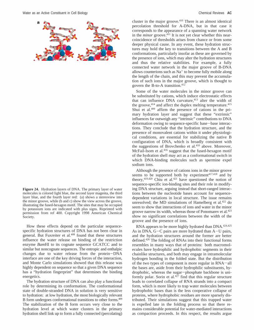
How these effects depend on the particular sequence-specific hydration structures of DNA has not been clear ingeneral. But Fuxreiter et al.408 found that these structuresinfluence the water release on binding of the restrictionenzymeBamHI to its cognate sequence GCATCC and tosimilar but noncognate sequences. The entropic and enthalpicchanges due to water release from the protein-DNAinterface are one of the key driving forces of the interaction,and Monte Carlo simulations showed that this release washighly dependent on sequence so that a given DNA sequencehas a “hydration fingerprint” that determines the bindingenergetics.
The hydration structure of DNA can also play a functionalrole by determining its conformation. The conformationalstate of double-stranded DNA in solution is very sensitiveto hydration: at low hydration, the most biologically relevantB form undergoes conformational transitions to other forms.409
The stabilization of the B form occurs very close to thehydration level at which water clusters in the primaryhydration shell link up to form a fully connected (percolating)
cluster in the major groove.410 There is an almost identicalpercolation threshold for A-DNA, but in that case itcorresponds to the appearance of a spanning water networkin theminor groove.411 It is not yet clear whether this near-coincidence of thresholds arises from chance or from somedeeper physical cause. In any event, these hydration struc-tures may hold the key to transitions between the A and Bconformations, particularly insofar as these are governed bythe presence of ions, which may alter the hydration structuresand thus the relative stabilities. For example, a fullyconnected water network in the major groove of B-DNAallows counterions such as Na+ to become fully mobile alongthe length of the chain, and this may prevent the accumula-tion of such ions in the major groove, which is thought togovern the B-to-A transition.412
Some of the water molecules in the minor groove canbe substituted by cations, which induce electrostatic effectsthat can influence DNA curvature,413 alter the width ofthe groove,414 and affect the duplex melting temperature.415
Shui et al.400 affirm the presence of cations in the pri-mary hydration layer and suggest that these “extrinsic”influences far outweigh any “intrinsic” contributions to DNAdeformation owing to sequence-specific base-base interac-tions. They conclude that the hydration structure, and thepresence of monovalent cations within it under physiologi-cal conditions, are essential for stabilizing the native Bconfiguration of DNA, which is broadly consistent withthe suggestions of Brovchenko et al.410 above. Moreover,McFail-Isom et al.416 suggest that the fused-hexagon motifof the hydration shell may act as a conformational switch inwhich DNA-binding molecules such as spermine expelsodium ions.
Although the presence of cations ions in the minor grooveseems to be supported both by experiment417,418 and bytheory,419,420 Chiu et al.421 have questioned the notion ofsequence-specific ion-binding sites and their role in modify-ing DNA structure, arguing instead that short-ranged interac-tions between the nucleotide bases account for sequence-dependent variations in local structure. The issue remainsunresolved; the MD simulations of Hamelberg et al.422 doseem to show that interactions of ions and water in the minorgroove narrow its width, whereas those of Ponomarev et al.423
show no significant correlations between the width of thegroove and the presence of ions.
RNA appears to be more highly hydrated than DNA.424,425
As in DNA, G-C pairs are more hydrated than A-U pairs,and the hydration structures around the former are betterdefined.426 The folding of RNAs into their functional formsresembles in many ways that of proteins: both macromol-ecules have hydrophilic and hydrophobic segments in theirchainlike structures, and both may engage in intramolecularhydrogen bonding in the folded state. But the distributionof the two types of component is more regular in RNAsallthe bases are, aside from their hydrophilic substituents, hy-drophobic, whereas the sugar-phosphate backbone is uni-formly polar. Sorin et al.427 find that this regular structureleads to correlated collapse of RNA strands into a compactform, which is more likely to trap water molecules betweenhydrophobic bases than is the less cooperative collapse ofproteins, where hydrophobic residues are more sparsely dis-tributed. Their simulations suggest that this trapped wateris expelled late in the folding process so that there re-mains considerable potential for water-mediated interactionsas compaction proceeds. In this respect, the results argue
Figure 24. Hydration layers of DNA. The primary layer of watermolecules is colored light blue, the second layer magenta, the thirdlayer blue, and the fourth layer red: (a) shows a stereoview intothe minor groove, while (b and c) show the view across the groove,illustrating the fused-hexagon motif. The sites that may be occupiedby potassium ions are indicated with plus signs. Reprinted withpermission from ref 400. Copyright 1998 American ChemicalSociety.
Water as an Active Constituent in Cell Biology Chemical Reviews AC

that explicit water molecules buried within the foldingmacromolecule can play an important role in mediatingcompaction, as proposed for proteins by Cheung et al.199 andPapoian et al.203
7. ConclusionsWater plays a wide variety of roles in biochemical
processes. It maintains macromolecular structure and medi-ates molecular recognition, it activates and modulates proteindynamics, it provides a switchable communication channelacross membranes and between the inside and outside ofproteins. Many of these properties do seem to depend, to agreater or lesser degree, on the “special” attributes of theH2O molecule, in particular its ability to engage in direc-tional, weak bonding in a way that allows for reorientationand reconfiguration of discrete and identifiable three-dimensional structures. Thus, although it seems entirely likelythat someof water’s functions in biology are those of ageneric polar solvent rather than being unique to water itself,it is very hard to imagine any other solvent that could fulfillall of its rolessor even all of those that help to distinguisha generic polypeptide chain from a fully functioning protein.The fact that fully folded proteins moved from an aqueousto a nonaqueous environment may retain some of theirfunctionality does not alter this and does not detract fromthe centrality of water for life on earth.
That, however, is not the same as saying that all life mustbe aqueous. At least with our present (incomplete) state ofknowledge about pivotal concepts such as the hydrophobicinteraction, it is not obvious that any one of the functions ofwater in biology has to stand as an irreducible aspect of a“living system”. It is certainly possible to imagine, and evento make,428 artificial chemical systems that engage in someform of information transfersindispensable for inheritanceand Darwinian evolutionsin nonaqueous media. Thoseproperties of water that do seem extremely rare, if not unique,in a solventssuch as rapid proton transfer via Grotthuss or“Moses” intermolecular hoppingsare clearly exploited byterrestrial cells but do not seem obviously indispensable forlife to exist.
Moreover, creating and sustaining life in water faces somesignificant obstacles429sperhaps most notably the solvent’sreactivity, raising the problem of hydrolysis of polymericstructures and of fundamental building blocks such as sugars.How the first pseudobiological macromolecules on the earlyEarth avoided this problem is still something of a puzzle. Itis also unclear whether a solvent capable of engaging inhydrogen bonding might initially help or hinder the use ofthis valuable, reversible noncovalent interaction for definingcomplex structures in macromolecules and their aggregates.Certainly, there is now reason to believe that such moleculescan utilize both hydrogen bonding and solvophobic effectsin acquiring well-defined structures without needing wateras their solvent.61
Attempts to enunciate the irreducible molecular-scalerequirements for something we might recognize as “life”have so far been rather sporadic429,430and are often hamperedby the difficulty of looking at the question through anythingother than aqua-tinted spectacles. From the point of view ofthinking about nonaqueous astrobiological solvents, a reviewof water’s roles in terrestrial biochemistry surely raises onekey consideration straight away: it is not sufficient, in thiscontext, to imagine a clear separation between the “molecularmachinery” and the solvent. There is a two-way exchange
of behaviors between them, and this literally erases anydividing line between the “biological components” and theirenvironment. Water is an extraordinarily responsive andsympathetic solvent, as well as being far more than merelya solvent. If living systems depend on that kind of exchange,for example so that molecular information can be transmittedbeyond the boundaries of the molecules that embody it, oneis tempted to conclude that these systems would need tomake use of water.
It is not just because of its molecular-scale structure thatwater has been characterized as “biophilic”swhen Hendersonfirst raised the intriguing notion of water’s “fitness” as life’smatrix in 1913,431 he had in mind the unusual macroscopicproperties such as its high heat capacity and densityanomalies. Nonetheless, even these have their origins inwater’s more or less unique set of molecular characteristics.Barring some unforeseen revelation from the exploration ofTitan, however, it is likely that we will have to rely onexperiment rather than discovery to put Henderson’s hy-pothesis to the test. Rather excitingly, with the advent ofsynthetic biology,432-434 along with chemical and biologicalsystems for exploring “alternative biochemistries”,435-439 itis now far from inconceivable that this test can be arranged.
8. AcknowledgmentsFor commenting on the draft manuscript and providing
additional material, I thank Austen Angell, Bruce Berne,David Chandler, Jan Engberts, Ariel Ferna´ndez, John Finney,Klaus Gerwert, Teresa Head-Gordon, John Ladbury, RichardPashley, Lawrence Pratt, Klaus Schulten, Jeremy Smith,Sergei Sukharev, Gregory Voth, and Loren Williams.
9. Note Added in ProofSignificant new findings in this field are arriving so
frequently that a review cannot be anything more than asnapshot. While the paper was in production, for example,there has been a thorough study of the structure of aqueoussalt solutions440 (which provides further evidence for the lackof utility of the picture of structure-making and structure-breaking), an exploration of the effect of hydrophobicinteractions on pressure- and temperature-induced proteindenaturation,441 a beautiful example of hydration as a tuningparameter in the redox behavior of iron-sulfur proteins,442
a study of the reliance of DNA hybridization on an aqueousenvironment,443 an illustration of decoupling of protein andhydration dynamics for membrane proteins,444 and a reviewof water structure at hydrophobic and hydrophilic surfaces.445
Such advances will continue to be reported in an informaland no doubt highly incomplete way on the blog “Water inBiology” (http://waterinbiology.blogspot.com).
10. Note Added after ASAP PublicationThis paper was published on the Web on December 21,
2007, with errors in Figure 1 and section 2.1. The correctversion was posted to the Web on December 28, 2007.
11. References(1) Szent-Gyo¨rgyi, A. In Cell-Associated Water; Drost-Hansen, W, Clegg,
J. S., Eds.; Academic Press: New York, 1979.(2) Jacobi, J., Ed.Paracelsus: Selected Writings; Princeton University
Press, 1979; p 13.(3) Gerstein, M.; Levitt, M.Sci. Am.1998, 279 (Nov), 101.
AD Chemical Reviews Ball
(4) Marchand, P. J.Life in the Cold, 3rd ed.; University Press of NewEngland: Hanover, NH, 1996.
(5) Bowles, D. J.; Lillford, P. J.; Rees, D. A.; Shanks, I. A.Philos. Trans.R. Soc. London, Ser. B2002, 357, 827.
(6) Block, W. Sci. Prog.2003, 86, 77.(7) Lebreton, J.-P.; Witasse, O.; Sollazzo, C.; Blancquaert, T.; Couzin,
P.; Schipper, A.-M.; Jones, J. B.; Matson, D. L.; Gurvits, L. I.;Atkinson, D. H.; Kazeminejad, B.; Pe´rez-Ayucar, M. Nature2005,438, 758.
(8) Enderby, J. E.; Neilson, G. W. InWater: A ComprehensiVe Treatise;Franks, F., Ed.; 1979; Vol. 6, p 1.
(9) Wernet, Ph.; Nordlund, D.; Bergmann, U.; Cavalleri, M.; Odelius,M.; Ogasawara, H.; Na¨slund, L. Å.; Hirsch, T. K.; Ojama¨e, L.;Glatzel, P.; Pettersson, L. G. M.; Nilsson, A.Science2004, 304, 995.
(10) Smith, J.; Cappa, C. D.; Wilson, K. R.; Messer, B. M.; Cohen, R.C.; Saykally, R. J.Science2004, 306, 851.
(11) Head-Gordon, T.; Johnson, M. E.Proc. Natl. Acad. Sci. U.S.A.2006,103, 7973.
(12) Prendergast, D.; Galli, G.Phys. ReV. Lett. 2006, 96, 215502.(13) Smith, J. D.; Cappa, C. D.; Messer, B. M.; Drisdell, W. S.; Cohen,
R. C.; Saykally, R. J.J. Phys. Chem. B2006, 110, 20038.(14) Smith, J. D.; Cappa, C. D.; Wilson, K. R.; Cohen, R. C.; Geissler,
P. L.; Saykally, R. J.Proc. Natl. Acad. Sci. U.S.A.2005, 102, 14171.(15) Sciortino, F.; Geiger, A.; Stanley, H. E.Nature1991, 354, 218.(16) Mishima, O.; Stanley, H. E.Nature1998, 396, 329.(17) Vedamuthu, M.; Singh, S.; Robinson, G. W.J. Phys. Chem.1994,
98, 2222.(18) Frank, H. F.; Franks, F.J. Chem. Phys.1968, 48, 4746.(19) Eaves, J. D.; Loparo, J. J.; Fecko, C. J.; Roberts, S. T.; Tokmakoff,
A.; Geissler, P. L.Proc. Natl. Acad. Sci. U.S.A.2005, 102, 13019.(20) Soper, A. K.; Neilson, G. W.; Enderby, J. E.; Howe, R. A.J. Phys.
C: Solid State Phys.1977, 10, 1793.(21) Cappa, C. D.; Smith, J. D.; Wilson, K. R.; Messer, B. M.; Gilles, M.
K.; Cohen, R. C.; Saykally, R. J.J. Phys. Chem. B2005, 109, 7046.(22) Cappa, C. D.; Smith, J. D.; Messer, B. M.; Cohen, R. C.; Saykally,
R. J.J. Phys. Chem. B2006, 110, 5301.(23) Krekeler, C.; Delle Site, L.J. Phys.: Condens. Matter2007, 19,
192101.(24) Omta, A. W.; Kropman, M. F.; Woutersen, S.; Bakker, H. J.Science
2003, 301, 347.(25) McLain, S. E.; Soper, A. K.; Watts, A.J. Phys. Chem. B2006, 110,
21251.(26) McLain, S. E.; Soper, A. K.; Terry, A. E.; Watts, A.J. Phys. Chem.
B 2007, 111, 4568.(27) Soper, A. K.; Castner, E. W.; Luzar, A.Biophys. Chem.2003, 105,
649.(28) Hayashi, Y.; Katsumoto, Y.; Omori, S.; Kishii, N.; Yasuda, A.J.
Phys. Chem. B2007, 111, 1076.(29) Rezus, Y. L. A.; Bakker, H. J.Proc. Natl. Acad. Sci. U.S.A.2006,
103, 18417.(30) Wallqvist, A.; Covell, D. G.; Thirumalai, D.J. Am. Chem. Soc.1998,
120, 427.(31) Tobi, D.; Elber, R.; Thirumalai, D.Biopolymers2003, 68, 359.(32) Mountain, R. D.; Thirumalai, D.J. Am. Chem. Soc.2003, 125, 1950.(33) Lee, M.-E.; van der Vegt, N. F. A.J. Am. Chem. Soc.2006, 128,
4948.(34) Bennion, B. J.; Daggett, V.Proc. Natl. Acad. Sci. U.S.A.2003, 100,
5142.(35) Shulgin, I. L.; Ruckenstein, E.J. Phys. Chem. B2007, 111, 3990.(36) Frank, H. S.; Franks, F.J. Chem. Phys.1968, 48, 4746.(37) Kokubo, H.; Pettitt, B. M.J. Phys. Chem. B2007, 111, 5233.(38) Tanford, C.The Hydrophobic Effect, 2nd ed.; Wiley: New York,
1980.(39) Dixit, S.; Crain, J.; Poon, W. C. K.; Finney, J. L.; Soper, A. K.Nature
2002, 416, 829.(40) Kauzmann, W.AdV. Protein Chem.1969, 14, 1.(41) Frank, H. S.; Evans, M. W.J. Chem. Phys.1945, 13, 507.(42) Glew, D. N.J. Phys. Chem.1962, 66, 605.(43) Cheng, Y.; Rossky, P. J.Nature1998, 392, 696.(44) Pauling, L. InHydrogen Bonding; Hadzi, D., Ed.; Pergamon: New
York, 1959; p 1.(45) Sloan, E. D.Clathrate Hydrates of Natural Gases; Marcel Dekker:
New York, 1998.(46) Blokzijl, W.; Engberts, J. B. F. N.Angew. Chem., Int. Ed.1993, 32,
1545.(47) Ashbaugh, H. S.; Paulaitis, M. E.J. Am. Chem. Soc.2001, 123,
10721.(48) Lucas, M.J. Phys. Chem.1976, 80, 359.(49) Lee, B.Biopolymers1985, 24, 813.(50) Lee, B.Biopolymers1991, 31, 993.(51) Reiss, H.; Frisch, H.; Lebowitz, J. L.J. Chem. Phys.1959, 31, 369.(52) Stillinger, F. H.J. Solution Chem.1973, 2, 141.
(53) Hummer, G.; Garde, S.; Garcia, A. E.; Paulaitis, M. E.; Pratt, L. R.J. Phys. Chem. B1998, 102, 10469.
(54) Hummer, G.; Garde, S.; Garcia, A. E.; Pratt, L. R.Chem. Phys.2000,258, 349.
(55) Hummer, G.; Garde, S.; Garcia, A. E.; Pohorille, A.; Pratt, L. R.Proc. Natl. Acad. Sci. U.S.A.1996, 93, 8951.
(56) Garde, S.; Hummer, G.; Garcia, A. E.; Paulaitis, M. E.; Pratt, L. R.Phys. ReV. Lett. 1996, 77, 4966.
(57) Gomez, M. A.; Pratt, L. R.; Hummer, G.; Garde, S.J. Phys. Chem.1999, 103, 3520.
(58) Ashbaugh, H. S.; Pratt, L. R.ReV. Mod. Phys.2006, 78, 159.(59) Rajamani, S.; Truskett, T. M.; Garde, S.Proc. Natl. Acad. Sci. U.S.A.
2005, 102, 9475.(60) Southall, N. T.; Dill, K. A.; Haymet, A. D. J.J. Phys. Chem. B2001,
106, 521.(61) Huang, K.; Wu, C. W.; Sanborn, T. J.; Patch, J. A.; Kirshenbaum,
K.; Zuckermann, R. N.; Barron, A. E.; Radhakrishnan, I.J. Am.Chem. Soc.2006, 128, 1733.
(62) Zuckermann, R. N.; Kerr, J. M.; Kent, S. B. H.; Moos, W. H.J. Am.Chem. Soc.1992, 114, 10646.
(63) de Jong, P. H. K.; Wilson, J. E.; Neilson, G. W.; Buckingham, A.D. Mol. Phys.1997, 91, 99.
(64) Filipponi, A.; Bowron, D. T.; Lobban, C.; Finney, J. L.Phys. ReV.Lett. 1997, 79, 1293.
(65) Bowron, D. T.; Weigel, R.; Filipponi, A.; Roberts, M. A.; Finney, J.L. Mol. Phys.2001, 99, 761.
(66) Chau, P. L.; Mancera, R. L.Mol. Phys.1999, 96, 109.(67) Ashbaugh, H. S.; Asthagiri, D.; Pratt, L. R.; Rempe, S. B.Biophys.
Chem.2003, 105, 321.(68) LaViolette, R.; Copeland, K. L.; Pratt, L. R.J. Phys. Chem. A2003,
107, 11267.(69) Bowron, D. T.; Filipponi, A.; Roberts, M. A.; Finney, J. L.Phys.
ReV. Lett. 1998, 81, 4164.(70) Finney, J. L.Philos. Trans. R. Soc. London, Ser. B2004, 359, 1145.(71) Roduner, E.; Tregenna-Piggott, P.; Dilger, H.; Ehrensberger, K.;
Senba, M.J. Chem. Soc., Faraday Trans.1995, 91, 1935.(72) Kirchner, B.; Stubbs, J.; Marx, D.Phys. ReV. Lett.2002, 89, 215901.(73) Atwood, J. L.; Hamada, F.; Robinson, K. D.; Orr, G. W.; Vincent,
R. L. Nature1991, 349, 683.(74) Raschke, T. M.; Levitt, M.Proc. Natl. Acad. Sci. U.S.A.2005, 102,
6777.(75) Allesch, M.; Schwegler, E.; Galli, G.J. Phys. Chem. B2007, 111,
1081.(76) Dixit, S.; Crain, J.; Poon, W. C. L.; Finney, J. L.; Soper, A. K.Nature
2002, 416, 829.(77) Dixit, S.; Soper, A. K.; Finney, J. L.; Crain, J.Europhys. Lett.2002,
59, 377.(78) Soper, A. K.; Finney, J. L.Phys. ReV. Lett. 1993, 71, 4346.(79) Finney, J. L.; Bowron, D. T.; Daniel, R. M.; Timmis, P. A.; Roberts,
M. A. Biophys. Chem.2003, 105, 391.(80) Lum, K.; Chandler, D.; Weeks, J. D.J. Phys. Chem. B1999, 103,
4570.(81) Wallqvist, A.; Berne, B. J.J. Phys. Chem.1995, 99, 2893.(82) Huang, X.; Margulis, C. J.; Berne, B. J.Proc. Natl. Acad. Sci. U.S.A.
2003, 100, 11953.(83) Koishi, T.; Yoo, S.; Yasuoka, K.; Zeng, X. C.; Narumi, T.; Susukita,
R.; Kawai, A.; Furusawa, H.; Suenaga, A.; Okimoto, N.; Futasugi,N.; Ebisuzaki, T.Phys. ReV. Lett. 2004, 93, 185701.
(84) ten Wolde, P. R.; Chandler, D.Proc. Natl. Acad. Sci. U.S.A.2002,99, 6539.
(85) Zhou, R.; Huang, X.; Margulis, C. J.; Berne, B. J.Science2004,305, 1605.
(86) MacCullum, J. L.; Maghaddam, M. S.; Chan, H. S.; Tieleman, D. P.Proc. Natl. Acad. Sci. U.S.A.2007, 104, 6206.
(87) Liu, P.; Huang, X.; Zhou, R.; Berne, B. J.Nature2005, 437, 159.(88) Hua, L.; Huang, X.; Liu, P.; Zhou, R.; Berne, B. J.J. Phys. Chem.
B 2007, 111, 9069.(89) Choudhury, N.; Pettitt, B. M.J. Am. Chem. Soc.2005, 127, 3556.(90) Choudhury, N.; Pettitt, B. M.J. Am. Chem. Soc.2007, 129, 4847.(91) Evans, R.; Marini Bettolo Marconi, U.; Tarazona, P.J. Chem. Phys.
1986, 84, 2376.(92) Bresme, F.; Wynveen, A.J. Chem. Phys.2007, 126, 044501.(93) Ball, P.Nature2003, 423, 25.(94) Jensen, T. R.; Jensen, M. Ø.; Reitzel, N.; Balashev, K.; Peters, G.
H.; Kjaer, K.; Bjørnholm, T.Phys. ReV. Lett. 2003, 90, 086101.(95) Mezger, M.; Reichert, H.; Scho¨der, S.; Okasinki, J.; Schro¨der, H.;
Dosch, H.; Palms, D.; Ralston, J.; Honkima¨ki, V. Proc. Natl. Acad.Sci. U.S.A.2006, 103, 18401.
(96) Poynor, A.; Hong, L.; Robinson, I. K.; Granick, S.; Zhang, Z.; Fenter,P. A. Phys. ReV. Lett. 2006, 97, 266101.
(97) Pertsin, A. J.; Hayashi, T.; Grunze, M.J. Phys. Chem. B2002, 106,12274.
Water as an Active Constituent in Cell Biology Chemical Reviews AE
(98) Ashbaugh, H. S.; Pratt, L. R.; Paulaitis, M. E.; Clohecy, J.; Beck, T.L. J. Am. Chem. Soc.2005, 127, 2808.
(99) Schwendel, D.; Hayashi, T.; Dahint, R.; Pertsin, A.; Grunze, M.;Steitz, R.; Schreiber, F.Langmuir2003, 19, 2284.
(100) Steitz, R.; Gutberlet, T.; Hauss, T.; Klo¨sgen, B.; Krastev, R.;Schemmel, S.; Simonsen, A. C.; Findenegg, G. H.Langmuir2003,19, 2409.
(101) Doshi, D. A.; Watkins, E. B.; Israelachvili, J. N.; Majewski, J.Proc.Natl. Acad. Sci U.S.A.2005, 102, 9458.
(102) Maccarini, M.; Steitz, R.; Himmelhaus, M.; Fick, J.; Tatur, S.; Wolff,M.; Grunze, M.; Janecek, J.; Netz, R. R.Langmuir2007, 23, 598.
(103) Dokter, A. M.; Woutersen, S.; Bakker, H. J.Proc. Natl. Acad. Sci.U.S.A.2006, 103, 15355.
(104) Jensen, M. Ø.; Mouritsen, O. G.; Peters, G. H.J. Chem. Phys.2004,120, 9729.
(105) Wiggins, P. M.Microbiol. ReV. 1990, 54, 432.(106) Stuart, S. J.; Berne, B. J.J. Phys. Chem.1996, 100, 11934.(107) Stuart, S.; Berne, B. J.J. Phys. Chem. A1999, 103, 10300.(108) Hofft, O.; Borodin, A.; Kahnert, U.; Kempter, V.; Dang, L. X.;
Jungwirth, P.J. Phys. Chem. B2006, 110, 11971.(109) Jungwirth, P.; Tobias, D. J.Chem. ReV. 2006, 106, 1259.(110) Petersen, P. B.; Saykally, R. J.Annu. ReV. Phys. Chem.2006, 57,
333.(111) Pegram, L. M.; Record, M. T., Jr.Proc. Natl. Acad. Sci. U.S.A.2006,
103, 14278.(112) Vaitheeswaran, S.; Thirumalai, D.J. Am. Chem. Soc.2006, 128,
13490.(113) Petersen, M. K.; Iyengar, S. S.; Day, T. J. F.; Voth, G. A.J. Phys.
Chem. B2004, 108, 14804.(114) Mucha, M.; Frigato, T.; Levering, L. M.; Allen, H. C.; Tobias, D.
J.; Dang, L. X.; Jungwirth, P.J. Phys. Chem. B2005, 109, 7617.(115) Iyengar, S. S.; Day, T. J. F.; Voth, G. A.Int. J. Mass Spectrom.
2005, 241, 197.(116) Petersen, P. B.; Saykally, R. J.J. Phys. Chem. B2005, 109, 7976.(117) Tarbuck, T. L.; Ota, S. R.; Richmond, G. L.J. Am. Chem. Soc.2006,
128, 14519.(118) Buch, V.; Milet, A.; Vacha, R.; Jungwirth, P.; Devlin, J. P.Proc.
Natl. Acad. Sci. U.S.A.2007, 104, 7342.(119) Collins, K. D.; Washabaugh, M. W.Q. ReV. Biophys.1985, 18, 323.(120) Kunz, W.; Henle, J.; Ninham, B. W.Curr. Opin. Colloid Interface
Sci.2004, 9, 19.(121) Franks, F.Water; Royal Society of Chemistry: London, 1983.(122) Hribar, B.; Southall, N. T.; Vlachy, V.; Dill, K. A.J. Am. Chem.
Soc.2002, 124, 12302.(123) Pegram, L. M.; Record, M. T., Jr.J. Phys. Chem. B2007, 111, 5411.(124) Pinna, M. C.; Salis, A.; Monduzzi, M.; Ninham, B. W.J. Phys. Chem.
B 2005, 109, 5406.(125) Salis, A.; Bilanicova´, D.; Ninham, B. W.; Monduzzi, M.J. Phys.
Chem. B2007, 111, 1149.(126) Sachs, J. N.; Woolf, T. B.J. Am. Chem. Soc.2003, 125, 8742.(127) Bowron, D. T.; Finney, J. L.Phys. ReV. Lett. 2002, 89, 215508.(128) Bowron, D. T.; Finney, J. L.J. Chem. Phys.2003, 118, 8357.(129) Finney, J. L.; Bowron, D. T.Curr. Opin. Colloid Interface Sci.2004,
9, 59.(130) Zangi, R.; Berne, B. J.J. Phys. Chem. B2006, 110, 22736.(131) Mayele, M.; Holz, M.Phys. Chem. Chem. Phys.2000, 2, 2429.(132) Zangi, R.; Hagen, M.; Berne, B. J.J. Am. Chem. Soc.2007, 129,
4678.(133) Israelachvili, J. N.; Pashley, R. M.Colloids Surf.1981, 2, 169.(134) Israelachvili, J. N.; Pashley, R. M.Nature1982, 300, 341.(135) Blake, T. D.; Kitchener, J. A.J. Chem. Soc., Faraday Trans. 11972,
68, 1435.(136) Parker, J. L.; Claesson, P. M.; Attard, P.J. Phys. Chem.1994, 98,
8468.(137) Pashley, R. M.; McGuiggan, P. M.; Ninham, B.; Evans, D. F.Science
1985, 229, 1088.(138) Attard, P.J. Phys. Chem.1989, 93, 6441.(139) Podgornik, R.J. Chem. Phys.1989, 91, 5840.(140) Tsao, Y. H.; Evans, D. F.; Wennerstro¨m, H. Langmuir1993, 9, 779.(141) Despa, F.; Berry, R. S.Biophys. J.2007, 92, 373.(142) Despa, F.; Ferna´ndez, A.; Berry, R. S.Phys. ReV. Lett. 2004, 93,
228104.(143) Attard, P.Langmuir1996, 12, 1693.(144) Carambassis, A.; Jonker, L. C.; Attard, P.; Rutland, M. W.Phys.
ReV. Lett. 1998, 80, 5357.(145) Tyrrell, J. W. G.; Attard, P.Phys. ReV. Lett. 2001, 87, 176104.(146) Tyrrell, J. W. G.; Attard, P.Langmuir2002, 18, 160.(147) Simonsen, A. C.; Hansen, P. L.; Klo¨sgen, B.J. Colloid Interface
Sci.2004, 273, 291.(148) Zhang, X. H.; Khan, A.; Ducker, W. A.Phys. ReV. Lett. 2007, 98,
136101.(149) Dammer, S. M.; Lohse, D.Phys. ReV. Lett. 2006, 96, 206101.
(150) Craig, V. S. J.; Ninham, B. W.; Pashley, R. M.Langmuir1999, 15,1562.
(151) Considine, R. F.; Hayes, R. A.; Horn, R. G.Langmuir 1999, 15,1657.
(152) Stevens, H.; Considine, R. F.; Drummond, C. J.; Hayes, R. A.; Attard,P. Langmuir2005, 21, 6399.
(153) Seo, H.; Yoo, M.; Jeon, S.Langmuir2007, 23, 1623.(154) Pashley, R. M.J. Phys. Chem. B2003, 107, 1714.(155) Snoswell, D. R. E.; Duan, J.; Fornasiero, D.; Ralston, J.J. Phys.
Chem. B2003, 107, 2986.(156) Meyer, E. E.; Lin, Q.; Israelachvili, J. N.Langmuir2005, 21, 256.(157) Meyer, E. E.; Lin, Q.; Hassenkam, T.; Oroudjev, E.; Israelachvili, J.
N. Proc. Natl. Acad. Sci. U.S.A.2005, 102, 6839.(158) Meyer, E. E.; Rosenberg, K. J.; Israelachvili, J. N.Proc. Natl. Acad.
Sci. U.S.A.2006, 103, 15739.(159) Lessard, R. R.; Zieminski, S. A.Ind. Eng. Chem. Fundam.1971,
10, 260.(160) Craig, V. S. J.; Ninham, B. W.; Pashley, R. M.Nature1993, 364,
317.(161) Claesson, P. M.; Blom, C. E.; Herder, P. C.; Ninham, B. W.J. Colloid
Interface Sci.1986, 114, 234.(162) Christenson, H. K.; Fang, J.; Ninham, B. W.; Parker, J. L.J. Phys.
Chem.1990, 94, 8004.(163) Rand, R. P.; Parsegian, V. A.Biochim. Biophys. Acta1989, 988,
351.(164) Horn, R. G.; Smith, D. T.; Haller, W.Chem. Phys. Lett.1989, 162,
404.(165) Israelachvili, J. N.Chem. Scr.1985, 25, 7.(166) Marcelja, S.; Radic, N.Chem. Phys. Lett.1976, 42, 129.(167) Attard, P.; Batchelor, M. T.Chem. Phys. Lett.1988, 149, 206.(168) Kornyshev, A. A.; Leikin, S.Phys. ReV. A 1989, 40, 6431.(169) Israelachvili, J.; Wennerstro¨m, H. Nature1996, 379, 219.(170) Leikin, S.; Parsegian, V. A.; Rau, D. C.; Rand, R. P.Annu. ReV.
Phys. Chem.1993, 44, 369.(171) McIntosh, T. J.; Simon, S. A.Annu. ReV. Biophys. Biomol. Struct.
1994, 23, 27.(172) Saiz, L.; Klein, M. L.Acc. Chem. Res.2002, 35, 482.(173) Marrink, S.-J.; Berkowitz, M. InPermeability and Stability of Lipid
Bilayers; Disalvo, E. A., Simon, S. A., Eds.; CRC: Boca Raton,FL, 1995.
(174) Cheng, J.-X.; Pautot, S.; Weitz, D. A.; Xie, X. S.Proc. Natl. Acad.Sci. U.S.A.2003, 100, 9826.
(175) Rhykerd, C. L., Jr.; Schoen, M.; Diestler, D. J.; Cushman, J. H.Nature1987, 330, 461.
(176) Ellis, R. J.; Minton, A. P.Nature2003, 425, 27.(177) Thompson, H.; Soper, A. K.; Ricci, M. A.; Bruni, F.; Skipper, N. T.
J. Phys. Chem. B2007, 111, 5610.(178) Tsukahara, T.; Hibara, A.; Ikeda, Y.; Kitamori, T.Angew. Chem.,
Int. Ed. 2007, 46, 1180.(179) Major, R. C.; Houston, J. E.; McGrath, M. J.; Siepmann, J. I.; Zhu,
X.-Y. Phys. ReV. Lett. 2006, 96, 177803.(180) Li, T.-D.; Gao, J.; Szoszkiewicz, R.; Landman, U.; Riedo, E.Phys.
ReV. B 2007, 75, 115415.(181) Golding, I.; Cox, E. C.Phys. ReV. Lett. 2006, 96, 098102.(182) Cheung, M. S.; Klimov, D.; Thirumalai, D.Proc. Natl. Acad. Sci.
U.S.A.2005, 102, 4753.(183) Minh, D. D. L.; Chang, C.-E.; Trylska, J.; Tozzini, V.; McCammon,
J. A. J. Am. Chem. Soc.2006, 128, 6006.(184) Stradner, A.; Sedgwick, H.; Cardinaux, F.; Poon, W. C. K.; Egelhaaf,
S. U.; Schurtenberger, P.Nature2004, 432, 492.(185) Gliko, O.; Pan, W.; Katsonis, P.; Neumaier, N.; Galkin, O.; Weinkauf,
S.; Velikov, P. G.J. Phys. Chem. B2007, 111, 3106.(186) Hill, T. L. Thermodynamics of Small Systems; W. A. Benjamin: New
York, 1963, 1964; Vols. 1 and 2.(187) Pollack, G. H.; Reitz, F. B.Cell. Mol. Biol. 2001, 47, 885.(188) Pollack, G. H.Cells, Gels, and the Engines of Life; Ebner & Sons:
Seattle, WA, 2001.(189) Damadian, R.Science1971, 171, 1151.(190) Fung, B. M.; Wassil, D. A.; Durham, D. L.; Chesnut, E. W.; Durham,
N. N.; Berlin, K. D. Biochim. Biophys. Acta1975, 385, 180.(191) Tehei, M.; Franzetti, B.; Wood, K.; Gabel, F.; Fabiani, E.; Jasnin,
M.; Zamponi, M.; Oesterhelt, D.; Zaccai, G.; Ginzburg, M.; Ginzburg,B.-Z. Proc. Natl. Acad. Sci. U.S.A.2007, 104, 766.
(192) Mirsky, A. E.; Pauling, L.Proc. Natl. Acad. Sci. U.S.A.1936, 22,439.
(193) Pauling, L.; Corey, R. B.; Branson, R. H.Proc. Natl. Acad. Sci. U.S.A.1951, 37, 205.
(194) Kohtani, M.; Jones, T. C.; Schneider, J. E.; Jarrold, M. F.J. Am.Chem. Soc.2004, 126, 7420.
(195) Langmuir, I.Proc. R. Inst. G. B.1938, 30, 483.(196) Bernal, J. D.Proc. R. Inst. G. B.1939, 30, 541.(197) Lesser, G. J.; Rose, G. D.Proteins: Struct. Funct. Genet.1990, 8,
6.
AF Chemical Reviews Ball

(198) Shea, J. E.; Onuchic, J. N.; Brooks, C. L.Proc. Natl. Acad. Sci.U.S.A.2002, 99, 16064.
(199) Cheung, M. S.; Garcia, A. E.; Onuchic, J. N.Proc. Natl. Acad. Sci.U.S.A.2002, 99, 685.
(200) Garcia, A. E.; Onuchic, J. N.Proc. Natl. Acad. Sci. U.S.A.2003,100, 13898.
(201) Zhang, O.; Forman-Kay, J. D.Biochemistry1997, 36, 3959.(202) Mok, Y.-K.; Kay, C. M.; Kay, L. E.; Forman-Kay, J.J. Mol. Biol.
1999, 289, 619.(203) Papoian, G. A.; Ulander, J.; Eastwood, M. P.; Luthey-Schulten, Z.;
Wolynes, P. G.Proc. Natl. Acad. Sci. U.S.A.2004, 101, 3352.(204) Zong, C.; Papoian, G. A.; Ulander, J.; Wolynes, P. G.J. Am. Chem.
Soc.2006, 128, 5168.(205) Harano, Y.; Kinoshita, M.Chem. Phys. Lett.2004, 399, 342.(206) Harano, Y.; Kinoshita, M.Biophys. J.2005, 89, 2701.(207) Otting, G.; Liepinsh, E.; Wu¨thrich, K. Science1991, 254, 974.(208) Likic, V. A.; Prendergast, F. G.Proteins2001, 43, 65.(209) Marakov, V.; Pettitt, B. M.; Feig, M.Acc. Chem. Res.2002, 35,
376.(210) Denisov, V. P.; Halle, B.J. Mol. Biol. 1995, 245, 698.(211) Denisov, V. P.; Halle, B.J. Mol. Biol. 1995, 245, 682.(212) Makarov, V. A.; Andrews, B. K.; Smith, P. E.; Pettitt, B. M.Biophys.
J. 2000, 76, 2966.(213) Luise, A.; Falconi, M.; Desideri, A.Proteins2000, 39, 56.(214) Makarov, V. A.; Feig, M.; Andrews, B. K.; Pettitt, B. M.Biophys.
J. 1998, 75, 150.(215) Smolin, N.; Winter, R.J. Phys. Chem. B2004, 108, 15928.(216) Merzel, F.; Smith, J. C.Proc. Natl. Acad. Sci.2002, 99, 5378.(217) Svergun, D. I.; Richard, S.; Koch, M. H. J.; Sayers, Z.; Kuprin, S.;
Zaccai, G.Proc. Natl. Acad. Sci. U.S.A.1998, 95, 2267.(218) Rand, R. P.Science1992, 256, 618.(219) Colombo, M. F.; Rau, D. C.; Parsegian, V. A.Science1992, 256,
655.(220) Kornblatt, J. A.; Hui Bon Hoa, G.Biochemistry1990, 29, 9370.(221) Klibanov, A. M.Trends Biochem. Sci.1989, 14, 141.(222) Dunn, R. V.; Daniel, R. M.Philos. Trans. R. Soc. London, Ser. B
2004, 359, 1309.(223) Rupley, J. A.; Careri, G.AdV. Protein Chem.1991, 41, 37.(224) Modig, K.; Liepinsh, E.; Otting, G.; Halle, B.J. Am. Chem. Soc.
2004, 126, 102.(225) Pal, S. K.; Peon, J.; Zewail, A. H.Proc. Natl. Acad. Sci. U.S.A.2002,
99, 15297.(226) Li, T.; Hassanali, A. A.; Kao, Y.-T.; Zhong, D.; Singer, S. J.J. Am.
Chem. Soc.2007, 129, 3376.(227) Bhattacharyya, S. M.; Wang, Z.-G.; Zewail, A. H.J. Phys. Chem. B
2003, 107, 13218.(228) Russo, D.; Murarka, R. K.; Copley, J. R. D.; Head-Gordon, T.J.
Phys. Chem. B2005, 109, 12966.(229) Frauenfelder, H.; Sligar, S. G.; Wolynes, P. G.Science1991, 254,
1598.(230) Mattos, C.; Ringe, D.Curr. Opin. Struct. Biol.2001, 11, 761.(231) Olano, L. R.; Rick, S. W.J. Am. Chem. Soc.2004, 126, 7991.(232) Dwyer, J. J.; Gittis, A. G.; Karp, D. A.; Lattman, E. E.; Spencer, D.
S. Biophys. J.2000, 79, 1610.(233) Okimoto, N.; Nakamura, T.; Suenaga, A.; Futatsugi, N.; Hirano, Y.;
Yamaguchi, I.; Ebisuzaki, T.J. Am. Chem. Soc.2004, 126, 13132.(234) Bizzarri, A. R.; Cannistraro, S.J. Phys. Chem. B2002, 106, 6617.(235) Russo, D.; Hura, G.; Head-Gordon, T.Biophys. J.2004, 86, 1852.(236) Frauenfelder, H.; Fenimore, P. W.; Chen, G.; McMahon, B. H.Proc.
Natl. Acad. Sci. U.S.A.2006, 103, 15469.(237) Knapp, E. W.; Fischer, S. F.; Parak, F.J. Am. Chem. Soc.1982, 86,
5042.(238) Rasmussen, B. F.; Stock, A. M.; Ringe, D.; Petsko, G. A.Nature
1992, 357, 423.(239) Tilton, R. F.; Dewan, J. C.; Petsko, G. A.Biochemistry1992, 31,
2469.(240) Borovykh, I. V.; Gast, P.; Dzuba, S. A.J. Phys. Chem. B2005, 109,
7535.(241) Reat, V.; Dunn, R.; Ferrand, M.; Finney, J. L.; Daniel, R. M.; Smith,
J. C.Proc. Natl. Acad. Sci U.S.A.2000, 97, 9961.(242) Bizzarri, A. R.; Paciaroni, A.; Cannistraro, S.Phys. ReV. E 2000,
62, 3991.(243) Wong, C. F.; Zheng, C.; McCammon, J. A.Chem. Phys. Lett.1989,
154, 151.(244) Arcangeli, C.; Bizzarri, A. R.; Cannistraro, S.Chem. Phys. Lett.1998,
291, 7.(245) Tournier, A. L.; Xu, J.; Smith, J. C.Biophys. J.2003, 85, 1871.(246) Hayward, J. A.; Smith, J. C.Biophys. J.2002, 82, 1216.(247) Tarek, M.; Tobias, D. J.Phys. ReV. Lett. 2002, 88, 138101.(248) Tarek, M.; Tobias, D. J.Biophys. J.2000, 79, 3244.(249) Daniel, R. M.; Smith, J. C.; Ferrand, M.; Hery, S.; Dunn, R.; Finney,
J. L. Biophys. J.1998, 75, 2504.
(250) Dunn, R. V.; Re´at, V.; Finney, J.; Ferrand, M.; Smith, J. C.; Daniel,R. M. Biochem. J.2000, 346 (2), 355.
(251) Fenimore, P. W.; Frauenfelder, H.; McMahon, B. H.; Parak, F. G.Proc. Natl. Acad. Sci. U.S.A.2002, 99, 16047.
(252) Chen, S.-H.; Liu, L.; Fratini, E.; Baglioni, P.; Faraone, A.; Mamontov,E. Proc. Natl. Acad. Sci. U.S.A.2006, 103, 9012.
(253) Ito, K.; Moynihan, C. T.; Angell, C. A.Nature1999, 398, 492.(254) Faraone, A.; Liu, L.; Mou, C.-Y.; Yen, C.-W.; Chen, S.-H.J. Chem.
Phys.2004, 121, 10843.(255) Liu, L.; Chen, S.-H.; Faraone, A.; Yen, C.-W.; Mou, C.-Y.Phys.
ReV. Lett. 2005, 95, 117802.(256) Kumar, P.; Yan, Z.; Xu, L.; Mazza, M. G.; Buldyrev, S. V.; Chen,
S.-H.; Sastry, S.; Stanley, H. E.Phys. ReV. Lett. 2006, 97, 177802.(257) Chen, S.-H.; Liu, L.; Chu, X.; Zhang, Y.; Fratini, E.; Baglioni, P.;
Faraone, A.; Mamontov, E. 2006, arXiv:cond-mat/0605294. arXiv.orge-Print archive.
(258) Chu, X.-Q.; Baglioni, P.; Faraone, A.; Chen, S.-H. 2007, arXiv:physics/0703166. arXiv.org e-Print archive.
(259) Pizzitutti, F.; Bruni, F.Phys. ReV. E 2001, 64, 052905.(260) Russo, D.; Murarka, R. K.; Hura, G.; Verschell, E.; Copley, J. R.
D.; Head-Gordon, T.J. Phys. Chem. B2004, 108, 19885.(261) Oleinikova, A.; Smolin, N.; Brovchenko, I.; Geiger, A.; Winter, R.
J. Phys. Chem. B2005, 109, 1988.(262) Smolin, N.; Oleinikova, A.; Brovchenko, I.; Geiger, A.; Winter, R.
J. Phys. Chem. B2005, 109, 10995.(263) Kurkal, V.; Daniel, R. M.; Finney, J. L.; Tehei, M.; Dunn, R. V.;
Smith, J. C.Biophys. J.2005, 89, 1282.(264) Hamaneh, M. B.; Buck, M.Biophys. J.2007, 92, L49.(265) Purkiss, A.; Skoulakis, S.; Goodfellow, J. M.Philos. Trans. R. Soc.
London, Ser. A2001, 359, 1515.(266) Marino, M.; Braun, L.; Cossart, P.; Ghosh, P.Mol. Cell 1999, 4,
1063.(267) Papoian, G. A.; Ulander, J.; Wolynes, P. G.J. Am. Chem. Soc.2003,
125, 9170.(268) Ferna´ndez, A.; Scott, R.Biophys. J.2003, 85, 1914.(269) Ferna´ndez, A.; Scheraga, H. A.Proc. Natl. Acad. Sci. U.S.A.2003,
100, 113.(270) Ferna´ndez, A. preprint.(271) Ferna´ndez, A.; Kardos, J.; Scott, L. R.; Goto, Y.; Berry, R. S.Proc.
Natl. Acad. Sci. U.S.A.2003, 100, 6446.(272) De Simone, A.; Dodson, G. G.; Verma, C. S.; Zagari, A.; Fraternali,
F. Proc. Natl. Acad. Sci. U.S.A.2005, 102, 7535.(273) Clackson, T.; Ultsch, M. H.; Wells, J. A.; de Vos, J. A.J. Mol. Biol.
1998, 277, 1111.(274) Bogan, A. A.; Thorn, K. S.J. Mol. Biol. 1998, 280, 1.(275) Ma, B.; Wolfson, H. J.; Nussinov, R.Curr. Opin. Struct. Biol.2001,
11, 364.(276) Moreira, I. S.; Fernandes, P. A.; Ramos, M. J.J. Phys. Chem. B
2007, 111, 2697.(277) Renzoni, D. A.; Zvelebil, M. J. J. M.; Lundba¨ck, T.; Ladbury, J. E.
In Structure-Based Drug Design: Thermodynamics, Modeling andStrategy; Ladbury, J. E., Connelly, P. R., Eds.; Landes Bioscience:Georgetown, TX, 1997; p 161.
(278) Tame, J. R. H.; Murshudov, G. N.; Dodson, E. J.Science1994, 264,1578.
(279) Tame, J. R. H.; Sleigh, S. H.; Wilkinson, A. J.; Ladbury, J. E.Nat.Struct. Biol.1996, 3, 998.
(280) Chung, E.; Henriques, D.; Renzoni, D.; Zvelebil, M.; Bradshaw, J.M.; Waksman, G.; Robinson, C. V.; Ladbury, J. E.Structure1998,6, 1141.
(281) McPhalen, C. A.; James, M. N. G.Biochemistry1988, 27, 6582.(282) Bhat, T. N.; Bentley, G. A.; Fischmann, T. O.; Boulot, G.; Poljak,
R. J.Nature1990, 347, 483.(283) Bhat, T. N.; Bentley, G. A.; Boulot, G.; Greene, M. I.; Tello, D.;
Dall’ Acqua, W.; Souchon, H.; Schwarz, F. P.; Mariuzza, R. A.;Poljak, R. J.Proc. Natl. Acad. Sci. U.S.A.1994, 91, 1089.
(284) Quiocho, F. A.; Wilson, D. K.; Vyas, N. K.Nature1989, 340, 404.(285) Lemieux, R. U.Acc. Chem. Res.1996, 29, 373.(286) Ladbury, J. E.Chem. Biol.1996, 3, 973.(287) Clarke, C.; Woods, R. J.; Gluska, J.; Cooper, A.; Nutley, M. A.;
Boons, G.-J.J. Am. Chem. Soc.2001, 123, 12238.(288) Talhout, R.; Villa, A.; Mark, A. E.; Engberts, J. B. F. N.J. Am.
Chem. Soc.2003, 125, 10570.(289) Dunitz, J. D.Science1994, 264, 670.(290) Dunitz, J. D.Chem. Biol.1995, 2, 709.(291) Baldwin, E. T.; Bhat, T. N.; Gulnik, S.; Liu, B.; Topol, I. A.; Kiso,
Y.; Mimoto, T.; Mitsu, H.; Erickson, J. W.Structure1995, 3, 581.(292) Louis, J. M.; Dyda, F.; Nashed, N. T.; Kimmel, A. R.; Davies, D. R.
Biochemistry1998, 37, 2105.(293) Hong, L.; Zhang, X. J.; Foundling, S.; Hartsuck, J. A.; Tang, J.FEBS
Lett. 1997, 420, 11.
Water as an Active Constituent in Cell Biology Chemical Reviews AG

(294) Kervinen, J.; Thanki, N.; Zdanov, A.; Tina, J.; Barrish, J.; Lin, P.F.; Colonno, R.; Riccardi, K.; Samanta, H.; Wlodawer, A.ProteinPept. Lett.1996, 3, 399.
(295) Lam, P. Y. S.; Jadhav, P. K.; Eyermann, C. J.; Hodge, C. N.; Ru,Y.; Bacheler, L. T.; Meek, J. L.; Otto, M. J.; Rayner, M. M.; Wong,Y. N.; Chang, C.-H.; Weber, P. C.; Jackson, D. A.; Sharpe, T. R.;Erickson-Viitanen, S.Science1994, 263, 380.
(296) Li, Z.; Lazaridis, T.J. Am. Chem. Soc.2003, 125, 6636.(297) Bergqvist, S.; Williams, M. A.; O’Brien, R.; Ladbury, J. E.J. Mol.
Biol. 2004, 336, 829.(298) Henriques, D. A.; Ladbury, J. E.Arch. Biochem. Biophys.2001, 390,
158.(299) Wang, H.; Ben-Naim, A.J. Med. Chem.1996, 39, 1531.(300) Barillari, C.; Taylor, J.; Viner, R.; Essex, J. W.J Am. Chem. Soc.
2007, 129, 2577.(301) Friesner, R. A.; Murphy, R. B.; Repasky, M. P.; Frye, L. L.; Halgren,
T. A.; Sanschagrin, P. C.; Mainz, D. T.J. Med. Chem.2006, 49,6177.
(302) Young, T.; Abel, R.; Kim, B.; Berne, B. J.; Friesner, R. A.Proc.Natl. Acad. Sci. U.S.A.2007, 104, 808.
(303) Ohno, K.; Kamiya, N.; Asakawa, N.; Inoue, Y.; Sakurai, M.J. Am.Chem. Soc.2001, 123, 8161.
(304) Okada, T.; Fujiyoshi, Y.; Silow, M.; Navarro, J.; Landau, E. M.;Shichida, Y.Proc. Natl. Acad. Sci. U.S.A.2002, 99, 5982.
(305) Autenrieth, F.; Tajkhorshid, E.; Schulten, K.; Luthey-Schulten, Z.J.Phys. Chem. B2004, 108, 20376.
(306) Royer, W. E., Jr.; Pardanani, A.; Gibson, Q. H.; Peterson, E. S.;Friedman, J. M.Proc. Natl. Acad. Sci. U.S.A.1996, 93, 14526.
(307) Collins, M. D.; Hummer, G.; Quillin, M. L.; Matthews, B. W.; Gruner,S. M. Proc. Natl. Acad. Sci. U.S.A.2005, 102, 16668.
(308) Stetefeld, J.; Jenny, M.; Schulthess, T.; Landwehr, R.; Engel, J.;Kammerer, R. A.Nat. Struct. Biol.2000, 7, 772.
(309) Yin, H.; Hummer, G.; Rasaiah, J. C.J. Am. Chem. Soc.2007, 129,7369.
(310) Koga, K.; Gao, G. T.; Tanaka, H.; Zeng, X. C.Nature2001, 412,802.
(311) Yoshizawa, M.; Kusukawa, T.; Kawano, M.; Ohhara, T.; Taaka, I.;Kurihara, K.; Niimura, N.; Fujita, M.J. Am. Chem. Soc.2005, 127,2798.
(312) van Amsterdam, I. M. C.; Ubbink, M.; Einsle, O.; Messerschmidt,A.; Merli, A.; Cavazzini, D.; Rossi, G. L.; Canters, G. W.Nat. Struct.Biol. 2001, 9, 48.
(313) Tezcan, F. A.; Crane, B. R.; Winkler, J. R.; Gray, H. B.Proc. Natl.Acad. Sci. U.S.A.2001, 98, 5002.
(314) Francisco, W. A.; Wille, G.; Smith, A. J.; Merkelr, D. J.; Klinman,J. P.J. Am. Chem. Soc.2004, 126, 13168.
(315) Lin, J.; Balabin, I. A.; Beratan, D. N.Science2005, 310, 1311.(316) Gray, H. B.; Winkler, J. R.Proc. Natl. Acad. Sci. U.S.A.2005, 102,
3534.(317) Liang, Z. X.; Kurnikov, I. V.; Nocek, J. M.; Mauk, A. G.; Beratan,
D. N.; Hoffman, B. M.J. Am. Chem. Soc.2004, 126, 2785.(318) Krauss, M.; Gilson, H. S. R.; Gresh, N.J. Phys. Chem. B2001, 105,
8040.(319) Erhardt, S.; Jaime, E.; Weston, J.J. Am. Chem. Soc.2005, 127, 3654.(320) Wang, L.; Yu, X.; Hu, P.; Broyde, S.; Zhang, Y.J. Am. Chem. Soc.
2007, 129, 4731.(321) Derat, E.; Shaik, S.; Rovira, C.; Vidossich, P.; Alfonso-Prieto, M.J.
Am. Chem. Soc.2007, 129, 6346.(322) de Grotthuss, C. J. T.Ann. Chim.1806, 58, 54.(323) Agmon, N.Chem. Phys. Lett.1995, 244, 456.(324) Day, T. J. F.; Schmitt, U. W.; Voth, G. A.J. Am. Chem. Soc.2000,
122, 12027.(325) Kornyshev, A. A.; Kuznetsov, A. M.; Spohr, E.; Ulstrup, J.J. Phys.
Chem. B2003, 107, 3351.(326) Swanson, J. M. J.; Maupin, C. M.; Chen, H.; Petersen, M. K.; Xu,
J.; Wu, Y.; Voth, G. A.J. Phys. Chem. B2007, 111, 4300.(327) Lapid, H.; Agmon, N.; Peterson, M. K.; Voth, G. A.J. Chem. Phys.
2005, 122, 014506.(328) Mezer, A.; Friedman, R.; Noivirt, O.; Nachliel, E.; Gutman, M.J.
Phys. Chem. B2005, 109, 11379.(329) Suzuki, T.; Sota, T.J. Phys. Chem. B2005, 109, 12603.(330) Saenger, W.Nature1979, 279, 343.(331) Ermler, U.; Fritzsche, G.; Buchanan, S. K.; Michel, H.Structure1994,
2, 925.(332) Seibold, S. A.; Mills, D. A.; Ferguson-Miller, S.; Cukier, R. I.
Biochemistry2005, 44, 10475.(333) Lu, D.; Voth, G. A.J. Am. Chem. Soc.1998, 120, 4006.(334) Cui, Q.; Karplus, M.J. Phys. Chem. B2003, 107, 1071.(335) Martinez, S. E.; Huang, D.; Ponomarev, M.; Cramer, W. A.; Smith,
J. L. Protein Sci.1996, 5, 1081.(336) Schlichting, I.; Berendzen, J.; Chu, K.; Stock, A. M.; Maves, S. A.;
Benson, D. E.; Sweet, R. M.; Ringe, D.; Petsko, G. A.; Sligar, S. G.Science2000, 287, 1615.
(337) Akeson, M.; Deamer, D. W.Biophys. J.1991, 60, 101.(338) Xu, J.; Voth, G. A.Proc. Natl. Acad. Sci. U.S.A.2005, 102, 6795.(339) Taraphder, S.; Hummer, G.J. Am. Chem. Soc.2003, 125, 3931.(340) Xu, J.; Sharpe, M. A.; Qin, L.; Ferguson-Miller, S.; Voth, G. A.J.
Am. Chem. Soc.2007, 129, 2910.(341) Lamoureux, G.; Klein, M. L.; Berne`che, S.Biophys. J.2007, 92,
L82.(342) Pome`s, R.; Roux, B.Biophys. J.1996, 71, 19.(343) Mann, D. J.; Halls, M. D.Phys. ReV. Lett. 2003, 90, 195503.(344) Dellago, C.; Naor, M. M.; Hummer, G.Phys. ReV. Lett. 2003, 90,
105902.(345) Hummer, G.; Rasaiah, J. C.; Noworyta, J. P.Nature2001, 414, 188.(346) Mashl, R. J.; Joseph, S.; Aluru, N. R.; Jakobsson, E.Nano Lett.2004,
3, 589.(347) Byl, O.; Liu, J.-C.; Wang, Y.; Yim, W.-L.; Johnson, J. K.; Yates, J.
T., Jr.J. Am. Chem. Soc.2006, 128, 12090.(348) Brewer, M. L.; Schmitt, U. W.; Voth, G. A.Biophys. J.2001, 80,
1691.(349) Luecke, H.; Schobert, B.; Richter, H.-T.; Cartailler, J.-P.; Lanyi, J.
K. J. Mol. Biol. 1999, 291, 899.(350) Sass, H. J.; Bu¨ldt, G.; Gessenich, R.; Hehn, D.; Neff, D.; Schlesinger,
R.; Berendzen, J.; Ormos, P.Nature2000, 406, 649.(351) Baudry, J.; Tajkhorshid, E.; Molnar, F.; Phillips, J.; Schulten, K.J.
Phys. Chem. B2001, 105, 905.(352) Grudinin, S.; Bu¨ldt, G.; Gordeliy, V.; Baumgaertner, A.Biophys. J.
2005, 88, 3252.(353) Rammelsberg, R.; Huhn, G.; Lu¨bben, M.; Gerwert, K.Biochemistry
1998, 37, 5001.(354) Garczarek, F.; Gerwert, K.Nature2006, 439, 109.(355) Spassov, V. Z.; Luecke, H.; Gerwert, K.; Bashford, D.J. Mol. Biol.
2001, 312, 203.(356) Garczarek, F.; Brown, L. S.; Lanyi, J. K.; Gerwert, K.Proc. Natl.
Acad. Sci. U.S.A.2005, 102, 3633.(357) Rousseau, R.; Kleinschmidt, V.; Schmitt, U. W.; Marx, D.Phys.
Chem. Chem. Phys.2004, 6, 1848.(358) Mathias, G.; Marx, D.Proc. Natl. Acad. Sci. U.S.A.2007, 104, 6980.(359) Garczarek, F.; Wang, J.; El-Sayed, M. A.; Gerwert, K.Biophys. J.
2004, 87, 2676.(360) Lee, Y.-S.; Krauss, M.J. Am. Chem. Soc.2004, 126, 2225.(361) Schobert, B.; Brown, L. S.; Lanyi, J. K.J. Mol. Biol. 2003, 330,
553.(362) Furutani, Y.; Shibata, M.; Kandori, H.Photochem. Photobiol. Sci.
2005, 4, 661.(363) Muneda, N.; Shibata, M.; Demura, M.; Kandori, H.J. Am. Chem.
Soc.2006, 128, 6294.(364) Agre, P.Angew. Chem., Int. Ed.2004, 43, 4278.(365) Tajkhorshid, E.; Nollert, P.; Jensen, M. Ø.; Miercke, L. J. W.;
O’Connell, J.; Stroud, R. M.; Schulten, K.Science2002, 296, 525.(366) de Groot, B. L.; Grubmu¨ller, H. Science2001, 294, 2353.(367) Chakrabarti, N.; Tajkhorshid, E.; Roux, B.; Pome`s, R.Structure2004,
12, 65.(368) de Groot, B. L.; Frigato, T.; Grubmu¨ller, H. J. Mol. Biol.2003, 333,
279.(369) Warshel, A.; Burykin, A.Biophys. J.2003, 85, 3696.(370) Chen, H.; Wu, Y.; Voth, G. A.Biophys. J.2006, 90, L73.(371) Chen, H.; Ilan, B.; Wu, Y.; Zhu, F.; Schulten, K.; Voth, G. A.
Biophys. J.2007, 92, 46.(372) Savage, D. F.; Egea, P. F.; Robles Colmenares, Y.; O’Connell, J. D.
PLoS Biol.2003, 1 (3), e2.(373) Beitz, E.; Wu, B.; Holm, L. M.; Schultz, J. E.; Zeuthen, T.Proc.
Natl. Acad. Sci. U.S.A.2006, 103, 269.(374) Li, J.; Gong, X.; Lu, H.; Li, D.; Fang, H.; Zhou, R.Proc. Natl. Acad.
Sci. U.S.A.2007, 104, 3687.(375) Dellago, C.; Hummer, G.Phys. ReV. Lett. 2006, 97, 245901.(376) Smondyrev, A. M.; Voth, G. A.Biophys. J.2002, 83, 1987.(377) Wu, Y.; Voth, G. A.FEBS Lett.2003, 552, 23.(378) Wu, Y.; Voth, G. A.Biophys. J.2005, 89, 2402.(379) Beckstein, O.; Biggin, P. C.; Sansom, M. S. P. A.J. Phys. Chem. B
2001, 105, 12902.(380) Sriraman, S.; Kevrekidis, I. G.; Hummer, G. 2005, arXiv:cond-mat/
0503491. arXiv.org e-Print archive.(381) Wan, R.; Li, J.; Lu, H.; Fang, H.J. Am. Chem. Soc.2005, 127, 7166.(382) Zimmerli, U.; Gonnet, P. G.; Walther, J. H.; Koumoutsakos, P.Nano
Lett. 2005, 5, 1017.(383) Jiang, Y.; Lee, A.; Chen, J.; Cadene, M.; Chait, B. T.; MacKinnon,
R. Nature2002, 417, 515.(384) Jiang, Y.; Ruta, V.; Chen, J.; Lee, A.; MacKinnon, R.Nature2003,
423, 42.(385) Sukharev, S.; Betanzos, M.; Chiang, C.-S.; Guy, H. R.Nature2001,
409, 720.(386) Anishkin, A.; Chiang, C. S.; Sukharev, S.J. Gen. Physiol.2005,
125, 155.(387) Anishkin, A.; Sukharev, S.Biophys. J.2004, 86, 2883.
AH Chemical Reviews Ball
(388) Bass, R. B.; Strop, P.; Barclay, M.; Rees, D. C.Science2002, 298,1582.
(389) Spronk, S. A.; Elmore, D. E.; Dougherty, D. A.Biophys. J.2006,90, 3555.
(390) Herkovits, T. T.; Hattington, J. P.Biochemistry1972, 11, 4800.(391) Turner, D. H. InNucleic Acids. Structure, Properties and Functions;
Bloomfield, V. A., Crothers, D. M., Tinoco, I., Eds.; UniversityScience Books: Sausalito, CA, 2000; p 308.
(392) Hofstadler, S. A.; Griffey, R. H.Chem. ReV. 2001, 101, 377.(393) Rueda, M.; Kalko, S. G.; Luque, F. J.; Orozco, M.J. Am. Chem.
Soc.2003, 125, 8007.(394) Cui, S.; Albrecht, C.; Ku¨hner, F.; Gaub, H. E.J. Am. Chem. Soc.
2006, 128, 6636.(395) Oesterhelt, F.; Rief, M.; Gaub, H. E.New J. Phys.1999, 1, 6.1.(396) Wing, R.; Drew, H.; Takano, T.; Broka, C.; Tanaka, S.; Itakura, K.;
Dickerson, R. E.Nature1980, 287, 755.(397) Drew, H. R.; Wing, R. M.; Takano, T.; Broka, C.; Itakura, K.;
Dickerson, R. E.Proc. Natl. Acad. Sci. U.S.A.1981, 78, 2179.(398) Kopka, M. L.; Fratini, A. V.; Drew, H. R.; Dickerson, R. E.J. Mol.
Biol. 1983, 163, 129.(399) Liepinsh, E.; Otting, G.; Wuthrich, K.Nucleic Acids Res.1992, 20,
6549.(400) Shui, X.; Sines, C. C.; McFail-Isom, L.; VanDerveer, D.; Williams,
L. D. Biochemistry1998, 37, 16877.(401) Shui, X.; McFail-Isom, L.; Hu, G. G.; Williams, L. D.Biochemistry
1998, 37, 8541.(402) Tereshko, V.; Minasov, G.; Egli, M.J. Am. Chem. Soc.1999, 121,
3590.(403) Liepinsh, E.; Leupin, W.; Otting, G.Nucleic Acids Res.1994, 22,
2249.(404) Woods, K. K.; Lan, T.; McLaughlin, L. W.; Williams, L. D.Nucleic
Acids Res.2003, 31, 1536.(405) Ha, J. H.; Capp, M. W.; Hohenwalter, M. D.; Baskerville, M.; Record,
M. T., Jr. J. Mol. Biol. 1992, 228, 252.(406) Robinson, C. R.; Sligar, S. G.J. Mol. Biol. 1993, 234, 302.(407) Sidorova, N. Y.; Rau, D. C.Proc. Natl. Acad. Sci. U.S.A.1996, 93,
12272.(408) Fuxreiter, M.; Mezei, M.; Simon, I.; Osman, R.Biophys. J.2005,
89, 903.(409) Saenger, W.Principles of Nucleic Acid Structure; Springer-Verlag:
Berlin, 1984.(410) Brovchenko, I.; Krukau, A.; Oleinikova, A.; Mazur, A. K.Phys. ReV.
Lett. 2006, 97, 137801.(411) Brovchenko, I.; Krukau, A.; Oleinikova, A.; Mazur, A. K.J. Phys.
Chem. B2007, 111, 3258.(412) Mazur, A. K.J. Am. Chem. Soc.2003, 125, 7849.(413) Hud, N. V.; Feigon, J.J. Am. Chem. Soc.1997, 119, 5756.(414) Young, M. A.; Jayaram, B.; Beveridge, D. L.J. Am. Chem. Soc.
1997, 119, 59.(415) Spink, C. H.; Chaires, J. B.Biochemistry1999, 38, 496.(416) McFail-Isom, L.; Sines, C. C.; Williams, L. D.Curr. Opin. Struct.
Biol. 1999, 9, 298.
(417) Woods, K.; McFail-Isom, L.; Sines, C. C.; Howerton, S. B.; Stephens,R. K.; Williams, L. D. J. Am. Chem. Soc.2000, 122, 1546.
(418) Hud, N. V.; Sklenar, V.; Feigon, J.J. Mol. Biol. 1999, 286, 651.(419) Young, M. A.; Beveridge, D. L.J. Mol. Biol. 1998, 281, 675.(420) Feig, M.; Pettitt, B. M.Biophys. J.1999, 77, 1769.(421) Chiu, T. K.; Kaczor-Grzeskowiak, M.; Dickerson, R. E.J. Mol. Biol.
1999, 292, 589.(422) Hamelberg, D.; McFail-Isom, L.; Williams, L. D.; Wilson, W. D.J.
Am. Chem. Soc.2000, 122, 10513.(423) Ponomarev, S. Y.; Thayer, K. M.; Beveridge, D. L.Proc. Natl. Acad.
Sci. U.S.A.2004, 101, 14771.(424) Egli, M.; Portmann, S.; Usman, N.Biochemistry1996, 35, 8489.(425) Rozners, E.; Moulder, J.Nucleic Acids Res.2004, 32, 248.(426) Auffinger, P.; Westhof, E.J. Biomol. Struct. Dyn.1998, 16, 693.(427) Sorin, E. J.; Rhee, Y. M.; Pande, V. S.Biophys. J.2005, 88, 2516.(428) Tjivikua, T.; Ballester, P.; Rebek, J., Jr.J. Am. Chem. Soc.1990,
112, 1249.(429) Benner, S. A.; Ricardo, A.; Carrigan, M. A.Curr. Opin. Chem. Biol.
2004, 8, 672.(430) Philos. Trans. R. Soc. London, Ser. B2004, 359, (No. 1448), 1 ff
special issue.(431) Henderson, L.The Fitness of the EnVironment; Macmillan: London,
1913.(432) Benner, S.Nature2003, 421, 118.(433) Ferber, D.Science2004, 303, 158.(434) Benner, S. A., Ed.Redesigning the Molecules of Life; Springer-
Verlag: Berlin, 1988.(435) Piccirilli, J. A.; Krauch, T.; Moroney, S. E.; Benner, S. A.Nature
1990, 343, 33.(436) Wang, L.; Brock, A.; Herberich, B.; Schultz, P. G.Science2001,
292, 498.(437) Chin, J. W.; Cropp, T. A.; Anderson, J. C.; Zhang, Z.; Schultz, P.
G. Science2003, 301, 964.(438) Kool, E. T.Acc. Chem. Res.2002, 35, 936.(439) Liu, H.; Gao, J.; Saito, D.; Maynard, L.; Kool, E. T.J. Am. Chem.
Soc.2004, 126, 1102.(440) Mancinelli, R.; Botti, A.; Bruni, F.; Ricci, M. A.; Soper, A. K.J.
Phys. Chem.2007, 111, 13570.(441) Patel, B. A.; Debenedetti, P. G.; Stillinger, F. H.; Rossky, P. J.
Biophys. J.2007, 93, 4116.(442) Dey, A.; Jenney, F. E., Jr.; Adams, M. W. W.; Babini, E.; Takahashi,
Y.; Fukuyama, K.; Hodgson, K. O.; Hedman, B.; Solomon, E. I.Science2007, 318, 1464.
(443) Cui, S.; Yu, J.; Kuhner, F.; Schulten, K.; Gaub, H. E.J. Am. Chem.Soc.2007, 129, 14710.
(444) Wood, K.; Plazanet, M.; Gabel, F.; Kessler, B.; Oesterhelt, D.; Tobias,D. J.; Zaccai, G.; Weik, M.Proc. Natl. Acad. Sci. U.S.A.2007, 104,18049.
(445) Maccarini, M.Biointerphases2007, 2, MR1.
CR068037A
Water as an Active Constituent in Cell Biology PAGE EST: 34.6 Chemical Reviews AI