waterhouse-friderichsenensues, with marked prostration and collapse, attended -by a falling blood...
TRANSCRIPT

THE WATERHOUSE-FRIDERICHSEN SYNDROME:REPORT OF THREE CASES IN ADULTS WITH NECROPSY FINDINGS*
ABBOTT A. NEWMAN
Three additional cases, with autopsy findings, occurring at theNew Haven Hospital during 1943-1944, are offered to the increas-ing number of reports dealing with the occurrence of the Water-house-Friderichsen syndrome in adults.
Voelker, in 1894, described a fulminating purpura attended bybilateral adrenal hemorrhages. Waterhouse, in 1911, is creditedwith one of the earliest accurate descriptions of the disease, but notuntil several years later was the important rOle of the meningococcusdiscovered. Although some score of adults are now included in the150 reported cases, there is a 90 per cent incidence in children under10 years of age, 70 per cent of the cases occurring in children under2 years of age.
The clinical features of the disease are becoming increasinglyfamiliar. There may be a preceding upper respiratory infection,but the onset is usually abrupt, with sudden chills, fever, malaise,and an early appearance of petechial and purpuric lesions. Cyanosisensues, with marked prostration and collapse, attended -by a fallingblood pressure and a vanishing pulse. Circulatory collapse withdeath usually follows in 12 to 48 hours after the first indication ofthe disease. Only a small percentage have meningeal reactions.Cultures of spinal fluid are frequently negative.
PathogenesisThe meningococcus has been recognized as the etiological agent
in at least 60 per cent of the cases,2 and the incidence of this organismin the disease is probably higher. H. influenzae and pneumococcihave been reported.2' 1 Bacterial stains of tissues have revealedmeningococci in adrenal glands,7 skin, spleen,5 renal glomeruli,lungs, and liver.5 They have also been demonstrated within thecoats of blood vessel walls, as well as in surrounding tissues.6
PathologyThe most striking pathological feature of the disease is the* From the Laboratory of Pathology, Yale University School of Medicine.

YALE JOURNAL OF BIOLOGY AND MEDICINE
widespread vascular damage, generally considered a direct resultof overwhelming toxemia."'6, 15 Hemorrhages are found in theskin as well as on visceral, serous, and mucosal surfaces. Thelesions range from simple dermal capillary engorgement9 to asso-ciated hemorrhage,12 and in rare instances capillary thrombi contain-ing meningococci.'4 Hemorrhages into the adrenal glands are themost frequent findings. They vary from pin-point to massivevarieties, and from focal types to those that involve the entiregland. They rarely rupture through the capsule. The rich supplyof capillaries in the adrenal medulla of infants is believed to bea contributing factor to the high incidence of the syndrome inchildren.6 In about 95 per cent of the cases the adrenal involve-ment is bilateral.10
Other incidental lesions have been reported. Levinson9 foundeosinophils, lymphocytes, plasma cells, and polymorphonuclearleukocytes beneath Glisson's capsule. Herbert and Manges6 notedswelling of liver cells, sinusoidal and perisinusoidal congestion,distortion of the hepatic structure, and diffuse leukocytic infiltration,which they consider on the basis of frequency, as part of the syn-drome. Thomas and Leiphart'5 demonstrated focal midzonalhepatic necrosis and swelling of the liver cells. In one of thecases to be discussed in this paper, an acute diffuse hepatitis wasobserved. The occurrence of this lesion in adults disposes of theconfusion in interpretation raised by this picture in infants, whereiit has occasionally been attributed to extramedullary hemopoiesis.
In most cases, enlargement of the thymus, lymph nodes, Peyer'spatches, and splenic follicles has been dbserved.2 10 11 12 The lesionsvary from swelling, edema, and congestion of lymphatic tissueto definite adenitis, as was found in two of the adults includedin this report. Acute -myocarditis has been observed by Kwedar,8by Morrison,'3 and by Thomas and Leiphart.'5
Other unusual lesions include hemorrhage complicating coagula-tive necrosis in the glandular portion of the pituitary,4 andhemorrhages in various organs, including the ovary, thyroid, brain,muscle, and intestinal wall. Exudative meningitis has not 'beendescribed in the great majority of cases. The severe neurologicalsigns are attributed by Banks and McCartney3 to an associatedencephalomeningitis.
Review of the literature indicates that only acute congestion or
32
THE WATERHOUSE-FRIDERICHSEN SYNDROME
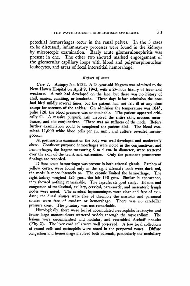
petechial hemorrhages occur in the renal pelves. In the 3 casesto.be discussed, inflammatory processes were found in the kidneysby microscopic examination. Early acute glomerulonephritis waspresent in one. The other two showed marked engorgement ofthe glomerular capillary loops with blood and polymorphonuclearleukocytes, and areas of focal interstitial hemorrhage.
Report of casesCase 1. Autopsy No. 6122. A 24-year-old Negress was admitted to the
New Haven Hospital on April 9, 1943, with a 24-hour history of fever andweakness. A rash had developed on the face, but there was no history ofchili, nausea, vomiting, or headache. Three days before admission the nosehad bled mildly several times, but the patient had not felt ill at any timeexcept for soreness of the ankles. On admission the temperature was 1040,pulse 120, the blood pressure was unobtainable. The patient appeared criti-cally ill. A massive purpuric rash involved the entire skin, mucous mem-brar-es, and the conjunctivae. There was no stiffness of the neck. Beforefurther examination could be completed the patient died. The blood con-tained 11,000 white blood cells per cu. mm., and culture revealed menin-gococci.
At postmortem examination the body was well developed and moderatelyobese. Confluent purpuric hemorrhages were noted in the conjunctivae, andhemorrhages, the largest measuring 3 to 4 cm. in diameter, were scatteredover the skin of the trunk and extremities. Only the pertinent postmortemfindings are recorded.
Diffuse acute hemorrhage was present in both adrenal glands. Patches ofyellow cortex were found only in the right adrenal; both were dark red,the medulla more intensely so. The capsule limited the hemorrhage. Theright kidney weighed 125 gms., the left 140 gms. Similar in appearance,they showed nothing remarkable. The capsules stripped easily. Edema andcongestion of mediastinal, axillary, cervical, para-aortic, and mesenteric lymphnodes were noted. The cerebral leptomeninges were clear and free of exu-date; the dural sinuses were free of thrombi; the mastoids and paranasalsinuses were free of exudate or hemorrhage. There was no cerebellarpressure cone. The pituitary was not remarkable.
Histologically, there were foci of accumulated neutrophilic leukocytes andfewer large mononuclears scattered widely through the myocardium. Thelesions were circumscribed and nodular, and resembled Aschoff nodules(Fig. 2). The liver cord cells were well preserved. A few focal collectionsof round cells and eosinophils were noted in the periportal zones. Diffusecongestion and hemorrhage involved both adrenals, particularly the medullary
33

YALE JOURNAL OF BIOLOGY AND MEDICINE
portions. Cell detail could be made out in only the outer third of the cortex.Capillary loops of a few glomeruli showed fibrinoid change and an accurpu-lation of numerous polymorphonuclear leukocytes (Fig. 1). Lymph nodesrevealed varying degrees of congestion and hyperplasia.
Case 2. Autopsy No. 6390. A 21-year-old white factory-workerdeveloped a slight cold with diarrhea and vomiting one week before admissionto the New Haven Hospital on December 17, 1943. On the night beforeadmission he suffered from headache, shaking chills, pain in the right knee,and a fever of 1040. On the day of admission nausea and vomiting recurred.On admission the temperature was 1040, pulse 120, respiration 32, and bloodpressure 70/40. The patient was a well-developed man appearing desperatelyill. There were several small petechial hemorrhages in the conjunc'tivae.The patient was given sulfamerazine intravenously and 20 cc. of corticalextract, but he died 6 hours after admission. His white count showed17,500 cells per cu. mm. of which 79 per cent were neutrophils. Numerousred blood cells, white blood cells, and casts were present in the urine. Bloodculture, as well as spinal fluid culture, yielded meningococci. The spinalfluid, however, was clear and colorless and the initial pressure was 150 mm.
At postmortem examination bilateral conjunctival hemorrhages werenoted. The right thorax yielded 200 cc. of amber fluid and 20 cc. of amberfluid were found in the pericardial sac. Serosal hemorrhages were noted inthe stomach and duodenum and intestines. The liver weighed 1840 gms.was extremely soft and of a homogeneous yellow-brown color presenting arough disorganized appearance on section. Some raised irregular areas andother depressed zones could be seen. The adrenal glands weighed 27 gms.together and both were destroyed by hemorrhage. The right kidney weighed185 gms., the left 190 gms. They were slightly swollen, large, and darkred. Numerous large hemorrhages could be seen in both testes. Themesenteric lymph nodes were swollen. The cerebral leptomeninges wereclear. There was no evidence of increased cranial pressure. The middle earsand the paranasal sinuses revealed no exudate.
There were many polymorphonuclear leukocytes in the interstitium ofthe myocardium with a rare zone of myocardial necrosis (Fig. 3). A diffusehepatitis with foci of necrosis was present. The adrenal gland was exten-sively hemorrhagic. The medulla could no longer be identified and thecortex was infiltrated with hemorrhage as far as the capsule. The renalglomeruli were markedly congested and there were accumulations of leu-kocytes in the glomerular tufts. Numerous interstitial hemorrhages of varyingsize were noted in 'both testes. There was an acute mesenteric lymphadenitis.
Case 3. Autopsy No. 6461. A 26-year-old white married womanwas admitted to the New Haven Hospital on February 11, 1944. On theafternoon of -the day of admission she suddenly had headaches and several
34
THE WATERHOUSE-FRIDERICHSEN SYNDROME
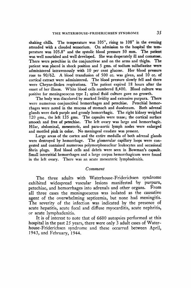
shaking chills. The temperature was 1050, rising to 1080 in the eveningattended with a clouded sensorium. On admission to the hospital the tem-perature was 105.80 and the systolic blood pressure 50 mm. The patientwas well nourished and well developed. She was desperately ill and comatose.There were petechiae in the conjunctivae and on the arms and thighs. Thepatient was placed in shock position and 5 gms. of sodium sulfadiazine wereadministered intravenously with 10 per cent glucose. Her blood pressurerose to 90/62. A blood transfusion of 500 cc. was given, and 10 cc. ofcortical extract were administered. The blood pressure slowly fell and therewere Cheyne-Stokes respirations. The patient expired 18 hours after theonset of her illness. White blood cells numbered 8,400. Blood culture waspositive for meningococcus type I; spinal fluid culture gave no growth.
The body was discolored by marked lividity and extensive purpura. Therewere numerous conjunctival hemorrhages and petechiae. Petechial hemor-rhages were noted in the mucosa of stomach and duodenum. Both adrenalglands were dark purple and grossly hemorrhagic. The right kidney weighed120 gms., the left 135 gms. The capsules were tense; the cortical surfacesmooath and free of petechiae. The left ovary was large and hemorrhagic.Hilar, abdominal, mesenteric, and para-aortic lymph nodes were enlargedand mottled pink in color. No meningeal exudate was present.
Large areas of the cortex and the entire medulla of both adrenal glandswere destroyed by hemorrhage. The glomerular capillary loops were con-gested and contained numerous polymorphonuclear leukocytes and occasionalfibrin plugs. Red blood cells and debris were seen in Bowman's capsule.Small interstitial hemorrhages and a large corpus hemorrhagicum were foundin !the left ovary. There was an acute mesenteric lymphadenitis.
Comment
The three adults with Waterhouse-Friderichsen syndromeexhibited widespread vascular lesions manifested by purpura,petechiae, and hemorrhages into adrenals and other organs. Fromall three cases the meningococcus was isolated as the causativeagent of the overwhelming septicemia, but none had meningitis.The severity of the infection was indicated by the presence ofacute hepatitis, acute focal and diffuse myocarditis, acute nephritis,or acute lymphadenitis.
It is of interest to note that of 6600 autopsies performed at thishospital in the past 25 years, there were only 3 adult cases of Water-house-Friderichsen syndrome and these occurred between April,1943, and February, 1944.
35

36 YALE JOURNAL OF BIOLOGY AND MEDICINE
Summary1. Three adults with Waterhouse-Friderichsen syndrome, bilateraladrenal hemorrhages, and overwhelming meningococcic septicemiaare described.2. Widespread vascular damage was present in all, as demon-strated by small and large hemorrhages in various organs.3. Early acute inflammatory reactions involved various organs,but the meninges were not included. Evidence of renal damagewas found histologically in all three cases.4. The three examples included in this report occurred during theperiod from April, 1943, to February, 1944, and are the onlyexamples of Waterhouse-Friderichsen syndrome encountered inadults during a 25-year period.
REFERENCES
1 Aegerter, E. E.: J. Am. Med. Asso., 1936, 706, 1715.2 Bachs, M. S.: Ann. Int. Med., 1937, 10, 1105-14.3 Banks, H. S., and J. E. McCartney: Lancet, 1943, i, 771-75.4 Gordon, W. H., and M. B. Simkin: J. Am. Med. Asso., 1943, 123, 147-48.5 Grace, W. H., C. V. Harrison, and T. B. Davie: Lancet, 1940, ii, 102-03.6 Herbert, P. A., and W. E. Manges: Arch. Path., 1943, 36, 413-22.7 Kunstadter, R. H.: Arch. Pediat., 1939, 56, 489-507.8 Kwedar, A. T.: Ann. Int. Med., 1942, 16, 787-91.9 Levinson, S. A.: J. Pediat., 1939, 14, 506-16.10 Lindsey, J. S., E. C. Rice, M. A. Selinger, and L. Robins: Am. J. Med. Sci.,
1941, 201, 263-70.11 Michael, P., and L. Jacobus: Arch. Pediat., 1942, 59, 141-47.12 Montfort, J. A., and J. H. Mehring: Am. J. Dis. Child., 1941, 62, 144-49.13 Morrison, J. E.: Lancet, 1943, i, 800-02.14 Ross, E. S., and J. R. Schenken: New Orleans Med. & Surg. J., 1940, 93,
294-300.15 Thomas, H. B., and C. D. Leiphart: J. Am. Med. Asso., 1944, 125, 884-90.

Fir. 1. (Case 1.) Section of kidne. Acute glonlmeltlonephritis. A gloiiei tilus containing red bloodcells, polymoiphonuclealar leukocytes, dn(d fiirin (leposits. X 400.
Fi4. 2. (Case 1.) Se(ction of iivos(>idimpii. A fse'd Iin a -dial1 lesihll illwhi0 h 1)(4ymlsrp(hllcells, inionionuclears, and n-sacrophages are seen. X 350.

it * MI W, 11§r or,s
:.!"
FiG. 3. (Case 2.) Section of mnwocardium with infiltration of polymorplhonuclear leuikocytes andswollen myocardial fibers. X 350.
-.NNW u.
+ ik:10. rf O%p
f,,,plft. 400
VW . qb lll-.Ib. 16.zVam.--.
'k -:...;,44W.A.Wr* "W.Ijw 4tkm
t . * .4f