wear behavior and in vitro cytotoxicity of wear debris...
TRANSCRIPT
Wear behavior and in vitro cytotoxicity of wear debris generated fromhydroxyapatite–carbon nanotube composite coating
Debrupa Lahiri,1 Ana Paula Benaduce,2 Francois Rouzaud,2 Jonathan Solomon,1,3
Anup Kumar Keshri,1 Lidia Kos,2 Arvind Agarwal1
1Plasma Forming Laboratory and High Temperature Tribology Laboratory, Mechanical and Materials Engineering, Florida
International University, Miami, Florida 331742Biological Sciences, Florida International University, Miami, Florida 331743Materials Science and Engineering, University of Florida, Gainesville, Florida 32611
Received 25 June 2010; revised 19 July 2010; accepted 21 July 2010
Published online 13 October 2010 in Wiley Online Library (wileyonlinelibrary.com). DOI: 10.1002/jbm.a.32952
Abstract: This work evaluates the effect of carbon nanotube
(CNT) addition to plasma-sprayed hydroxyapatite (HA) coating
on its tribological behavior, biocompatibility of the coating, and
cytotoxicity of CNT-containing wear debris. Biological response
of the CNT-containing wear debris is critical for osteoblasts, the
bone-forming cells, and macrophages, the cells that clear up
wear debris from blood stream. The addition of 4 wt % CNTs
to HA coating reduces the volume of wear debris generation by
80% because of the improved elastic modulus and fracture
toughness. CNT reinforcement has a pronounced effect on the
particle size in the wear debris and subsequent biological
response. There was a slight increase in the numbers and via-
bility of osteoblasts grown on HA–CNT compared with HA
alone. The cytotoxic effect of HA and HA–CNT debris to macro-
phages and osteoblasts was similar, demonstrating that loose
CNT does not pose a problem to these cells. VC 2010 Wiley Periodi-
cals, Inc. J Biomed Mater Res Part A: 96A: 1–12, 2011.
Key Words: carbon nanotube, hydroxyapatite, wear debris,
cytotoxicity, ostoeblast, macrophage
INTRODUCTION
Hydroxyapatite (HA) is a clinically proven and commerciallyused coating material for orthopedic implants because of itsunique bioactive and osteoconductive properties. The chem-ical composition of HA (Ca10(PO4)6(OH)2), its crystal struc-ture, and calcium to phosphate ratio (1.67) are similar tothat of apatite found in the human skeletal system.1–3 How-ever, certain mechanical properties of HA are insufficient formajor load-bearing implants. The fracture toughness ofdense HA (1 MPa.m0.5) is just half of the minimum reportedfracture toughness of cortical bone (2–12 MPa.m0.5).2 Poorwear resistance of HA is another limiting factor for its suc-cessful application in orthopedic implants.2–6 Low fracturetoughness and wear resistance of HA can be improved bycontrolling the HA grain size7 and/or adding second-phasereinforcements, such as carbon nanotubes (CNTs), that pos-sess high elastic modulus (200–1000 GPa8) and tensilestrength (11–63 GPa9). Previous studies have shown thatCNT is an effective reinforcement to HA for improving thefracture toughness and wear resistance.5,6,10
The coated implants stay inside the human body for along time. For example, �90% of the total hip implants staywithin the living body for >20 years.11 Wear debris is gen-
erated from the coated surface during the service period.Hence, it is important to assess the cytotoxicity of the weardebris for the safe use of the coated orthopedic implants.The issue becomes even more critical in case of HA–CNTcomposite coatings, because debris may contain embeddedand loose CNTs. Although the cytotoxicity of CNTs is stillunder debate,12 studies have shown CNTs to be biocompati-ble to bone cells.13–16 Usui et al.14 have shown that CNTspossess good bone–tissue compatibility without any toxiceffect. CNTs were implanted in the bone with the collagen,which resulted in an accelerated bone growth.14
Wear debris could be introduced into the blood streamin the long run. Wear debris, when generated, is suspendedin the body fluid near the bone or synovial fluid around thejoints. The debris could get introduced into the bloodstream from the body fluid at the finer capillary portion ofthe blood vessels through intercellular space between endo-thelial cells.17 In the blood, the debris is first attacked bythe macrophages, the security guard of the blood from anyforeign element. Macrophages engulf the debris in the bloodstream through a vesicular internalization process known asphagocytosis to clear them out from the blood. For effectiveremoval of the debris from the blood, the macrophages
Correspondence to: A. Agarwal; e-mail: [email protected]
Contract grant sponsor: NSF CAREER Award; contract grant number: NSF-DMI-0547178 (A.A)
Contract grant sponsor: Office of Naval Research; contract grant number: N00014-08-1-0494 (A.A)
Contract grant sponsor: DURIP program; contract grant number: N00014-06-0675 (A.A)
Contract grant sponsors: Dissertation Evidence Acquisition Fellowship by University Graduate School of Florida International University (D.L),
Dissertation Year Fellowship by University Graduate School of Florida International University (A.K.K)
VC 2010 WILEY PERIODICALS, INC. 1
need to ingest the debris without being harmed. Thus, theevaluation of cytotoxicity of wear debris to macrophages isimportant. Contradictory results have been reported on thecytotoxic effect of CNTs on macrophage cells. Chen et al.have found multiwall CNT causing incomplete phagocytosisand mechanically piercing through the plasma membranethat results in oxidative stress and macrophage cell death.18
But, at the same time, Cherukuri et al.19 have shown thatsingle-wall CNT is successfully ingested by the macrophagewithout any cytotoxic effect. At this point, it is worth men-tioning that until now no study has addressed the issue ofcytotoxicity of HA–CNT or any other CNT-containing weardebris to osteoblasts and macrophages. The size and mor-phology of wear debris are also important while assessingthe cytotoxic effect of the coating.
In view of the present scenario, the main objective ofthis study is to investigate the wear behavior and biologicalresponse of the wear debris generated from plasma-sprayedHA and HA–CNT coatings. This study thus involves under-standing the cytotoxicity of the wear debris on osteoblastand macrophage cells. The effect of CNT addition on thewear resistance, coefficient of friction (CoF), and tougheningof HA is also investigated. The size and morphology of thewear debris are correlated with the wear mechanism andthe biological response of osteoblast and macrophage cells.
EXPERIMENTAL
Powder preparation and coating synthesisHA nanorods (length: 100–325 nm, diameter: 25–50 nm, anddensity: 3.2 g/cm3) and multi-wall CNTs (95% purity; 40–70nm OD; 1–3 lm in length; density: 2.1 g/cm3) were procured
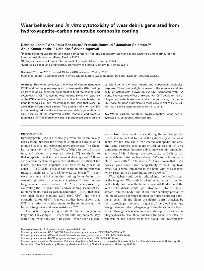
from Inframat Corporation (Willington, CT) and spray driedtogether [Fig. 1(a)] to synthesize the composite agglomerateswith a size of 15–55 lm [Fig. 1(b)]. During spray drying, HAnanorods and CNTs were dispersed in a water-soluble organicbinder. The suspension was sprayed in an atomized chamberand dried subsequently to obtain micron size porous sphericalagglomerates. Uniform distribution of CNT in the spray-driedpowder [Fig. 1(c)] enables a higher degree of homogeneity ofCNT distribution in the HA matrix.
Spray-dried HA and HA–CNT powder was plasmasprayed using SG 100 gun (Praxair Surface Technology, Dan-bury, CT) on 2.5-mm thick Ti-6Al-4V medical grade alloysubstrate. Plasma spraying is US food and drug administra-tion–approved process for coating deposition on orthopedicimplants. 23 kW plasma power was used with a powderfeeding rate of 4.5 g/min and standoff distance as 0.1 m. Ar-gon was used as the primary gas (flow rate: 30 slpm) withhelium as an auxiliary gas (28 slpm). Argon was also usedas powder carrier gas (25.5 slpm). The deposited coatinghad a uniform thickness of �150 lm. The coating densitywas measured using Archimedes principle. The coating wasstripped off from the substrate for the densitymeasurement.
CharacterizationThe cross-section of plasma-sprayed HA and HA–CNT coat-ings was metallographically polished for nanoindentation,microhardness, and microstructural characterization. JEOLJSM-633OF field emission scanning electron operating at 15kV with a working distance of �37–38 mm was used forthe characterization of powders, coatings, and wear debris.Hysitron Triboindenter (Hysitron, Minneapolis, MN) with
FIGURE 1. (a) Schematic showing spray drying of HA–CNT composite powder. (b) Agglomerated HA–CNT composite powder particles of dough-
nut shape. (c) High-magnification image of agglomerate showing distribution of CNTs in HA nanorods. [Color figure can be viewed in the online
issue, which is available at wileyonlinelibrary.com.]
2 LAHIRI ET AL. WEAR AND IN VITRO CYTOTOXICITY OF HA-CNT WEAR DEBRIS

100-nm Berkovich pyramidal tip was used in quasi-static in-dentation mode to measure the elastic modulus of the coat-ings. Indentation was performed on the polished cross-sec-tion of the coatings with a constant loading/unloading ratefor 10 s and 3 s hold at the peak load of 1200 lN. Elasticmodulus (E) was calculated from the load-displacementcurves using Oliver-Pharr method.20 Microhardness wasmeasured on the polished cross-section using a microhard-ness tester (Shanghai Taiming Optical Instrument Co., modelHXD-1000 TMC, Shanghai) with Vickers probe at 300g loadfor 15 s. For an accurate measurement of the radial cracklength, the indents were observed under scanning electronmicroscope (SEM). Fracture toughness was calculated usingthe method suggested by Anstis et al.21 Micro-Raman spec-troscopy of the spray-dried powder and coating was per-formed using a Spectra Physics (Model 3900S, CA) with Ti-sapphire crystal as target, a laser power of 18 mW and thedetector from Kaiser Optical Systems (MI).
Wear studyBall-on-disk tribometer (Nanovea, CA) was used to evaluatethe wear resistance and CoF of plasma-sprayed HA and HA–CNT coatings. The top surface of coatings was polished to aroughness (Ra) of 0.5 lm or less. Wear studies were per-formed at 50 RPM speed with a circular track of 3 mm radiusand a total travel distance of 100 m. An alumina ball of 3 mmdiameter was used as the counter surface. The wear load andsliding speed were chosen considering the condition of animplant inside the living body. A detailed discussion on thechoice of wear parameters is provided later in the ‘‘choice ofwear parameters’’ section. All wear tests were performed inthe ambient conditions without any lubricant. Three weartracks were made on each coating. The lateral force betweenthe alumina ball and the coating surface has been measuredby the linear variable differential transformer sensor. The CoFdata were acquired at a frequency of 16.67 Hz. Wear trackprofiles in across the tracks were obtained using NanoveaST400 Optical Profiler. Wear volume was computed using thedepth profile from the wear tracks. ImageJ22 software wasused for the quantitative analysis of the particle size in weardebris from the SEM images.
Osteoblast and macrophage cell culture andcytotoxicity testHuman osteoblasts ATCC CRL-11372 (ATCC, Manassas, VA)were seeded at a density of 1000 cells per well in six-wellpolystyrene petridishes (Corning, NY) at 310 K (37�C), 5%CO2 in a 1:1 mixture of Ham’s F12 Medium Dulbecco’smodified eagle’s medium, with 2.5 mM L-glutamine. Thephenol red–free base media was supplemented with 10%fetal bovine serum (Atlanta Biologicals, Lawrenceville, GA),100 UI/mL of penicillin, and 100 lg/mL of streptomycin(MP Biomedicals, Irvine, CA). Murine macrophages (J774Eclone, provided by Dr. M.A. Barbieri, Florida InternationalUniversity) were seeded in the same manner in Dulbecco’smodified eagle’s medium supplemented with 5% fetal bo-vine serum, 1% sodium pyruvate (Atlanta Biologicals), 100UI/mL of penicillin, and 100 lg/mL of streptomycin (MP
Biomedicals) at 310 K (37�C). Osteoblasts and macrophageswere allowed to attach to the plastic surface for 24 h, afterwhich the medium was replaced by a fresh medium supple-mented by HA and HA–CNT wear debris at 1 lg/mL con-centration (typically 2 mL of medium was added to thethree experimental wells). Before mixing in the culture me-dium, the wear particles were washed with 95% ETOH,washed three times with fresh medium, and left for 3 h in ahood under UV light. Both types of cells were cultured for 3days with the wear debris before the cytotoxicity test, whichwas performed with the CytoTox 96 Non-Radioactive Cyto-toxicity Assay kit (Promega, Madison, WI) following themanufacturer’s recommendations. Cytotoxicity test per-formed is a colorimetric assay that quantitatively measureslactate dehydrogenase (LDH), a stable cytosolic enzymereleased into the culture medium on cell lysis. ReleasedLDH in culture supernatant is measured with a coupled en-zymatic assay, which results in the formation of a red form-azan product that can be measured at 490 nm with a spec-trophotometer and is proportional to the number of cellslysed. Student t test was performed to find the 95% confi-dence interval. The absorbance value of the culture mediumwithout any cells or debris was considered as the back-ground and subtracted from the experimental absorbancevalues obtained for the cells cultured with and without de-bris. LDH absorbance was obtained for cells that died overthe 3-day experimental period. Cells that remained alive bythe end of the experiment were detached and lysed for therelease of LDH, which was then measured. The reportednumbers from the cytotoxicity tests (Table I) are the %LDHabsorbance value for the live cells.
Osteoblast viability test on the coatingsOsteoblasts were cultured in the same way as describedabove. The coatings on the substrates (5 mm � 5 mm sur-face area) were washed with 95% ETOH, washed threetimes with fresh medium, and left for 3 h in a hood underUV light. They were then placed into six-well polystyrenepetridishes (Corning, NY). For cell viability studies, osteo-blasts were seeded at a density of 5000 cells per well in 2.5mL of medium and grown in an incubator at 310 K (37�C)and 5% CO2. After 3 days, cells grown on the coatings werestained for 2 min with a phosphate buffer saline 1� solu-tion containing 15 lg/mL of fluorescein diacetate (FDA)(MP Biomedicals) and 4.5 lg/mL of propidium iodide (PI)(Fisher Scientific, Waltham, MA)23 before visualization on aLeica Leitz DM RB fluorescent microscope (Leica, Bannock-burn, IL). Digital pictures were captured with a Leica DM500 camera. Live (green) versus dead (red) cells countingwas manually assessed. Student t test was performed tofind the 95% confidence interval for the viability data.
RESULTS AND DISCUSSION
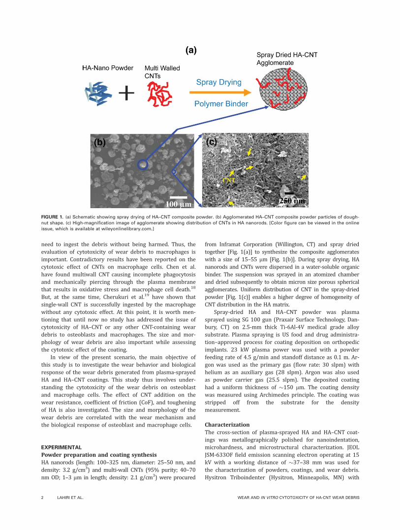
Microstructural characterization of coatingThe polished cross-section of HA–CNT coating reveals typi-cal signatures of plasma-sprayed structure, such as wavysplats, lamellar cracks, and porosity [Fig. 2(a)]. Density ofHA and HA–CNT coatings, measured using Archimedes
ORIGINAL ARTICLE
JOURNAL OF BIOMEDICAL MATERIALS RESEARCH A | JAN 2011 VOL 96, ISSUE 1 3
principle and water as immersion medium, is 93.7 and 94.0% TD, respectively. The densities are comparable for HAand HA–CNT coatings. Fracture surface of the HA–CNT com-posite coating [Fig. 2(b)] shows uniformly dispersed andembedded CNTs protruding out of HA matrix. The homoge-neous dispersion of CNT in HA at powder stage in spray-dried agglomerates [Fig. 1(c)] is carried forward to theplasma-sprayed coating. Thus, spray drying route for com-posite powder preparation is an effective method for homo-geneous dispersion of CNTs,24 which is required to obtainuniform mechanical properties. High-magnification SEMimage of the HA–CNT coating reveals attachment of fine HAparticles on the individual CNT surface. Higher thermal con-ductivity of CNT makes the HA nucleation and precipitationeasier on the CNT surface.10
High-temperature exposure during plasma sprayingmakes it necessary to check the survival of CNT structure inthe final coating. Micro-Raman spectrum (Fig. 3) of HA–CNTpowder and coating shows the presence of D and G peaks
coming from the CAC bond in CNT. The signature from theRaman spectrum along with the tubular structure visible inthe fracture surface [Fig. 2(b)] ensures the presence of CNTin the plasma-sprayed composite coating. Even after beingexposed to several thousand degrees, the survival of CNTsduring plasma spraying could be justified for the followingreasons. The short exposure (milliseconds) to high tempera-ture is not sufficient for the oxidation and destruction ofCNTs. In addition, the ceramic melts during plasma sprayingand forms protective coating on the CNT surface.24–25 Theinert carrier gas (argon) also creates a shroud over thesprayed particles, which acts as a shield against oxidation. Areduction in the peak intensity ratio (ID/IG) in plasma-sprayed coating (0.39) from powder (0.60) is observed,which indicates a decrease of defect density in CNT in thecoating. Similar observations have been reported by Keshriet al.26 for alumina–CNT plasma-sprayed coatings. Thedecrease in defects in CNT has been attributed to theincreasing degree of graphitization in CNT due to high-tem-perature exposure. Decrease in the strain energy duringannealing decreases the interlayer spacing between CNTwalls. Consequently, the defect density in CNT decreasesduring annealing.27,28
Tribological behavior of the coatingsConcern about the biological response of wear debris isdirectly related to the volume of the debris generated.
TABLE I. Percentage LDH Absorbance Values From Live Cells
Indicating Cytotoxicity Level of Wear Debris (p < 0.05)
Cell Type
%LDH Absorbance From Live Cells
With HA Debris With HA–CNT Debris
Osteoblasts 0.19 6 0.04 0.48 6 0.03Macrophages 0.50 6 0.06 0.46 6 0.01
FIGURE 2. (a) Cross-section of plasma-sprayed HA–CNT coating showing the splat structure, cracks, and porosity. (b) Fracture surface showing
homogeneous distribution of CNT in HA matrix. (c) Precipitation of HA crystals on CNTs.
4 LAHIRI ET AL. WEAR AND IN VITRO CYTOTOXICITY OF HA-CNT WEAR DEBRIS

Generation of more volume of debris results in higheramount of foreign element in contact with the bone cellsand in body fluid, which is always undesired. The amount ofdebris and its local concentration is reported to haveadverse effect on osteoblasts’ viability at the implant sur-face.29 Moreover, the wear debris can stimulate cellularresponses, which in turn may cause excess osteoclastic dif-ferentiation, reduced numbers of bone-forming cells, orstimulation of osteoblastic cells to release bone-resorbingmediators.30–34 All these phenomena lead to osteolysis andas a result loosening and failure of the implant. Hence, con-trolling the amount of debris generated is extremely impor-tant. The volume of debris generated is inversely related tothe wear resistance of the coating. Furthermore, the mor-phology of the wear debris is directly related to the wearmechanism controlling the tribological behavior of the coat-ings. The size and morphology of the wear debris are im-portant factors that regulate the cytotoxic response of osteo-blast and macrophage cells.
Choice of wear parameters. Parameters for the tribologicalstudy were selected considering the wear conditions of anorthopedic implant inside the human body. Hip joint is oneof the major load-bearing parts that face severe frictionalforces during movement. HA is usually coated on the stemof the femoral part and outer surface of the acetabular cupof the hip joint. The frictional forces faced by these partsare much lesser than the mating surface of the femoralhead and the inside surface of acetabular cup. The wear pa-rameters for the current study are selected based on thewear conditions faced by the femoral head inside the ace-tabular cup. The stress in a hip joint during walking is 0.8–2.5 MPa.35 HA coating is expected to withstand high fric-tional forces for a minimum of 20 years.11 Because wearexperiments cannot be continued for such a long period, thenormal load during the test was kept at a high value of 5 N,to obtain significant wear loss data. Considering the wearprobe as a ball of 3-mm diameter, the stress exerted on thewear track (coating) is determined using the calculationscheme adopted in our previous work.36 The calculatedstress on the wear track for 5 N normal load is �12 GPa,
which is three orders of magnitude higher than the actualloading condition at the hip joint.36 Wear speed wasselected by considering a normal walking speed of a healthyadult as 4 km/h. The average step size of a 6-ft tall man is0.91 m. Assuming the swinging action of a 25-mm diameteracetabular cup as the cause for wear during walking, thecalculated speed for wear is 955 mm/min. Hence, the wearspeed of the current study is fixed to �950 mm/min (50RPM). We acknowledge that movement in the hip implantmay best be simulated by fretting wear.37 However, the cur-rent study uses ball-on-disk wear technique, which is anaccepted technique, because several researchers have usedball/pin as counter surface along a circular track to evaluatewear characteristics of HA.38–42 Our experiments were con-ducted in dry condition as opposed to physiological solutionbecause dry condition is mechanically more aggressive toevaluate wear and would provide a conservative estimate ofthe wear resistance.
CoF. CoF was obtained continuously during sliding of thealumina ball on the wear track. Figure 4 shows the CoF forplasma-sprayed HA and HA–CNT coatings with 5 N normalload up to 100 m of distance. CoF decreases from 0.9 to0.68 with the CNT addition to HA matrix. The decrease inCoF in the presence of CNT is due to the lubrication offeredby the peeled-off graphite layers from the CNT surface.36
Removal of a single graphite layer from multiwall CNTrequires a tensile force �11 GPa along its axial direction.9
Lateral force applied in the wear causes shearing removal ofmass on the surface, which causes tensile stress along thesurface of wear track. Our previous study has showed thatcomputed tensile stress in the wear track was �12 GPa,which is sufficient to remove graphite layer from the CNTwithin the wear track.36 Hence, it is possible to havepeeled-off graphite layers in wear track of HA–CNT coating.
Wear resistance. Figure 5 shows the 3D profile of theentire wear track and 2D line profile across the wear trackfor HA and HA–CNT coatings. The volume of the wear track,calculated from the profiles, is 1.23 and 0.24 mm3 for HA
FIGURE 3. Raman spectrum of HA–CNT powder and plasma-sprayed
HA–CNT coating showing D and G peaks of CNT. [Color figure can be
viewed in the online issue, which is available at wileyonlinelibrary.com.]
FIGURE 4. Coefficient of friction for HA and HA–CNT coatings plotted
against sliding distance. [Color figure can be viewed in the online
issue, which is available at wileyonlinelibrary.com.]
ORIGINAL ARTICLE
JOURNAL OF BIOMEDICAL MATERIALS RESEARCH A | JAN 2011 VOL 96, ISSUE 1 5

and HA–CNT, respectively. Wear volume calculated for threetracks in each sample shows a standard deviation of <10%.Addition of CNT results in decrease in wear volume of HAcoating by 80%. The improvement in wear resistance meansgeneration of less volume of debris, resulting in reducedprobability of disturbance in the biological environmentaround the implant. Two major factors are responsible for theincrease in wear resistance of HA–CNT coating. The first fac-tor is the decrease in CoF. With decreasing CoF, the effectivelateral force on wear track is small, resulting in low wear vol-ume. The second and dominating factor is the toughening ofthe HA coating with CNT reinforcement, which makes the re-moval of mass difficult. To assess the toughening of HA coat-ing with CNT addition, the elastic modulus (E) and fracturetoughness (KIC) of HA–CNT composite coating has been eval-uated and compared with the HA coating.
Elastic modulus (E) of the coatings has been evaluatedusing nanoindentation, a well established technique for brit-tle ceramic coatings.43 Plasma-sprayed composite coating isheterogeneous in nature. Because the single indent provideslocalized mechanical property, more than 100 indents weremade at randomly chosen regions throughout the polishedcross-section of the coatings to obtain the bulk mechanicalproperty. In each region, the indents were made at a dis-tance of 9 lm from each other. The total area covered bythe indents was >5000 lm2 in each coating. Figure 6(a)shows a typical load-displacement curve obtained throughnanoindentation of HA and HA–CNT composite coatings. Thestatistical distribution of the elastic modulus [Fig. 6(b)],measured from the individual indent, thus provides the me-chanical property of the composite coating at macro-scalelength. The elastic modulus for HA and HA–CNT coating is
51 6 4 GPa and 88 6 10 GPa, respectively. A 72.5%improvement in elastic modulus is attributed to the high Evalue of CNT and effective reinforcement of CNT in HA ma-trix. Effective reinforcement is justified in terms of uniformdistribution of CNT in HA matrix [Fig. 2(b)] and good inter-facial bonding of each CNT with the HA matrix. Figure 6(c)shows a protruded CNT from the HA matrix. Strong bondingat HA–CNT interface is inferred from the absence of anycrack or gap at the interface.
Fracture toughness (KIC) of the coatings is evaluatedusing the length of the radial crack generated in microin-dentation. The impression of Vickers indent was observedunder SEM for an accurate measurement of the radial cracklengths. Figure 7 shows SEM images of indents on HA andHA–CNT coatings. The microhardness of HA and HA–CNTcoatings is 1.30 6 0.01 GPa and 2.43 6 0.02 GPa, respec-tively. Fracture toughness of the composite structures hasbeen evaluated using Anstis’ equation21 expressed as:
KIC ¼ 0:016E
H
� �1=2 P
c3=2(1)
where, P is the applied load, E is the elastic modulus, H isVickers hardness, and c is radial crack length (measuredfrom the center of the indent). E values, measured by nano-indentation, were used to compute KIC of the composite.The radial crack length in HA was >125 lm. But, the radialcracks are of much smaller length (�50 lm) in HA–CNTcoating. The decrease in radial crack length causes a higherfracture toughness of 3 MPa.m0.5 for HA–CNT comparedwith 0.64 MPa.m0.5 for HA. CNT addition improved the
FIGURE 5. Three-dimensional optical profile of the wear tracks and a two-dimensional profile across the track on (a) HA and (b) HA–CNT at 5 N
load and 100 m of sliding distance. [Color figure can be viewed in the online issue, which is available at wileyonlinelibrary.com.]
6 LAHIRI ET AL. WEAR AND IN VITRO CYTOTOXICITY OF HA-CNT WEAR DEBRIS

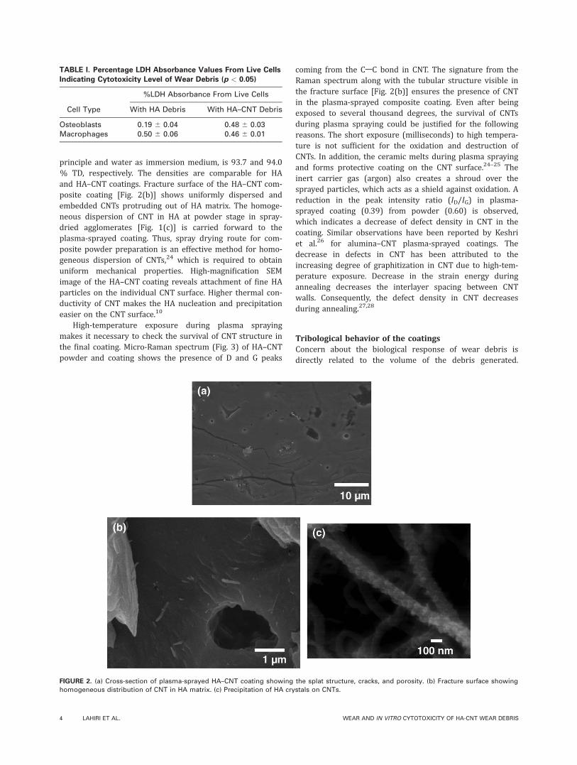
fracture toughness of HA coating by �350%, which isattributed to (i) higher E value with CNT addition and (ii)crack bridging effect offered by CNT. Figure 8 shows high-magnification SEM image of CNT bridges within a radialcrack generated during microindentation on HA–CNT coat-ing. CNTs act as bridges restricting the crack propagation. Aprevious study by our group on spark plasma-sintered HA–CNT composite has shown that the CNT pull-out energy inHA matrix is higher than the fracture energy of HA.36 Hence,crack propagating through HA matrix gets restricted whenit comes in vicinity of a CNT reinforcement.
Wear volume loss is a combined function of mechanicalproperties of material, such as KIC, E, and H. The model pro-
posed by Evans and Marshall estimates the wear volumeloss for brittle ceramics as a function of their mechanicalproperties.44 The relationship is as follows:
V ¼ P1:125K�0:5IC H�0:625 E
H
� �0:8
S (2)
where, V is the wear volume loss, P is normal load, KIC isthe fracture toughness, H is the hardness, E is the elasticmodulus, and S is the total travelling distance on weartrack. The computed volume loss for the current studyshows a 70% reduction in the wear volume with CNT addi-tion. The computed improvement shows a good match with80% wear resistance improvement for HA–CNT, obtainedexperimentally. The slight discrepancy between computedand experimental results can be explained by the lubricationavailable from the peeling of graphite layer, which is notaccounted by the computation model suggested by Evansand Marshall.
Wear debris size distribution and prediction of wearmechanisms. It is possible to predict the wear mechanismfrom the size and morphology of wear debris and the weartrack. Figure 9 shows a statistical ‘box-plot’ presenting thesize distribution of wear debris for HA and HA–CNT coat-ings. The debris from HA coating shows a wide range ofsize distribution (0.3–9.5 lm) with an average particle sizeof �3 lm. The average debris particle size in HA–CNT ismuch lower (0.6 lm) with a smaller size range (0.1–3.1lm). The wide variation in the HA debris size indicates thatwear in HA coating is governed by abrasion as well as frac-ture and chipping. Finer particles are generated during ab-rasive wear, whereas the larger particles are the result offracture and chipping. In case of HA–CNT coating, the major-ity of the debris particle is fine, indicating predominantlyabrasive wear. The images of wear track [Fig. 10(a,b)] revealsmaller size of craters and large area of abraded surface inHA–CNT wear track compared with HA. The crater is cre-ated by fracture and chipping, whereas the rough surface isthe result of abrasive wear. The mode of wear and debrissize is directly related to the fracture toughness of the
FIGURE 6. (a) Load versus displacement curves for HA and HA–CNT
coatings obtained by nanoindentation. (b) Statistical distribution of E
value in HA and HA–CNT measured for >100 nanoindents in each sam-
ple. (c) Protruding CNT from HA–CNT fracture surface showing absence
of crack or gap at matrix/CNT interface. [Color figure can be viewed in
the online issue, which is available at wileyonlinelibrary.com.]
FIGURE 7. SEM images of radial cracks emerging from microindents on (a) HA and (b) HA–CNT coatings.
ORIGINAL ARTICLE
JOURNAL OF BIOMEDICAL MATERIALS RESEARCH A | JAN 2011 VOL 96, ISSUE 1 7
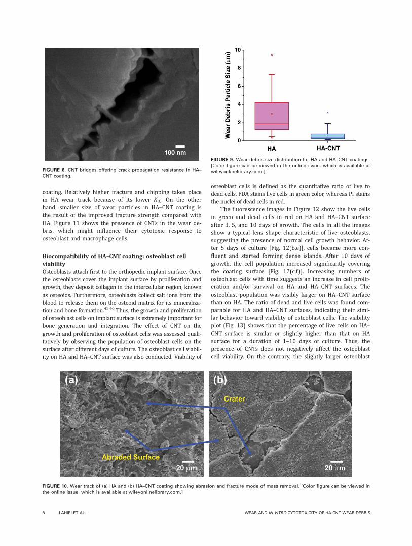
coating. Relatively higher fracture and chipping takes placein HA wear track because of its lower KIC. On the otherhand, smaller size of wear particles in HA–CNT coating isthe result of the improved fracture strength compared withHA. Figure 11 shows the presence of CNTs in the wear de-bris, which might influence their cytotoxic response toosteoblast and macrophage cells.
Biocompatibility of HA–CNT coating: osteoblast cellviabilityOsteoblasts attach first to the orthopedic implant surface. Oncethe osteoblasts cover the implant surface by proliferation andgrowth, they deposit collagen in the intercellular region, knownas osteoids. Furthermore, osteoblasts collect salt ions from theblood to release them on the osteoid matrix for its mineraliza-tion and bone formation.45,46 Thus, the growth and proliferationof osteoblast cells on implant surface is extremely important forbone generation and integration. The effect of CNT on thegrowth and proliferation of osteoblast cells was assessed quali-tatively by observing the population of osteoblast cells on thesurface after different days of culture. The osteoblast cell viabil-ity on HA and HA–CNT surface was also conducted. Viability of
osteoblast cells is defined as the quantitative ratio of live todead cells. FDA stains live cells in green color, whereas PI stainsthe nuclei of dead cells in red.
The fluorescence images in Figure 12 show the live cellsin green and dead cells in red on HA and HA–CNT surfaceafter 3, 5, and 10 days of growth. The cells in all the imagesshow a typical lens shape characteristic of live osteoblasts,suggesting the presence of normal cell growth behavior. Af-ter 5 days of culture [Fig. 12(b,e)], cells became more con-fluent and started forming dense islands. After 10 days ofgrowth, the cell population increased significantly coveringthe coating surface [Fig. 12(c,f)]. Increasing numbers ofosteoblast cells with time suggests an increase in cell prolif-eration and/or survival on HA and HA–CNT surfaces. Theosteoblast population was visibly larger on HA–CNT surfacethan on HA. The ratio of dead and live cells was found com-parable for HA and HA–CNT surfaces, indicating their simi-lar behavior toward viability of osteoblast cells. The viabilityplot (Fig. 13) shows that the percentage of live cells on HA–CNT surface is similar or slightly higher than that on HAsurface for a duration of 1–10 days of culture. Thus, thepresence of CNTs does not negatively affect the osteoblastcell viability. On the contrary, the slightly larger osteoblast
FIGURE 8. CNT bridges offering crack propagation resistance in HA–
CNT coating.
FIGURE 9. Wear debris size distribution for HA and HA–CNT coatings.
[Color figure can be viewed in the online issue, which is available at
wileyonlinelibrary.com.]
FIGURE 10. Wear track of (a) HA and (b) HA–CNT coating showing abrasion and fracture mode of mass removal. [Color figure can be viewed in
the online issue, which is available at wileyonlinelibrary.com.]
8 LAHIRI ET AL. WEAR AND IN VITRO CYTOTOXICITY OF HA-CNT WEAR DEBRIS

population and viability on HA–CNT surface with increasingnumber of days indicates the positive effect of CNTs inosteoblast proliferation. The density of both HA (93.7 % TD)and HA–CNT (94 % TD) coatings being similar in this studydoes not pose any differential effect on osteoblast viabilityand population. Studies on the biocompatibility of HA–CNTcomposites by other research groups have also suggestedthat the presence of CNTs in HA promotes osteoblast cellproliferation.10,47–49 The protein expression profile of osteo-blasts grown on HA–CNT surface was shown to be distinctfrom that of cells grown on HA and resulted in differences
in proliferation.50,51 These differences observed in cellsgrown on HA–CNTs may be explained by recent studiesindicating that CNTs can absorb various molecules on theirsurface.15,16 Akasaka et al.16 have shown that both single-wall nanotube (SWNT) and multiwall CNTs absorb proteinsfrom the serum (in cell culture medium) on their surfaceand positively influence osteoblast cell proliferation.Matsuoka et al.15 have further observed that the adhesionof osteoblasts on multiwall CNT surface is higher than onSWNT because of the rough and curled surface of the for-mer. More interestingly, both of these studies15,16 did notuse any surface treatment or functionalization for the CNTs.
The presence of CNTs can also play an important roleduring new bone formation and integration to the coatedimplant. During bone formation, osteoblasts deposit crystallinesalts on the collagen matrix, which combines to form HAcrystals. Balani et al.10 have shown that CNT surface is suita-ble for apatite mineralization. HA crystal binds on the CNTsurface with the favorable crystallographic orientation, whichresults into coherent bonding with a minimal lattice strain.36
Figure 2(c) shows the precipitation and attachment of HAcrystals on CNT surface during plasma spraying. Moreover,CNTs are reported to inhibit osteoclast differentiation, whichin turn controls osteoclastic resorption of bone.52 This is anadded advantage because the presence of CNTs in HA coatingcan prevent osteolysis at the implant surface, which couldotherwise lead to loosening and failure of the implant.
Cytotoxicity evaluation of wear debrisWear debris was collected and used for studying in vitro cyto-toxicity to osteoblast and macrophage cells. The cytotoxicity
FIGURE 11. Presence of CNTs in the wear debris generated from HA–
CNT coating.
FIGURE 12. Fluorescent images of osteoblast cells grown for 3, 5, and 10 days on (a–c) HA and (d–f) HA–CNT coatings. The live cells are stained in
green color with FDA and dead cells in red with PI. [Color figure can be viewed in the online issue, which is available at wileyonlinelibrary.com.]
ORIGINAL ARTICLE
JOURNAL OF BIOMEDICAL MATERIALS RESEARCH A | JAN 2011 VOL 96, ISSUE 1 9

assay used in this study allows for the rapid and accuratequantification of released LDH, a stable cytosolic enzymereleased from lysed cells. The amount of LDH released is pro-portional to the number of dead cells, which is quantifiedthrough a colorimetric assay by measuring the absorbance at490 nm. The percentage absorbance value reported in thisstudy is calculated using the LDH released from the live cellsin the medium (after they were lysed), with respect to thetotal LDH released from all the cells (live and dead) in themedium after 3 days of culture with debris.
Cytotoxicity response of osteoblasts. The cytotoxicity ofwear debris to osteoblasts is important because these cellsstay in direct contact with the implants during the bonegrowth stage. If osteoblasts die on the implant surfacebecause of the presence of debris, then new bone will notbe generated. Our results show that the level of cytotoxicityto osteoblasts was smaller when osteoblasts were culturedin the presence of debris from HA–CNT than with debrisfrom HA alone (Table I). A possible explanation for theseresults is the fact that the presence of CNTs in the debrismay have the potential for increasing osteoblast viability asdiscussed earlier in this study. Another reason could be thesize of wear debris. Osteoblasts can phagocytose solid par-ticles available in the culture medium.29 Bigger particles(0.3–9.5 lm) present in HA wear debris, if internalized, cancause more disturbance and harm to osteoblasts. Thus,smaller particle (0.1–3.1 lm) size in HA–CNT wear debriswill cause lesser disturbance if phagocytosed, resulting inhigher population of live osteoblasts in the medium.
Cytotoxicity response of macrophages. If internalization ofthe wear debris can cause harm or death of macrophages,the blood stream would be more prone to contaminationand infection. Hence, cytotoxicity of macrophages needs tobe assessed. The results of the cytotoxicity assay revealslightly higher level in the presence of HA–CNT debris thanHA (Table I). The difference, however, was not statisticallysignificant. This observation indicates that the presence of
CNTs in the debris does not alter the cytotoxicity responseof macrophages. HA–CNT debris are as biocompatible as HAdebris to macrophages. Despite the difference in the debrisparticle size for HA and HA–CNT, they show similar cyto-toxic behavior to macrophages. Olivier et al.53 have shownthat the death of macrophages occur at the same rate forboth smaller (0.45 lm) and larger (3.53 lm) polystyreneparticles, although the underlying mechanisms were differ-ent. The smaller particles caused apoptosis, whereas largerones induced necrosis of macrophages. Another aspect thatneeds to be taken into consideration is fiber geometry, givenits effect on the process of macrophage phagocytosis. Fiberswith a length of <17 lm were found to be noncytotoxic tomurine alveolar macrophages (�13 lm diameter).54 CNTsused in this study were much smaller (1–3 lm) than thecritical fiber length (17 lm) required shown to cause cyto-toxicity. Murine macrophages (J774 Eclone) used in thisstudy are larger (�20 lm diameter) than alveolar macro-phages, which should decrease the chances of cytotoxiceffect of loose CNTs in the wear debris. The recent study byKagan et al.55 reported that human myeloperoxidaseenzymes, contained in macrophages, can biodegrade CNTs(SWNT). Thus, it is unlikely that loose CNTs in debris posea threat to macrophages.
Wear particles can induce some behavioral changes inmacrophages, leading to adverse effect on biocompatibilityof implant.34 Metallic wear particles stimulate the macro-phages to release hormonal factors such as prostaglandin E2and interlukin-1.30,56,57 These factors induce osteoclastic dif-ferentiation of precursor cells.58–60 Increase in the osteo-clast formation aggravates the bone resorption. The pres-ence of CNTs in the composite coating would be beneficialin such a situation because it can reduce bone resorptionby inhibiting osteoclast proliferation and growth.52 Furtherstudies on the effects of wear debris produced in physiolog-ical solution, on macrophages and osteoblasts, can providemore insight on different aspects of cytocompatibility of HAand HA–CNT coatings for orthopedic implants.
CONCLUSION
The addition of 4 wt % CNTs to HA improves the wear re-sistance by 80% and results in less volume of debris gener-ation. The elastic modulus and fracture toughness of theplasma-sprayed coating increases by 72.5 and 350%,respectively, with CNT addition. Release of the graphite layerfrom CNT provides lubrication and decreases the CoF onHA–CNT surface. Presence of CNTs decrease the size ofwear particles (HA: 0.3–9.5 lm and HA–CNT: 0.1–3.1 lm).Cytotoxicity of osteoblast cells on HA–CNT surface is lowerthan on HA. This result could be explained as a cumulativeeffect of increased osteoblast viability in the presence ofCNT and decreasing particle size of HA–CNT debris. Osteo-blast cell population and viability is found slightly higher onHA–CNT surface up to 10 days of growth time. CNT surfaceis also found favorable for HA-crystal precipitation duringbone mineralization. Macrophage cytotoxicity response toHA–CNT debris is similar to that of HA. Presence of CNTsdoes not affect the cytotoxicity of wear debris. Thus, HA–
FIGURE 13. Osteoblast cell viability on HA and HA–CNT coatings for
1, 3, 5, and 10 days of culture (p < 0.05). [Color figure can be viewed
in the online issue, which is available at wileyonlinelibrary.com.]
10 LAHIRI ET AL. WEAR AND IN VITRO CYTOTOXICITY OF HA-CNT WEAR DEBRIS
CNT coatings on orthopedic implants are predicted to per-form better than HA, both mechanically and biologically,during their long stay inside the living body.
ACKNOWLEDGMENTS
The authors acknowledge the support fromMr. Neal Ricks andthe research facilities at Advanced Materials Engineering andResearch Institute (AMERI) at Florida International University.Ms. Sushmita Mustafi and Dr. M.A. Barbieri, Biological Sciencesat FIU, are acknowledged for providing the murine macro-phages. D. L. acknowledges valuable discussion with Dr. SisirMondal. The authors are thankful to the Center for study ofMatters in Extreme Conditions (CeSMEC) and Dr. S. Saxena forthe use of Micro-Raman Spectroscopy facility.
REFERENCES1. Gu YW, Loha NH, Khor KA, Tor SB, Cheang P. Spark plasma sin-
tering of hydroxyapatite powders. Biomaterials 2002;23:37–43.
2. White AA, Best SM, Kinloch IA. Hydroxyapatite-carbon nanotube
composites for biomedical applications: A review. Int J Appl
Ceram Tech 2007;4:1–13.
3. Yu LG, Khor KA, Li H, Cheang P. Effect of spark plasma sintering
on the microstructure and in vitro behavior of plasma sprayed HA
coatings. Biomaterials 2003;24:2695–2705.
4. Gu YW, Khor KA, Cheang P. Bone-like apatite layer formation on
hydroxyapatite prepared by spark plasma sintering (SPS). Bioma-
terials 2004;25:4127–4134.
5. Balani K, Chen Y, Harimkar SB, Dahotre NB, Agarwal A. Tribologi-
cal behavior of plasma-sprayed carbon nanotube-reinforced hy-
droxyapatite coating in physiological solution. Acta Biomater
2007;3:944–951.
6. Chen Y, Zhang TH, Gun CH, Yu G. Wear studies of hydroxyapatite
composite coating reinforced by carbon nanotubes. Carbon 2007;
45:998–1004.
7. Wang J, Shaw LL. Nanocrystalline hydroxyapatite with simultane-
ous enhancements in hardness and toughness. Biomaterials
2009;30:6565–6572.
8. Singh S, Pei Y, Miller R, Sundararajan PR. Long-range, entangled
carbon nanotube networks in polycarbonate. Adv Funct Mater
2003;13:868–872.
9. Yu MF, Lourie O, Dyer MJ, Moloni K, Kelly TF, Ruoff RS. Strength
and breaking mechanism of multiwalled carbon nanotubes under
tensile load. Science 2000;287:637–640.
10. Balani K, Anderson R, Laha T, Andara M, Tercero J, Crumpler E,
Agarwal A. Plasma-sprayed carbon nanotube reinforced hydroxy-
apatite coatings and their interaction with human osteoblasts in
vitro. Biomaterials 2007;28:618–624.
11. Neumann L, Freund KG, Sorenson KH. Long-term results of
Chernley total hip replacement. J Bone Joint Surg Br 1994;76:
245–251.
12. Fiorito S. Carbon nanotube: Angels or demons? Singapore: Pan
Stanford Publishing; 2008. ISBN: 9814241016, 9789814241014.
13. Kalbacova M, Kalbac M, Dunsch L, Hempel U. Influence of single-
walled carbon nanotube films on metabolic activity and adher-
ence of human osteoblasts. Carbon 2007;45:2266–2272.
14. Usui Y, Aoki K, Narita N, Murakami N, Nakamura I, Nakamura K,
Ishigaki N, Yamazaki H, Horiuchi H, Kato H, Taruta S, Kim YA,
Endo M, Saito N. Carbon nanotubes with high bone-tissue com-
patibility and bone-formation acceleration effects. Small 2008;4:
240–246.
15. Matsuoka M, Akasaka T, Totsuka Y, Watari F. Strong adhesion of
Saos-2 cells to multi-walled carbon nanotubes. Mater Sci Eng B
2010;173:182–186.
16. Akasaka T, Yokoyama A, Matsuoka M, Hashimoto T, Watari F.
Thin films of single-walled carbon nanotubes promote human
osteoblastic cells (Saos-2) proliferation in low serum concentra-
tions. Mater Sci Eng C 2010;30:391–399.
17. Pappas GD, Blanchette EJ. Transport of colloidal particles from
small blood vessels correlated with cyclic changes in permeabil-
ity. Invest Opthalmol 1965;4:1026–1035.
18. Cheng C, Muller KH, Koziol KK, Skepper JN, Midgley PA, Welland
ME, Porter AE. Toxicity and imaging of multi-walled carbon nano-
tubes in human macrophage cells. Biomaterials 2009;30:4152–4160.
19. Cherukuri P, Bachilo SM, Litovsky SH, Weisman RB. Near-infrared
fluorescence microscopy of single-walled carbon nanotubes in
phagocytic cells. J Am Chem Soc 2004;126:15638–15639.
20. Oliver WC, Pharr GM. An improved technique for determining
hardness and elastic modulus using load and displacement sens-
ing indentation experiments. J Mater Res 1992;7:1564–1583.
21. Anstis GR, Chantikul P, Lawn BR, Marshall DB. A critical evalua-
tion of indentation techniques for measuring fracture toughness.
I. Direct crack measurements. J Am Ceram Soc 1981;64:533–538.
22. Abramoff MD, Magelhaes PJ, Ram SJ. Image processing with
ImageJ. Biophotonics Int 2004;11:36–42.
23. Valdes JJ, Weeks OI. Estradiol and lithium chloride specifically al-
ter NMDA receptor subunit NR1 mRNA and excitotoxicity in pri-
mary cultures. Brain Res 2009;1268:1–12.
24. Balani K, Bakshi SR, Chen Y, Laha T, Agarwal A. Role of powder
treatment and carbon nanotube dispersion in the fracture tough-
ening of plasma-sprayed aluminum oxide-carbon nanotube nano-
composite. J NanoSci Nanotech 2007;7:1–10.
25. Balani K, Agarwal A. Wetting of carbon nanotubes by aluminum
oxide. Nanotechnology 2008;19:165701 (8 pp).
26. Keshri AK, Huang J, Singh V, Choi W, Seal S, Agarwal A. Synthe-
sis of aluminum oxide coating with carbon nanotube reinforce-
ment produced by chemical vapor deposition for improved
fracture and wear resistance. Carbon 2010;48:431–442.
27. Ci L, Zhu H, Wei B, Xu C, Wu D. Annealing amorphous carbon
nanotubes for their application in hydrogen storage. Appl Surf
Sci 2003;205:39–43.
28. Huang W, Wang Y, Luo G, Wei F. 99% Purity multi-walled carbon
nanotubes by vacuum high-temperature annealing. Carbon 2003;
41:2528–2590.
29. Pioletti DP, Takei H, Kwon SY, Wood D, Sung KLP. The cytotoxic
effect of titanium particles phagocytosed by osteoblasts. J Biomed
Mater Res 1999;46:399–407.
30. Haynes DR, Hay SJ, Rogers SD, Ohta S, Howie DW, Graves SE.
Regulation of bone cells by particle-activated mononuclear phag-
ocytes. J Bone Joint Surg Br 1997;79:988–994.
31. Neale SD, Hayes DR, Howie DW, Murray DW, Athanasou NA. The
effect of particle phagocytosis and metallic wear particles on
osteoclast formation and bone resorption in vitro. J Arthroplasty
2000;15:654–662.
32. Lohmann CH, Dean DD, Koster G, Casasola D, Buchhorn GH, Fink U,
Schwartz Z, Boyan BD. Ceramic and PMMA particles differentially
affect osteoblast phenotype. Biomaterials 2002;23:1855–1863.
33. Wijenayaka AK, Colby CB, Atkins GJ, Majewski P. Biomimetic hy-
droxyapatite coating on glass coverslips for the assay of osteo-
clast activity in vitro. J Mater Sci: Mater Med 2009;20:1467–1473.
34. Goodman SB, Ma T. Cellular chemotaxis induced by wear par-
ticles from joint replacements. Biomaterials 2010;31:5045–5050.
35. Ipavec M, Iglic A, Iglic VK, Srakar K. Stress distribution on the hip
joint articular surface during gait. Eur J Physiol 1996;431(Suppl):
R275–R276.
36. Lahiri D, Singh V, Keshri AK, Seal S, Agarwal A. Carbon nanotube
toughened hydroxyapatite by spark plasma sintering—microstruc-
tural evolution and multi-scale tribological properties. Carbon
2010:48;3103–3120.
37. Viceconti M, Ruggeri O, Toni A, Giunti A. Design related fretting
wear in modular neck hip prosthesis. J Biomed Mater Res 1996;
30:181–186.
38. Younesi Y, Bahrololoom ME, Fooladfar H. Development of wear
resistant NFSS-HA novel biocomposites and study of their tribo-
logical properties for orthopedic applications. J Mech Behav
Biomed Mater 2010;3:178–188.
39. Sun T, Wang M. Mechanical performance of apatite/TiO2 compos-
ite coating formed on Ti and NiTi shape memory alloy. Appl Surf
Sci 2008;255:404–408.
40. Wang Q, Ge S, Zhang D. Nano-mechanical properties and biotri-
bological behaviors of nanosized HA/partially-stabilized zirconia
composites. Wear 2005;259:952–957.
41. Younesi M, Bahrloloom ME. Optimization of wear resistance and
toughness of hydroxyapatite nickel free stainless steel new bio-
ORIGINAL ARTICLE
JOURNAL OF BIOMEDICAL MATERIALS RESEARCH A | JAN 2011 VOL 96, ISSUE 1 11

composites for using in a total joint replacement. Mater Des 2010;
31:234–243.
42. Park JE, Ozturk A, You SH, Park SS, Bae WT, Shin DW. Effect of
microstructure on the tribological properties of apatite-wallaston-
ite glass ceramic. J Ceram Proc Res 2008;9:230–233.
43. Agarwal A, Dahotre NB. Mechanical properties of laser-deposited
composite boride coating using nanoindentation. Met Trans A
2003;31:401–408.
44. Evans AG, Marshall B. Wear mechanisms in ceramics. In: Rigney
DA, editor. Fundamentals of Friction and Wear of Material. Mate-
rials Park, Ohio: ASM; 1981. p 439–452.
45. Bronner F, Farach-Carson MC. Bone Formation. London:
Springer-Verlag; 2004. ISBN: 1-85233-717-6.
46. Kumar V, Abbas AK, Nelson F, Aster JC. Robbins and Cotran Petho-
logic Basis of Disease. 8th ed, Philadelphia: Saunders Elsevier;
2010. ISBN: 978-1-4160-3121-5.
47. Lin C, Han H, Zhang F, Li A. Electrophoretic deposition of HA/
MWNTs composite coating for biomaterial applications. J Mater
Sci: Mater Med 2008;19:2569–2574.
48. Hahn BD, Lee JM, Park DS, Choi JJ, Ryu J, Yoon WH, Lee BK,
Shin DS, Kim HE. Mechanical and in vitro biological performan-
ces of hydroxyapatite-carbon nanotube composite coatings de-
posited on Ti by aerosol deposition. Acta Biomater 2009;5:
3205–3214.
49. Xu JL, Khor KA, Sui JJ, Chen WN. Preparation and characteriza-
tion of a novel hydroxyapatite/carbon nanotubes composite and
its interaction with osteoblast-like cells. Mater Sci Eng C 2009;29:
44–49.
50. Xu J, Khor KA, Sui J, Zhang J, Tan TL, Chen WN. Comparative
petromics profile of osteoblasts on dissimilar hydroxyapatite bio-
materials: An iTRAQ-coupled 2-D LC-MS/MS analysis. Petromics
2008;8:4249–4258.
51. Xu JL, Khor KA, Sui JJ, Chen WN. Investigation on multiwall car-
bon nanotube modified hydroxyapatite on human osteoblast cell
line using iTRAQ technology. Key Eng Mater 2008;361–363:
1047–1050.
52. Narita N, Kobayashi Y, Nakamura H, Maeda K, Ishihara A, Mizo-
guchi T, Usui Y, Aoki K, Simizu M, Kato H, Ozawa H, Udagawa N,
Endo M, Takahashi N, Saito N. Multiwalled carbon nanotubes
specifically inhibit osteoclast differentiation and function. Nano
Lett 2009;9:1406–1413.
53. Olivier V, Duva JL, Hindi M, Pouletaut P, Nagel MD. Comparative
particle-induced cytotoxicity toward macrophages and fibroblasts.
Cell Biol Toxicol 2003;19:145–159.
54. Blake T, Castranova V, Schwegler-Berry S, Baron P, Deye GJ, Ci
L, Jones W. Effect of fiber length on glass microfiber cytotoxicity.
Toxicol Environ Health 1998;54:243–259.
55. Kagan VE, Kondur NV, Feng W, Allen BL, Conroy J, Volkov Y, Vla-
sova II, Belikova NA, Yanamala N, Kapralov A, Tyurina YY, Shi J,
Kisin ER, Murray AR, Franks J, Stolz D, Gou P, Klein-Seetharaman
J, Fadeel B, Star A, Shvedova A. Carbon nanotubes degraded by
neutrophil myeloperoxidase induce less pulmonary inflammation.
Nat Nanotech 2010;5:354–359.
56. Haynes DR, Rogers SD, Hay S, Pearcy MJ, Howie DW. The differ-
ences in toxicity and release of bone-resorbing mediators induced
by titanium and cobalt-chromium-alloy wear particles. Bone Joint
Surg Am 1993;75:825–834.
57. Glant TT, Jacob JJ, Molnar G, Shanbag AS, Valyon M, Galante
JO. Bone resorption activity of particulate-stimulated macro-
phages. J Bone Miner Res 1993;8:1071–1079.
58. Wani MR, Fuller K, Kim NS, Choi Y. Prostaglandin E2 cooperates
with TRANCE in osteoclast induction from hemopoietic precur-
sors: Synergistic activation of differentiation, cell spreading, and
fusion. Endocrinology 1999;140:1927–1935.
59. Kobayashi Y, Mizoguchi T, Take I, Kurihara S, Udagawa N, Taka-
hashi N. Prostaglandin E2 enhances osteoclastic differentiation of
precursor cells through protein kinase A-dependent phosphoryla-
tion of TAK1. J Biol Chem 2005;280:11395–11403.
60. Yao Z, Xing L, Qin C, Schwarz EM, Boyce BF. Osteoclast precur-
sor interaction with bone matrix induces osteoclast formation
directly by an interleukin-1-mediated autocrine mechanism. J Biol
Chem 2008;283:9917–9924.
12 LAHIRI ET AL. WEAR AND IN VITRO CYTOTOXICITY OF HA-CNT WEAR DEBRIS