· web view(2) the magnitude of the doppler shift does not depend on the relative velocity between...
TRANSCRIPT

2014 SONO
Q1.Which of the following statements is true regarding the Doppler effect?(1) The Doppler shift is the difference between the transmitted frequency and the observed frequency(2) The magnitude of the Doppler shift does not depend on the relative velocity between the source and the receiver (3) The maximal Doppler shift will occur when the direction of ultrasound wave propagation is perpendicular to the motion (4) All of the above
Q2.If the physical size of a cyst is 2 cm in diameter, what is the depicted size in the image? Assume that the acoustic velocity in the cyst is 1000 m/s? (1) 1.3 cm(2) 2 cm(3) 3.1 cm(4) 6.2 cm
Q3.Which of the following properties of ultrasound forms the basis for intermittent imaging with contrast agents? (1) Harmonic scattering from microbubbles (2) Solid (3) Nonlinear propagation (4) Stress/strain of tissue
Q4.Which of the following properties of ultrasound forms the basis for elastic imaging? (1) Harmonic scattering from microbubbles(2) Tissue-dependent attenuation (3) Nonlinear propagation (4) Stress/strain of tissue
Q5.Which of the following represents depth vs. time scan (1) A mode(2)B mode(3) C mode(4) M mode
頁 1 / 22
Q6.Increase frequency will (1) increase resolution(2) increase penetration(3) increase refraction (4) 1 and 3
Q7.Which of the following determines the operating frequency of the ultrasound transducer? (1) element diameter (2) element thickness (3) element impedance(4) all of the above
Q8.When angle correction applied on the color Doppler display, the Nyquist limits on the color map will (1) increase (2) decrease (3) not change (4) irrelevant
Q9.Increasing the PRF will (1) improve detail resolution(2) decrease frame rate (3) decrease maximum unambiguous depth(4) increase penetration
Q10.Tissue mimicking phantoms are composed of materials designed to exhibit(1) the same density and acoustic impedance of tissue(2)the same acoustic velocity as ultrasound in tissue without regard to attenuation so that distance accuracy can be evaluated(3) the same attenuation rate without regard to velocity of propagation so that sensitivity can be evaluated (4) the same acoustic properties for ultrasound propagation through tissue, including attenuation, velocity, and scattering
頁 2 / 22

Q11.Quality control testing indicates that the distance measurements in the horizontal direction are not accurate. What is the most likely cause?(1) Velocity calibration is not 1540 m/s (2) Problems with beam steering (3) Geometric distortion from nonlinear propagation (4)High-frequency components associated with broadband transducers
Q12.As the supervisor, what should you do in response to complaints of poor clinical image quality by a single sonographer? (1) Call for service (2) Purchase a new scanner (3) Investigation if the problem is associated with a particulartransducer or sonographer (4) Providing skills course to sonographer
Q13.Which of the following is used to evaluate gray-scale contrast on the display monitor? (1) Sensitometer(2) Hydrophone (3) Microbalance(4) SMPTE test pattern
Q14.The aliasing artifact appears:(1) CW Doppler(2) M-mode (3) PW Doppler(4) compound B-mode
Q15.According to the American Institute of Ultrasound in Medicine (AIUM) Statement on Mammalian In Vivo Biological Effects, no independently confirmed biological effects have occurred with spatial peak, temporal average intensities below for tissues exposed in vivo. (1) 1 W/cm2 (2) 1,000 mW/cm2 (3) 500 dB (4) 100 mW/cm2
頁 3 / 22

Q16.What is the artifact of this image (1) reverberation artifact(2) grating lobe; side lobe (3) ghost image(4) acoustic enhancement
Q17.The arrow in Figure indicates (1) reverberation artifact (2) ghost image (3) section thickness artifact (4) acoustic enhancement
Q18.Which of the following can cause improper location of objects on a display except? (1) shadowing(2) speed error (3) mirror image(4) refraction
頁 4 / 22
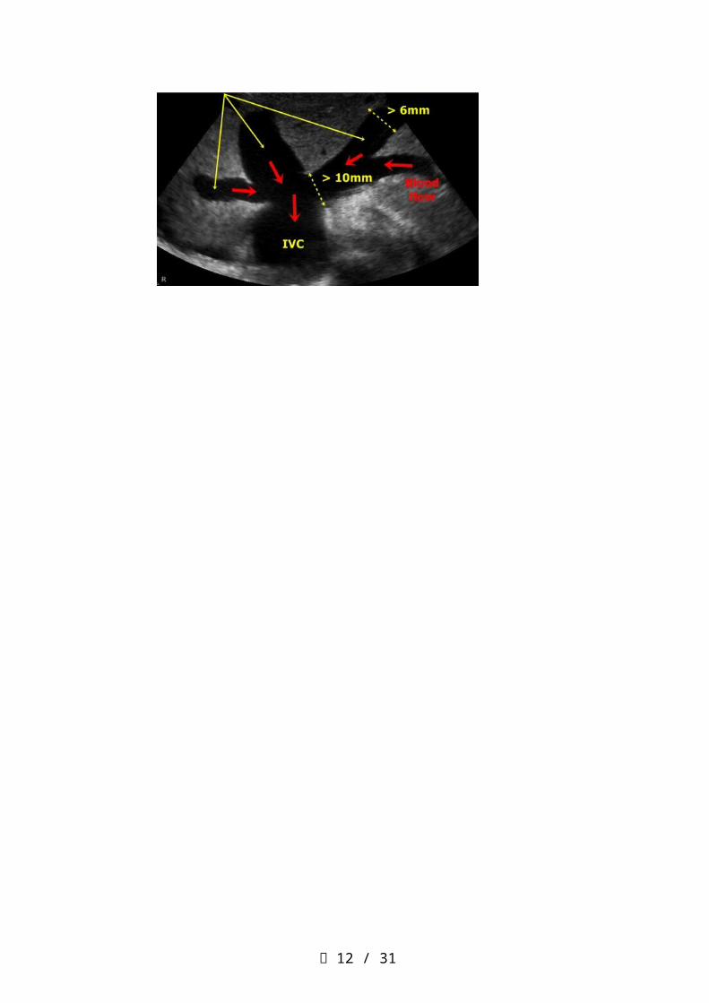
Q19.The best way to distinguish hepatic veins from portal veins is by (1) size, the hepatic veins are much small than the portal veins (2) tracing them to their point of origin (3) visualizing the pulsations of the hepatic veins (4) visualizing the wall thickness of the vessels knowing that hepatic veins have thicker walls
Q20.The arrow in Figure points to(1) LHv (2) IVC(3) RHv(4) Pv
Q21.You are scanning a patient with a known mass in the left medial segment of the liver. What anatomical landmark can you use to identify the left medial segment separate from the right anterior segment of the liver? (1) left portal vein (2) ligamentum teres (3) ligamentum venosum (4) middle hepatic vein
Q22.A patient is referred for a liver ultrasound with the clinical history of a raised serum alpha-fetoprotein level. What should you look for? (1) HCC(2) fatty liver(3) gallstone(4) focal nodular hyperplasia
頁 5 / 22

Q23.You are reviewing lab work prior to performing an abdominal ultrasound exam. Elevated lab values in GGT and ALP. Which statement is true? (1) elevations in both of these lab values is highly specific for HCC (2) elevation of both ALP and GGT is a sensitive indicator of Pancreatitis(3) concomitant elevation of both GGT and ALP indicates the source of the elevated ALP is the liver (4) all of the above
Q24.The most common cause of a hyperechoic liver mass on ultrasound is: (1) Hepatoma(2) Simple cyst (3) Hemangioma (4) Echinococcal cyst
Q25.A patient is referred with right upper quadrant pain and tenderness. Patient has a history of oral contraceptive use. A solid, hypoechoic mass is identified in the right lobe of the liver. Color Doppler reveals hypervascularity of the mass. Which of the following scenarios is most likely?(1) hepatic lipoma (2) hepatic adenoma (3) hepatic abscess (4) fatty liver
Q26.You are scanning a patient with suspected liver cirrhosis. All of the following are sonographic features of cirrhosis except: (1) surface nodularity (2) shrunken caudate lobe (3) altered echo texture (4) ascites
Q27.Compare the echogenicities of the following structures and place them in decreasing echogenic order. (1) renal sinus > pancreas > liver > spleen > renal parenchyma (2) renal sinus > liver > spleen > pancreas > renal parenchyma (3) pancreas > liver > spleen > renal sinus > renal parenchyma (4) renal parenchyma > spleen > liver > pancreas > renal sinus
頁 6 / 22

Q28.What is the arrow showed at the bottom image? (1) hemangioma(2) HCC (3) metastasis(4) mirror image
Q29.What is possible diagnosis at the sonography image? (1) right-side heart failure(2) fatty liver(3) HCC(4) portal hypertension
頁 7 / 22

Q30.If the ultrasound beam passes through a fatty tumor within the liver, and we know that the speed of sound in fat is lower than in soft tissue, where will this fatty tumor be placed? (1) further away than it really is(2) closer than it really is (3) its true position (4) all of the above, depanding upon which frequency transducer is used
Q31.When the gallbladder fundus is folded over on itself, this is referred to as a (1) junctional fold(2) Hartmann’s pouch (3) phrygian cap(4) all of the above
頁 8 / 22
Q32.Name the "sign" associated with a dilated common bile duct. Dilated Common Bile Duct (1) Trademark(2) Too many tubes (3) Murphy's(4) Shotgun
Q33.Direct bilirubin is increased in (1) extrahepatic Obstruction (2) bile duct obstruction (3) intrahepatic disruption (4) all of the above
Q34.Acute cholecystitis is associated with all of the following except(1) gallbladder wall thickening greater than 3 mm (2) obstruction of the cystic duct (3) jaundice(4) tender enlarged gallbladder (Murphy’s sign)
Q35.Carcinoma of the gallbladder would most likely appear as (1) thin-walled enlarged gallbladder (2) small gallbladder with thickened walls (3) large gallbladder with a halo effect (4) a diffusely thickened gallbladder with gallstones
頁 9 / 22

Q36.Ultrasound of a 40 year old female demonstrates numerous ring down artefacts (comet tail artifact) from the gallbladder wall. What is the mostly diagnosis (1) gallstone(2) cyst(3) carcinoma (4) adenomyomatosis
Q37.Choledochal cyst is a (1) cyst within the gallbladder (2) focal dilatation of the biliary tree(3) complication of a pseudocyst (4) associated with adenomyomatosis
Q38.Nonmobile internal echoes without shadowing in gallbladder could be (1) gallstone(2) blood clot (3) gallbladder sludge(4) polyp
Q39.Diffuse thickening of the gallbladder wall can be seen with all of the following except (1) acute cholecystitis(2) hepatitis (3) congestive heart failure(4) elevated portal pressure
頁 10 / 22

Q40.On the transverse sonogram, the common bile duct is located to the head of the pancreas and lies to the inferior vena cava.(1) anterior; medial (2) posterior; posterior (3) posterior; anterior (4) anterior; posterior
Q41.High levels of serum amylase may be a result of all of the following except(1) liver disease (2) acute pancreatitis (3) blockage of the pancreatic duct by a stone within the common bile duct(4) pseudocyst
Q42.A 34-year-old male presents with right-upper quadrant pain and recurrent attacks of pancreatitis. His laboratory values could indicate a(n) (1) increase in BUN (2) decrease in serum amylase (3) increase in lipase (4) increase in indirect bilirubin
Q43.Which is hyperechoic during scanning pancreas? (1) acute pancreatitis(2) adenocarcinoma (3) pseudocyst (4) chronic pancreatitis
Q44.Acute pancreatitis may led to all of the following except (1) gallstone (2) pancreatic pseduocyst (3) common bile duct obstruction (4) pancreatic abscess
頁 11 / 22
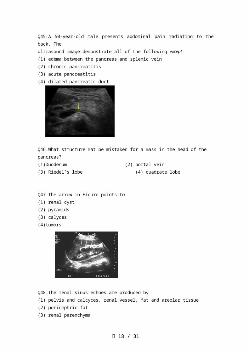
Q45.A 50-year-old male presents abdominal pain radiating to the back. The ultrasound image demonstrate all of the following except (1) edema between the pancreas and splenic vein (2) chronic pancreatitis (3) acute pancreatitis (4) dilated pancreatic duct
Q46.What structure mat be mistaken for a mass in the head of the pancreas? (1)Duodenum (2) portal vein (3) Riedel’s lobe (4) quadrate lobe
Q47.The arrow in Figure points to(1) renal cyst(2) pyramids(3) calyces(4)tumors
Q48.The renal sinus echoes are produced by (1) pelvis and calcyces, renal vessel, fat and areolar tissue (2) perinephric fat (3) renal parenchyma (4) all of the above
頁 12 / 22

Q49.Column of Bertin may be confused with psedotumor. These may be found (1) in the cortex surrounding and separating the renal pyramids and are usually large (2) in the major calyces as a rudimentary ureter (3) in the major calyces as a rudimentary calix (4) outside the renal capsule
Q50.The left renal vein runs (1) parallel to the portal vein (2) posterior to the aorta (3) posterior to the inferior vena cava (4) between the aorta and the superior mesenteric artery
Q51.The type of renal calculi that may be non-opaque radiographically but appear as echogenic foci on sonography includes (1) a calcium oxalate (2) calcium bilirubinate (3) cholesterol (4) none of the above
Q52.While performing an ultrasound examination, the sonographer finds that both kidneys measure 5 cm in length. They are very echogenic. One should consider the possibility of all of the following except (1) chronic glomerulonephritis (2) chronic pyelonephritis (3) renal vascular disease (4) renal vein thrombosis
53.This ultrasound image shows increased echogenicity of the renal medulla (the pyramids are normally hypoechoic to cortex). What is the possible diagnosis? (1) renal stone(2) RCC (3) medullary nephrocalcinosis(4) renal abscess
頁 13 / 22

54.A 3-year-old boy presents with hematuria and palpable left flank mass. Sonography depicts a solid renal mass. This finding would mostly likely represent (1) hypernephroma (2) Wilm’s tumor (3) infantile polycystic kidney disease (4) neuroblastoma
55.The possible diagnosis in this abdominal image is (1) hydronephrosis (2) ascites (3) RCC (4) renal stone
Q56.The best approach for the evaluation of the left kidney is(1)supine(2) prone(3) RAO(4) coronal
Q57.Anatomic landmarks helpful in locating the left adrenal gland are (1) aorta, spleen, and left kidney (2) gastric antrum, left kidney and inferior vena cava (3) left kidney, spleen, and inferior vena cava (4) left kidney, left psoas muscle, and left hemi-diaphragm
頁 14 / 22

Q58.The sonographic texture of the normal testis is (1) heterogeneous with high-intensity echoes (2) homogeneous with low-level echo (3) homogeneous with medium-level echogenicity (4) variable echo pattern
Q59.The possible diagnosis in this image (testes) is (1) testicular cancer (2) hydrocele(3) varicocele(4) trauma
Q60.Which of the following statements regarding the prostate are false? (1) The size of the prostate is 4 × 3 × 4 cm (2) The largest zone and the majority of cancers occur central zone (3) The prostate lies inferior to the seminal vesicles (4) The prostate lies posterior to the symphysis pubis
Q61.The possible diagnosis from this TRUS images is(1) BPH(2) acute prostatitis(3) prostate calcification (4) seminal vesicle calculi
頁 15 / 22

Q62.Which of the following statements regarding the transrectal scanning are false?(1) For transrectal scanning a high-frequency (7 to 10 MHz) linear array probe is preferred (2) Transrectal scanning was performed in the lateral position (3) Patients were instructed to full their bladders before the procedure (4) Scanning was performed in both the transverse and longitudinal planes
Q63.Which of the following statements regarding the spleen are false? (1) a prominent bulge along the medial surface of the spleen can be seen in normal patients (2) the normal-sized spleen should not extend caudal to the midportion of the left kidney (3) the spleen is a hyperechoic organ (4) the sonographic texture of the normal spleen is homogeneous
Q64.The best sonographic window to the left hemi-diaphragm is the (1) spleen(2) kidney(3) pancreas(4) kidney
Q65.What is the possible diagnosis (arrow)? (1) splenomegaly(2) splenic infarct(3) splenic calcification(4) splenic abscess
頁 16 / 22

Q66.Cysts of the spleen are(1) rare(2) common(3) of no clinical significant(4) usually congenital
Q67.A 60-year-old male is suffering from cirrhosis. This spleen ultrasound image demonstrates (1) metastasis(2) splenomegaly(3) hematoma(4) splenic abscess
Q68.Which of the following is not retroperitoneal (1) kidney(2) aorta(3) psoas muscle(4) spleen
Q69.The minimum anterior-posterior diameter for the diagnosis of abdominal aortic aneurysm is (1) 1 cm(2) 2 cm(3) 3 cm(4) above 3 cm
頁 17 / 22
Q70.The celiac trunk originates within the first 2 cm from the diaphragm and immediately branches into all of the following except (1) common hepatic artery (2) left gastric artery (3) gastroduodenal artery (4) splenic artery
Q71.All of the following describe the inferior vena cava except (1) Valsalva maneuver results in a change in the diameter of the inferior vena cava (2) it lies immediately anterior to the surface of the spine, to the right of the aorta (3) the caliber of the inferior vena cava (4) it passes through the caval hiatus of the diaphragm to enter the left artrium
Q72.The possible diagnosis of this obese hypertension patient is (1) congestive liver(2) abdominal aortic aneurysm (3) HCC(4) CBD stone
Q73.Anterior displacement of the splenic vein can be caused by(1) pancreatitis(2) pseudocyst (3) left adrenal hyperplasia(4) aneurysm
頁 18 / 22

Q74.The inferior vena cava increases in diameter superior to the entrance of the(1) portal vein(2) superior mesenteric artery (3) splenic vein(4) renal vein
Q75.The gastroduodenal artery supplies the (1) duodenum, part of the stomach, and the head of pancreas and arises from the common hepatic artery (2) small bowel, duodenum, , and the head of pancreas and arises from the common splenic artery (3) duodenum, arises from the celiac axis (4) duodenum, part of the stomach, and the head of pancreas and arises from the superior mesenteric artery
Q76.Which of the following is typical sign for carcinoma in GI (1) target sign(2) hump sign (3) pseudokidney sign(4) star sign
Q77.What are the dimensions of the normal appendix as seen on ultrasound? (1) 7 cm × 2 cm(2) 7 mm × 2 mm(3) 7 inches × 2 inches(4) None of the above
Q78.Sonographically, the gastroesophageal junction can be visualized(1) proximal common bile duct (2) distal common bile duct(3) common hepatic duct(4) pancreatic duct
頁 19 / 22

Q79.In order to identify and measure the ovaries, the recommended technique is(1) measure length × width × thickness and divide by 6 (2) identify the internal iliac vessels running along the posterolateral aspect of the ovary(3) identify the internal iliac vessels running along the anteriolateral aspect of the ovary(4) all of the above
Q80.The range of transducer frequencies used in transvaginal sonography (1) 3.5 to 5.0 MHz(2) 5.0 to 7.5 MHz(3) 2.5 to 3.5 MHz(4) 12 MHz
Q81.What is number 6?(1) Right lobe(2) Longus coli(3) Strap muscles(4) SCM
Q82.The position of the parathyroid glands are variable. Most often they are Located(1) posterior to the thyroid lobes and medial to the common carotid Arteries(2) posterior to the thyroid lobes and anterior to the strap muscles(3) anterior to the thyroid lobes and lateral to the anterior scalene Muscles(4) lateral to the thyroid lobes
頁 20 / 22

Q83.An increased resistivity index (RI) in the common carotid artery may Indicate(1) stenotic disease proximal to the sample size(2) stenotic disease distal to the sample size(3) disease at the sample site(4) sample volume placement too close to the arterial wall
Q84.A 58-year-old male had elastography of thyroid. What is possible diagnosis?(1) hemorrhagic cyst(2) thyroid carcinoma(3) goiter(4) adenoma
Q85.Enlarged lymph nodes will usually appear sonographically as a(1) heterogeneous mass with poor through transmission(2) homogeneous mass with poor through transmission(3) heterogeneous mass with excellent through transmission(4) none of the above
Q86.Carotid bruit is heard because of(1) arteriovenous malformation(2) disease of the great vessels(3) audible turbulence localized at the carotid bifurcation(4) valvular stenosis
頁 21 / 22

Q87.The halo sign as it pertains to thyroid masses is defined as a rim of sonolucency surrounding an intrathyroidal mass. It is most commonly encountered in(1) cyst(2) carcinoma(3) thyroiditis(4) adenoma
Q88.How to perform this normal 3rd and 4th ventricles image?(1) sagittal(2) anterior coronal(3) medial coronal(4) Using the 4 MHz convex probe
Q89.All the following statements about infantile hypertrophic pyloric stenosis are true except(1) it causes projectile (2) it is predominantly a disorder of male infants (3) the hypertrophied pyloric muscle measures less than 3 mm(4) it appears sonographically as a target-shaped lesion
Q90.A 6-year-old female child presents with recurrent fever, right upper quadrant pain, and jaundice. A sonogram showed a 2 cm cyst at medial and separate from the gallbladder but communicating with CBD. This cystic structure mostly probably represents (1) choledochal cyst(2) a pseduocyst(3) an aortic aneurysm (4) a mucocele
頁 22 / 22