documents.cap.org · web viewmei l, smith sc, faber ac, et al. gastrointestinal stromal tumors: the...
TRANSCRIPT

Template for Reporting Results of Biomarker Testing of Specimens From Patients With Gastrointestinal Stromal TumorsVersion: GIST Biomarkers 1.0.0.2 Template Posting Date: February 2020
This biomarker template is not required for accreditation purposes but may be used to facilitate compliance with CAP Accreditation Program checklist requirements.
AuthorsJavier A. Laurini, MD, FCAP*, Meera Hameed, MD, FCAP; Christopher L. Corless, MD, PhD*; Suzanne George, MD; Jason L. Hornick MD, PhD, FCAP; Sanjay Kakar, MD, FCAP; Alex Lazar, MD, PhD, FCAP; Laura Tang, MD, FCAPWith guidance from the CAP Cancer and CAP Pathology Electronic Reporting Committees.* Denotes primary author. All other contributing authors are listed alphabetically.
Summary of ChangesV1.0.0.2The following data elements were modified:Revised the Explanatory Notes for Immunohistochemistry and Molecular analysis
© 2020 College of American Pathologists (CAP). All rights reserved. For Terms of Use please visit www.cap.org/cancerprotocols.

CAP Approved GIST • Biomarkers • 1.0.0.2
GIST Biomarker Reporting Template
Template web posting date: February 2020
Completion of the template is the responsibility of the laboratory performing the biomarker testing and/or providing the interpretation. When both testing and interpretation are performed elsewhere (eg, a reference laboratory), synoptic reporting of the results by the laboratory submitting the tissue for testing is also encouraged to ensure that all information is included in the patient’s medical record and thus readily available to the treating clinical team.
GASTROINTESTINAL STROMAL TUMOR (GIST)
Select a single response unless otherwise indicated.
Note: Use of this template is optional. If some studies were performed on different specimen(s), the specimen number(s) should be provided.
RESULTS
Immunohistochemical Studies (Note A)___ KIT (CD117)
___ Positive___ Negative
___ DOG1 (ANO1)___ Positive___ Negative
___ SDHB ___ Intact___ Deficient
___ SDHA ___ Intact___ Deficient
___ Other (specify): _______________________________ Positive___ Negative
Molecular Genetic Studies (eg, KIT, PDGFRA, BRAF, SDHA/B/C/D, or NF1 mutational analysis) (Note B)___ Submitted for analysis; results pending___ Performed, see separate report: _______________________________ Performed
Specify method(s) and results: _______________________________ Not performed
KIT Mutational Analysis (Note C)___ No mutation detected ___ Mutation identified (specify:)_______________________ Cannot be determined (explain): __________________________
PDGFRA Mutational Analysis (Note D)___ No mutation detected ___ Mutation identified (specify): _______________________ Cannot be determined (explain): __________________________
2

CAP Approved GIST • Biomarkers • 1.0.0.2
BRAF Mutational Analysis (Note E)__ No BRAF mutation detected ___ BRAF V600E (c.1799T>A) mutation___ Other BRAF mutation (specify): _______________________ Cannot be determined (explain): __________________________
SDHA/B/C/D Mutational Analysis (Note F)___ No mutation detected ___ Mutation identified (specify): _______________________ Cannot be determined (explain): __________________________
NF1 Mutational Analysis (Note G)___ No mutation detected ___ Mutation identified (specify): _______________________ Cannot be determined (explain): __________________________
METHODS
Dissection Method(s) (select all that apply) (Note H)___ Laser capture microdissection___ Manual under microscopic observation___ Manual without microscopic observation___ Cored from block___ Whole tissue section (no tumor enrichment procedure employed)
KIT Mutational Analysis (Note C)
Exons Assessed (select all that apply)___ Exon 9___ Exon 11___ Exon 13___ Exon 14___ Exon 17___ Other (specify): _________________________
Testing Method(s)#
Specify name of method used and exons tested: __________________________# Please specify if different testing methods are used for different exons. PDGFRA Mutational Analysis (Note D)
Exons Assessed (select all that apply)___ Exon 12___ Exon 14___ Exon 18___ Other (specify): __________________________
Testing Method(s)#
Specify name of method used and exons tested: __________________________# Please specify if different testing methods are used for different exons.
BRAF Mutational Analysis (Note E)
Exons Assessed
3

CAP Approved GIST • Biomarkers • 1.0.0.2
___ Exon 15___ Other (specify): _________________________
Testing Method(s) Specify name of method used and exons tested: __________________________
SDH A/B/C/D Mutational Analysis (Note F)___ Exons assessed (specify): ___________________________
Testing Method(s)#
Specify name of method used and exons tested: __________________________# Please specify if different testing methods are used for different exons.
NF1 Mutational Analysis (Note G)Exons assessed (specify): __________________________
Testing Method(s)#
___ Sanger___ Next-generation sequencing (NGS)___ Other (specify): _______________________Specify name of method used: __________________________# Please specify if different testing methods are used for different exons.
COMMENT(S)____________________________________________________________________
Note: Fixative type, time to fixation (cold ischemia time), and time of fixation should be reported if applicable in this template or in the original pathology report.
Gene names should follow recommendations of The Human Genome Organisation (HUGO) Nomenclature Committee (www.genenames.org; accessed February 16, 2015). (Note I)
All reported gene sequence variations should be identified following the recommendations of the Human Genome Variation Society (www.hgvs.org/mutnomen/; accessed February 16, 2015). (Note I)
4

Background Documentation GIST • Biomarkers • 1.0.0.2
Explanatory Notes
A. Immunohistochemical AnalysisBecause of the advent of small-molecule kinase inhibitor therapy in the treatment of GIST (see the following), it has become imperative to distinguish GIST from its histologic mimics, mainly leiomyoma, leiomyosarcoma, schwannoma, and desmoid fibromatosis.1,2 Immunohistochemistry is instrumental in the workup of GIST. For the initial work up of GIST, a basic immunohistochemical panel including CD117 (KIT), DOG1 (Ano1), Desmin, S100 protein and CD34 is recommended. GISTs are immunoreactive for KIT (CD117) (approximately 95%) and/or DOG1(>99%).3-5 KIT immunoreactivity is usually strong and diffuse but can be more focal in unusual cases (Figure 1, A and B). It is not unusual for GISTs to exhibit dot-like perinuclear staining (Figure 1, C), while less commonly, some cases exhibit membranous staining (Figure 1, D). These patterns do not clearly correlate with mutation type or response to therapy. Most KIT-negative / DOG1 positive GISTs are gastric or extra-visceral GISTs and almost invariably harbor a platelet-derived growth factor receptor A (PDGFRA) mutation.6 DOG1 expression is not related to mutational status in GISTs, and it may be a useful marker to identify a subset of patients with CD117-negative GISTs, who might benefit from targeted therapy 4,5. Approximately 70% of GISTs are positive for CD34, 30% to 40% are positive for smooth muscle actin, 5% are positive for S100 protein (usually focal), 5% are positive for desmin (usually focal), and 1% to 2% are positive for keratin (weak/focal).7
Since succinate dehydrogenase (SDH)-deficient GISTs have specific implications (see the following), it is recommended to screen all gastric GISTs for loss of SDH by immunohistochemistry, usually best accomplished by staining for SDHB, which is loss in all subtypes of SDH-deficient GISTs. 8-11 Mutations in SDHA are detected in 30% of SDH-deficient GISTs and loss of expression of SDHA specifically identifies tumors with SDHA mutations; other SDH-deficient GISTs show normal (intact) cytoplasmic staining for SDHA.12,13 Patients with SDH-deficient GIST should be referred to a genetic counselor for appropriate work up.
Figure 1. Patterns of KIT staining in gastrointestinal stromal tumor (GIST). A, Diffuse and strong immunoreactivity in a typical GIST. B, Focal and weak pattern in an epithelioid gastric GIST with a PDGFRA mutation. C, Dot-like perinuclear staining. D, Membranous pattern. (Original magnification X400.)
5

Background Documentation GIST • Biomarkers • 1.0.0.2
References1. Hornick JL, Fletcher CD. Immunohistochemical staining for KIT (CD117) in soft tissue sarcomas is very
limited in distribution. Am J Clin Pathol. 2002;117(2):188-193.2. Miettinen M, Sobin LH, Sarlomo-Rikala M. Immunohistochemical spectrum of GISTs at different sites and
their differential diagnosis with a reference to CD117 (KIT). Mod Pathol. 2000;13(10):1134-1142.3. Sarlomo-Rikala M, Kovatich AJ, Barusevicius A, Miettinen M. CD117: a sensitive marker for gastrointestinal
stromal tumors that is more specific than CD34. Mod Pathol. 1998;11(8):728-734.4. Espinosa I, Lee CH, Kim MK, et al. A novel monoclonal antibody against DOG1 is a sensitive and specific
marker for gastrointestinal stromal tumors. Am J Surg Path. 2008;32(2):210–218.5. Miettinen M, Wang ZF, Lasota J. DOG1 antibody in the differential diagnosis of gastrointestinal stromal
tumors: a study of 1840 cases. Am J Surg Pathol. 2009;33:1401–1408.6. Medeiros F, Corless CL, Duensing A, et al. KIT-negative gastrointestinal stromal tumors: proof of concept
and therapeutic implications. Am J Surg Pathol. 2004;28(7):889-894.7. Fletcher CD, Berman JJ, Corless C, et al. Diagnosis of gastrointestinal stromal tumors: a consensus
approach. Hum Pathol. 2002;33(5):459-465.8. Mei L, Smith SC, Faber AC, et al. Gastrointestinal Stromal Tumors: The GIST of Precision Medicine. Trends
Cancer. 2018;4:74-91.9. Gill AJ. Succinate dehydrogenase (SDH) and mitochondrial driven neoplasia. Pathology. 2012
Jun;44(4):285-92.10. Gill AJ, Benn DE, Chou A, et al. Immunohistochemistry for SDHB triages genetic testing of SDHB, SDHC,
and SDHD in paraganglioma-pheochromocytoma syndromes. Hum Pathol. 2010 Jun;41(6):805-1411. Doyle LA, Nelson D, Heinrich MC, et al. Loss of succinate dehydrogenase subunit B (SDHB) expression is
limited to a distinctive subset of gastric wild-type gastrointestinal stromal tumours: a comprehensive genotype-phenotype correlation study. Histopathology. 2012;61(5):801-809.11.
12. Wagner AJ, Remillard SP, Zhang YX, et al. Loss of expression of SDHA predicts SDHA mutations in gastrointestinal stromal tumors. Mod Pathol. 2013;26(2):289-294.12.
13. Dwight T, Benn DE, Clarkson A, et al. Loss of SDHA expression identifies SDHA mutations in succinate dehydrogenase-deficient gastrointestinal stromal tumors. Am J Surg Pathol. 2013;37(2):226-233.
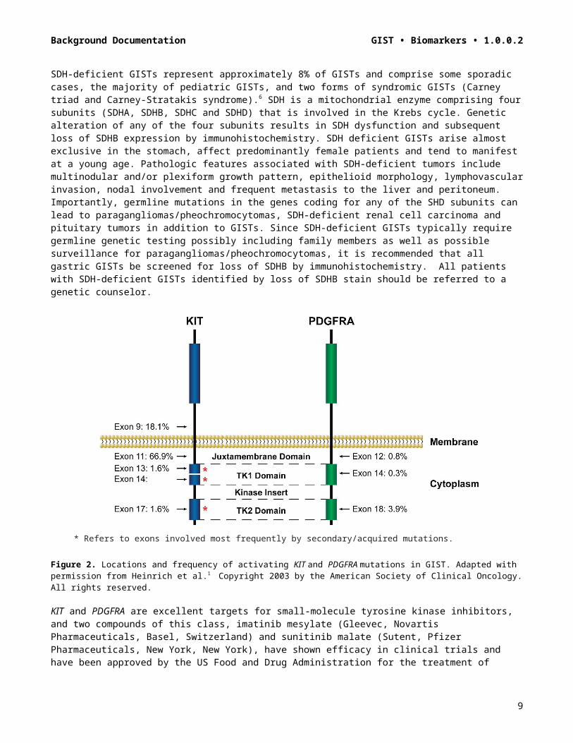
B. Molecular AnalysisApproximately 75% of GISTs possess activating mutations in the KIT gene, whereas another 10% have activating mutations in the PDGFRA gene.1-4 These mutations result in virtually full-length KIT proteins that exhibit ligand-independent activation. KIT and PDGFRA each contain 21 exons. However, mutations cluster within “hotspots”: exons 9, 11, 13, and 17 in KIT, and exons 12, 14, and 18 in PDGFRA (Figure 2). About 5% to 10% of GISTs appear to be negative for both KIT and PDGFRA mutations. The most recent NCCN Task Force on GIST strongly encourages that KIT and PDGFRA mutational analysis be performed if tyrosine kinase inhibitors (TKIs) are considered as part of the treatment plan for unresectable or metastatic disease and that mutational analysis be considered for patients with primary disease, particularly those with high-risk tumors. KIT and PDGFRA mutation status can be determined easily from paraffin-embedded tissue. Secondary or acquired mutations can be associated with development of tumor resistance in the setting of long-term imatinib mesylate treatment. These are usually point mutations that occur most commonly in KIT exons 13, 14, and 17.5 The clinical utility of these mutations is an evolving concept, but it is important not to confuse them with the primary or initial mutation in GIST.
Recent studies focusing on the molecular classification of GISTs recognized two major subgroups : succinate dehydrogenase (SHD)-competent and SDH-deficient GISTs, both of which can arise in the sporadic or familiar setting.6.7 SDH-competent GISTs include tumors with mutations of KIT and PDGFRA as well of a subset of wild-type GISTs with mutations mainly in NF1 and BRAF genes. On the other hand, SDH-deficient GISTs include tumors with a genetic alteration in any of the SDH subunits leading to SDH dysfunction.
SDH-deficient GISTs represent approximately 8% of GISTs and comprise some sporadic cases, the majority of pediatric GISTs, and two forms of syndromic GISTs (Carney triad and Carney-Stratakis syndrome).6 SDH is a mitochondrial enzyme comprising four subunits (SDHA, SDHB, SDHC and SDHD) that is involved in the Krebs cycle. Genetic alteration of any of the four subunits results in SDH dysfunction and subsequent loss of SDHB expression by immunohistochemistry. SDH deficient GISTs arise almost exclusive in the stomach, affect
6

Background Documentation GIST • Biomarkers • 1.0.0.2
predominantly female patients and tend to manifest at a young age. Pathologic features associated with SDH-deficient tumors include multinodular and/or plexiform growth pattern, epithelioid morphology, lymphovascular invasion, nodal involvement and frequent metastasis to the liver and peritoneum. Importantly, germline mutations in the genes coding for any of the SHD subunits can lead to paragangliomas/pheochromocytomas, SDH-deficient renal cell carcinoma and pituitary tumors in addition to GISTs. Since SDH-deficient GISTs typically require germline genetic testing possibly including family members as well as possible surveillance for paragangliomas/pheochromocytomas, it is recommended that all gastric GISTs be screened for loss of SDHB by immunohistochemistry. All patients with SDH-deficient GISTs identified by loss of SDHB stain should be referred to a genetic counselor.
* Refers to exons involved most frequently by secondary/acquired mutations.
Figure 2. Locations and frequency of activating KIT and PDGFRA mutations in GIST. Adapted with permission from Heinrich et al.1 Copyright 2003 by the American Society of Clinical Oncology. All rights reserved.
KIT and PDGFRA are excellent targets for small-molecule tyrosine kinase inhibitors, and two compounds of this class, imatinib mesylate (Gleevec, Novartis Pharmaceuticals, Basel, Switzerland) and sunitinib malate (Sutent, Pfizer Pharmaceuticals, New York, New York), have shown efficacy in clinical trials and have been approved by the US Food and Drug Administration for the treatment of GIST.8-10 SDH-deficient GISTs are usually resistant to imatinib but may have a higher probability of response to sunitinib.6 Because different tyrosine kinase inhibitors (TKIs) may have more efficacy in genetic subsets of GIST, oncologists may want to know the mutation status of each GIST, because this may impact which drug each patient should receive.1,11 Secondary resistance mutations may also affect drug selection as their significance is further defined.
References1. Heinrich MC, Corless CL, Demetri GD, et al. Kinase mutations and imatinib response in patients with
metastatic gastrointestinal stromal tumor. J Clin Oncol. 2003;21(23):4342-4349.2. Heinrich MC, Corless CL, Duensing A, et al. PDGFRA activating mutations in gastrointestinal stromal
tumors. Science. 2003;299(5607):708-710.3. Hirota S, Isozaki K, Moriyama Y, et al. Gain-of-function mutations of c-kit in human gastrointestinal stromal
tumors. Science. 1998;279(5350):577-580.4. Rubin BP, Singer S, Tsao C, et al. KIT activation is a ubiquitous feature of gastrointestinal stromal tumors.
Cancer Res. 2001;61(22):8118-8121.5. Heinrich MC, Corless CL, Blanke CD, et al. Molecular correlates of imatinib resistance in gastrointestinal
stromal tumors. J Clin Oncol. 2006;24(29):4764-4774.
7
Background Documentation GIST • Biomarkers • 1.0.0.2
6. Mei L, Smith SC, Faber AC, et al. Gastrointestinal Stromal Tumors: The GIST of Precision Medicine. Trends Cancer. 2018;4:74-91.
7. Gill AJ. Succinate dehydrogenase (SDH) and mitochondrial driven neoplasia. Pathology. 2012 Jun;44(4):285-92.
8. Demetri GD, Benjamin RS, Blanke CD, et al; NCCN Task Force. NCCN Task Force report: management of patients with gastrointestinal stromal tumor (GIST)--update of the NCCN clinical practice guidelines. J Natl Compr Canc Netw. 2007;5(Suppl 2):S1-S29.
9. Demetri GD. Targeting the molecular pathophysiology of gastrointestinal stromal tumors with imatinib: mechanisms, successes, and challenges to rational drug development. Hematol Oncol Clin North Am. 2002;16(5):1115-1124.
10. Demetri GD, van Oosterom AT, Garrett CR, et al. Efficacy and safety of sunitinib in patients with advanced gastrointestinal stromal tumour after failure of imatinib: a randomised controlled trial. Lancet. 2006;368(9544):1329-1338.
11. Corless CL, Schroeder A, Griffith D, et al. PDGFRA mutations in gastrointestinal stromal tumors: frequency, spectrum and in vitro sensitivity to imatinib. J Clin Oncol. 2005;23(23):5357-5364.
C. KIT Mutational Analysis The most common mutations affect the juxta membrane domain encoded by exon 11 (two-thirds of GIST). These mutations include in-frame deletions, substitutions, and insertions. Deletions (in particular codon 557 and/or 558) are associated with shorter progression free and overall survival.1-6 The vast majority of exon 11-mutated GISTs are located in the stomach. 5 About 7% to 10% of the tumors harbor mutations in the extracellular domain encoded by exon 9 (most commonly insAY502-503).5,7 Exon 9-mutant GISTs arise predominantly in the small bowel and have reduced sensitivity to imatinib which could be overcome by using higher doses. 5 Primary mutations in the activation loop (exon 17) and ATP binding region (exon 13) are uncommon (1%). The majority of these mutations are substitutions.8 KIT exon 8 mutations are extremely rare (0.15%).9 Secondary or resistance mutations occur commonly in tumors harboring primary exon 11 mutations. These newly acquired secondary mutations are always located in exons encoding tyrosine kinase domain (exons 13, 14, 17).10
References1. Gastrointestinal Stromal Tumor Meta-Analysis Group (MetaGIST). Comparison of two doses of imatinib for
the treatment of unresectable or metastatic gastrointestinal stromal tumor: a meta-analysis of 1640 patients. J Clin Oncol. 2010;28(7):1247-1253.
2. Heinrich MC, Corless CL, Blanke CD, et al. Molecular correlates of imatinib resistance in gastrointestinal stromal tumors. J Clin Oncol. 2006;24(29):4764-4774.
3. Andersson J, Bumming P, Meis-Kindblom JM, et al. Gastrointestinal stromal tumors with KIT exon 11 deletions are associated with poor prognosis. Gastroenterology. 2006;130(6):1573-1581.
4. Liu XH, Bai CG, Xie Q, et al. Prognostic value of KIT mutation in gastrointestinal stromal tumors. World J Gastroenterol. 2005;11(25):3948-3952.
5. Mei L, Smith SC, Faber AC, et al. Gastrointestinal Stromal Tumors: The GIST of Precision Medicine. Trends Cancer. 2018;4:74-91.
6. Wozniak A, Rutkowski P, Schöffski P, et al. Tumor genotype is an independent prognostic factor in primary gastrointestinal stromal tumors of gastric origin: a European multicenter analysis based on ConticaGIST. Clin Cancer Res. 2014 Dec 1;20(23):6105-16.
7. Wardelmann E, Losen I, Hans V, et al. Deletion of Trp-557 and Lys-558 in the juxtamembrane domain of the c-kit protooncogene is associated with metastatic behavior of gastrointestinal stromal tumors. Int J Cancer. 2003;106(6):887-895.
8. Lux ML, Rubin BP, Biase TL, et al. KIT extracellular and kinase domain mutations in gastrointestinal stromal tumors. Am J Pathol. 2000;156(3):791-795.
9. Huss S, Künstlinger H, Wardelmann E, et al. A subset of gastrointestinal stromal tumors previously regarded as wild-type tumors carries somatic activating mutations in KIT exon 8 (p. D419del). Mod Pathol. 2013;26(7):1004-1012.
10. Lasota J, Corless CL, Heinrich MC, et al. Clinicopathologic profile of gastrointestinal stromal tumors (GISTs) with primary KIT exon 13 or 17 mutations: a multicenter study of 54 cases. Mod Pathol. 2008;21(4):476-484
D. PDGFRA Mutational Analysis
8
Background Documentation GIST • Biomarkers • 1.0.0.2
More than 80% of KIT-negative GISTS have PDGFRA mutations. The majority of PDGFRA-mutated GISTs arise in the stomach, usually with epithelioid or mixed epithelioid and spindle cell morphology and often with myxoid stromal changes. 1,2 PDGFRA-mutated GISTs tend to have a lower risk of recurrence. 1,3 Activation of PDGFRA is seen in GISTs harboring mutations in juxta membranous domain (exon 12), the ATP binding domain (exon 14), or the activation loop (exon 18).1,2 Mutations include substitutions and deletions. Primary resistance to imatinib is seen with the most common PDGFRA exon 18 D842V mutation.1
References1. Mei L, Smith SC, Faber AC, et al. Gastrointestinal Stromal Tumors: The GIST of Precision Medicine. Trends
Cancer. 2018;4:74-91.2. LaCosta J, Miettinen M. Clinical significance of oncogenic KIT and PDGFRA mutations in gastrointestinal
stromal tumors. Histopathology. 2008;53(3):245-266.3. Barnett CM, Corless CL, Heinrich MC. Gastrointestinal stromal tumors: molecular markers and genetic
subtypes. Hematol Oncol Clin North Am. 2013 Oct;27(5):871-88.
E. BRAF Mutational AnalysisActivating mutations of BRAF (V600E) have been identified in a small subset (7%) of KIT/PDGFRA wild-type GISTs. These tumors show a predilection for small bowel location, arise in middle-aged females, exhibit a high mitotic rate and are associated with early metastasis.1,2 BRAF-mutated GISTs show primary resistance to imatinib but may respond to BRAF inhibitors.2
References1. Agaram NP, Wong GC, Guo T, et al. Novel V600E BRAF mutations in imatinib-naive and imatinib-resistant
gastrointestinal stromal tumors. Genes Chromosomes Cancer. 2008;47(10):853–859.2. Mei L, Smith SC, Faber AC, et al. Gastrointestinal Stromal Tumors: The GIST of Precision Medicine. Trends
Cancer. 2018;4:74-91.
F. SDH A/B/C/D Mutational AnalysisThe succinate dehydrogenase (SDH) complex (mitochondrial complex II) participates in both the Krebs cycle and the electron transport chain of oxidative phosphorylation. About 8% of GISTs (all lacking mutations in KIT and PDGFRA) are caused by dysfunction of the SDH complex ("SDH-deficient GISTs"). Around 50% of patients affected by such tumors harbor germline mutations in one of the SDH subunit genes (SDHA/B/C or D). SDHA-inactivating mutations are most common, detected in about 30% of SDH-deficient GISTs. Mutations involve exons 2, 3, 5, 6, 7, 8, 9, 10, 11, 13, 14 of SDHA; exons 1, 2, 3, 4, 6, 7 of SDHB; exons 1, 4, 5 of SDHC; and exons 4 and 6 of SDHD. While the majority of the mutations are substitutions, deletions, splice-site mutations, frame shift, and duplications have also been reported.1-4
References1. Doyle LA, Hornick JL. Gastrointestinal stromal tumours: from KIT to succinate dehydrogenase.
Histopathology. 2014;64(1):53-67.2. Wagner AJ, Remillard SP, Zhang YX, et al. Loss of expression of SDHA predicts SDHA mutations in
gastrointestinal stromal tumors. Mod Pathol. 2013;26(2):289-294.3. Fletcher CD, Berman JJ, Corless C, et al. Diagnosis of gastrointestinal stromal tumors: a consensus
approach. Hum Pathol. 2002; 33(5):459-465.4. Nannini, M, Biasco B, Astolfi A, et al. An overview on molecular biology of KIT/PDGFRA wild type (WT)
gastrointestinal stromal tumours (GIST). J Med Genet. 2013;50(10):653-661.
G. Neurofibromatosis Type 1 (NF1) Mutational AnalysisNF1 is an inherited, autosomal dominant disease characterized by multiple café au lait spots, Lisch nodules, freckling, and development of neurofibromas. GISTs in NF1 patients arise predominantly from the small intestine, including duodenum, can be multicentric, lack KIT and PDGFRA mutations and are associated with Cajal cell hyperplasia.1,2 Only a minority (approximately 7%) of NF-1 patients develop NF1-mutated GISTs, therefore, molecular testing for canonical mutations in KIT and PDGFRA is recommended for GISTs arising in the setting of neurofibromatosis.2
References
9

Background Documentation GIST • Biomarkers • 1.0.0.2
1. Nannini, M, Biasco B, Astolfi A, et al. An overview on molecular biology of KIT/PDGFRA wild type (WT) gastrointestinal stromal tumours (GIST). J Med Genet. 2013;50(10):653-661.
2. Mei L, Smith SC, Faber AC, et al. Gastrointestinal Stromal Tumors: The GIST of Precision Medicine. Trends Cancer. 2018;4:74-91.
H. Dissection Method:While in majority of cases GIST samples show tumor percentage (%) well above the analytical sensitivity of Sanger sequencing (>50% neoplastic cell percentage/20% to 25% mutant allele percentage), in cases of mutation analysis of treated samples, careful macro/microdissection may be necessary to avoid false negative results.
I. Reporting NomenclatureConsistent gene mutation nomenclature is essential for efficient and accurate reporting.1 Following are examples as recommended by Human Genome Variation Society (HGVS) for description of variant changes.2 It is also preferred that protein alterations are mentioned in the report in addition to genomic coordinates.
Examples of DNA, RNA, and Protein NomenclatureDNA: A, G, C, T (example: c.957A>T)RNA: a, g, c, u (example: r.957 a>u)Protein: 3-letter amino acid code, X= Stop codon (example: p. Glu78Gln)
Examples of Nomenclatures for Types of Sequence VariantsTypes of Variation Examples Substitution c.123A>GDeletion c.123delA, c.586_591delTGGTCA or c.586_591del6Duplication c.123dupA, c.586_591dupTGGTCA or c.586_591dup6Insertion c.123_124insC, c.1086_1087insGCGTGAFrame shift p. Arg83 fs or p. Arg83Ser fsX15Deletion/insertions “indel” c.112_117delAGGTCAinsTG
References1. Ogino S, Gulley M, den Dunneb JT, et al. Standard mutation nomenclature in molecular diagnostics:
practical and educational challenges. J Mol Diagn. 2007;9(1):1-6.2. den Dunnen JT, Antonarakis SE. Mutation nomenclature. Curr Protoc Hum Genet. 2003;Chapter 7, Unit
7.13. http://www.currentprotocols.com/WileyCDA/CPUnit/refId-hg0713.html. Accessed October 29, 2014.
10