weber-christian panniculitis and auto-immune disease
TRANSCRIPT
J. clin. Path., 1976, 29, 144-149
Weber-Christian panniculitis and auto-immune disease:a case reportT. G. ALLEN-MERSH1
From the Department ofPathology, Ninewells Hospital and Medical School, Dundee
SYNOPSIS A case is described of Weber-Christian panniculitis accompanied by a gammaglobulindisturbance which preceded by five years the diagnosis of an autoimmune hepatitis and pancyto-penia. Also associated was the onset of diabetes mellitus, found at necropsy to be related topancreatic islet amyloid deposition. This case reinforces the view that Weber-Christian panniculitismay be an adipose response to a variety of immunological stimuli.
The Weber-Christian type of panniculitis is arecurrent febrile illness characterized by multiplesymmetrical subcutaneous nodules or plaques whichresolve, leaving characteristically depressed atrophicareas (Cairns, 1968). Histologically (Lever, 1967)there are three phases: (1) degeneration of fat cellsaccompanied by an inflammatory infiltrate com-posed of neutrophils, lymphocytes, and macro-phages: abscess formation does not occur; (2)infiltration with macrophages, many laden with fatand some multinucleate: in some areas macrophageswith foamy cytoplasm completely replace fat cells;(3) fibroblasts intermingled with lymphocytes andplasma cells replace foam cells: collagen is laid down,resulting in fibrosis.
Steinberg (1953) reviewed 43 cases with systemicsymptoms and signs, and he concluded these to bemalaise, fatigability, generalized aching, chills,night sweats, fever, nausea or vomiting, hepato-rnegaly, splenomegaly, normochromic normocyticanaemia, leucopenia or leucocytosis, and elevatedor depressed platelet count. Milner and Mitchinson(1965) reviewed 11 published necropsy reports onpatients with systemic involvement and reported afurther case. In these cases there was involvementof subcutaneous and retroperitoneal, mesenteric orepicardial adipose tissue. They recommended thename systemic Weber-Christian disease where thetypical lesions are present in subcutaneous andvisceral adipose tissue, and Weber-Christian panni-culitis where the lesions are confined to the sub-cutaneous adipose tissue. This terminology isadhered to herein.
'Present address: National Heart Hospital, WestmorelandStreet, London WIReceived for publication 28 July 1975
Case Summary
A 7-year-old girl was first seen in October 1960 withtenderness, swelling, and redness of the skin behindboth knees, over the right foot (fig 1), and over thecutaneous surface of the right tibia of a few days'duration. The mother remarked that her daughterhad recently been feverish. One year previously shehad suffered similar pain, tenderness, and rednessbehind the knees, but this had settled spontaneously;otherwise she had always been healthy. She had anolder brother who was an insulin-dependent diabeticand had necrobiosis lipoidica diabeticorum. Apartfrom the lesions mentioned, there was no abnor-mality on examination. Laboratory investigationrevealed a white cell count of 8 5 x 10⁹/l, an ESRof 20 mm in the first hour (Wintrobe), and a totalplasma protein of 72 5 g/l (albumin 34 g/l, globulin38 5 g/l) with an increased gammaglobulin com-ponent on electrophoresis. Thymol turbidity was4 units (normal range 0-4 units) and cephalinflocculation 3 + (normal range 0-1 +). LE cellswere not seen in the blood. An oral and an intravenousglucose tolerance test were normal, serum choles-terol was 7-4 mmol/l. A biopsy (fig 2) taken fromthe lesion on the foot showed 'areas of fat necrosis,accumulation of leucocytes including neutrophilpolymorphonuclear leucocytes, and macrophageswith foamy cytoplasm. Histologically the appearancesare those of a Weber-Christian disease' (Dr W.Guthrie). She was treated with oral prednisolone,initially 60 mg/day which was tailed off over thenext five months. Four months after the onset of theprevious lesions (February 1961), while on steroid,she suffered a similar lesion on the right wrist andleft forearm. This settled and, apart from fatatrophy of the affected areas (fig 3), she remained
144
copyright. on O
ctober 24, 2021 by guest. Protected by
http://jcp.bmj.com
/J C
lin Pathol: first published as 10.1136/jcp.29.2.144 on 1 F
ebruary 1976. Dow
nloaded from

Weber-Christian panniculitis and auto-immune disease: a case report
Fig 1 Area of Weber-Christianpanniculitis on dorsum of rightfoot: the lesion was red andpainful, andfelt hard.
- 'W*"#
Fig 2 Skin biopsyfrom dorsum ofrightfoot. Adipose tissue is infiltrated with inflammatory cells andfat-laden macrophages (Haematoxylin and eosin x 335).
well. Four years later (January 1965) she suffered atwo-week feverish illness with cough and coryza.A week later an area of swelling, 7 5 cm in diameter,appeared on the lateral aspect of the left ankle,which on biopsy was histologically similar to the
earlier biopsy. There was no other clinical abnor-mality. Laboratory investigation showed a hypo-chromic anaemia (Hb 9-8 g/dl), general leucopenia(WBC 3-1 x 10⁹/l), plasma protein 73 g/l (albumin35 g/l, globulin 38 g/l), and plasma electrophoresis
145
copyright. on O
ctober 24, 2021 by guest. Protected by
http://jcp.bmj.com
/J C
lin Pathol: first published as 10.1136/jcp.29.2.144 on 1 F
ebruary 1976. Dow
nloaded from
T. G. Allen-Mersh
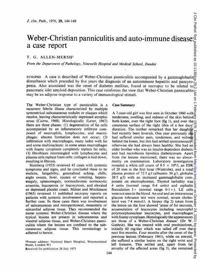
Fig 3 Loss of subcutaneousadipose tissue at site ofan oldlesion on left forearm.
suggestive of increased beta and gammaglobulins.ESR was 23 mm in the first hour (Wintrobe). Anti-bodies to thyroid, stomach, and blood vessels werenegative at this time (Dr J. S. Beck), and theWassermann reaction was negative. She recoveredfrom this attack without treatment.She remained well for five years, but in April 1970
when aged 16 years she complained of thirst andpolyuria. On examination splenomegaly (3 fingers)was noted but no other abnormality. Laboratoryinvestigations revealed: glycosuria, anESR of90 mmin the first hour (Westergren), a serum glutamicoxaloacetic transferase of 86 SF units (normal range0-33), a plasma bilirubin of 22 ,umol/l, a haemo-globin of 7-4 g/dl (hypochromic microcytic film), awhite cell count of 2-8 x 10⁹/1 (no primitive cellsseen), and a platelet count of 45 x 109/1. Australiaantigen and antibody were negative. A sternalmarrow examination showed erythloid hyperplasiawith depleted iron stores. Red cell half-life was 18days (slightly reduced) but there was no sign ofsplenic sequestration. She was diagnosed as havingdiabetes mellitus and hepatic cirrhosis.
She developed skin hypersensitivity to all formsof insulin except Actrapid (porcine base neutral pH).Her diabetes was eventually controlled with thisagent.
In April 1971, one year after splenomegaly hadbeen noted, the direct antiglobulin test was found tobe positive, and antibody (subsequently shown tobe IgG, C3d) was demonstrated on the red cellsurface: IgM antibody, which agglutinated enzyme-treated erythrocytes (titre 1:128) and lysed the same
cells (titre 1 :64), was found in the serum (Dr S. M.Woriledge, Royal Postgraduate Medical School,London). At this time the patient suffered from asevere crop of perineal warts, and two years later(February 1973) she developed herpes genitalis.
Right-sided abdominal pain became very trouble-some and shewas investigated byProfessor Sherlock'sUnit at the Royal Free Hospital, London (February1973). The following results were obtained at thistime: plasma IgG 29-7 g/l (5-0-16-0 g/l normal range),IgA 3-15 g/l (1 254-25 g/l normal range), and IgM3-3 g/l (0-5-1-8 g/l normal range). The lipid profilewas normal. The antimitochondrial antibodywas weakly positive. The anti DNA antibody was'slightly raised', but the antinuclear factor wasnegative. The gastric parietal cell antibody waspositive. The Wassermann reaction was positive butthe Treponema immobilization test was negative.Liver biopsy showed intact lobular architecturewith expanded portal tracts infiltrated with a mixtureof inflammatory cells, including many plasma cells,combined with bile duct proliferation and disruptionof the limiting plate. The parenchyma showed mildfatty change and nuclear vacuolation. There was nocholestasis nor siderosis. The liver disease was placedsomewhere in the spectrum of autoimmune liverdisease, between primary biliary cirrhosis and activechronic hepatitis (Professor S. Sherlock).
Subsequently she developed progressive liver cellfailure and in April 1975 she died.
PATHOLOGICAL SUMMARYThe body was of a young icteric adult female.
146
copyright. on O
ctober 24, 2021 by guest. Protected by
http://jcp.bmj.com
/J C
lin Pathol: first published as 10.1136/jcp.29.2.144 on 1 F
ebruary 1976. Dow
nloaded from

Weber-Christian panniculitis and auto-immune disease: a case report
Fig 4 Section ofliver showing increasedfibrous tissue, distortion oflobular architecture, bile ductproliferation, and somefatty change ofhepatocytes (H andE x 65).
41~'4~~!l
Fig 5 Section ofpancreas stained with sulphated alcian blue/van Gieson. The islet (centre) containeda blue amorphous material (appearing black in this photomicrograph) which had the stainingcharacteristics ofamyloid ( x 335).
147
.AR
copyright. on O
ctober 24, 2021 by guest. Protected by
http://jcp.bmj.com
/J C
lin Pathol: first published as 10.1136/jcp.29.2.144 on 1 F
ebruary 1976. Dow
nloaded from

T. G. Allen-Mersh
Except for some patchy atrophy over the calvesthere was no abnormality of fat distribution. Theliver showed a macronodular cirrhosis, the spleenweighed 1570 g and measured 30 x 15 x 10 cm,and there were moderately severe oesophagealvarices. The pancreas appeared macroscopicallynormal. Red marrow was present throughout thefemoral shaft. Macroscopically no sign of inflam-mation of retroperitoneal, mesenteric or epicardialfat was noted. Grey pus (from which enterococciwere cultured) was found underlying insulin in-jection sites on the thighs.
Histological examination of the liver showed amacronodular cirrhosis, with bile duct proliferation(fig 4). The limiting plates of hepatic lobules werewell defined. The spleen was congested, but macro-phage activity was not prominent. Pancreatic acinartissue appeared normal, but the islets contained ahyalin material which showed dichroic birefringencewith a Congo red stain and was blue with an alcianstain (fig 5): this was thought to be amyloid material.Adipose tissue from the periureteric area showedoccasional small foci of invasion by plasma cells andfat-laden macrophages. Mesenteric and epicardialadipose tissue showed no abnormality. Other organsshowed no significant histological abnormality.
Discussion
The aetiology of the adipose damage in Weber-Christian panniculitis and systemic Weber-Christiandisease is unknown. An immune mechanism mayinitiate or perpetuate the adipose damage. It hasbeen described in association with tuberculosis(Tilden et al, 1940; Beerman, 1953; Macdonaldand Feiwel, 1968), glomerulonephritis (Spain andFoley, 1944), myositis (Kiernan and Burger, 1960),intrahepatic sclerosing cholangitis, vasculitis, andpositive LE cells (Hellstrom and Perez-Stable, 1966),leucopenia (Friedman, 1945), leucopenia and leuc-agglutinins (Rosenstock, 1968), pancytopenia (Wyatt1969), hypersplenism, megaloblastosis, and pancyto-penia (Mitsutani et al, 1973), and following with-drawal of steroids given in treatment of rheumaticfever (Smith and Good, 1956). A histologicallysimilar condition (lupus erythematosus profundus)has also been described in association with systemiclupus erythematosus (Macoul, 1967), and Tuffanelli(1971) has demonstrated IgG antivascular antibody,IgM, and complement at the dermal epidermaljunction in this condition. Experimentally Goddard(1947) produced lesions histologically similar toWeber-Christian panniculitis by the injection ofsublethal doses of ovalbumen into previouslysensitized guinea-pigs: he regarded this as an Arthusphenomenon. The clinical and histological features
of Weber-Christian panniculitis have also beenproduced by the repeated self-injection of milk(Ackerman et al, 1966). Weber-Christian panniculitisis also reported in association with insulin-dependentdiabetes mellitus (Machacek, 1948) in four cases,of which two were also hyperthyroid (one withmultiple hypersensitivities).
In the case reported here, histologically typicalWeber-Christian panniculitis accompanied by aplasma globulin elevation (elevated IgG component)preceded by five years the diagnosis of an auto-immune hepatitis with autoimmune haemolyticanaemia, leucopenia, thrombocytopenia, and as-sociated herpes genitalis. It similarly preceded theonset of diabetes mellitus, which was related tolocalized amyloid deposit in pancreatic islets. Thepresence of amyloid in pancreatic islets was foundto occur in 50% of diabetics over 50 years of age(Ehrlich and Ratner, 1961) but is rare in juvenilediabetes (Bell, 1952). This islet cell abnormalitymay be related to immune complex disease resultingfrom insulin treatment (Rao et al, 1974) or it maybe that an islet cell antibody (MacLaren et al, 1975)was present as well as the other antibodies noted.Macdonald (1970) has described Weber-Christian
panniculitis as a disorder of unknown aetiologyvarying greatly in clinical presentation and outcome,and including cases which have been acute, chronic,fulminating, transient, febrile, and nonfebrile. Inhis original description, Weber (1925) suggested thatthe condition was allied to other forms of panniculitissometimes found in erythema induratum anderythema nodosum. In the case discussed here, it istempting to speculate that Weber-Christian panni-culitis is not a nosological entity but rather anadipose component of a severe autoimmune disease.
I am indebted to Dr R. Semple for permission toreport this case, to Professor J. S. Beck for helpfuladvice and criticism, to Mr R. Fawkes for technicaladvice and assistance, and to Miss J. Towns forsecretarial assistance.
References
Ackerman, A. B., Mosher, D. T., and Schwamm, H. A. (1966).Factitial Weber-Christian syndrome. J. Amer. med. Ass.,198, 73 1-736.
Beerman, H. (1953). Weber-Christian syndrome. Amer. J.med. Sci., 225, 446-462.
Bell, E. T. (1952). Hyalinization of the islets of Langerhansin diabetes mellitus. Diabetes, 1, 341-344.
Cairns, R. J. (1968). Weber-Christian panniculitis. In Text-book of Dermatology, edited by A. Rook, D. S. Wilkinson,and F. J. G. Ebling, pp. 1307-1310. Blackwell, Oxford.
Ehrlich, J. C. and Ratner, I. M. (1961). Amyloidosis of theislets of Langerhans. Amer. J. Path., 38, 49-59.
Friedman, N. B. (1945). Fatal panniculitis. Arch. Path., 39,42-46.
148
copyright. on O
ctober 24, 2021 by guest. Protected by
http://jcp.bmj.com
/J C
lin Pathol: first published as 10.1136/jcp.29.2.144 on 1 F
ebruary 1976. Dow
nloaded from
Weber-Christian panniculitis and auto-immune disease: a case report
Goddard, J. W. (1947). Granuloma, a characteristic 'qualit-ative' change in focal anaphylactic inflammation. Amer.J. Path., 23, 943-966.
Hellstrom, H. R. and Perez-Stable, E. C. (1966). Retro-peritoneal fibrosis with disseminated vasculitis and intra-hepatic sclerosing cholangitis. Amer. J. Med., 40, 184-187.
Kiernan, P. J., and Burger, H. G. (1960). Fatal panniculitis.Med. J. Aust., 47, Pt 1, 966-970.
Lever, W. F. (1967). Relapsing febrile nodular nonsuppura-tive panniculitis (Weber-Christian disease). In Histopatho-logy of the Skin, 4th ed., pp. 246-249. Pitman, London.
Macdonald, A. (1970). Inflammatory disorders of the sub-cutaneous fat. Geriatrics, 25, (11), 156-174.
Macdonald, A. and Feiwel, M. (1968). A review of theconcept of Weber-Christian panniculitis with a report offive cases. Brit. J. Derm., 80, 355-361.
Machacek, G. F. (1948). Nodular nonsuppurative panni-culitis complicating diabetes melitus. J. invest. Derm., 10,215-224.
MacLaren, N. K., Huang, S. W., and Fogh, J. (1975).Antibody to cultured human insulinoma cells in insulin-dependent diabetes. Lancet, 1, 997-999.
Macoul, K. L. (1967). Panniculitis, vasculitis, and a positivelupus erythematosus cell test. J. Amer. med. Ass., 199,428-430.
Milner, R. D. G., and Mitchinson, M. J. (1965). SystemicWeber-Christian disease. J. clin. Path., 18, 150-156.
Mitsutani, S., Hiraide, S., and Matsumoto, H. (1973).Systemic Weber-Christian disease complicated with pan-cytopenia and megaloblastosis: report of a case with
autopsy. (Japanese). Jap. J. clin. Haemat., 14, 1054-1059.Rao, K. J., Faulk, W. P., Koram, J. H., Grodsky, G. M.,and Forsham, P. H. (1974). Evidence in support of theconcept of immune complex disease in insulin treateddiabetics. In Immunity and Auto-immunity in DiabetesMellitus, edited by P. A. Bastenie, and W. Gepts, pp.255-263. Excerpta Medica, Amsterdam.
Rosenstock, H. A. (1968). Weber-Christian disease. Reportof a case documenting the presence of leukogglutinins.J. Amer. med. Ass., 203, 890-891.
Smith, R. T., and Good, R. A. (1956). Sequelae of Prednisonetreatment of acute rheumatic fever. Clin. Res. Proc., 4,156-157.
Spain, D. M. and Foley, J. M. (1944). Nonsuppurativenodular panniculitis (Weber-Christian's disease). Amer. J.Path., 20, 783-787.
Steinberg, B. (1953). Systemic nodular panniculitis. Amer. J.Path., 29, 1059-1081.
Tilden, I. L., Gotshalk, H. C., and Avakian, E. V. (1940).Relapsing febrile nonsuppurative panniculitis. Arch. Derm.Syph. (Chic.), 41, 681-689.
Tuffanelli, D. L. (1971). Lupus erythematosus panniculitis(profundus); clinical and immunologic studies. Arch.Derm., 103,231-246.
Weber, F. P. (1925). A case of relapsing non-suppurativenodular panniculitis, showing phagocytosis of sub-cutaneous fat-cells by macrophages. Brit. J. Derm., 37,301-311.
Wyatt, E. H. (1969). Panniculitis and pancytopenia. Brit. J.clin. Pract., 23, 473-476.
149
copyright. on O
ctober 24, 2021 by guest. Protected by
http://jcp.bmj.com
/J C
lin Pathol: first published as 10.1136/jcp.29.2.144 on 1 F
ebruary 1976. Dow
nloaded from