webinar #8 2020 orthotic management of ulnar and/ or
TRANSCRIPT

6/16/2020
1
© Orfit Industries2018
Webinar #8 2020
Orthotic Management of Ulnar and/ or Median Nerve Dysfunction
Deborah A. Schwartz, OTD, OTR/L, CHT
Product and Educational Specialist, Physical Rehabilitation
Orfit Industries America
© Orfit Industries2018
At the conclusion of this session, participants will be able to:
1. Recognize peripheral nerve dysfunction of the hand and fingers
that benefit from orthotic management.
2. Learn tips and tricks for working with low temperature
thermoplastic materials that benefit specific orthotic fabrication.
3. Identify the steps of fabrication for 2-4 custom orthoses for the
thumb and fingers to address the above conditions.
4. Understand the current levels of evidence to support these
orthoses as therapeutic interventions.
Learning Objectives
1
2

6/16/2020
2
© Orfit Industries2018
Peripheral Nerve Dysfunction
Causes:
1. Infection or disease– polio, leprosy
2. Neurologic – Charcot-Marie-Tooth, spinal muscular atrophy
3. Congenital – absence of thenar muscles
4. Trauma – cervical spine, brachial plexus, lacerations
5. Compression or entrapment
© Orfit Industries2018
The Ulnar Nerve
Google Images
3
4

6/16/2020
3
© Orfit Industries2018
Causes:
• Cubital tunnel syndrome
• Impact to the ulnar nerve at the medial epicondyle
• Excessive valgus stress at the elbow (throwing athletes)
• Compression by flexor carpi ulnaris
• Bony spurs at the olecranon and medial epicondyle
• Carpal bone dislocation
• Colles fracture or humeral fracture
Ulnar Nerve Dysfunction
Google Images
© Orfit Industries2018
Low Nerve Injury
Loss of flexion of the proximal phalanges -paralysis of the interossei and other intrinsic muscles.
Clawing results from the extrinsic muscles hyperextending the proximal phalanges and from the pull FDP muscle, which contributes to poor grasp.
Ulnar Nerve Dysfunction
Google Images
5
6

6/16/2020
4
© Orfit Industries2018
High Nerve InjuryFDP muscle is also without innervation DIP joints are no longer flexed in digits 4 and 5.
Milder appearing hand deformity.
Over time, both types (high and low) have deformities that become fixed.
Ulnar Nerve Dysfunction
Google Images
© Orfit Industries2018
Ulnar Nerve DysfunctionFlattening of the normal arches of
the hand
Hyper-extension of MCP and flexion in PIP and DIP of 4, 5th
Unable to ABD and ADD fingers
Claw hand deformity: MCP joint hyper flexion and PIP joint flexion is caused by loss of intrinsic muscles to combat force of extrinsic flexors- imbalance between extrinsic and intrinsic muscle forces
Google Images
Froment’s Sign:
Compensatory thumb
MP hyperextension and
flexion by FPL during
pinch
7
8
6/16/2020
5
© Orfit Industries2018
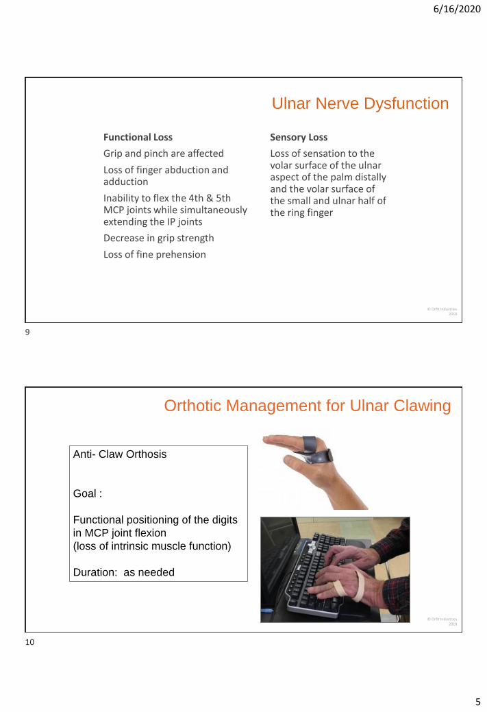
Functional Loss
Grip and pinch are affected
Loss of finger abduction and adduction
Inability to flex the 4th & 5th MCP joints while simultaneously extending the IP joints
Decrease in grip strength
Loss of fine prehension
Sensory Loss
Loss of sensation to the volar surface of the ulnar aspect of the palm distally and the volar surface of the small and ulnar half of the ring finger
Ulnar Nerve Dysfunction
© Orfit Industries2018
Anti- Claw Orthosis
Goal :
Functional positioning of the digits
in MCP joint flexion
(loss of intrinsic muscle function)
Duration: as needed
Orthotic Management for Ulnar Clawing
9
10

6/16/2020
6
© Orfit Industries2018
Fabrication of the Ulnar Claw Orthosis
https://www.youtube.com/watch?v=LhOvT3NjNr4
© Orfit Industries2018
Materials:
Orfit Strips 3.2mm x 12”
or
Orficast 6 cm- folded lengthwise
(either in half or tripled)
11
12
6/16/2020
7
© Orfit Industries2018
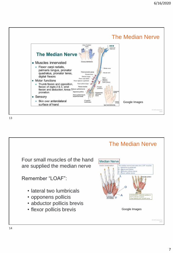
The Median Nerve
Google Images
© Orfit Industries2018
Four small muscles of the hand
are supplied the median nerve
Remember “LOAF”:
• lateral two lumbricals
• opponens pollicis
• abductor pollicis brevis
• flexor pollicis brevis Google Images
The Median Nerve
13
14

6/16/2020
8
© Orfit Industries2018
Sites of compression:
Pronator syndrome – between the 2 heads of pronator teres
(tenderness in forearm, pain with repetitive pronation)
AIN compression – motor branch off median nerve (unable to make an ok sign)
Carpal tunnel syndrome (nocturnal symptoms, numbness and tingling, atrophy)
The Median Nerve
Google Images
© Orfit Industries2018
Differential Diagnosis:
The clinical evaluation of CTS vs Pronator Teres syndrome differs in the following ways:
Tinel sign is typically absent at the wrist, but may be positive over the proximal anterior forearm
Phalen’s test is usually negative in pronator syndrome
Palpation demonstrates tenderness over the pronator teres and likely over the medial epicondyle
Median Nerve Dysfunction
15
16

6/16/2020
9
© Orfit Industries2018
High Nerve Injury
Above origin of AIN
Low Nerve Injury
Thenar intrinsic muscles paralyzed
Deborah A. Schwartz
Median Nerve Dysfunction
Google Images
© Orfit Industries2018
Median Nerve Dysfunction
Symptoms include
•Weakness and /or loss of functional grip and pinch
•Atrophy of thenar muscles
•Sensory Loss- thumb, index finger, long finger, and the radial aspect of the ring finger
Google Images
17
18

6/16/2020
10
© Orfit Industries2018
ADLs are affected including all fine motor tasks
• Difficulty grasping, pinching
• Problems with opening containers
• Pain with holding objects
• Decreased power grip
• Lumbrical muscles of index and middle finger are weak
Median Nerve Dysfunction
Google Images
© Orfit Industries2018
Orthotic Management
Recommended Orthoses:
Hand based thumb spica with MCP joint included
Hand based CMC joint orthosis w/out MCP joint
Goal of the Orthosis:
Positioning for maintaining the
first web space to provide
support, and improve ADL
function
Duration: as needed
19
20

6/16/2020
11
© Orfit Industries2018
Orthotic Options for the Thumb
© Orfit Industries2018
Fabrication of a Thumb OrthosisMaterials:
Orficast More 15 cm / 6”
Orfit Precuts
Sheet Materials:
Classic
Orfilight
Orfit Colors NS
21
22
6/16/2020
12
© Orfit Industries2018
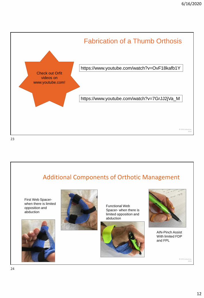
Fabrication of a Thumb Orthosis
https://www.youtube.com/watch?v=OvF18kafb1Y
https://www.youtube.com/watch?v=7GrJJ2jVa_M
Check out Orfit
videos on
www.youtube.com!
© Orfit Industries2018
Additional Components of Orthotic Management
First Web Spacer-
when there is limited
opposition and
abduction
AIN-Pinch Assist
With limited FDP
and FPL
Functional Web
Spacer- when there is
limited opposition and
abduction
23
24

6/16/2020
13
© Orfit Industries2018
Combined Median Nerve/ Ulnar Nerve Dysfunction
Characteristics:
• Clawing of all four digits
• Adduction of thumb
• Inability to oppose and abduct
the thumb
• Inability to extend PIP joints
© Orfit Industries2018
Indications:
• Spinal Cord Injuries
• Charcot Marie Tooth
• ALS
• Trauma
25
26

6/16/2020
14
© Orfit Industries2018
Create a Functional Orthosis
Without thumb
With thumb
© Orfit Industries2018
Create a Functional Orthosis
Dynamic thumb attachment
27
28
6/16/2020
15
© Orfit Industries2018
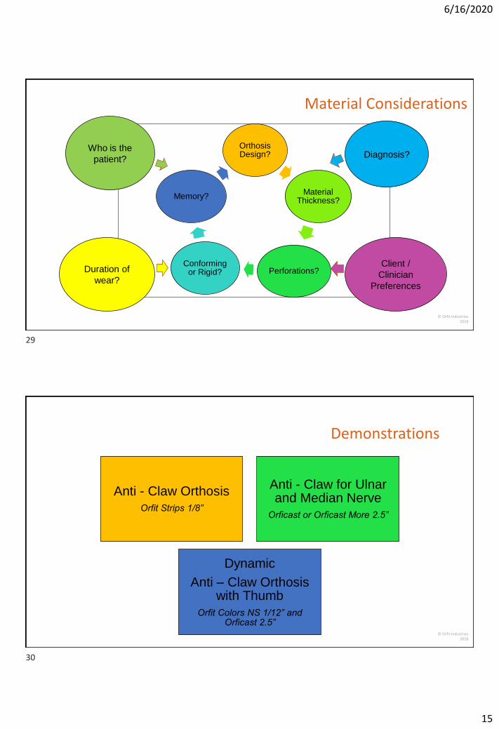
Orthosis Design?
Material Thickness?
Perforations?Conforming
or Rigid?
Memory?
Client /
Clinician
Preferences
Duration of
wear?
Diagnosis?Who is the
patient?
Material Considerations
© Orfit Industries2018
Demonstrations
Anti - Claw Orthosis
Orfit Strips 1/8”
Anti - Claw for Ulnar and Median Nerve
Orficast or Orficast More 2.5”
Dynamic
Anti – Claw Orthosis with Thumb
Orfit Colors NS 1/12” and Orficast 2.5”
29
30

6/16/2020
16
© Orfit Industries2018
For Show
Thumb and Index
Figure of 8 Orthoses for enhanced pinch
Orfit Strips 1/12”
Thumb Abduction Strap
Orficast or Orficast More 2.5”
First Web Spacer
Orficast 2.5”
© Orfit Industries2018
Evidence
Chan, R. K. (2002). Splinting for peripheral nerve injury in upper limb. Hand Surgery, 7(02), 251-259.
Choi, J. S., Mun, J. H., Lee, J. Y., Jeon, J. H., Jung, Y. J., Seo, C. H., & Jang, K. U. (2011). Effects of modified dynamic metacarpophalangeal joint flexion orthoses after hand burn. Annals of rehabilitation medicine, 35(6), 880.
Colditz, J. C. (2002). Splinting the hand with a peripheral nerve injury. In Rehabilitation of the hand and upper extremity (pp. 622-634). Mosby, Inc, St. Louis, MO.
Dauzère, Florence & Delclaux, S. & Pham, T.T. & Rongières, Michel & Mansat, Pierre. (2018). Combined Median and Ulnar Nerve Palsy Complicating Distal Radius Fractures. Orthopaedics & Traumatology: Surgery & Research. 104. 10.1016/j.otsr.2018.04.026.
Dell, P. C., & Sforzo, C. R. (2005). Ulnar intrinsic anatomy and dysfunction. Journal of Hand Therapy, 18(2), 198-207.
31
32

6/16/2020
17
© Orfit Industries2018
Evidence
Gajiwala, K. J., Sams, S. B., Pandya, N., & Wagh, A. (1991). A new dynamic lumbrical simulating splint for claw hand deformity. Plastic and reconstructive surgery, 87(1), 170-173.
McKee, P., & Rivard, A. (2004). Orthoses as enablers of occupation: client-centered splinting for better outcomes. Canadian Journal of Occupational Therapy, 71(5), 306-314.
Ruijs, A. C., Jaquet, J. B., Kalmijn, S., Giele, H., & Hovius, S. E. (2005). Median and ulnar nerve injuries: a meta-analysis of predictors of motor and sensory recovery after modern microsurgical nerve repair. Plastic and reconstructive surgery, 116(2), 484-494.
Seu, M., & Pasqualetto, M. (2012). Hand therapy for dysfunction of the intrinsic muscles. Hand clinics, 28(1), 87-100.
Sousa, G. G., & de Macêdo, M. P. (2015). Effects of a dynamic orthosis in an individual with claw deformity. Journal of Hand Therapy, 28(4), 425-428.
Watanabe, H., Ogata, K., Okabe, T., & Amano, T. (1978). Hand orthosis for various finger impairments—the KU finger splint. Prosthetics and orthotics international, 2(2), 95-100.
© Orfit Industries2018
Tips for Increasing Your Client’s Compliance with Orthotic Wear
•Make sure client understand purpose of orthosis
•Make sure client understands wearing schedule
•Make sure client has some say in final design (choice of color of material, straps)
•Have client keep a log of orthotic use
•Assess functional status with and without the orthosis
**Measure active and passive range of motion and/or perform a functional assessment prior to orthotic intervention.
33
34

6/16/2020
18
© Orfit Industries2018
Photo courtesy of
Orfit Industries
Thank you for your attention!
© Orfit Industries2018
Join Orfit on Social Media!
Orfit Splinting &
Rehabilitation Group
www.facebook.com/orfit.industries
https://www.facebook.com/groups/Orfit.splinting
Twitter @Orfit
LinkedIn www.linkedin.com/companies/orfit-industries
YouTube www.youtube.com/orfitindustries
Instagram https://www.instagram.com/orfitindustries/
Blog Blog.orfit.com
35
36

6/16/2020
19
© Orfit Industries2018
www.orfit.com
37