week 1 lesson plan - dental assistant school · week 1 lesson plan outcomes: upon ... orthodontics:...
TRANSCRIPT

Week 1 Lesson Plan
Outcomes:Upon completion of the unit, students will be able to describe eight dental specialties, describe the role of the dental office personnel, identify the major areas and pieces of equipment in the dental office, identify the primary and permanent teeth and identify the surfaces and parts of teeth. Students will also be able to identify the basic components of a patient chart and gather patient data including personal and financial information and medical and dental histories.
Instructional Aids:Video: Essentials of Effective Dental AssistingWeek 1 Checklist
Topics: Strategies (see lecture notes):History of Dentistry Founders of modern dentistryThe Dental Health Team Explain members of dental teamThe Dental Specialties List specialties and discussThe Dental Office Explain rooms and functions of areasDental Office Equipment Explain equipmentCoronal Polish Demonstrate and take turns doing coronal
Practice polishing on each other (nice introduction)Parts and Tissues of the Teeth Use diagram of tooth to show anatomyLandmarks of the mouth Use handout and dexterTypes of teeth Draw on whiteboardSurfaces of teeth Use set of teeth for typodontCharting symbols Draw on whiteboard and do charting
exercises together.Gathering patient information Show New Patient Forms and Medical
History formWatch video: Essentials of Effective Dental Assisting

Lecture Notes: Chapter 1History of DentistryPierre Fauchard is the founder of modern dentistry.GV Black made many contributions to dentistry including the perfection of amalgam.He is often called the father of dentistry.Kells is credited with the use of dental assistants (ladies in attendance).William Roentgen discovered x-rays and radiographs.
The Dental Team:Four-handed dentistry is also known as team dentistry.The dentist is ultimately responsible, legally and ethically for the entire dental team.The clinical dental assistant is directly involved in patient care by assisting the dentist.The dental hygienist often does preventative procedures such as scaling and root planning, sealants, fluoride rinses, and regular prophylaxes (cleanings).Business assistants largely take care of the business office.Laboratory technicians create prosthetics (crowns, bridges, dentures, etc).A dentist is either a DDS or DMD.
Dental Specialties:Dental public health: studies community dental health and makes suggestions for improvements.Endodontics: concerned with the prevention and treatment of disease and injury to the pulp. (Root canal)Oral and Maxillofacial Radiology: uses sophisticated imaging techniques to diagnose tumors, disease and TMJ disorders.Oral and Maxillofacial Surgery: specialty in surgery (complicated tooth extractions).Oral Pathology: specialty of the nature of disease affecting the oral cavity.Orthodontics: specialty in diagnosis and treatment of all forms of malocclusion.Pedodontics: specialty concerned with all oral health of children.Periodontics: specialty of disease of oral tissues.Prosthodontics: specialty of restoration of teeth.
The Dental Office:The reception area: previously referred to as the waiting room. Place where patients gather to wait for treatment.Business office: room or space where office employees work on scheduling, handling accounts, maintaining records, billing.Central sterilization area: room or space where soiled or contaminated instruments are sterilized for reuse. This space would always include a contaminated and clean area.Treatment rooms: also known as operatories. The place or room where treatment takes place.Laboratories: work areas for basic laboratory procedures.
Dental Associations:
ADA OSAP EPAADHA OSHA FDAADAA CDC DANB
Chapter 2Ethics, Regulations and LicensingEthics involves codes of behavior surrounding the dental profession. All dental personnel are bound by a Code of Ethics, taught in dental and hygiene school.Legal aspects deal with the law and regulations of the governing body of that profession.
Civil Law: quality/standard of careCriminal Law: violation of a license, inappropriate use of drugs, insurance or other fraud.Contract Law: violation of contractsTort Law: violation of torts
State Board of Dental Examiners publishes the Dental Practice Act, which specifies rules and regulations, and enforces them.Licensure: Dentists and Hygienists must have active and current licenses to practice in the state. Dental Assistants require registration and certifications in some states for certain procedures.
Registration can require a course, exam and registering. Certification can require a course and exam.Certified Dental Assistant is a DANB national certification, used in some states to allow
dental assistants to perform certain procedures. The DANB specific national and state certifications through exams in radiology and infection control are used in certain states for specific state certifications.
Risk ManagementMalpractice is professional negligence.Act of Omission occurs when the dentist fails to act.Act of Commission occurs when the dentist did something unreasonable or unacceptable.Consent means has accepted or agrees to treatment, etc. There is implied or informed consent.
Clinical RecordsDental charts are patient records, containing diagnosis, radiographs, consent forms, medical histories, lab scripts, correspondences and progress notes. A dental chart or patient record is a legal document.Broken appointments or cancellations should be noted in the patient’s record.Ownership of the chart is the dentist’s, although patients have a right to view and access the chart.If an error is made while making a note in the chart, draw a line through the error, date and initial.
Chapter 3Terms of the Body PlanesSagittal plane is any vertical plane that divides the body into top, bottom, left, right The Coronal PlaneMidsagittal Plane is the midline, can be called the frenum.
Head and Neck AnatomyThe maxillary bone forms the upper jawThe mandibular bone forms the lower jawThe temperomandibular joint is located where the temporal bone and the mandible join.Movements of the TMJ are hinge action and gliding action movements.The muscle that raises the mandible, closes the jaws and occludes the teeth is the masseter.The zygomatic muscle draws the angles of the mouth upward and backward.
TongueSmall elevated structures of mucosa called the lingual papillae are associated with taste.The dorsal surface is the top of the tongue.Lateral surfaces are the sides.The underside is called the ventral.The frenum is a narrow band of tissue that connects two structures.The maxillary labial frenum is between the upper incisors.The mandibular labial frenum passes between the lower anterior teeth.The lingual frenum passes from the floor of the mouth to the midline of the undersurface of the tongue.The buccal frenum is located in the molar areas and it passes from the gingival to the inner sides of the cheek.
Hard and Soft PalatesThe soft palate is in the posterior area.The hard palate is in the anterior area and may be covered with rugae.
Chapter 4
Dental AnatomyAnatomic crown: the portion of the crown covered with enamel.Clinical crown: the portion of the crown visible in the mouth.Root: the portion of the tooth normally embedded in the alveolar process covered with cementum.Root formation: bifurcation, trifurcation, apex, apical periapical.Cervix: where the root meets the crown (cementoenamel junction or CEJ).
Tissues of the ToothEnamel: hardest material in the body; it is translucent and is made up of millions of calcified enamel prisms or enamel rods.Dentin: Makes up the main portion of the inside of the tooth, it is yellow in color and somewhat transparent. It is mineralized and is made up of microscopic dentinal tubules. Inside each tubule, is a dentinal fiber that transmits pain to the pulp.Cementum: is not as hard as enamel or dentin, it protects the root of the tooth. It is light yellow in shade, darker than enamel. It can be exposed due to gingival recession.Periodontal ligament : a dense connective tissue that connects the cementum to the alveolar bone.Pulp: the inner aspect of the dentin forms the boundaries of the pulp chamber. It is made up of blood vessels and nerves.
Types of TeethThere are 32 total permanent teeth.There are 20 primary/deciduous teeth.Incisors: single rooted teeth with a sharp thin edge located in the front (8 total).Canines (cuspids) are located at the corners of the arch and are long and thick. (4 total).Premolars: (bicuspids): have two cusps with a broader chewing surface. (8 total).Molars: have more cusps, usually four, and are located in the posterior. (12 total).
Dental ArchesMandibular: the lower archMaxillary: the upper arch
Quadrants and sextantsQuandrants divide the arch in half.Sextants divide the arch in thirds.Anterior and Posterior: front versus back.
Tooth SurfacesFacial (buccal)Lingual

OcclusalMesialDistalIncisal
Anatomical FeaturesCingulum: a bulge of enamel found on the cervical third of the lingual surface of an anterior tooth.Cusp: a pronounced elevation on the occlusal surface.Cusp of Carabelli: the fifth cusp on an upper first molar.Fissure: a fault occurring along a developmental groove caused by incomplete joining of lobes.Pit: where two fissures cross.Fossa: a rounded or angular depression on the surface of the tooth.Lingual Fossa: a shallow depression on the lingual surface of an incisor or cuspid.Central Fossa: deep angular valley in the central portion of the occlusal surface of a mandibular molar.Triangular Fossa: a shallow depression on the occlusal surface of the posterior teeth.Groove: a small linear depression on the surface of the tooth.Developmental groove: a groove formed by the union of the two lobes during development of the crown.Supplemental groove: these often give the occlusal surface a wrinkled appearance.Incisal: edge formed by the junction of the facial and lingual surfaces.Lobe: a developmental segment of the tooth.Mamelon: ridges on anterior incisal edges.Ridge: a linear elevation on the surface of the tooth.
Descriptive FeaturesA contact is the region of the mesial or distal surfaces that touch. A contact point is the actual point where the teeth touch. Embrassures are triangular spaces between the proximal surfaces of two adjoining teeth.Occlusion is the contact between the maxillary and mandibular teeth.The cusps of the teeth in one arch fit into the fossae of the teeth in the opposing arch.Dentition refers to the natural teeth in the dental arches.The term edentulous mean without teeth.The primary dentition consists of twenty teeth that are in place shortly after the age of two.Eruption is the movement of a tooth through the bone and the gingival tissue into position.Primary teeth are also called deciduous teeth and are exfoliated in order for the permanent teeth to come into place.The roots of the deciduous teeth are resorbed during the process.
Tooth numbering systemsThe system approved by the ADA is the Universal Numbering System.
Teeth are numbered 1-32 starting with the upper right, moving to the upper left, then down to the lower left and across to the lower right.For primary teeth in this system, letters A-T are used in the same direction as the permanent system.There are other systems, however, they are not widely accepted.
Chapter 12Components of the dental examination.The purpose of the dental examination is the diagnosis or identification of disease and the recommendation of a treatment plan.The examination begins after a thorough medical history and vital signs are obtained.The exam consists of radiographs, impressions for diagnostic casts, oral examination, periodontal examination, and examination of the teeth.Chartings are taking a physical description and writing it down systematically in a diagram type of form – either geometric or anatomic.The oral examination includes a soft tissue exam of the neck, face, lips, and all soft tissues of the head and neck areas.A periodontal charting is done with a perio probe using a six number measuring system.
Charts are Legal Documents.Red and Black pens are used. Red is for treatment required and black for existing conditions.Standard cavity classifications are used to describe the types and locations of decay.A treatment plan is written diagnosis of all dental needs of the patient following the exam.Levels of treatment are: Emergency Care (level 1), Standard Care (level 2), Optimum Care (level 3).
Chapter 11
The patient record.It is a legal document.A patient registration form is used to obtain the responsible party, insurance information, clinical information and medical history.
Medical HistoriesMedical histories include questions regarding the patient’s past and present physical conditions, chronic conditions, allergies, and current medications taken.It alerts the dentist to medical conditions that may complicate treatment.It aids the dentist in identifying any special treatment needs, as well as potential medical emergencies.This document must be signed by the patient to insure accuracy.The dentist may also consult the patient’s physician regarding health problems.It is necessary to know of allergies to medications as well as latex.Antibiotics are often prescribed to patients who have heart defects of prosthetic joints.

Medical alerts are posted on charts to inform the doctor of potential problems.

DENTAL ABBREVIATIONS
M………..mesial ANT…………anteriorD………..distal POST………..posteriorB………..buccal DEC…………deciduousI…………incisal MAX………..maxillaryO………..occlusal
PT……...patientNP……..new patient RHH…….review health historyCC…….chief concern HBP……..high blood pressureAPPT….appointment CA……….cancerEX or E..examination HX……….historyTX……..treatment HH……….health historyDIAG….diagnosis RX……….prescriptionBWX….bitewing x-ray ANES……anesthesiaPA……..periapical x-ray CARP……carpuleFMX…..full mouth series x-ray LIDO……lidocainePO……..postoperative CARBO…carbocaineEXT……extraction EPI………epinephrineAMAL…amalgam COMP….compositeGING…..gingivalPREP…..preparationSEAT….final seat of prosthesisCRN……crownPFM……porcelain fused to metalFGC……full gold crownBR……..bridgeRCT……root canal therapyIMP……impressionSM…….study modelTEMP…temporary (provisional)FUD…..full upper dentureFLD…..full lower denture


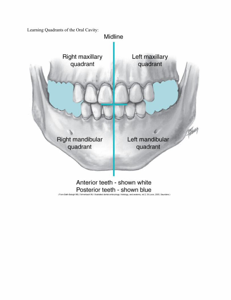
Learning Quadrants of the Oral Cavity:

Charting Abbreviations
Single Surface Abbreviations – ie: “O” for occlusal surface.Combination of Surfaces – When two tooth surfaces are involved, such as distal and occlusal, the combined surfaces are referred to as “DO” for distal-occlusal. Three surfaces combined are also used: “MOD” for mesial-occlusal-distal. The letters are pronounced separately, ie: D-O caries or M-O-D restoration.
Charting Symbols
Amalgam: Outline the surfaces that are involved and color in the area.
Composite: outline the surfaces involved.
Porcelain fused to metal: outline the coronal portion of the tooth and either add diagonal lines to indicate gold or use abbreviation if another metal is used.
Gold: outline the crown of the tooth and place diagonal lines.
Sealant: place an “S” on the occlusal surface.
Stainless steel crown: outline the crown of the tooth and place “SS” on the occlusal surface.
To Be Extracted: draw a red diagonal line through the tooth. An alternative method is to draw two red vertical lines through the tooth.
Missing Tooth: draw a blue/black “X” through the tooth. Whether the tooth was extracted or whether it just never erupted does not make a difference in the charting. If a quadrant or arch is edentulous, make an “X” over area where teeth would otherwise be.
Impacted or Unerupted: draw a red circle around the whole tooth, including the root.
Decay: depending on the caries classification, outline and color the area for amalgam, or outline the area for composite.
Recurrent Decay: outline the existing restoration in red to indicate decay in the area.
Root Canal: draw a line through the center of each root involved.
Periapical Abscess: draw a red circle at the apex of the root to indicate infection.
Post and Core: draw a line through the root that requires a post, then continue the line into the gingival third of the crown, making a triangular shape.
Rotated tooth: if a tooth has rotated in its position, indicate the direction the tooth has turned by placing a red arrow along the side of the tooth.
Diastema: where there is considerable space between two teeth, draw two red vertical lines between the areas.Fixed Bridge: draw an “X” through the roots of the missing tooth or teeth. Then draw a line to connect each of the teeth that make up the bridge. The type of material used to fabricate the bridge will determine whether you would outline the crowns for porcelain, use diagonal lines for gold, or use a combination of the two.
Full Crown: outline the complete crown if it is to be a porcelain crown, or outline and place diagonal lines for a gold crown.
Drifting: place a red arrow pointing in the direction of drift of a tooth.
Implant: Draw horizontal lines through the root or roots of a tooth.
Bonded Veneer: veneers cover only the facial surface of a tooth. Outline the facial portion only and use abbreviation “V”.
Fractured tooth or root: draw a red zigzag line where the fracture occurred.

Tooth Designation System
Maxillary Teeth
1. Maxillary right third molar (wisdom tooth)2. Maxillary right second molar3. Maxillary right first molar4. Maxillary right second premolar/bicuspid5. Maxillary right first premolar/bicuspid6. Maxillary right canine/cuspid7. Maxillary right lateral incisor8. Maxillary right central incisor9. Maxillary left central incisor10. Maxillary left lateral incisor11. Maxillary left canine/cuspid12. Maxillary left first premolar/bicuspid13. Maxillary left second premolar/bicuspid14. Maxillary left first molar15. Maxillary left second molar16. Maxillary left third molar (wisdom tooth)
Mandibular Teeth
17. Mandibular left third molar (wisdom tooth)18. Mandibular left second molar19. Mandibular left first molar20. Mandibular left second premolar/bicuspid21. Mandibular left first premolar/bicuspid22. Mandibular left canine/cuspid23. Mandibular left lateral incisor24. Mandibular left central incisor25. Mandibular right central incisor26. Mandibular right lateral incisor27. Mandibular right canine/cuspid28. Mandibular right first premolar/bicuspid29. Mandibular right second premolar/bicuspid30. Mandibular right first molar31. Mandibular right second molar32. Mandibular right third molar (wisdom tooth)
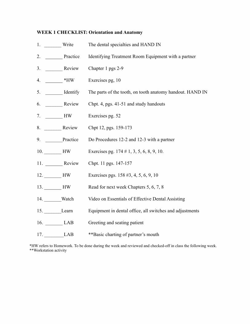
WEEK 1 CHECKLIST: Orientation and Anatomy
1. _______ Write The dental specialties and HAND IN
2. _______ Practice Identifying Treatment Room Equipment with a partner
3. _______ Review Chapter 1 pgs 2-9
4. _______ *HW Exercises pg, 10
5. _______ Identify The parts of the tooth, on tooth anatomy handout. HAND IN
6. _______ Review Chpt. 4, pgs. 41-51 and study handouts
7. _______ HW Exercises pg. 52
8. _______ Review Chpt 12, pgs. 159-173
9. _______Practice Do Procedures 12-2 and 12-3 with a partner
10. _______ HW Exercises pg. 174 # 1, 3, 5, 6, 8, 9, 10.
11. _______ Review Chpt. 11 pgs. 147-157
12. _______ HW Exercises pgs. 158 #3, 4, 5, 6, 9, 10
13. _______ HW Read for next week Chapters 5, 6, 7, 8
14. _______Watch Video on Essentials of Effective Dental Assisting
15. _______Learn Equipment in dental office, all switches and adjustments
16. _______ LAB Greeting and seating patient
17. ________LAB **Basic charting of partner’s mouth
*HW refers to Homework. To be done during the week and reviewed and checked-off in class the following week. **Workstation activity

QUIZ #1 – ORIENTATION, ANATOMY AND CHARTING
Name_________________________________ Date______________Session______________________________
1. Disease of the dental pulp are treated by an/a ______________.a. Endodontistb. Oral surgeonc. Periodontistd. Prosthodontist
2. The dental degree DDS means__________.a. Dental Doctor and Specialistb. Degree in Dental Sciencec. Doctor of Dental Specialtiesd. Doctor of Dental Surgery
3. What does ADAA stand for? ____________________________________________4. What does ADA stand for? _____________________________________________
5. The dental specialty concerned with the nature of the diseases affecting the oral structures and adjacent regions is ____________________.
a. dental hygieneb. dental public healthc. oral and maxillofacial surgeryd. oral pathology
a.6. A/An ______________________ legally performs tasks such as fabricating bridges, as
specified by the written prescription of the dentist.a. dental laboratory technicianb. expanded-functions dental assistantc. registered dental hygienistd. A and B
7. Identify the Dental Specialties and define them (8 of 9)
a.
b.
c
d.

e.
f.
g.
h.
8. Normally there are ___________ molars in the permanent dentition.a. tenb. twelvec. eight
9. The adult dentition has _______ teeth in an arch.a. 8b. 10c. 16d. 30
10. _______________ have four or more cusps to form a broad occlusal (chewing) surface.a. Molarsb. Premolarsc. Incisorsd. Cuspids
11. Define Edentulous. ________________________________________________
12. One diagonal line in blue across a tooth on a chart means that the tooth a. is missing b. has been extractedc. needs a fillingd. has cariese. both a and b

Quiz Answer Key #1 – Orientation to Dentistry, Tooth Anatomy1. A.2. D3. American Dental Assistants Association4. American Dental Association5. D6. A7. Endodontics, pedodontics, periodontics, prosthodontics, orthodontics and dentofacial orthopaedics, oral and
maxillofacial surgery, oral pathology, public health8. B9. C10. A11. Without teeth12. E