wenig - thyroid carcinoma criteria.ppt - mainweb-v.musc.edu - 04... · bulging tumor nests ... this...
TRANSCRIPT

1
Criteria for Diagnosing Thyroid Carcinoma
MUSC Pathology Multi-Specialty Course Kiawah Island, SC
April 19, 2018
Bruce M. Wenig, MD
Moffitt Cancer Center
Tampa, FL
• Dr. Wenig has no conflict(s) of interest to disclose.
ACCME/Disclosures
Minimal Criteria for Diagnosing Thyroid Carcinoma
• Diagnostic Criteria:
– Invasion
– Cytomorphologic findings
– Mitoses and necrosis
– Metastatic disease
2
Minimal Criteria for Diagnosing Thyroid Carcinoma
• Tumor types:
– Follicular Carcinoma
– Papillary Carcinoma
– Poorly-Differentiated Thyroid Carcinoma
– Anaplastic Thyroid Carcinoma
– Medullary Carcinoma
– Malignant Lymphoma
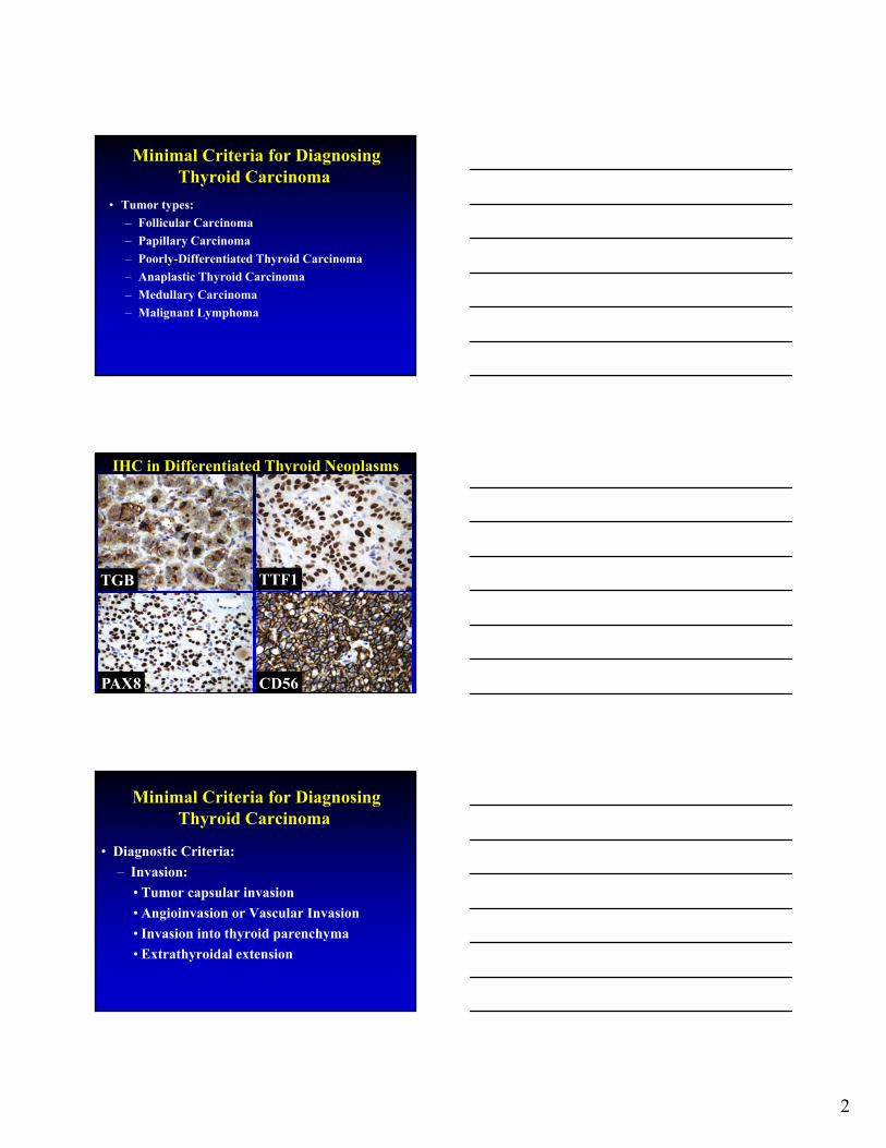
IHC in Differentiated Thyroid Neoplasms
TGB
PAX8
TTF1
CD56
Minimal Criteria for Diagnosing Thyroid Carcinoma
• Diagnostic Criteria:
– Invasion:
• Tumor capsular invasion
• Angioinvasion or Vascular Invasion
• Invasion into thyroid parenchyma
• Extrathyroidal extension

3

4
Invasion = Carcinoma

5
Follicular Adenoma vs Follicular Carcinoma
• A diagnosis of follicular carcinoma is predicated on the presence of invasive growth:
– capsular invasion
– angioinvasion
– invasion into adjacent thyroid parenchyma or beyond
Capsular Invasion• Extent of capsular invasion is contentious:
– any degree of invasion into the capsule qualifies categorization as minimally invasive follicular carcinoma
– tumor has to penetrate the entire thickness of the capsule to be regarded as unequivocal evidence of capsular invasion
• Special stains of questionable utility
Capsular Invasion*
*From Chan JKC. In: Fletcher CDM, ed. Diagnostic Histopathology of Tumors; 2013:1204.

6
Capsular Invasion
Capsular Invasion
Capsular Invasion?

7
Post-FNAB Tract
Fibrous Capsule
• *Benign tumors grow as cohesive expansile masses remaining localized to their site of origin and do not have capacity to infiltrate, invade, or metastasize
• *Benign tumors grow and expand slowly develop rim of compressed connective tissue – fibrous capsule: – separates tumor from host tissue – derived largely from extracellular matrix of native
tissue due to atrophy of normal parenchymal cells under pressure of expanding tumor
– encapsulation does not prevent tumor growth but keeps benign tumors as discrete mass
* Robbins & Cotran. Pathologic Basis of Disease. 2010:268
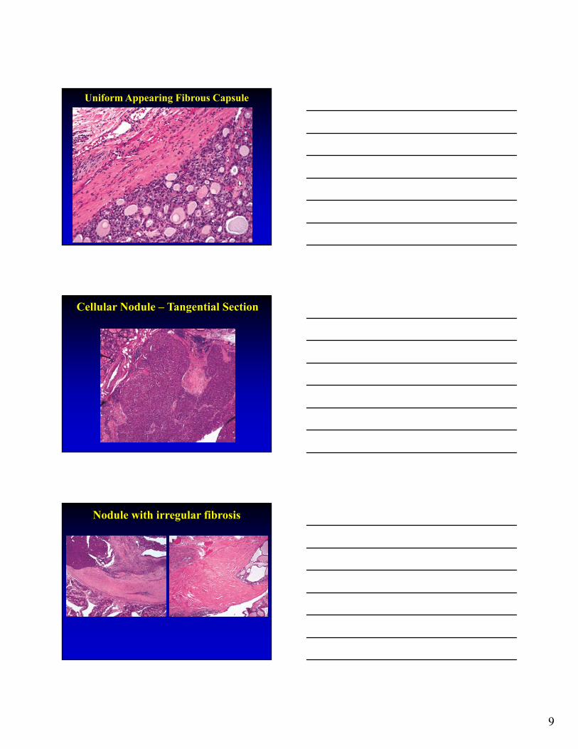
Fibrous Capsule
• Histologic features:
– Uniformity in thickness
– Fibers run in parallel

8
Fibrous Capsule
Fibrous Capsule
Capsular Invasion• Problematic features relative to diagnostic
interpretation: – irregular contour(s) of the tumor– tangential sectioning– separate nodule(s) lying outside capsule of main
nodule– serial sections to determine whether there is or is not
connection• continuity between main nodule and nodule(s)
outside the capsule = invasion (carcinoma)• absence of any connection does not exclude a
diagnosis of carcinoma• may be indicative of multiple adenomatoid
nodules
9
Uniform Appearing Fibrous Capsule
Cellular Nodule – Tangential Section
Nodule with irregular fibrosis

10
Nodule with retrogressive changes
Nodule with retrogressive changes
Adapted from: Meissner & Warren: Tumors of the Thyroid Gland. AFIP Fascicle 4; Second Series; 1969: 50.
Adenomatoid Nodule(s)
• Multiple nodules
• Poor encapsulation
• Variable structure
• Comparable growth pattern in adjacent gland
• No compression of adjacent gland
• Retrogressive changes common (post-FNAB)
• Polyclonal; reports of monoclonality
Follicular Adenoma
• Solitary nodule
• Good encapsulation
• Uniform structure
• Different growth pattern from adjacent gland
• Compression of adjacent gland
• Retrogressive changes less common (post-FNAB) (except oncocytic cell dominant)
• Monoclonal
11
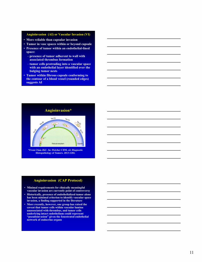
Angioinvasion (AI) or Vascular Invasion (VI)
• More reliable than capsular invasion• Tumor in vasc spaces within or beyond capsule• Presence of tumor within an endothelial-lined
space:– presence of tumor adherent to wall with
associated thrombus formation – tumor cells protruding into a vascular space
with an endothelial layer identified over the bulging tumor nests
• Tumor within fibrous capsule conforming to the contour of a blood vessel (rounded edges) suggests AI
Angioinvasion*
*From Chan JKC. In: Fletcher CDM, ed. Diagnostic Histopathology of Tumors. 2013:1202.
Angioinvasion (CAP Protocol)
• Minimal requirements for clinically meaningful vascular invasion are currently point of controversy
• Historically, presence of endothelialized tumor alone has been minimal criterion to identify vascular space invasion, a finding supported in the literature
• More recently, however, one group has raised the caveat that tumor cells within vascular lumina unassociated with thrombus, and tumor cells underlying intact endothelium could represent “pseudoinvasion” given the fenestrated endothelial network of endocrine organs

12
Angioinvasion (CAP Protocol)
• Using more rigorous criteria, namely invasion of tumor cells through a vessel wall as well as thrombus formation in association with tumor, this group demonstrated that over one-third of tumors that fulfilled these criteria had distant metastases
• It is acknowledged that the risk of metastasis when these criteria are not fulfilled by a focus in vessels is not entirely absent
Follicular NeoplasmsAngioinvasion
• Mete and Asa (Modern Pathology 2011;24:1545-52)
– Strict criteria:
• Tumor cells invade through vessel wall
• Thrombus adherent to intravascular tumor
• Found in 118 of 4000 lesions (3%)
• Follow-up in 98 cases: 35% developed metastases
• Application of rigid criteria for vascular invasion predicts distant metastasis in thyroid carcinoma especially well-differentiated thyroid carcinoma
Angioinvasion*
*From Seethala RR, et al. CAP Protocol –Carcinomas of the Thyroid Gland 2014.

13
Angioinvasion*
**** D represents a common but contentious scenario among experts, in light of these new proposed criteria for significant VI. This endothelialized tumor deposit is juxtaposed to the vessel wall. As this is somewhat similar to C, and there is no obvious thrombus, technically this would not count as significant VI. One counterargument is that the endothelialized appearance represents “organization” of a tumor thrombus and is thus still significant. While deeper levels may help, this scenario may still be considered a “JUDGMENT CALL” based on current level of evidence
VI with fibrin thrombus
VI with fibrin thrombus

14
VI with fibrin thrombus
Angioinvasion*
*From Seethala RR, et al. CAP Protocol –Carcinomas of the Thyroid Gland 2014.
VI without fibrin thrombus

15
VI without fibrin thrombus
VI without fibrin thrombus
CD31
VI without fibrin thrombus

16
VI without fibrin thrombus
Is this VI or not?
Follicular carcinoma with capsular invasion and foci worrisome for VI
Is this VI or not?
NOT!

17
Is this VI or not?
CD31
NOT!
Angioinvasion*
**** D represents a common but contentious scenario among experts, in light of these new proposed criteria for significant VI. This endothelialized tumor deposit is juxtaposed to the vessel wall. As this is somewhat similar to C, and there is no obvious thrombus, technically this would not count as significant VI. One counterargument is that the endothelialized appearance represents “organization” of a tumor thrombus and is thus still significant. While deeper levels may help, this scenario may still be considered a “JUDGMENT CALL” based on current level of evidence
Follicular Carcinoma Categorization

18
2017 CAP Protocol
• Angioinvasion (vascular invasion)
___ Not identified
___ Present
+ Extent:
+ ___ Focal (less than 4 vessels)
+ ___ Extensive (4 or more vessels)
___ Cannot be determined
• Lymphatic Invasion
___ Not identified
___ Present
___ Cannot be determined
Immunohistochemical staining using CD31 and podoplanin (D2-40) may
be useful in differentiating capillary sized vascular spaces from lymphatic
spaces
Follicular Adenoma v Follicular CarcinomaTissue Sectioning
• Ideally submit entire lesion• Not practical for larger tumors:
– minimum of 10 blocks– International Workshop on Thyroid Pathology:
• Encapsulated follicular neoplasm - at least 5:– Low cellularity, large follicles, edematous
stroma and no invasion = FA – Increased cellularity and/or other suspicious
features – at least 5 additional blocks
Follicular Tumor of Uncertain Malignant Potential (FT-UMP)
• Introduced for those tumors in which there is limited capsular invasion (absence of complete capsular transgression), absence of angioinvasion, absence of nuclear features of papillary thyroid carcinoma
• Follicular adenoma with atypical features

19
Well-Differentiated Tumor of Uncertain Malignant Potential (WDT-UMP)
• Introduced for those tumors in which there are questionable (incomplete) nuclear features of papillary thyroid carcinoma
• Follicular adenoma with atypical features
Follicular Carcinoma Histologic Types
• Oncocytic (Hürthle cell)• Signet ring cell• Clear cell • Mucinous variant• Hyalinizing trabecular carcinoma
2017 CAP Protocol - Histologic Type Follicular Thyroid Carcinoma
• ___ Follicular carcinoma, minimally invasive • ___ Follicular carcinoma, encapsulated angioinvasive• ___ Follicular carcinoma, widely invasive • ___ Follicular carcinoma, minimally invasive, oncocytic (Hürthle cell) • ___ Follicular carcinoma, encapsulated angioinvasive, oncocytic
(Hürthle cell) • ___ Follicular carcinoma, widely invasive, oncocytic (Hürthle cell) • ___ Follicular carcinoma, minimally invasive, other variant (specify)• ___ Follicular carcinoma, encapsulated angioinvasive, other variant • ___ Follicular carcinoma, widely invasive, other variant (specify) • ___ Follicular carcinoma
20
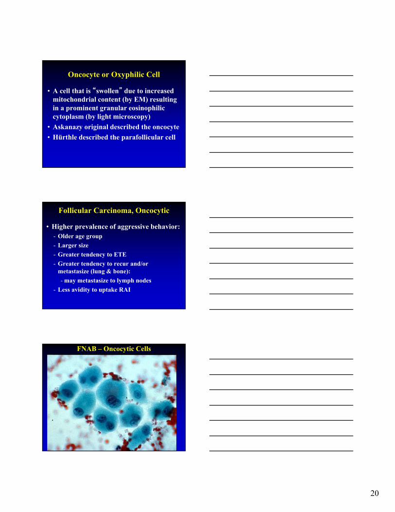
Oncocyte or Oxyphilic Cell
• A cell that is “swollen” due to increased mitochondrial content (by EM) resulting in a prominent granular eosinophilic cytoplasm (by light microscopy)
• Askanazy original described the oncocyte
• Hürthle described the parafollicular cell
Follicular Carcinoma, Oncocytic
• Higher prevalence of aggressive behavior:- Older age group
- Larger size
- Greater tendency to ETE
- Greater tendency to recur and/or metastasize (lung & bone):
- may metastasize to lymph nodes
- Less avidity to uptake RAI
FNAB – Oncocytic Cells
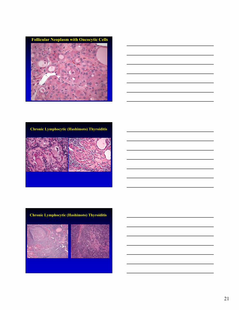
21
Follicular Neoplasm with Oncocytic Cells
Chronic Lymphocytic (Hashimoto) Thyroiditis
Chronic Lymphocytic (Hashimoto) Thyroiditis

22
Thyroid Lesions with Oncocytic Cells
• Nonneoplastic Lesions:– Lymphocytic thyroiditis– Adenomatoid nodules – Graves disease– Post-radiation; aging
• Neoplasms:– Follicular adenoma/carcinoma (Hürthle
cell adenoma/carcinoma); Papillary Thyroid Carcinoma
Minimal Criteria for Diagnosing Thyroid Carcinoma
• Diagnostic Criteria:
– Invasion
– Cytomorphologic findings
– Mitoses and Necrosis
– Metastatic disease

23
Diagnosis of Thyroid Carcinoma Based on Cell Type
• Papillary Thyroid Carcinoma
• Medullary Thyroid Carcinoma
• Poorly-differentiated Thyroid Carcinoma
• Anaplastic Thyroid Carcinoma
• Malignant Lymphoma
• In general, follicular adenoma and follicular carcinoma cannot be differentiated based on cell type
Papillary Thyroid Carcinoma (PTC) Definition
• Malignant thyroid follicular epithelial cell neoplasm characterized by distinctive nuclear features
PTC – Papillary Growth

24
Papillary Thyroid Carcinoma Pathologic Features
• Cytopathologic (Nuclear) features:– Nuclear enlargement and/or elongation with
irregularities in size and shape– Dispersed (very fine) to optically clear
appearing chromatin– Crowding and overlapping – Nuclear grooves– Cytoplasmic invagination into nucleus
(inclusions)
PTC – “Orphan Annie” Nuclei
Hapke MR, Dehner LP. The optically clear nucleus. A reliable sign of papillary carcinoma of the thyroid? AJSP 1979;3:31-38.
PTC – Diagnostic Nuclei
25
PTC – Nuclear Inclusions
Inclusion in Follicular Adenoma
“Bubble artifact” ≠ Inclusions

26
PTC can be diagnosed by FNAB
Psammoma Bodies
Calcified/inspissated colloid –not psammoma body

27
Endocrine Atypia
Papillary Growth in Adenomatoid Nodule
Papillary Thyroid CarcinomaHistologic Types/Variants
• Usual or conventional
• Papillary microcarcinoma
• Encapsulated
• Follicular
• Macrofollicular
• Oncocytic or oxyphilic
• Clear cell

28
PTC, Oncocytic Variant
Papillary Thyroid CarcinomaHistologic Types/Variants Cont’d
• Warthin tumor-like
• Diffuse (Multinodular) Follicular
• PTC with nodular fasciitis-like stroma
• PTC with spindle cell metaplasia
• PTC with lipomatous stroma
Papillary Thyroid CarcinomaHistologic Types/Variants Cont’d
• Solid and Radiation-Induced
• Cribriform-Morular
• “Hobnail” (AJSP 2010;34:44-52)
• “Aggressive” variants:
– Diffuse sclerosing
– Columnar cell
– Tall cell
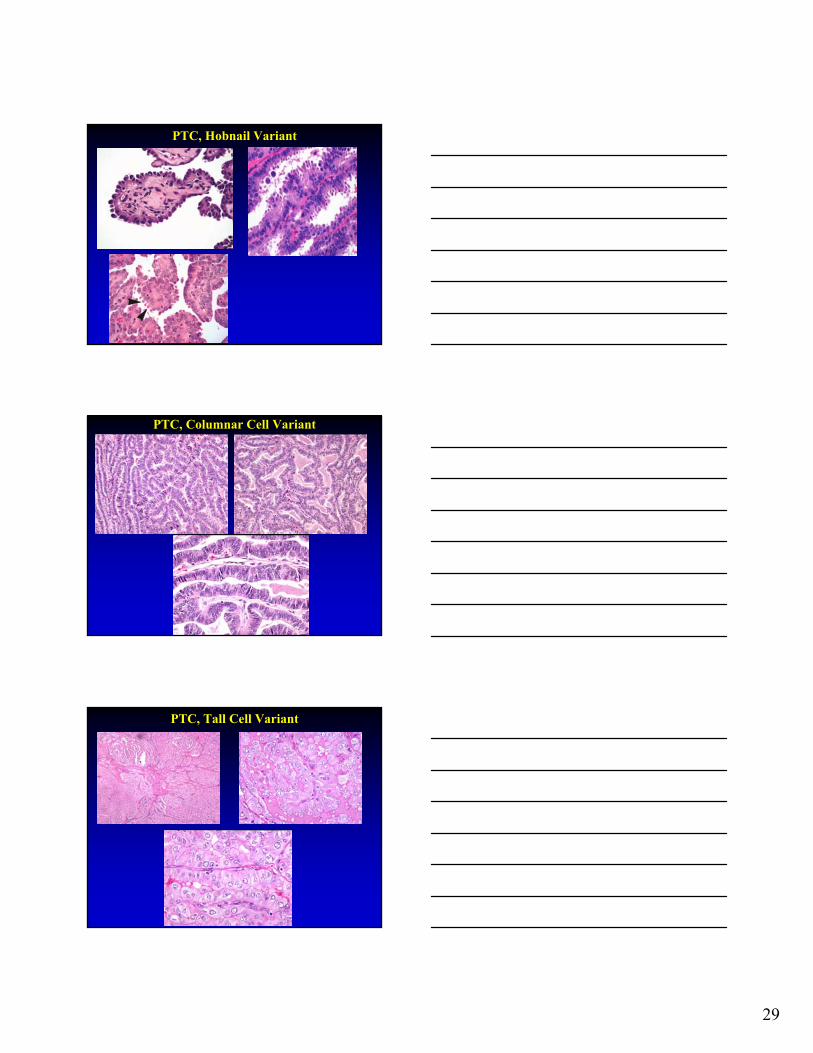
29
PTC, Hobnail Variant
PTC, Columnar Cell Variant
PTC, Tall Cell Variant

30
PTC, Tall Cell Variant
• WHO 2017 criteria:
– Cells 2-3x tall as wide
– Abundant eosinophilic (oncocytic-like) cytoplasm
– Typical nuclear features for PTC
– Account for ≥30% of all tumor cells
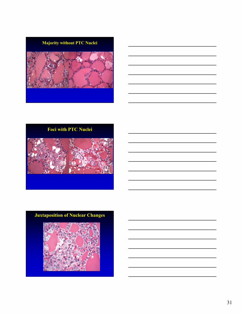
Follicular Variant of Papillary Thyroid Carcinoma
(FVPTC)
• Subset of papillary carcinoma entirely composed of follicular growth lacking papillary architecture lined by cells having the nuclear features of PTC
Circumscribed Follicular Pattern Lesion
31
Majority without PTC Nuclei
Foci with PTC Nuclei
Juxtaposition of Nuclear Changes

32
FVPTC Observer Variation*
• 10 reviewers; 87 tumors
• Concordant Diagnosis
• Most important criteria for diagnosis
• Less important criteria for diagnosis
* Lloyd et al: AJSP 2004;28:1336-40
Summary of Diagnoses
Reviewer FVPTC FA FCA Other
1 100 0 0 0
2 74.7 12.6 0 12.6
3 85.1 13.8 1.1 0
4 77 20.7 1.1 1.1
5 91.9 4.7 0 3.5
6 100 0 0 0
7 91.9 1.1 0 6.9
8 98.9 0 1.1 0
9 46.6 37.9 12.6 3.5
10 60.9 11.5 1.2 26.4
FVPTC Observer Variation
• Concordant diagnosis with a cumulative frequency of 39%
• Only 51% were diagnosed as follicular variant by all pathologists
• Metastatic disease in 24.1% affirming need to differentiate follicular variant of PTC from benign thyroid lesions

33
FVPTC Observer Variation*
• 6 reviewers; 15 cases• Interobserver and intraobserver
variation• Nuclear features of TPC not well
developed or only focally developed
* Elsheikh TM, et al: AJCP 2008;130:736-744
FVPTC Observer Variation
• Unanimous agreement FVPTC in 13% (2 cases)
• Majority agreement on benign and malignant diagnoses in 27% (4 cases)
• Majority agreement on malignant diagnosis in 53% (8 cases)
• Intraobserver agreement ranged 17-100%
• Lack of agreement on minimal criteria needed to diagnose FVPTC
FVPTCIssues
• Isolated or limited foci of PTC in an otherwise nondescript follicular lesion:
– Is there a percentage of the lesion below which not PTC but beyond which it is PTC?
– Does IHC assist in the diagnosis and DDX?
– What diagnostic term(s) should be used if not PTC?
– How to treat?
34
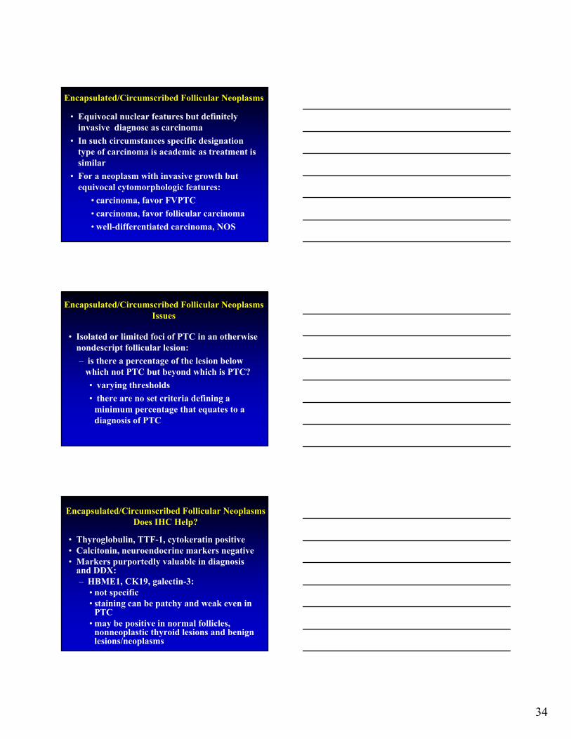
Encapsulated/Circumscribed Follicular Neoplasms
• Equivocal nuclear features but definitely invasive diagnose as carcinoma
• In such circumstances specific designation type of carcinoma is academic as treatment is similar
• For a neoplasm with invasive growth but equivocal cytomorphologic features:
• carcinoma, favor FVPTC
• carcinoma, favor follicular carcinoma
• well-differentiated carcinoma, NOS
Encapsulated/Circumscribed Follicular Neoplasms Issues
• Isolated or limited foci of PTC in an otherwise nondescript follicular lesion:
– is there a percentage of the lesion below which not PTC but beyond which is PTC?
• varying thresholds
• there are no set criteria defining a minimum percentage that equates to a diagnosis of PTC
Encapsulated/Circumscribed Follicular NeoplasmsDoes IHC Help?
• Thyroglobulin, TTF-1, cytokeratin positive• Calcitonin, neuroendocrine markers negative• Markers purportedly valuable in diagnosis
and DDX: – HBME1, CK19, galectin-3:
• not specific• staining can be patchy and weak even in
PTC• may be positive in normal follicles,
nonneoplastic thyroid lesions and benign lesions/neoplasms
35
Adenomatoid Nodule
Adenomatoid NoduleFalse positive HBME-1 Staining
HBME-1 IHC in Follicular Adenoma

36
Papillary Thyroid Carcinoma
• Does IHC assist in the diagnosis?
– at present there are no IHC markers that can reliably differentiate PTC from other follicular lesions (e.g., adenoma, carcinoma, adenomatoid nodules)
Isolated foci of PTC in an otherwise nondescript follicular lesion
• What diagnostic term should be used if is PTC?
–Follicular variant of PTC (FVPTC)
• Treatment:
– Total thyroidectomy and postoperative
radioactive iodine
Isolated foci of PTC in an otherwise nondescript follicular lesion
• What diagnostic term(s) should be used if not PTC? –Follicular adenoma (atypical)–FT-UMP–WDT-UMP
• Treatment:–Subtotal thyroidectomy

37
Isolated foci of PTC in an otherwise nondescript follicular lesion
• What diagnostic term should be used if you are unsure of the diagnosis?
– tendency to overdiagnose FVPTC
– err on the side of benignancy (follicular adenoma or atypical follicular adenoma)
– Treat conservatively
FVPTCMolecular Biology
• Molecular profile much closer to follicular adenoma and follicular carcinoma than to classical papillary carcinoma

38
Biologic Behavior of FVPTC
• Liu J, et al. Cancer. 2006;107:1255-64:
–No recurrence, lymph node metastasis
• Rivera M, et al. Mod Pathol 2010;23:1191-200:–Encapsulated/noninvasive tumors
extremely low recurrence rate–Metastatic nodal pattern:
• Noninvasive similar to follicular adenoma
• Infiltrative similar to classical PTC
Molecular Classification of PTC
• Giordano T, et al. Integrated genomic characterization of papillary thyroid carcinoma - Cancer Genome Atlas Research Network. Cell 2014;159:676-690
• Noninvasive: among RAS-like tumors rather than BRAF V600E-like tumors
• Invasive: among BRAF V600E-like tumors rather than RAS-like tumors
JAMA Oncology April 2016
39
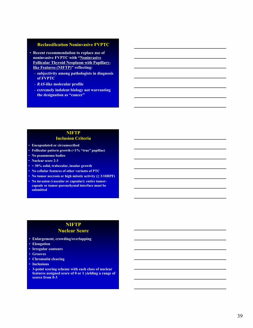
Reclassification Noninvasive FVPTC
• Recent recommendation to replace use of noninvasive FVPTC with “Noninvasive Follicular Thyroid Neoplasm with Papillary-like Features (NIFTP)” reflecting:
- subjectivity among pathologists in diagnosis of FVPTC
- RAS-like molecular profile
- extremely indolent biology not warranting the designation as “cancer”
NIFTPInclusion Criteria
• Encapsulated or circumscribed
• Follicular pattern growth (<1% “true” papillae)
• No psammoma bodies
• Nuclear score 2-3
• < 30% solid, trabecular, insular growth
• No cellular features of other variants of PTC
• No tumor necrosis or high mitotic activity (≥ 3/10HPF)
• No invasion (vascular or capsular): entire tumor-capsule or tumor-parenchymal interface must be submitted
NIFTPNuclear Score
• Enlargement, crowding/overlapping• Elongation• Irregular contours• Grooves• Chromatin clearing • Inclusions - 3-point scoring scheme with each class of nuclear
features assigned score of 0 or 1 yielding a range of scores from 0-3

40
NIFTP• Reclassification as a close entity to the follicular
adenoma/carcinoma group:- No adverse events in 109 patients- Treatment by lobectomy alone even in the
presence of adverse demographic prognostic factors (e.g., > 45 yrs, > 4 cm)
- Countless number of patients with non-invasive follicular variant spared unnecessary therapy with associated morbidity, financial costs and the psychological impact of “cancer” diagnosis
NIFTP – WHO 2017
• Definition: NIFTP is a noninvasive neoplasm of thyroid follicular cells with a follicular growth pattern and nuclear features of PTC that has extremely low malignant potential
– formerly referred to as non-invasive FVPTC
• 10-20% of all thyroid “cancers”
• F:M 3-4:1; wide age range most common in 4th-6th decades
• Cytology:
– about 50% = Bethesda IV;
– Most of remainder: Bethesda V or Bethesda III;
– Rarely: Bethesda VI “reliable distinction between NIFTP & PTC cannot be made in cytological preparations”
NIFTP• Since NIFTP initially reported:
- Several patients with locoregional nodal (micro)metastasis reported in primary tumors meeting proposed diagnostic criteria for NIFTP
• Re-evaluation of criteria for NIFTP:- No well formed papillae- Presence of diffuse nuclear features of PTC →
examination of entire tumor (not just the tumor-to-capsule/parenchymal interface)
- NIFTPs typically show moderate expression of diagnostic nuclear features of PTC
- Presence of BRAF V600E or other BRAF-like mutations (RET/PTC fusions) or high-risk mutations (TERT; TP53) → search for exclusionary features (e.g., true papillae; invasion)
41
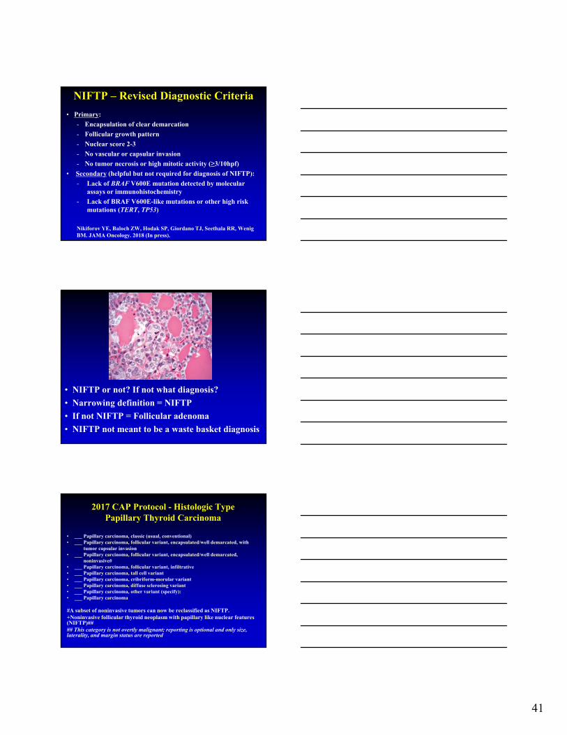
NIFTP – Revised Diagnostic Criteria
• Primary:
- Encapsulation of clear demarcation
- Follicular growth pattern
- Nuclear score 2-3
- No vascular or capsular invasion
- No tumor necrosis or high mitotic activity (≥3/10hpf)
• Secondary (helpful but not required for diagnosis of NIFTP):
- Lack of BRAF V600E mutation detected by molecular assays or immunohistochemistry
- Lack of BRAF V600E-like mutations or other high risk mutations (TERT, TP53)
Nikiforov YE, Baloch ZW, Hodak SP, Giordano TJ, Seethala RR, Wenig BM. JAMA Oncology. 2018 (In press).
• NIFTP or not? If not what diagnosis?
• Narrowing definition = NIFTP
• If not NIFTP = Follicular adenoma
• NIFTP not meant to be a waste basket diagnosis
2017 CAP Protocol - Histologic Type Papillary Thyroid Carcinoma
• ___ Papillary carcinoma, classic (usual, conventional) • ___ Papillary carcinoma, follicular variant, encapsulated/well demarcated, with
tumor capsular invasion • ___ Papillary carcinoma, follicular variant, encapsulated/well demarcated,
noninvasive# • ___ Papillary carcinoma, follicular variant, infiltrative • ___ Papillary carcinoma, tall cell variant • ___ Papillary carcinoma, cribriform-morular variant • ___ Papillary carcinoma, diffuse sclerosing variant • ___ Papillary carcinoma, other variant (specify): • ___ Papillary carcinoma
#A subset of noninvasive tumors can now be reclassified as NIFTP. +Noninvasive follicular thyroid neoplasm with papillary like nuclear features (NIFTP)## ## This category is not overtly malignant; reporting is optional and only size, laterality, and margin status are reported

42
Pre-invasive Stage of Invasive FVPTC
Hodak S, et al. Thyroid 2016;26:869-71
• Encapsulated, circumscribed (nonencapsulated)
• Low % intralesional sclerosis
• No psammoma bodies
• No ETE
• RAS mutation, PAX8/PPARγtranslocation
• Low to no incidence of metastasis
• If invasive → Invasive FVPTC →
behavior that of FTC
• Usually nonencapsulated
• ↑ % marked intralesional sclerosis
• Psammoma bodies may be present
• ↑ % ETE
• BRAF mutation, RET/PTCtranslocation
• Increased incidence of metastasis (nodal)
NIFTP Invasive FVPTC
43
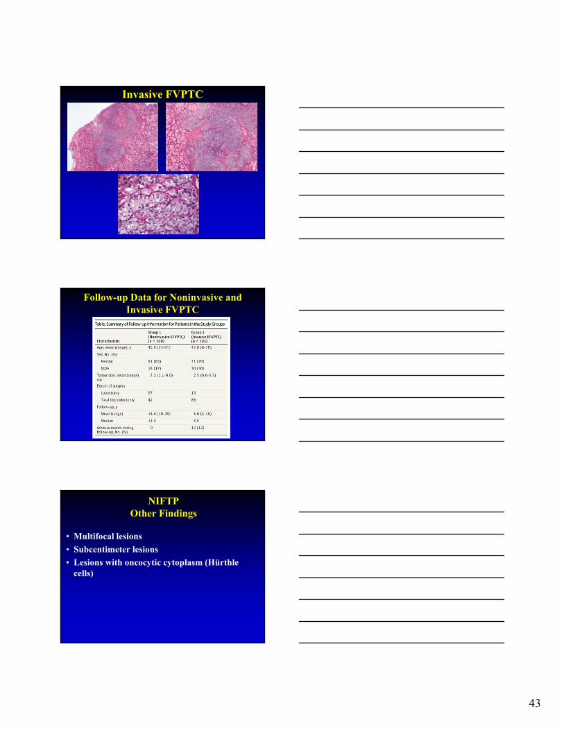
Invasive FVPTC
Follow-up Data for Noninvasive and Invasive FVPTC
NIFTPOther Findings
• Multifocal lesions
• Subcentimeter lesions
• Lesions with oncocytic cytoplasm (Hürthle cells)

44
Minimum Diagnostic Criteria for Thyroid Cancer
• Diagnostic Criteria:
– Invasion
– Cytomorphologic findings
– Mitoses and Necrosis
– Metastatic disease
Practical Genotype-Phenotype Correlations
Tall Cell VariantBRAF-V600E-like
BRAF-V600EStrong MAPK
OutputReduced Thyroid
DiffHigh risk
Follicular VariantRAS-like
RAS, PPARG fusionWeak MAPK
OutputHigh Thyroid Diff
Low risk
Classic TypeBRAF-V600E-like
BRAF-V600E, RETfusion
Inter. MAPK OutputInter. Thyroid DiffIntermediate risk
Practical Genotype-Phenotype Correlations
Hobnail VariantBRAF-V600E
High risk
Diffuse Sclerosing Variant
Mostly RET/PTC Some BRAF-V600E
Mixed risk
Cribriform-Morular VariantFAP associated
Nuclear B-catenin on IHC

45
Emerging view of
thyroid cancer pathogenesis

46
Poorly-Differentiated Thyroid Carcinoma (PDTC)
Definition
• Thyroid neoplasm with histologic and biologic features intermediate between those of differentiated thyroid carcinomas and undifferentiated (anaplastic) carcinoma
• Synonym: Insular Carcinoma
PDTCTurin Proposal*
• Presence of solid, trabecular or insular growth
• Absent nuclear features diagnostic for PTC
• Presence of at least one of the following:–Convoluted nuclei–Mitotic activity ≥3 mitoses per 10 HPF–Tumor necrosis
* Volante et al. AJSP 2007;31:1256-1264
PDTC – Insular, Trabecular and Solid
47
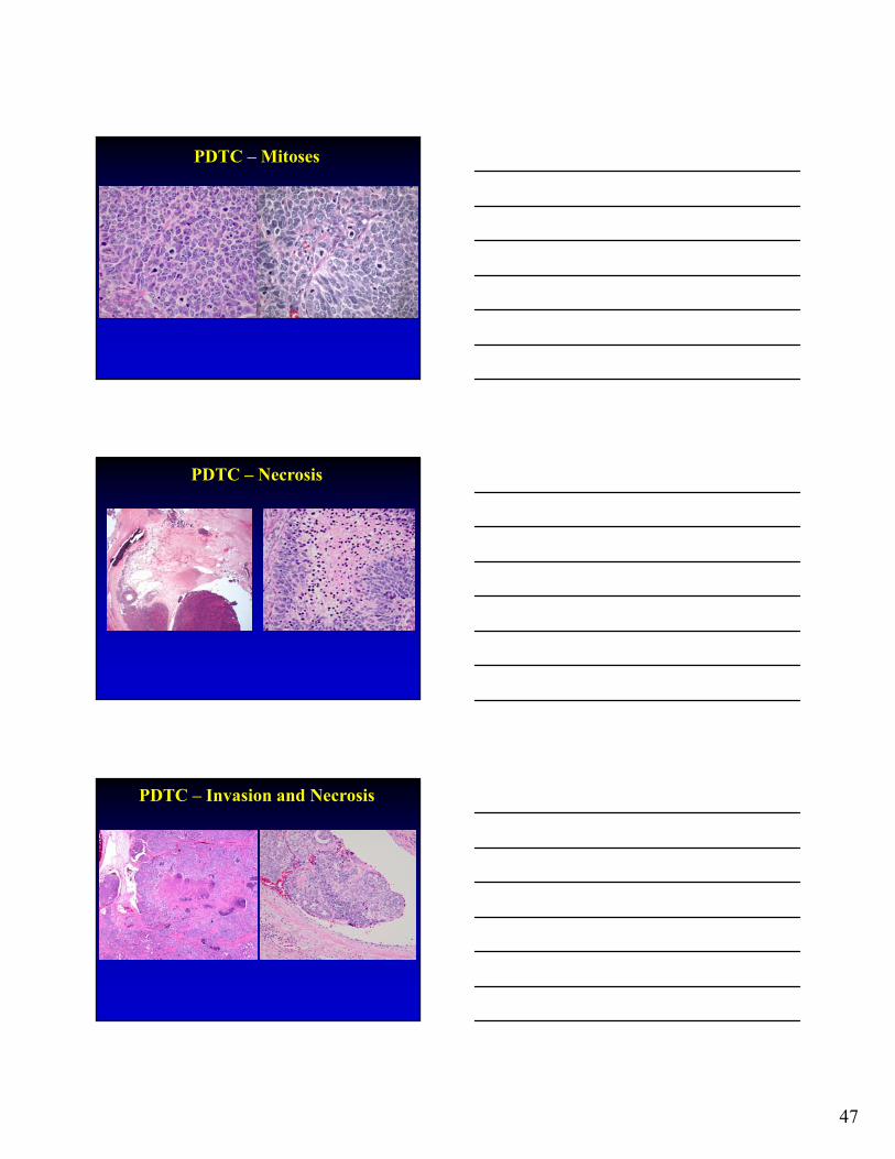
PDTC – Mitoses
PDTC – Necrosis
PDTC – Invasion and Necrosis
48
PDTC – Extrathyroidal Extension
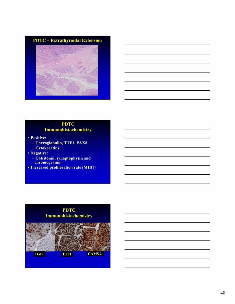
PDTCImmunohistochemistry
• Positive:– Thyroglobulin, TTF1, PAX8– Cytokeratins
• Negative:– Calcitonin, synaptophysin and
chromogranin• Increased proliferation rate (MIB1)
TGB
PDTCImmunohistochemistry
TTF1 CAM5.2
49
PDTC
• PDTC not limited to tumors with insular/solid/trabecular growth:– Hiltzik D, et al. Cancer
2006;106:1286-95:• PDTC defined on basis of ↑ mitotic activity and/or tumor necrosis
– Rivera M, et al. Cancer 2008;113:48-56:• Necrosis and/or mitotic index (≥ 5 x 10HPF)
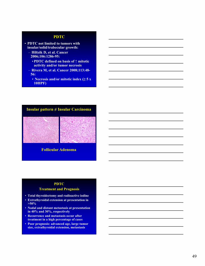
Insular pattern ≠ Insular Carcinoma
Follicular Adenoma
PDTCTreatment and Prognosis
• Total thyroidectomy and radioactive iodine • Extrathyroidal extension at presentation in
>50%• Nodal and distant metastasis at presentation
in 40% and 30%, respectively• Recurrence and metastasis occur after
treatment in a high percentage of cases• Poor prognosis: advanced age, large tumor
size, extrathyroidal extension, metastasis

50
PDTCDifferential Diagnosis
• Papillary Thyroid Carcinoma, Solid Variant
• Undifferentiated (Anaplastic) Thyroid Carcinoma
• Medullary Thyroid Carcinoma
PTC, Solid Variant
• PTC > 50% solid growth
• Common in children including those with exposure to radiation (adults, too)
• Solid sheets of tumor cells insular pattern and diagnostic nuclear features for PTC:
- lack increased mitotic activity, necrosis
- TGB, TTF1 +; CAL, NE markers negative
• Lymph-vascular invasion, extrathyroidal extension and nodal metastases
PTC, Solid Variant

51
PTC, Solid Variant
TGB TTF1
PAX8
Anaplastic Thyroid Carcinoma (ATC)
• Older patients
• Rapidly enlarging neck mass
• Long-standing history of thyroid-based mass
• Pathology:
- Absence of follicular differentiation by light microscopy and IHC
• Rapid death due to locally uncontrollable disease:
- median survival 3 - 4 months
- 5-year survival 3.6 - 10%
ATC

52
ATCIHC
AE1/AE3 PAX8
Medullary Thyroid Carcinoma (MTC)
• Malignant thyroid neuroendocrine tumor with C-cell differentiation
• Occurrence:
- Sporadic: 70-80% of all cases
- Hereditary: 20-30% of all cases:
• Germline mutation of RET in 85% of families
• MEN2A > Familial MEN > MEN2B
• Prognosis:
- 5-yr survival: approximately 70-80%
- 10-yr survival: approximately 50-78%
- 15-yr survival: approximately 65%
MTC

53
CAL
MTC
SYN
CHR TTF1
Minimum Diagnostic Criteria for Thyroid Cancer
• Diagnostic Criteria:
– Invasion
– Cytomorphologic findings
– Mitoses and Necrosis
– Metastatic disease

54
Thyroid Gland Development
Thyroid Follicles in Lymph Nodes
• When is metastatic carcinoma and when is it something else?:
– Thyroid inclusions
– Lymphocytic thyroiditis
Benign Thyroid Inclusions in Lymph Nodes
• Diagnostic Criteria:
– Midline or para-midline lymph nodes
– Localized to capsule or subcapsular region
• Metastatic PTC:
– Involvement of nodal parenchyma
– Replacement of ≥1/3 of the node
– Several nodes affected
– Not identified in nodes lateral to great vessels
– Diagnostic nuclei for PTC
– Psammoma bodies
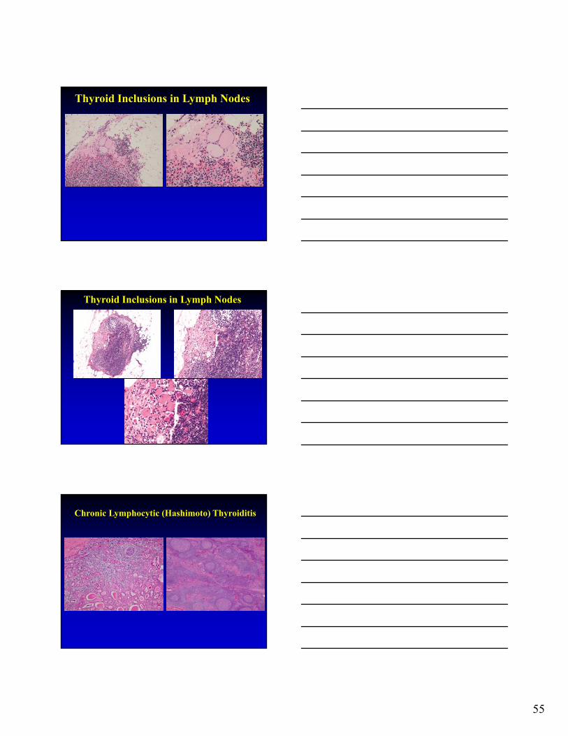
55
Thyroid Inclusions in Lymph Nodes
Thyroid Inclusions in Lymph Nodes
Chronic Lymphocytic (Hashimoto) Thyroiditis

56
Thyroid CarcinomaSummary
• Diagnosis based on constellation of histologic criteria coupled by IHC and molecular genetics:- Despite established criteria interpretation can be
contentious and subjective - Beware of potential pitfalls
• Specific types have distinct pathology but not necessarily distinct clinical features
• Histology (e.g., cell type, growth patterns) does not necessarily portend specific biology behavior
• Prognosis and treatment predicated on variety of parameters• NIFTP replaces NI-FVPTC but still evolving category • Molecular findings playing greater role in diagnosis of
follicular neoplasms
H. Lee Moffitt Cancer Center andResearch Institute
Questions?