westmead intensive care unit neurological … intensive care unit neurological learning package aim...
TRANSCRIPT

Westmead Intensive Care Unit
Neurological Learning Package Aim of the Package To provide the registered nurse with the opportunity to acquire the level of knowledge, through self directed learning, on which to base the nursing skills necessary for safe practice. Objectives of the Package
1. Identify the relevant gross anatomy of the brain 2. Discuss the pathophysiology of brain injury 3. Outline the importance of the prevention or minimization of secondary brain
injury through nursing care 4. Understand the pathophysiology of raised intracranial pressure and provide a
rational for the nursing and medical management of a patient with the potential for further brain injury as a result of raised intracranial pressure
5. Discuss organ donation in the context of brain death and donation after cardiac death
What to do with the Package
1. Attempt all questions
2. You will need to exceed 80% correct answers to pass the package, if this is
not achieved you will need to repeat the package
3. Your package will reviewed by a CNE and then you will be provided with the
package answers to allow further review
Useful Resources References found in this package will provide a guide for answering questions.
This list is by no means exhaustive and if you wish to do your own research, please do so.
Textbooks are available in the CNE nook CIAP can be access on all the computers through the intranet site

NEURO ANATOMY Question 1 Label the following diagrams.
Figure 1. Bones of the skull. From Hickey, J.V. (1986) The clinical practice of
neurological and neurosurgical nursing. J.B. Lippincott, Philadelphia
Figure 3. Bones of the base of skull. From Hickey (1986, p.20)
Mandible
Mental foramen
Maxilla
Mastoid process
Styloid process
Occipital bone
Lacrimal bone
Sphenoid bone
Zygomatic bone
Frontal bone
Parietal bone
Temporal bone
Nasal bone
External acoustic meatus
Squamosal suture
Lambdoidal suture
Coronal suture
Zygomatic arch
Frontal bone
Parietal bone
Occipital bone
Frontal sinus
Foramen magnum
Optic foramen
Crista galli
Sella turcica
Small wing of sphenoid
Great wing of sphenoid
Internal acoustic meatus
Cribriform plate of ethmoid bone
Petrous portion of temporal bone

Question 2 What are the main functions of the following areas of the brain: Frontal, Parietal, Occipital, and Temporal lobes? Corpus callosum? Basal ganglia? Mid brain, Pons and Medulla? Thalamus and Hypothalamus? Question 3
Frontal lobe
Parietal lobe
Occipital lobe
Temporal lobe
Corpus callosum
Cerebellum
Medulla oblongata
Pons
Spinal cord
Pituitary gland
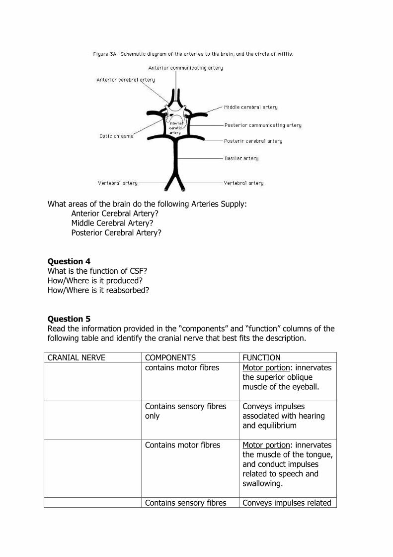
What areas of the brain do the following Arteries Supply: Anterior Cerebral Artery? Middle Cerebral Artery? Posterior Cerebral Artery? Question 4 What is the function of CSF? How/Where is it produced? How/Where is it reabsorbed? Question 5 Read the information provided in the “components” and “function” columns of the following table and identify the cranial nerve that best fits the description.
CRANIAL NERVE COMPONENTS FUNCTION
contains motor fibres Motor portion: innervates the superior oblique muscle of the eyeball.
Contains sensory fibres only
Conveys impulses associated with hearing and equilibrium
Contains motor fibres Motor portion: innervates the muscle of the tongue, and conduct impulses related to speech and swallowing.
Contains sensory fibres Conveys impulses related
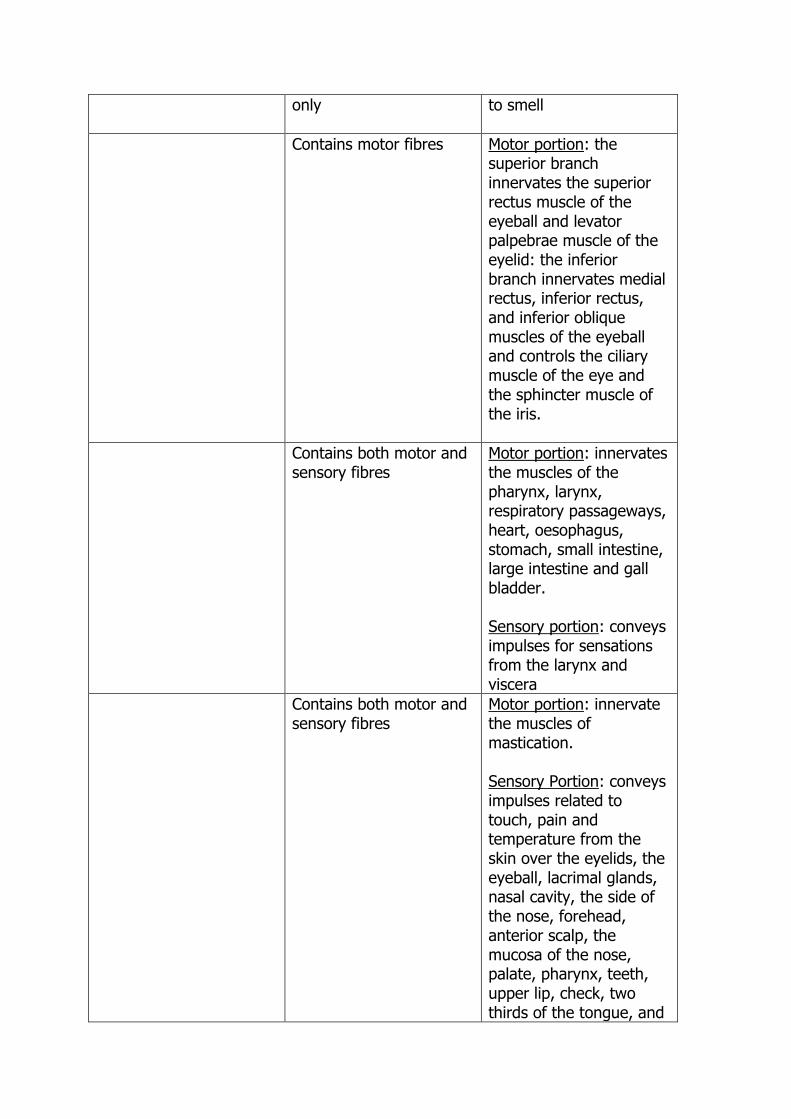
only to smell
Contains motor fibres Motor portion: the superior branch innervates the superior rectus muscle of the eyeball and levator palpebrae muscle of the eyelid: the inferior branch innervates medial rectus, inferior rectus, and inferior oblique muscles of the eyeball and controls the ciliary muscle of the eye and the sphincter muscle of the iris.
Contains both motor and sensory fibres
Motor portion: innervates the muscles of the pharynx, larynx, respiratory passageways, heart, oesophagus, stomach, small intestine, large intestine and gall bladder. Sensory portion: conveys impulses for sensations from the larynx and viscera
Contains both motor and sensory fibres
Motor portion: innervate the muscles of mastication. Sensory Portion: conveys impulses related to touch, pain and temperature from the skin over the eyelids, the eyeball, lacrimal glands, nasal cavity, the side of the nose, forehead, anterior scalp, the mucosa of the nose, palate, pharynx, teeth, upper lip, check, two thirds of the tongue, and

skin over the mandible, side of the head and floor of the mouth.
Contains sensory fibres only
Conveys impulses initiated by the rods and cones of the retina.
Contains motor fibres Motor Portion: innervates the voluntary muscles of the pharynx, larynx and soft palate as well as conveying impulses to the sternocleidomastoid and trapezium muscles.
Contains motor fibres Motor Portion: innervates the lateral rectus muscle of the eyeball.
Contains both motor and sensory fibres
Motor Portion: innervate the swallowing muscles of the pharynx and parotid gland. Sensory Portion: conduct impulses from the pharynx and taste buds of the posterior third of the tongue, and the carotid sinus: also conducts impulses for muscle sense (proprioperception) for the lateral rectus muscle.
Contains both motor and sensory fibres
Motor Portion: innervate the facial and scalp muscles, the sublingual and submandibular glands. Sensory Portion: conduct impulses from the taste buds of the anterior two thirds of the tongue: also conducts impulses for muscle sense (proprioception) for the

face and scalp.
ALTERED LEVEL OF CONSCIOUSNESS (LOC) Question 6 What are the neurological signs of Brain hypoxia? Question 7 List 6 potential causes of decreased LOC? Question 8 What is Diabetes Insipidus? Outline the difference between Neurogenic and Nephrogenic causes. NEUROLOGICAL ASSESSMENT Question 9 Strictly speaking, if you ask a patient with a Endotracheal Tube insitu what is their name, location, and current time, and they write the correct answers on a piece of paper what do they score for the voice section of the GCS? Question 10 What methods are used to illicit a painful stimulus to a patient whilst carrying out the GCS assessment? What are the pros and cons of each? Question 11 Identify factors which will inhibit the accuracy of the GCS? PRINCIPLES OF RAISED INTRACRANIAL PRESSURE
The primary goals in the management of the patient with increased intracranial pressure (ICP) in the intensive care unit are to prevent secondary neurological injury and limit possible complications that could occur in other organs systems (Butterworth & Prough 1991 p 1467). Secondary injury refers to events of complications that contribute to further brain injury after the initial primary event. Events that may cause secondary brain injury include systemic hypotension, hypoxaemia, hypercapnia, sustained increased intracranial pressure and sustained cerebral oedema. In order to understand the means of preventing secondary injury it is necessary to have a comprehensive understanding of what causes raised intracranial pressure and the basis for preventing secondary injury are based on the Monroe Kellie doctrine. Question 12

Define the “Munro-Kellie Doctrine” and explain what mechanisms allow for normal compensation (autoregulation) of increased ICP and at what point will decompensation occur? Further Reading: Andrus, C, 1991, “Intracranial Pressure: dynamics and nursing management”, Journal of Neuroscience Nursing, vol 23, no 2, pp85 –87 Question 13 Indicate on the “volume Pressure Curve” figure 4 where compensation and decompensation are occurring?
Question 14 What is the most significant problem of Increased ICP? Question 15 What is meant by the term Cerebral perfusion pressure (CPP) and how do we calculate it? What is normal? Question 16 Describe what effect increases and decreases of the following potentially have on ICP/CPP?
Cerebral metabolism Temperature PaCO2 PaO2 Venous return
Further reading:
Lyons, M, and Meyer, F, 1990, “Cerebrospinal fluid physiology and the
management of increased intracranial pressure”, Mayo Clinic Proc, vol65, pp 684-
705.

pH What nursing/medical interventions maybe used/altered to optimize these in a head injured patient? Cerebral Oedema Cerebral Oedema is an abnormal accumulation of water or fluid, either local or generalized, in the intracellular space, the extracellular space or both and is associated with an increase in brain tissue volume. Cerebral oedema may cause deterioration in brain function through the compression of brain tissue and blood vessels. The compression of blood vessels may cause deterioration in brain function by reducing oxygen delivery to the brain. Cerebral oedema may also block the flow of CSF, contributing to a rise in ICP. Cerebral oedema often reaches its maximum 48-72 hours post injury. Question 17 The three main types of cerebral oedema are vasogenic, cytotoxic and interstitial. Briefly describe how each of these occur? Question 18 Describe how mannitol and frusemide decrease ICP. Brain Herniation Increased intracranial pressure caused by cerebral oedema or a space occupying lesion will lead to herniation of the brain tissue if left untreated. Brain herniation can be simply defined as the protrusion of brain tissue outside of its normal compartment. Question 19 What are some of the impending signs for brain herniation in the conscious patient and those patients heavily sedated in the ICU? Subarachnoid Haemorrhage (SAH)/ Cerebral Vasospasm Subarachnoid haemorrhage is bleeding in the subarrachnoid space within the cranial vault. The causes of SAH include ruptured cerebral arteriovenous malformation and herpertensive haemorrhage. The major cause of SAH is a cerebral aneurysm.
Further Reading
Mauldin, R, and Coleman, L, 1983, “Intracerebral herniation”, Journal of
Neurosurgical Nursing, Vol. 15, No. 5. pp 287 – 290.
Marano Morrison, C, “Brain Herniation Syndromes”, Critical Care Nurse, vol 7, no.
5, pp 34 – 38.

Question 20 Hunt and Hess have developed a clinical grading of SAH based on patients presenting level of consciousness. The scale is presented below. Rearrange the definitions so they correctly match the Hunt and Hess grading. Hunt and Hess’s Clinical Grade of Aneurysms (Manifold p 63)
Grade 0 Stupor, moderate to severe hemiparesis, possibly early decerebrate rigidity and vegetative disturbances.
Grade 1 Drowsiness, confusion, or mild focal deficit.
Grade 2 Deep coma, decerebrate rigidity, moribund appearance.
Grade 3
Unruptured Aneurysm
Grade 4 Moderate to severe headache, nuchal rigidity, no neurological deficit other than third nerve palsy
Grade 5
Asymptomatic or minimal headache and slight nuchal rigidity

Question 21 Cerebral vasospasm is a major complication following a SAH.
What is it? What problems does it cause? How can it be prevented? How can in be treated should it occur?
Intracranial Pressure Monitoring The aim of ICP monitoring is to provide a constant and accurate measurement of ICP. There are many different sites used for monitoring ICP, some examples and the advantages and disadvantages these systems are listed below.
Site Advantages Disadvantages
Ventricular drains More accurate measurement of ICP
Able to drain CSF
Increased risk of infection
Risk of unintentional loss of CSF
Insertion may be difficult
Intraparenchymal Ease of placement Non fluid filled
system
Potential for brain injury
Risk of infection No CSF drainage
Subarachnoid Ease of placement No brain
penetration
Less risk of infection
Questionable accuracy
No CSF drainage
Fluid filled system Brain tissue
obstruction
Epidural Dura remains intact
Non fluid filled Ease of insertion
Questionable accuracy
No CSF drainage
Further Reading
Sikes, P, and Nolan, S, 1993, “Pharmacologic management of cerebral vasospasm”,
Critical Care nurse Quarterly, Vol. 15 No. 4, pp 78-88
Dorsch, N, 1992, “Cerebral aneurysms and subarachnoid haemorrhage”, Australian
Critical Care, Vol 5, No. 3, pp17-19
Stewart Amideri, C, 1989, “Hypervolemic hemodilution: a new approach to subarachnoid
haemorrhage”, Neurologic Care vol. 18, no. 6, pp590-597
Further Reading
Cummings, R, 1992, “Understanding external ventricular drainage”, Journal of
Neuroscience Nursing, vol.24, no.2, pp84-87
Wisinger, D, and Mest Beck, L, 1990, “ Ventriculostomy: a guide to nursing
management”, Journal of Neuroscience Nursing, vol.22, no.6.

Question 22 The medical order for your patient is to suspend the ventricular drainage bag 20cms above the tragus. Indicate, on the following diagram, where the tragus is and what reference point you would use on the ventricular system.
Question 23 During suctioning you note that your patients ICP rises, what actions would you take? Question 24 You detect a sustained elevation in ICP, above predetermined limits. Indicate appropriate nursing actions. Question25 Your patients CPP is below the acceptable parameters, what may cause this decrease?

Question 26 Infection is a recognized complication of intracranial catheters. List the risk factors associated with intracranial catheters and appropriate nursing actions to minimize the risk of infection.
ICP Waveform The ICP waveform is a modified arterial pressure trace and shows characteristic waveforms (Figure 2). The first peak (P1) is called the “percussive” wave and results from arterial pressure transmitted from the choroid plexus. The second peak (P2) is the “tidal” wave and its amplitude varies with brain compliance. Decreasing brain compliance increases P2 amplitude, which may then exceed P1. P3 represents the dicrotic notch, and is therefore caused by closure of the aortic valve.
Further Reading
Hickman, K, Mayer, B, Muwaswes, 1990, “ Intracranial pressure monitoring: review of
risk factors associated with infection”, Heart & Lung, vol. 19 no.1.
Franges, E, Biedman, M, 1988, “Infections related to intracranial pressure monitoring”,
Journal of Neuroscience Nursing, vol. 20, no. 2,pp94-103.
Smith, K, 1987, “Head trauma; comparison of infection rates for different methods of
intracranial pressure monitoring”, Journal of Neuroscience Nursing, vol.19, no. 6,
pp310-314.

Figure 2
Definition of Death and Brain Death
The statutory definition of death that applies in New South Wales is that “a person is dead when there is irreversible cessation of circulation of blood in the body of the person, or when there is irreversible cessation of all function of the brain of the person. The expression “brain death” is used to describe death which has occurred because of irreversible cessation of all function of the brain.” (Health Care Committee, National Health and Medical Research Council, November 1993 Draft guidelines for donation of organs and tissue for transplantation)
Further Reading
Pollack-Latham C.L., 1987, “Intracranial pressure monitoring. Part 1 Physiologic
principles” Critical Care Nurse, Vol 7 (5).
Pollack-Latham C.L., 1987, “Intracranial pressure monitoring. Part 2, Patient
care”’ Critical Care Nurse, Vol7 (6).
Mitchell, P, 1986, “Intracranial hypertension: influence of nursing care activities”,
Nursing Clinics of North America, vol21, no.4 pp. 563-575.
Drummond, B, 1990, Drummond, B, L, 1990, “Preventing increased intracranial
pressure: nursing care can make the difference”, focus on Critical Care, vol.17,
no.2, pp116-122.

Diagnostic Test for the Confirmation of Brain Death Before a diagnosis of brain death is made there is a number of exclusion criteria that need to be made, these include;
- A known cause for state of unconsciousness is known. - The patient is not under the influence of sedatives or other chemicals that
may mask level of consciousness. - All electrolytes are within normal level - The patient is normothermic
Once these exclusion criteria have been satisfied then the clinical testing of the brain death can begin. Brain death is diagnosed when all brain stem reflexes are absent. This includes the following:
1. The pupils are fixed in diameter and do not respond to sharp changes in the intensity of incident light. (III cranial nerve)
2. There is no corneal reflex. (V and VII cranial nerve) 3. The vestibule-ocular reflexes are absent. (VIII cranial nerve) 4. No motor response within cranial nerve distribution can be elicited by
adequate stimulation of any somatic area. (VII cranial nerve) 5. There is no gag reflex or reflex response to bronchial stimulation by a suction
catheter passed down the trachea. (IX and X cranial nerve) 6. No respiratory movements occur when the patient is disconnected from the
mechanical ventilator for long enough to ensure that the arterial carbon dioxide tension rises above the threshold for stimulation of respiration.
(The Lancet, November 1976 p 1069-1070)

Donor Criteria
Multi – Organ Criteria: 1. Age: No limit, dependent on Donor. 2. Has suffered complete and irreversible brain stem damage resulting in brain
stem death. 3. Is maintained on a ventilator. 4. Has no malignancy except primary brain tumor. 5. Has no major systemic sepsis. 6. The patient has to be tested for Hepatitis B, C and HIV, positive results are
considered in relation to the recipient. With the onset of tentorial herniation and subsequent brain death the following clinical manifestation are frequently seen;
1. Loss of vasomotor tone, with resultant fall in systolic blood pressure. 2. Diabetes insipidus contributing to hypervolaemia 3. Loss of homeostatic temperature control and therefore hyperthermia. 4. Neurogenic pulmonary oedema.
Once brain death has been diagnosed the above clinical manifestations need be treated if the patient is to donate their organs.
1. Give colloid rapidly until blood pressure is restored. This may require 4 litres or more of fluid. Maintain CVP 8-12 cmH2O, and urine output of 0.5ml/kg/hr.
2. If blood pressure does not respond start vasoconstrictor therapy Metaraminol or Noradrenaline to maintain systolic pressure of 80 – 100mmHg.
3. If in diuretic phase give fluids (and potassium if required) replacing previous hours urine output 100mls. Vasopressin (DDAVP or pritressin for example)
(Red Cross Transfusion Service) Question 27 What is the legal time of death for a patient undergoing brain death tests?
Further Reading
Pearson, 1993 The potential organ donor. The Medical Journal of Australia. Vol158, (4), pp 45-47.

Donation after Cardiac Death Donor Selection Criteria The following donor selection criteria are proposed: 1. The donor falls within Maastricht category 3 or 4. 2. Age: more than 5 years and less than/equal to 65 years. Preclusion of children less than 5 years relates to difficulty in this age group in establishing irreversibility of cessation of circulation and cardiac output within the short time frame required to proceed with DCD. 3. Catastrophic, irreversible cardiorespiratory or neurological injury, not fulfilling brain death criteria, where withdrawal of life sustaining treatment is considered appropriate and following which rapid progression to death is anticipated. 4. Expectation that death is likely to occur within 60 minutes once the patient is removed from the ventilator and other supportive measures. Multi-organ retrieval is possible, although retrieval of the liver in this setting usually requires that the patient decease within 30 minutes. 5. No history of malignant melanoma, metastatic malignancy, or non-curable malignancy. Some early stage malignancies that have undergone successful treatment may be considered. 6. No active HIV infection, or risk behaviours for HIV in the last 12 months. 7. No untreated bacterial, viral or fungal infection. Treatment for any suspected sepsis should be provided for at least 24 hours before suitability for donation is considered. 8. Patient identity is known.
The ‘Maastricht’ 8 categories for non-heart-beating donation, now termed ‘donation after cardiac death’ (DCD), have been developed as a way to categorise potential donors on a clinical basis and are widely accepted internationally.17, 18 Category 1: Dead on scene (out of hospital) - Unknown warm ischaemic time: ‘Uncontrolled’ Category 2: Unsuccessful resuscitation- Known warm ischaemic time: ‘Uncontrolled’ Category 3: Waiting cardiac death after planned treatment withdrawal - Known and limited warm ischaemic time: ‘Controlled’ Category 4: Cardiac arrest after confirmation of brain death but before planned organ procurement - Known and potentially limited warm ischaemic time: ‘Uncontrolled’
Organ Donation after Cardiac Death: NSW Guidelines 19-Jun-2007 Department of Health, NSW http://www.health.nsw.gov.au/policies/

References Andrews,B.T. and Pitts,L.H. 1991 Traumatic Transtentorial Herniation and its management. Futura Publishing Company. Inc, New York. Andrus, C, 1991, “Intracranial pressure: dynamics and nursing management”, Journal of Neuroscience Nursing, April, 1991 vol23, no 2, pp85 –92. Butterworth, J and Prough, D, 1991, “Head Trauma” in Intensive Care medicine, 2nd ed., J, Rippe, R, Irwin, J, Alpert, M, Fink, M, Little, Brown and Company, Boston. Dorsch, N, 1992, “Cerebral aneurysms and subarrachnoid haemorrhge”, Australian Critical Care, Vol 5, no. 3, pp 17-19. Franges, E, Biedman, M, 1988, “Infections related to intracranial pressure monitoring”’ Journal of Neuroscience Nursing, vol.20, no.2, pp94-103. Germon,K. Intracranial pressure monitoring in the 1990’s. Critical Care Nurse Quarterly vol17, (1) pp21-32. Hall, J, 1992, “The central nervous system”, in Principles of Critical care, J, Hall, G, Schmidt, L, Wood ed, Mc Raw Hill Inc, New York Hickey, J, 1986, The Clinical Practice of Neurological and Neurosurgical Nursing, 2nd ed, Lipponcott, Philadelphia. Hickman, K, Mayer, B, Munswaswes, 1990, “Intracranial pressure monitoring: review of risk factors associated with infection”, Heart & Lung, vol. 19, no.1. Hudak,C.M., Gallo,B.M. and Lohr,T. 1986 Critical Care Nursing; a holistic approach. J.B. Lippincott and Co, London. Pearson,I.Y. 1993 The potential organ donor. The Medical Journal of Australia. Vol158, (4), pp45-47. Pollack-Latham C.L., 1987, “Intracranial pressure monitoring. Part 1 Physiologic principles” Critical Care Nurse, Vol 7 (5). Pollack-Latham C.L., 1987, “Intracranial pressure monitoring. Part 2, Patient care”, Critical Care Nurse, Vol 7 (6) Marano Morrison, “Brain herniation syndromes”, Critical Care Nurse, vol. 7, no. 5, pp 34-38. March, K, Mitchell, P, Grady, S, Winn, R, 1990, “Effect of backrest position on intracranial and cerebral perfusion pressures”, Journal of Neuroscience Nursing, vol 22, no.6,pp375-381.