what causes a herniated disc? - top pain centertoppaincenter.com/treatment.pdf · what causes a...
TRANSCRIPT
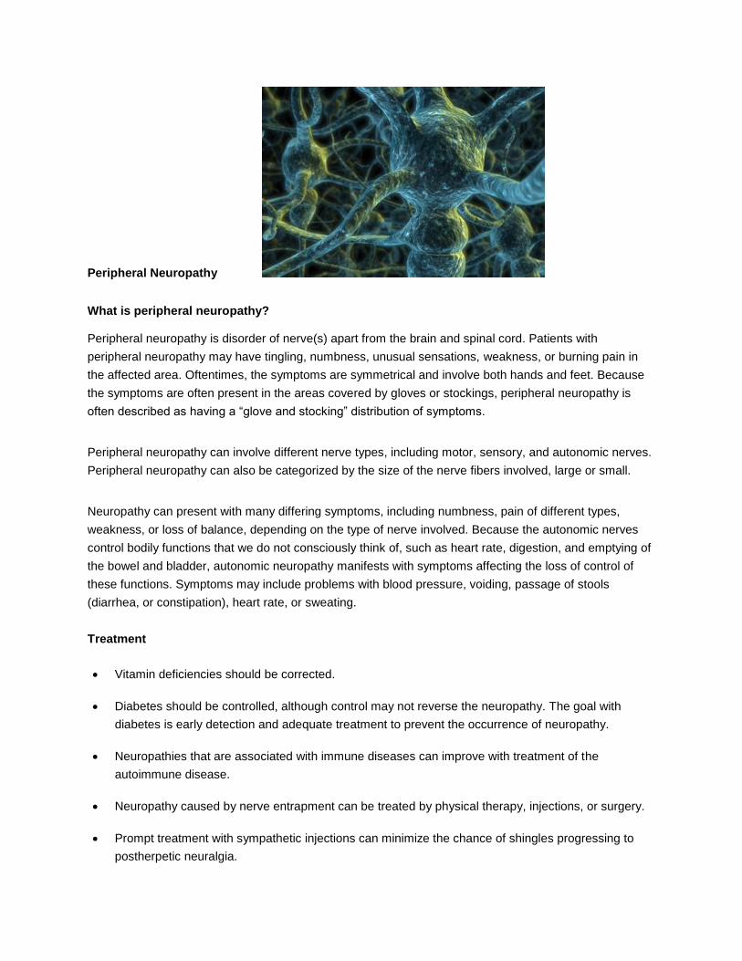
Herniated Disc
What is a herniated disc?
The bones (vertebrae) that form the spine in your back are cushioned by small, spongy discs. When these discs are healthy, they act as shock absorbers for the spine and keep the spine flexible. But when a disc is damaged, it may bulge or break open. This is called a herniated disc. It may also be called a slipped or ruptured disc.
You can have a herniated disc in any part of your spine. But most herniated discs affect the lower back (lumbar spine). Some happen in the neck (cervical spine) and, more rarely, in the upper back (thoracic spine).

What causes a herniated disc?
A herniated disc may be caused by:
Wear and tear of the disc. As you age, your discs dry out and aren't as flexible.
Injury to the spine. This may cause tiny tears or cracks in the hard outer layer of the disc. When this happens, the gel inside the disc can be forced out through the tears or cracks in the outer layer of the disc. This causes the disc to bulge, break open, or break into pieces.
What are the symptoms?
When a herniated disc presses on nerve roots, it can cause pain, numbness, and weakness in the area of the body where the nerve travels. A herniated disc in the lower back can cause pain and numbness in the buttock and down the leg. This is called sciatica. Sciatica is the most common symptom of a herniated disc in the low back.
If a herniated disc is not pressing on a nerve, you may have a backache or no pain at all.
If you have weakness or numbness in both legs, along with loss of bladder or bowel control, seek medical care right away. This could be a sign of a rare but serious problem called cauda equina syndrome.
How is a herniated disc diagnosed?
Your doctor may diagnose a herniated disc by asking questions about your symptoms and examining you. If your symptoms clearly point to a herniated disc, you may not need tests.
Sometimes a doctor will do tests such as an MRI or a CT scan to confirm a herniated disc or rule out other health problems.
How is it treated?
Symptoms from a herniated disc usually get better in a few weeks or months. To help you recover:
Rest if you have severe pain. Otherwise, stay active. Staying in bed for more than 1 or 2 days can weaken your muscles and make the problem worse. Walking and other light activity may help.
Try using a heating pad on a low or medium setting for 15 to 20 minutes every 2 or 3 hours. Try a warm shower in place of one session with the heating pad. You can also buy single-use heat wraps that last up to 8 hours. You can also try an ice pack for 10 to 15 minutes every 2 to 3 hours.
Do the exercises that your doctor or physical therapist suggests. These will help keep your back muscles strong and prevent another injury.
Steroid Injections can be helpful to reduce the pain, facilitate function restoration
Surgery is the last option if the patient experiences neurological deterioration.

Spinal Stenosis
Spinal stenosis is narrowing of the spinal column that causes pressure on the spinal cord, or narrowing of the openings (called neural foramina) where spinal nerves leave the spinal column.
Causes
Spinal stenosis usually occurs as a person ages and the disks become drier and start to shrink. At the same time, the bones and ligaments of the spine swell or grow larger due to arthritis or long-term swelling (inflammation).
Spinal stenosis may also be caused by:
Arthritis of the spine, usually in middle-aged or elderly people
Bone diseases, such as Paget's disease of bone and achondroplasia
Defect or growth in the spine that was present from birth (congenital defect)
Herniated or slipped disk, which often happened in the past
Injury that causes pressure on the nerve roots or the spinal cord
Tumors in the spine
Symptoms
Numbness, cramping, or pain in the back, buttocks, thighs, or calves, or in the neck, shoulders, or arms
Weakness of part of a leg or arm
Symptoms are more likely to be present or get worse when you stand or walk. They will often lessen or disappear when you sit down or lean forward. Most people with spinal stenosis cannot walk for a long period of time.
More serious symptoms include:
Difficulty or poor balance when walking
Problems controlling urine or bowel movements
How is lumbar spinal stenosis diagnosed?

Your doctor can tell if you have lumbar spinal stenosis by asking questions about your symptoms and
past health and by doing a physical exam. You will probably need imaging tests such as an MRI, a CT
scan, and sometimes X-rays.
How is it treated?
You can most likely control mild to moderate symptoms with pain medicines, exercise, and physical
therapy. If the conservative treatments fail, series of epidural steroid injections are usually helpful to
reduce the pain and weakness.
You may need surgery if your symptoms get worse.

Facet Arthritis
What are Facet Joints?
Facet joints are found in the posterior of the spine. There are 24 vertebrae which form the human spine.
There are two facet joints between the vertebrae of each spinal segment along the spinal column. A facet
joint has two bony surfaces with cartilage between them and a capsule of ligaments surrounding it.
Synovial fluid lubricates the joints as is the case with any joint.
What is Facet Arthropathy?
It is degenerative arthritis affecting the facet joints in the spine. In the area of the spine where there are
facet joints, arthritis pain can develop.
What Causes Facet Arthropathy?:
Arthritis in the facet joints can develop from:
wear and tear (decreases space between vertebrae causing facet joints to rub together)
previous back injury
fractures
torn ligaments
disc problems
What are the Symptoms of Facet Arthropathy?
Pain is the main symptom associated with facet arthopathy. The pain is typically worse following sleep or
rest. Pain associated with facet arthropathy may be exacerbated by twisting or bending backwards. Low
back pain is the most frequent complaint but it does not typically radiate down the arms, legs or buttocks,
unless spinal stenosis is also involved.
How is Facet Arthropathy Diagnosed?
X-rays, CAT scans, and Magnetic Resonance Imaging (MRI) may be used to help diagnose facet
arthropathy. A facet block is very helpful in making the diagnosis. Series of steroid injections usually offer

very good relief. If the facet joint is injected and pain relief is the result, that serves to confirm the
diagnosis of facet arthropathy.
How is Facet Arthropathy Treated?
Initially the doctor may recommend a period of rest in an effort to relieve the symptoms. Sleep positions
which take pressure off facet joints may be recommended.
Oral medication may be prescribed
Strengthening and aerobic exercise
Water therapy
If conservative measures fail:
injections of an anesthetic or steroid medicine into the facet joint or nerves that go to the facet
joint may be tried
Facet nerve ablation which destroys nerves with radiofrequency and facet fusion may be an
option
Facet Fusion

Compression Fractures
A compression fracture is a collapse of a vertebra. It may be due to trauma or due to a weakening of the vertebra. This weakening is seen in patients with osteoporosis or osteogenesis imperfecta, lytic lesions from metastatic or primary tumors, or infection
Treatment
Kyphoplasty and vertebroplasty, minimally invasive procedures designed to treat pain from osteoporotic compression fractures and sometimes other forms of fracture, such as a fracture caused by certain types of cancer. There is conflicting data in the literature about the effectiveness of vertebroplasty.

Sacroiliac Joint Pain:
The sacroiliac joint is formed by the connection between the sacrum and the iliac bone on either side. The sacrum consists of five fused vertebrae below the lumbar (lower) spine, where the iliac bone is the large bone that makes up the pelvis. A thick band of ligaments hold this joint together. There is very little normal motion between these two joints, however this joint does support much of the weight of our upper body when we are in the upright position. When this joint becomes injured or inflamed, pain can result, a condition called sacroiliac dysfunction.
Causes:
A variety of conditions can result in the pain of sacroiliac dysfunction. Arthritis affects many joints of the
body, and the sacroiliac joint is of no exception. When the joint’s cartilage is damaged or worn away, the
bones begin to rub on each other, and osteoarthritis results. This is the most common cause of SI joint
dysfunction. Direct trauma to the joint, such as a fall can also result in sacroiliac joint pain. Pregnancy is
another frequent cause of sacroiliac joint dysfunction, possibly because of changes in posture or
hormonal effects on joint laxity.
Leg length discrepancy, having one leg shorter than the other, is another condition that results in
excessive motion at the sacroiliac joint as well as pain. Various systemic disorders also affect the
sacroiliac joint. These include ankylosing spondylitis, psoriasis, rheumatoid arthritis and gout.
Symptoms:
Sacroiliac joint dysfunction causes pain in the lower back region that radiates down the posterior buttock
region. The pain increases during walking or standing and is less when lying down. Symptoms can
sometimes occur in the hips or groin region as well. Symptoms can be replicated when the joint is
stressed during an examination by a health care professional.
Treatments:
Treatment of sacroiliac joint dysfunction can include oral anti-inflammatory medicine, joint injections and
physical therapy exercises. Joint injections involve placing a solution of local anesthetic and steroids into
the joint. Therapeutic exercises involve lumbar stabilization and low back stretches.

Complex Regional Pain Syndrome/RSD
Complex Regional Pain Syndrome (CRPS), also known as Reflex Sympathetic Dystrophy, is a chronic neurological syndrome characterized by:
severe burning pain pathological changes in bone and color changes in skin excessive sweating tissue swelling extreme sensitivity to touch
Causes
It is usually caused by injuries or surgeries.
Diagnosis
Diagnosing CRPS can be difficult, but early diagnosis is very important. The doctor will take a medical history and do a physical examination. Other tests may necessary:
A test to show temperature changes and lack of blood supply in the affected limb (thermography)
Bone scans
Nerve conduction studies
X-rays
Treatment
There is no cure for CRPS, but the disease can be slowed. The main focus is on relieving the symptoms and helping people with this syndrome live as normal a life as possible.
Physical and occupational therapy should be started as early as possible. Starting an exercise program and learning to keep joints and muscles moving may prevent the disease from getting worse and help you perform everyday activities.
Medications may be used, including pain medicines, steroids, certain blood pressure medicines (Such as Clonidine), bone loss medications (such as Fosamax and Actonel), and antidepressants.
Cognitive behavioral therapy or psychotherapy, can help teach the skills you need to live with chronic pain.
Interventional or surgical treatment may be necessary:

Injected medicine that numbs the affected nerves or pain fibers around the spinal column (nerve block)
Internal pain pump that directly delivers medications to the spinal cord (intrathecal drug pump)
Spinal cord stimulator, which involves placing electrodes (electrical leads) next to the spinal cord. A low-level electrical current is used to create a pleasant or tingling sensation in the painful area is the best way to reduce pain in some patients.
Surgery that cuts the nerves to destroy the pain (surgical sympathectomy), although it is unclear how many patients this helps. It may also make some patients' symptoms worse.

Myofascial Pain
Myofascial pain is caused by abnormal stress on the muscles. It is a chronic condition that affects the
fascia (connective tissue that covers the muscles). Myofascial pain syndrome can be confused with
fibromyalgia and may also accompany it. Unlike fibromyalgia, myofascial pain tends to occur in trigger
points, as opposed to tender points, and typically there is no widespread, generalized pain".
Myofascial pain is often caused by tension, spasm, or fatigue of the muscles that allow a person to chew,
called the masticatory muscles. Grinding of the teeth and jaw clenching are related to myofascial pain and
can lead to headaches.
It is common for myofascial pain to limit jaw movement and to affect muscles in the neck, back, and
shoulder. Actually, myofascial pain can affect any skeletal muscle in the body. It is not limited to the
muscles of mastication (chewing).
Diagnosis:
Myofascial pain is diagnosed after a physical examination reveals trigger points. Locating the trigger
points is important to the diagnostician. X-rays are not helpful in diagnosing myofascial pain. Onset of
myofascial pain can be acute following injury or chronic following poor posture or overuse of the muscles.
Myofascial pain is a common condition. Considering that 14.4% of the general U.S. population have
chronic musculoskeletal pain, it has been estimated that 21% to 93% of patients complaining of regional
pain actually have myofascial pain.
Treatments
Myofascial pain is not considered fatal but it can significantly affect quality of life. Treatment is important
and can include:
Oral medications include muscle relaxants, sleep aids, NSAIDs, Tylenol.
Trigger point injections with local anesthetic or steroids or Botox are usually effective to relieve muscle spasm.
Physical therapy, relaxation, and biofeedback can also be helpful modes of treatment for myofascial pain.

Cancer Pain
The majority of people with cancer will experience pain at some time or another. The pain can result from
the cancer itself, or from the cancer's treatment. In addition, some people who have been cured of their
cancer can continue to suffer from pain.
Cancer pain, or the discomfort that stems from cancer and its treatment, can be controlled most of the
time. There are many different medicines and methods available to control cancer pain. People who have
cancer and are feeling pain need to inform their doctor immediately. The earlier pain treatment is started,
the more effective it is.
What Causes Cancer Pain?
There are many causes of cancer pain, but most cancer pain occurs when a tumor presses on nerves or
body organs or when cancer cells invade bones or body organs. Cancer treatments such as
chemotherapy, radiation, or surgery also may cause pain.
What Are the Symptoms of Cancer Pain?
The symptoms of cancer pain vary from person to person. The amount of pain present may depend on
the type of cancer, the stage or extent of the disease, and the person's pain threshold (tolerance for pain).
Pain can range from mild and occasional to severe and constant.
What Medicines Are Used To Treat Cancer Pain?
Mild Pain
Mild pain relievers: Tylenol and a group of pain relievers called nonsteroidal anti-inflammatory drugs
(NSAIDs) such as aspirin andibuprofen (Motrin and Aleve) can treat mild to moderate pain. Many of these
are over-the-counter drugs that do not require a prescription, but some do require a prescription. Patients
should check with a physician before using these medicines, especially if chemotherapy is being
administered. NSAIDs can slow blood clotting.
Moderate to Severe Pain
Narcotic pain relievers: These drugs include morphine,Actiq, Duragesic, Dilaudid, oxycodone (sold under
the brand names OxyContin,Percocet, and Tylox) and codeine. Narcotic pain relievers require a
prescription and may be used along with mild pain relievers for moderate to severe pain.
Breakthrough Pain

Onset narcotic pain relievers: Onset narcotic pain relievers, which require a prescription, are used to
treat breakthrough pain (a flare-up of pain characterized by rapid onset, severe intensity and short
duration). Immediate-release oral morphine is among these drugs.
Tingling and Burning Pain
Antidepressants: Antidepressants are used to relieve pain regardless of if the person is depressed.
Elavil, Pamelor, Norpramine are antidepressants prescribed to treat pain.
Anticonvulsants (anti-seizure medications): Despite the name, anticonvulsants are used not only for
seizures, but also to control burning and tingling pain, painful symptoms of nerve damage. Tegretol
andNeurontin require prescriptions
Other drugs: Corticosteroids are used to lessen swelling to reduce the pain
Reducing a tumor size by surgery or radiation or chemotherapy can be used along with medicine to
provide additional pain relief.
Interventional treatments, such as blocking the involved nerves are often effective. Intrathecal pump
implantation needs to be considered early, after medical and surgical treatments fail.
Peripheral Neuropathy
What is peripheral neuropathy?
Peripheral neuropathy is disorder of nerve(s) apart from the brain and spinal cord. Patients with
peripheral neuropathy may have tingling, numbness, unusual sensations, weakness, or burning pain in
the affected area. Oftentimes, the symptoms are symmetrical and involve both hands and feet. Because
the symptoms are often present in the areas covered by gloves or stockings, peripheral neuropathy is
often described as having a “glove and stocking” distribution of symptoms.
Peripheral neuropathy can involve different nerve types, including motor, sensory, and autonomic nerves.
Peripheral neuropathy can also be categorized by the size of the nerve fibers involved, large or small.
Neuropathy can present with many differing symptoms, including numbness, pain of different types,
weakness, or loss of balance, depending on the type of nerve involved. Because the autonomic nerves
control bodily functions that we do not consciously think of, such as heart rate, digestion, and emptying of
the bowel and bladder, autonomic neuropathy manifests with symptoms affecting the loss of control of
these functions. Symptoms may include problems with blood pressure, voiding, passage of stools
(diarrhea, or constipation), heart rate, or sweating.
Treatment
Vitamin deficiencies should be corrected.
Diabetes should be controlled, although control may not reverse the neuropathy. The goal with
diabetes is early detection and adequate treatment to prevent the occurrence of neuropathy.
Neuropathies that are associated with immune diseases can improve with treatment of the
autoimmune disease.
Neuropathy caused by nerve entrapment can be treated by physical therapy, injections, or surgery.
Prompt treatment with sympathetic injections can minimize the chance of shingles progressing to
postherpetic neuralgia.

Spinal or peripheral nerve stimulation or intrathecal pump implantation should be considered for
chronic severe pain that failed conservative treatments.

Spasticity due to stroke, multiple sclerosis, spinal cord injury or cerebral palsy
Definition
Spasticity is often described as tight, stiff muscles or spasms that may make movement, posture, and balance difficult. It may affect your ability to move one or more of your limbs, or to move one side of your body. Sometimes spasticity is so severe that it gets in the way of daily activities, sleep patterns, and caregiving.
Causes
Spasticity is caused by damage or injury to the part of the central nervous system (the brain or spinal cord) that controls voluntary movement. This damage disrupts important signals between the nervous system and muscles, creating an imbalance that increases muscle activity or spasms.
Associated Conditions
Severe spasticity is associated with the following conditions:
Brain injury
Cerebral palsy
Multiple sclerosis
Spinal cord injury
Stroke
Symptoms
Increased muscle tone
Overactive reflexes
Involuntary movements, which may include spasms (brisk and/or sustained involuntary muscle contraction) and clonus (series of fast involuntary contractions)
Pain
Decreased functional abilities and delayed motor development
Difficulty with care and hygiene
Abnormal posture
Contractures (permanent contraction of the muscle and tendon due to severe persistent stiffness and spasms)
Bone and joint deformities
Diagnosis
Spasticity is typically diagnosed after the onset of a neurological condition. Your doctor will review your medical history and perform a physical evaluation. Your doctor may consider medications you have taken and whether there is a history of neurological or muscular disorders in your family. To confirm the diagnosis, tests can be performed to evaluate arm and leg movements, muscular activity, passive and active range of motion, and ability to perform self-care activities.
Treatment
There is currently no cure for severe spasticity. However, there are a number of treatment options available for managing your symptoms. They include:
Rehabilitation Therapy
Rehabilitation therapy usually takes place in a clinic, a hospital, or at home. It can include any combination of physical, occupational, or speech therapy.
Oral Medication
Oral medications may help some people treat the symptoms of spasticity.
Injection Therapy
Injection therapy is usually intended for specific muscle groups (for example, one hand, one foot, one shoulder).
Orthopedic Surgery
Orthopedic surgeries include soft tissue procedures like tendon transfers and osteotomies (cutting a bone to change its alignment).
ITB Therapy (Baclofen Pump)
The baclofen pump is an adjustable, reversible treatment for severe spasticity. A surgically placed pump and catheter deliver liquid baclofen directly to the fluid around the spinal cord, where it’s needed most. This maximizes the effectiveness and minimizes the side effects of Baclofen.

Peripheral Vascular Pain
Atherosclerosis underlies most peripheral arterial disease. Narrowed vessels that cannot supply sufficient blood flow to exercising leg muscles may cause claudication, which is brought on by exercise and relieved by rest. As vessel narrowing increases, critical limb ischemia can develop when the blood flow does not meet the metabolic demands of tissue at rest. While critical limb ischemia may be due to an acute condition such as an embolus or thrombosis, most cases are the progressive result of a chronic condition, most commonly atherosclerosis.
Treatment
Smoking cessation,
Blood pressure, sugar and cholesterol control
Antiplatelet therapy with aspirin has been shown to substantially decrease the risk of myocardial infarction, stroke and death in patients with peripheral vascular disease
Surgical intervention includes revascularization and amputation
If the patient is no longer a candidate for revascularization, spinal cord stimulator may be a good choice to improve the circulation and reduce the pain

Brochure download
Link: http://www.valleyambulatory.com/pain-management/yuan-chen-md/
Treatment offerred
Epidural Steroid Injections Cervical Thoracic Lumbar
Facet Joint Injections Cervical Thoracic Lumbar ( click on each ward into link page)
Sacroiliac joint injections
Radiofrequency ablation of facets: Cervical Thoracic Lumbar
Discography
Trigger Point Injection
Joint injections
Acupuncture
Sympathetic blocks:
Peripheral nerve blocks:
Facet fusion
Mild Procedure:
Spinal/peripheral nerve stimulator
Intrathecal opioid/Baclofen pumps