what does the brain tumor board do? - cedars-sinai · what does the brain tumor board do? john s....
TRANSCRIPT

What does the Brain Tumor
Board do?
John S. Yu, M.D. Professor and Vice Chair
Department of Neurosurgery
Director, Brain Tumor Center of Excellence
Director, Surgical Neuro-Oncology
Cedars Sinai Medical Center

A multidisciplinary group of health-care workers
reviewing patient histories and data to make
recommendations
Neurosurgeons
Neuro-radiologists
Neuro-pathologists
Neuro-oncologists
Radiation oncologists
Medical oncologists
Nurses

What tumors do we discuss?
Statistics
Of all primary brain tumors:
35% are Astrocytomas including Glioblastomas
27% are Meningiomas
8% are Nerve sheath tumors (acoustic neuromas, vestibular schwannomas,
neurilemmomas)
7% are Pituitary tumors
3% are Lymphomas
3% are Oligodendrogliomas
2% are Medulloblastomas/embryonal/primitive
Metastatic brain tumors are the most common brain tumor, with an
annual incidence more than 4 x greater that that of primary brain tumors.
Cancers most commonly metastasize to the brain are lung and breast.

Clinical History of Brain Tumors
Symptoms of brain tumors are usually associated
with increased ICP
Headache
Generalized
Worse in the am
Aggravated by stooping, bending, and coughing
Vomiting
usually in the morning
with acute rise in ICP

Clinical Features of Brain Tumors
Frontal Lobe
Contralateral weakness
Expressive dysphasia
Personality changes
Parietal Lobe
Disturbed
sensation
Visual field defect
lower quadrantanopia
Gerstmann’s syndrome (Dominant hemisphere)
Right/left confusion, finger agnosia, acalculia, agraphia
(Non dominant) Dress apraxia,
geographic agnosia, Construction
apraxia, anosognosia
Signs of focal damage from tumor
Disturbed
Cerebral
Function
Occipital Lobe
Visual field defect
homonymous hemianopia
Temporal Lobe
Receptive dysphasia
Visual field defect
upper quadrantanopia

Astrocytomas - Imaging
Low grade astrocytoma
grade I/grade II
Hyperintense on T2
Hypointense on T1
Little, or no enhancement
Pilocytic astrocytomas –
Contrast enhancing often
cystic with mural nodule
Little, if any edema
Little, or no mass effect
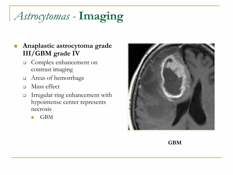
Astrocytomas - Imaging
Anaplastic astrocytoma grade III/GBM grade IV
Complex enhancement on contrast imaging
Areas of hemorrhage
Mass effect
Irregular ring enhancement with hypointense center represents necrosis
GBM
GBM

Pathological Classification of
Brain Tumors In 1979 the World Health
Organization (WHO) drew up an internationally agreed classification of intracranial tumors based on the tissue of origin.
9 types of CNS tumors
Tumors of neuroepithelial tissue
Tumors of the meninges
Tumors of cranial and spinal nerves
Hematopoietic neoplasms
Germ cell tumors
Cysts and tumor-like lesions
Tumors of the sellar region
Local extensions from regional tumors
Metastatic tumors

Pathology of Brain Tumors
Primary brain tumors can
be classified as either:
Benign
Very slow growing cells
Distinct borders, rarely
spreads
Well differentiated (cells
appear almost normal)

Pathology of Brain Tumors
Malignant
Rapid growth
Poor differentiation
Increased cellularity,
mitosis, necrosis and
vascular proliferation
However, metastases to
extracranial sites rarely
occur.

Possible Causes of Brain Tumors and
Risk Factors - Genetic Factors
Genetic Factors
Transformation of normal cells to malignant growth
probably results from a variety of different processes:
Normal cell growth and differentiation controlled by
Proto-oncogenes
Expression is altered resulting in oncogenes
Alters encoded proteins transforming cell into malignant state
Inactivation of expression of tumor suppressor genes
Over expression of genes controlling growth factor.

Mutations leading to infiltrative
astrocytic tumors. Molecular studies have identified some of the genetic
changes that underlie the pathologic differences among
astrocytic tumors; progression in tumor grade is associated
with an ordered accumulation of mutations

Astrocytoma - Treatment
Depends on a number of
factors:
Site of lesion
Degree of malignancy
+/- Elevated ICP
Degree of disability and
effect of steroid therapy
Suspected nature of tumor
on imaging
Patient’s age
Patient’s wishes

Common Brain Tumors –
Astrocytoma - Treatment
Grade I and Grade II
Surgery
Complete surgical resection if possible
Biopsy or partial resection is recommended in almost all cases to
determine pathology
Radiation Therapy
Fractionated XRT to residual tumor postop
Chemotherapy
Only with tumor progression
PCV (procarbazine, CCNU, vincristine) to stabalize growth.

Common Brain Tumors –
Astrocytoma - Treatment
Grade III and Grade IV
Standard against which other treatments are compared:
Surgical Resection
Followed by external beam radiation (EBRT)
40 Gy whole brain + 15-20 Gy to tumor bed =60 Gy
Median survival of
17 weeks after BX + XRT,
versus 30 weeks for SX
and XRT.

Common Brain Tumors –
Astrocytoma - Treatment
Chemotherapy
Alkylating agents benefit ~
10% of patients
Carmustine (BCNU)
Cisplatinum (Cisplatin)
Temozolomide (Temodar)
FDA approved for
treatment of initial relapse
of AA and progression
Used (off label) for newly
dx’d GBM and AA

Treatment of Brain Tumors
Treatment of Edema
Dexamethasone
Mannitol
Seizure Prophylaxis
Dilantin
Valproic Acid
Tegretol
Keppra
Neurosurgery
Surgical Resection
Biopsy
CSF access procedures
Radiation therapy
SRT
SRS
Chemotherapy
Oral
Alkylating agents
Intracranial wafers
Clinical Trials
Immunotherapy

Treatment of Brain Tumors
Immunotherapy Immunotherapy
T-cell mediated antitumor immunity
Pt’s with gliomas demonstrate impaired immune function.
Glioma cells down regulate surface expression of MHC molecules, depriving infiltrating immune cells of signals needed to recognize and clear tumor cells.
Dendritic cells (antigen presenting cells) are pulsed with tumor protein to make a vaccine.
DC introduces tumor associated antigen (TAA) to T-cells.
Activated T-cells eliminate tumor cells.

Brain Tumor Board
Questions?