what we know about grain safety - usda ars
TRANSCRIPT

1

2

3
What We Know About Grain Safety
Donald D. Kasarda, Ph.D.Research Chemist (Retired)
Agricultural Research ServiceU. S. Department of Agriculture
Albany, CA
Learning Objective: To review the relationship of the harmful grains in celiac disease toother grains that might suitably be included in the diet of celiac patients and to discusshow the proteins of these grains relate to celiac disease and allergy.
Celiac Disease and Allergy—DefinitionsCeliac disease and food allergy are often classified as hypersensitivity reactions
(Johansson et al. 2004; Janeway et al. 1999;). Here I shall be concerned only withhypersensitivities that involve immune responses to normally innocuous foods,substances that do not cause a problem for most people.
Celiac disease is often classified as a Type IV hypersensitivity (Janeway et al.1999) involving cellular mechanisms (T-cells play an important role). People with celiacdisease (gluten-sensitive enteropathy) must avoid eating wheat, rye, and barley storageproteins in order to avoid adverse changes to their intestinal mucosa that can lead toserious malabsorption of almost all nutrients (Green and Jabri 2003; Kasarda 2000;Feighery 1999; Maki and Collin 1997; Wieser 1995). The symptoms of celiac diseasefrequently include diarrhea and other gastrointestinal problems, although these symptomsare not always present. Because active disease produces damage to the epithelial layerlining the small intestine, which is the site of absorption of almost all nutrients, includingvitamins, minerals, proteins, carbohydrates, and lipids, clinical manifestations can bewide-ranging , including anemia, osteoporosis, peripheral neuropathy, and others. Thelarge number of observed manifestations, along with considerable variation from patient-to-patient, makes celiac disease difficult to diagnose. Dermatitis herpetiformis is aclosely related condition that differs mainly by having skin manifestations in addition tothe more usual characteristics of celiac disease. Hives and respiratory distress, which arecommon symptoms of allergy, are not characteristic of celiac disease.
Food allergy is often classified as a Type I hypersensitivity (Janeway et al. 1999)mediated by E-type immunoglobulins (IgE antibodies). The symptoms of food allergymay include gastrointestinal disturbances, such as diarrhea, but hives and other skinmanifestations are also common, as are respiratory symptoms, such as bronchospasm,(Sampson 2004). In rare cases, severe generalized anaphylactic shock may occur.Almost all grains, whether having celiac disease toxicity or not, appear to have proteinscapable of producing an allergic response in at least a small percentage of the humanpopulation (Breiteneder and Radauer 2004), particularly those populations thatcommonly consume the grain in question. Unlike celiac disease, allergy does not seem to
4
result from proteins of only a few closely related grain species and response to foodproteins is highly variable among populations. When a patient has gastrointestinalsymptoms, it is important to distinguish food allergy from celiac disease. In allergy, themucosal architecture is frequently normal (Veres et al. 2001), in contrast to the villousatrophy of the mucosa that is a usual characteristic of celiac disease.
Wheat, rye, and barleySince the discovery by W. K. Dicke in 1950 that wheat was a key environmental
factor that triggered celiac disease in susceptible individuals, the relationship of thedisease to ingestion of wheat gluten proteins has become an essential part of thedefinition. By and large, if wheat doesn’t trigger enteropathy (or at least, changes in themucosa that presage enteropathy), it isn’t celiac disease. Most reviews of celiac diseasetend to avoid the question of toxicity—or lack thereof—in grains, seeds, or foods otherthan wheat, to a considerable extent because studies of these grains are lacking orinadequate. This may be reasonable from a scientific standpoint, but patients, dietitians,and primary care physicians would like something more. Only wheat and, in recentyears, oats have been extensively studied with modern approaches (such as measurementof intraepithelial lymphocyte infiltration and cytokine production) for their toxicity inceliac disease—with wheat obviously being toxic, whereas conclusions regarding thetoxicity of oats are not straightforward and will be discussed in a further section. Ryeand barley have many nearly identical storage proteins to those in wheat, although theylack an important wheat protein type, the α-gliadins. Despite minimal testing, thesestrong protein sequence similarities between the proteins of wheat and those of rye andbarley, combined with the experience of celiac patients over many years with thesegrains, are supportive of some degree of toxicity for these grains in celiac disease. It isvery difficult to quantify the toxicity of any given grain, but it is at least possible that thelack of α-type gliadins (one of the most studied toxic protein fractions) in rye and barleyresults in lesser toxicity for these two grains in comparison with wheat. Furthermore, it isalso very difficult to quantify the toxicity of a given gluten protein relative to a differentgluten protein type. It appears at present that all classes of wheat gluten proteins havetoxicity. Although no studies have been carried out to determine toxicity for each of thetypes of proteins found in rye and barley grain, their sequence similarity to equivalentproteins in wheat suggests that they are likely to be toxic.
OatsIn the case of oats, some early work in which patients were not biopsied indicatedtoxicity for oats. Dicke et al. (1953) stated that rice flour, maize starch, wheat starch, andpotatoes were safe, but that wheat, rye, and oats were harmful. More recent, extensivework that may be accorded greater confidence has provided impressive evidence for alack of oats toxicity in celiac disease and dermatitis herpetiformis (Hogberg et al. 2004;Peraaho et al. 2004a; Peraaho et al. 2004b; Storsrud, S et al. 2003; Kilmartin et al. 2003;Picarelli et al. 2001; Janatuinen et al. 2000; Hoffenberg et al. 2000; Hardman et al. 1999;Srinivasan et al., 1999; Reunala et al., 1998; Janatuinen et al., 1995; Hardman et al. 1997;Srinivasan et al., 1996; Dissanayake et al. 1974). The cross-reactions between antibodiesto oat or wheat storage proteins has muddied the waters at times, suggesting a closer

5
sequence relationship than may be warranted. Oat avenins correspond in a general way(sequence similarity/homology) to the C-terminal domains (approximately the C-terminal
half) of α- and γ-gliadins, which is not the region primarily considered responsible fortoxicity in celiac disease (Arentz-Hansen et al., 2002; Anderson et al., 2001). Aveninslack the large proline-, glutamine-rich repetitive domain most strongly associated withtoxicity (Kasarda 1997). There are, however, some proline- and glutamine-containingsequences in oats near the N-terminal end of the protein polypeptide chain, that appear tobe toxic for a few patients and which may correspond to a vestigial repeat region. Lundinet al. (2003) and Arentz-Hansen et al. (2004) have described three patients whoapparently have a reaction to pure oats that cannot be distinguished from celiac disease.Given the large number of studies cited above in which the final conclusion was that oatsare safe for celiac patients, the results of Lundin et al. (2003) and Arentz-Hansen et al.(2004) are surprising. Arentz-Hansen et al. (2004) state, “Our observations demonstratethat even if oats seem to be well tolerated by many celiac patients, there are patients whohave an intestinal T-cell response to oats. Until the prevalence of oat intolerance in celiacdisease patients is established, clinical follow-up of celiac disease patients eating oats isadvisable. Clinicians should be aware that oat intolerance may be a reason for villousatrophy and inflammation in patients with celiac disease who are eating oats butotherwise are adhering to a strict gluten-free diet.”
Peräaho et al. (2004), while citing the results of Lundin et al. (2003), concludethat, provided safe (pure) oat products are available, the majority of celiac disease anddermatititis herpetiformis patients tolerate oats well. It should be noted, however, that inthe study of Peraaho et al. (2004b) the oats-containing gluten-free diet caused moreintestinal symptoms than the traditional diet. Peräaho et al. (2004) noted that mucosalintegrity was not disturbed in their subjects, but more inflammation was evident in the

6
oats group. Allergic reactions to oats may occur in some patients as is the case for allgrains.
The question of whether the amount of contamination of oats by wheat,particularly in the US, is sufficient to cause harm to celiac patients remains to beanswered. Lundin et al. (2003) have noted serious contamination of a particularcommercial oats sample in Norway.
Rice and corn (maize)Rice and corn have generally been considered safe grains for celiac patients.
Once again there has been lack of rigorous, controlled, scientific study of these grains inrelation to celiac disease, especially with up-to-date methods. I am not aware, however,of any major evidence against their safety for celiac patients during the past 50 years.There are people who are sensitive to rice and corn and probably to any grain. Somehave clearly allergic reactions, such as hives or respiratory distress in response to thesegrains, but gastrointestinal symptoms, such as diarrhea, may result from allergy as well(Janeway et al. 1999). If we accept celiac disease as being properly classified as a TypeIV hypersensitivity mediated by T cell responses, there remains the possibility that celiacdisease might be combined in a given patient with immediate hypersensitivities, such asallergies (Type I), to wheat or to any other grains, including rice and corn. Intestinalbiopsy might pick up the celiac disease, but not necessarily other sensitivities. As far as Iknow, these potential complications are not thoroughly understood although significantprogress is being made (Sampson 2004). It seems unreasonable, however, to suggest to aceliac patient who indicates that he or she responds badly to a particular grain or foodother than wheat, rye, or barley, that he or she is imagining things. Adverse reactions towhat I shall somewhat arbitrarily term safe grains for celiac patients may not be common,but they do exist. Such adverse reactions need more research to clarify the mechanismsinvolved.
Plant classification, protein sequences, and grain safetyWe do not have a unifying theory based on rigorous scientific investigation that
will include or exclude various bad reactions to the ‘safe’ grains in celiac disease, but Isuggest that it is unhelpful at this time to try to include responses to grains or otherfoodstuffs that occur in some people with celiac disease, but not in others, as part of theceliac disease syndrome. In the absence of solid scientific studies of the toxicity of mostgrains or seeds, I suggested in 1991 (see: Kasarda 2001) that plant classification mightprovide useful guidance in separating safe grains from unsafe grains. I update thatapproach here.
I suggest that an alternative to extensive testing (which may not be forthcoming)is to combine our knowledge of wheat and oats, which is fairly solid, with plantclassification or taxonomy, and with protein sequence data to provide recommendationsconcerning which grains or grain-like seeds are likely to be harmful to celiac patients andwhich are likely to be safe (Kasarda 2001). We have learned a great deal during the past50 years, but our knowledge is far from complete. Because studies of food reactions inceliac disease usually require human subjects, and biopsies (before and after challenge)are usually also required, it is extremely difficult and expensive to carry out suchstudies—first of all, even to recruit a reasonable number of clearly diagnosed celiac

7
patients is a considerable challenge. It is not likely that studies will be carried out in thenear future on many of the grains, seeds, or foods of possible interest to celiac patients.As scientific investigation continues, of course some of my conclusions may have to bemodified. New information about oats toxicity provides an example (Arentz-Hansen et al.2004).
Attachment II Kasarda
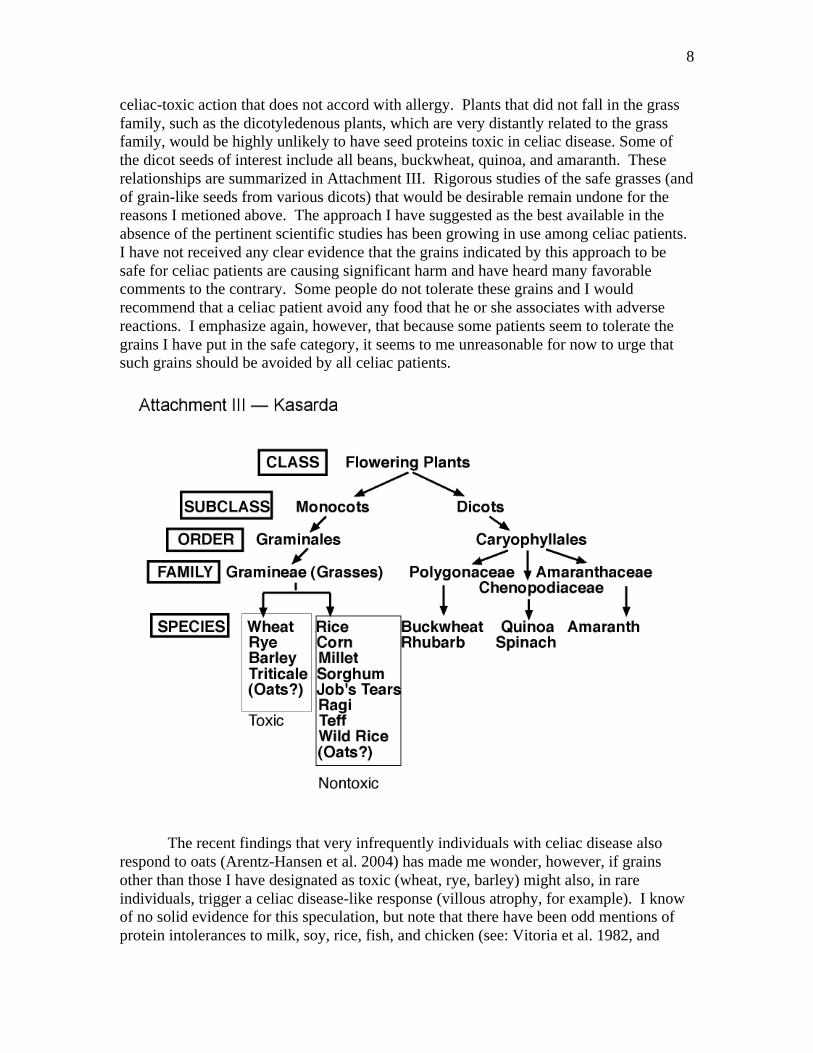
Given that wheat is toxic and assuming that rice and corn (maize) are not toxic, Isuggested that grains that were closer in their taxonomic relationships to corn or rice thanto wheat would not be toxic in celiac disease. Such grains included millet, sorghum,Job’s tears, ragi, teff, and wild rice. Furthermore, in plant classification, wheat, rye, andbarley are included in the tribe of the grass family called the Hordeae or Triticeae, whileoats fall in a separate tribe. There is, however, a significant similarity in protein sequencebetween oat avenins and some major wheat gliadin proteins, indicating relative closenessof the two tribes. Although oats now seem to be toxic for rare individuals (Arentz-Hansen 2004; Lundin et al. 2003), it appears that many celiac patients tolerate oats. Ifoats are included in the nontoxic category, then all toxic grains would be found in asingle tribe, the Triticeae (see attachments I and II). Such grains include bread wheat,durum wheat (used in pasta), spelt wheat (spelta), polonicum (Polish wheat), Kamut,monoccum (einkorn), farro, triticale (a cross between wheat and rye), and many wildgrass species not usually consumed by man. All grasses not in the tribe, Triticeae, wereconsequently classified as safe, including rice, corn, various millets, ragi, teff, Job’s tears,wild rice, and oats (see attachment III), although, as discussed, oats may have a rare
8
celiac-toxic action that does not accord with allergy. Plants that did not fall in the grassfamily, such as the dicotyledenous plants, which are very distantly related to the grassfamily, would be highly unlikely to have seed proteins toxic in celiac disease. Some ofthe dicot seeds of interest include all beans, buckwheat, quinoa, and amaranth. Theserelationships are summarized in Attachment III. Rigorous studies of the safe grasses (andof grain-like seeds from various dicots) that would be desirable remain undone for thereasons I metioned above. The approach I have suggested as the best available in theabsence of the pertinent scientific studies has been growing in use among celiac patients.I have not received any clear evidence that the grains indicated by this approach to besafe for celiac patients are causing significant harm and have heard many favorablecomments to the contrary. Some people do not tolerate these grains and I wouldrecommend that a celiac patient avoid any food that he or she associates with adversereactions. I emphasize again, however, that because some patients seem to tolerate thegrains I have put in the safe category, it seems to me unreasonable for now to urge thatsuch grains should be avoided by all celiac patients.
The recent findings that very infrequently individuals with celiac disease alsorespond to oats (Arentz-Hansen et al. 2004) has made me wonder, however, if grainsother than those I have designated as toxic (wheat, rye, barley) might also, in rareindividuals, trigger a celiac disease-like response (villous atrophy, for example). I knowof no solid evidence for this speculation, but note that there have been odd mentions ofprotein intolerances to milk, soy, rice, fish, and chicken (see: Vitoria et al. 1982, and
9
references therein; also Ferguson et al. 1982 and references therein) that producedchanges in mucosal morphology reminiscent of celiac disease, although the authorsindicated that these intolerances were mostly not permanent in contrast to celiac disease.Research is needed to examine more carefully those celiac patients who have digestiveproblems with maize (corn), buckwheat, and other presumably safe grains to provide abetter understanding of the nature of their responses. In a few cases where unresponsiveceliac disease was diagnosed, an elemental diet was found to elicit improvement(personal communication, K. E. A. Lundin), which might be a consequence of the patientreacting to some food protein other than those associated with celiac disease.Nevertheless, I suggest that the rarity of such reactions, while worthy of note, does notnegate the general conclusions I have drawn in my attempt to classify toxic vs. safegrains.
Protein sequencesThe gluten proteins, as is typical of proteins, are made up of about 20 different
amino acids strung together through peptide bonds like beads on a string into longpolymer chains, called polypeptides. There are many gluten proteins, which vary in size,incorporating from about 250 to 850 amino acids in the polypeptide chain. The numberof each type of amino acid and the sequence of incorporation of these amino acids intothe polypeptide chain largely defines any given protein. When a gluten proteinpolypeptide is broken down into smaller polypeptides (often called just ‘peptides’) bydigestive enzymes, some of the peptides, incorporating from about 12-33 amino acids,are quite resistant to digestion by digestive tract enzymes (Shan et al. 2002; Bronstein etal. 1966; Frazer et al. 1959) and this is an important aspect of their toxicity, along withthe presence of key glutamine and proline residues. A few of these peptides have beenshown to be toxic to celiac patients by instillation studies of the equivalent syntheticpeptides (Fraser et al. 2003; Marsh et al., 1997; Marsh et al. 1995; Sturgess et al., 1994).It is unlikely that all toxic peptide sequences have been identified at this time. The aminoacid sequences of the known toxic peptides do not, however, seem to have exactduplicates in proteins other than those of grains falling in the tribe Triticeae, particularlywhen proteins that are likely to appear in organisms at more than trace levels, such assignal transduction proteins, are considered (Kasarda 1997). Thus, these sequencesprovide support for the taxonomic classification approach. I have not taken into accountthe extent to which similar, but not identical, sequences may exhibit toxicity in celiacdisease because I don’t think we have enough of a handle on what the degree of variationin peptide sequence is that is compatible with toxicity.
Peptides that trigger allergic reactions have also been described. For example,Matsuo et al. (2004) reported that the sequences QQIPQQQ, QQFPQQQ, QQSPEQQ,and QQSPQQQ from ω-5 gliadin were dominant epitopes for triggering wheat-dependent exercise-induced anaphylaxis. Maruyama et al. (1998) found that the QQQPPmotif in certain low-molecular-weight glutenin subunits was primarily responsible forIgE binding when tested with sera from patients with wheat-associated allergy.Disclaimer
Although retired, I maintain a relationship with the U. S. Department ofAgriculture as a Collaborator. The material presented here represents my personal

10
interpretations and does not represent official policy of the U. S. Department ofAgriculture.
Anderson, O. D., Hsia, C. C., Adalsteins, A. E., Lew, E. J.-L., and Kasarda, D. D. 2001.Identification of several new classes of low-molecular-weight wheat gliadin-relatedproteins and genes. Theor. Appl. Genet. 103:307-315.
Arentz-Hansen, H., Fleckenstein, B., Molberg, Ø., Scott, H., Koning, F., Jung, G.,Roepstorff, P., Lundin, K., and Sollid, L. M. 2004. The molecular basis for oatintolerance in celiac disease patients. PloS Medicine, PLoS Med. 2004 Oct;1(1):e1. Epub2004 Oct 19.
Arentz-Hansen, H., McAdam, S. N., Molberg, Ø., Fleckenstein, B. Lundin, K.E.A.,Jørgensen, J. D., Jung, G., Roepstorff, P., and Sollid, L. M. 2002. Celiac lesion T cellsrecognize epitopes that cluster in regions of gliadins rich in proline residues. Gastroent.123:803-809.
Bronstein, H. D., Haeffner, L. J., and Kowlessar, O. D. 1966. Enzymatic digestion ofgliadin: The effect of the resultant peptides in celiac disease. Clin. Chim. Acta 14:141-155.
Dicke, W. K., Weijers, H. A., and van de Kamer, J. H. 1953. Coeliac disease. II. Thepresence in wheat of a factor having a deleterious effect in cases of celiac disease. ActaPasediatrica (Stockholm) 42:34-42.
Dissanayake, A. S., Truelove, S. C., and Whitehead, R. 1974. Lack of harmful effect ofoats on small-intestinal mucosa in coeliac disease. British Medical Journal 4:189-191.
Feighery, C. 1999. Fortnightly review: celiac disease. BMJ 319:236-239.
Ferguson, A., Mowat, A. M., Strobel, S., and Barnetson, R. StC. 1982. Induction andexpression of cell-mediated immunity in the small intestine. Redultion of the ImmuneResponse. Proceedings of the 8th International Convocation on Immunology, Amherst,N.Y,. (Karger, Basel, 1983), pp. 288-298.
Fraser, J. S., Engel, W., Ellis, H. J., Moodie, S. J., Pollock, E. L., Wieser, H., andCiclitira, P. 2003. Coeliac disease: in vivo toxicity of the putative immunodominantepitope. Gut 52:1698-1702
Frazer, A. C., Fletcher, R. F., Ross, C. A. C., Shaw, B., Sammons, H. G., and Schneider,R. 1959. Gluten-induced enteropathy: The effect of partially digested gluten. Lancet ii,252-255.
Greene, H. R., and Jabri, B. 2003. Coeliac disease. Lancet 362:383-391.
11
Hardman, C., Fry, L., Tatham, A., and Thomas, H. J. 1999. Absence of toxicity if aveninin patients with dermatitis herpetiformis. N. Engl. J. Med. 340:321.
Hardman, C. MN., Garioch, J. J., Leonard, J. N., Thomas, H. J., Walker, M. M., Lortan,J. E., Lister, A., and Fry, L. 1997. Absence of toxicity of oats in patients with dermatitisherpetiformis. N. Engl. J. Med. 337:1884-1887.
Hoffenberg, E. J., Haas, J., Drescher, A., Barnhurst, R., Osberg, I, Bao, F, and Eisebarth;,G. 2000. A trial of oats in children with newly diagnosed celiac disease. J. Pediatr.137:361-366.
Hogberg, L, Laurin, P, Falth-Magnusson, K, Grant, C, Grodzinsky, E, Jansson, G,Ascher, H, Browaldh, L, Hammersjo, JA, Lindberg, E, Myrda,l U, Stenhammar, L. 2004.Oats to children with newly diagnosed coeliac disease: a randomised double blind study.Gut 53:649-54.
Janatuinen, E. K., Kemplpainen, T. A., Pikkarainen, P. H. Holm, K. H., Kosma, V. AM.,Uusitupa, M. I., Mäki, M., and Julkunen, R. J. 2000. Lack of cellular and humoralresponses to oats in adults with coeliac disease. Gut 46:327-331.
Janatuinen, E. K., Pikkarainen, P. H., Kemppainen, T. A., Kosma, V. M., Jarvinen, R. M.,Uusitupa, M. I., and Julkunen, R. J. 1995. A comparison of diets with and without oats inadults with celiac disease. N. Engl. J. Med. 333:1033-1037.
Janeway, C. A., Travers, P., and Walport, M, with the assistance of Capra J. D. 1999.Immunobiology: The Immune System in Health and Disease, Fourth Edition, ElsevierScience/Garland Publishing, New York.
Johansson, S. G., Bieber, T., Dahl, R., Friedmann, P. S., Lanier, B. Q., Lockey, R. F.,Motala, C., Ortega Martell, J. A., Platts-Mills, T. A., Ring, J., Thien, F., VanCauwenberge, P., and Williams, H. C. 2004. Revised nomenclature for allergy for globaluse: Report of the Nomenclature Review Committee of the World Allergy Organization,October 2003. J Allergy Clin Immunol. 113:832-6.
Kasarda, D. D. 2001.Grains in relation to celiac disease. Cereal Foods World 46:209-210.
Kasarda, D. D. 2000. Celiac Disease. In: The Cambridge World History of Food, Vol. I,(Eds. K. F. Kiple and K. Coneè Ornelas), pp. 1008-1022, Cambridge University Press,Cambridge, UK.
Kasarda, D. D. 1997. Gluten and gliadin: precipitating factors in coeliac disease. In:Coeliac Disease (Proceedings of the 7th International Symposium on Coeliac Disease,1996, Tampere, Finland), Eds. M. Maki, P. Collin, and J.K. Visakorpi. Published byCoeliac Disease Study Group, Tampere, pp. 195-212
Kaukinen K, Collin P, Holm K, Rantala I, Vuolteenaho N, Reunala T, Maki M. 1999.
12
Wheat starch-containing gluten-free flour products in the treatment of coeliac disease anddermatitis herpetiformis. A long-term follow-up study. Scand J Gastroenterol. 34:163-169.
Kilmartin, C., Lynch, S., Abuzakouk, M., Wieser, H., and Feighery, C. 2003. Aveninfails to induce a Th1 response in coeliac tissue following in vitro culture. Gut 52:47-52
Lundin, KEA, Nison, EM, Scott, HG, Løberg, EM, Gjøen, A, Bratlie, J., Skar, V.Mendez, E. Løvik, A, Kett, K. 2003. Oats induced villous atrophy in celiac disease. Gut52:1649-1652.
Mäki, M., and Collin, P. 1997. Coeliac Disease. Lancet 349:1755-1759.
Marsh, M. N., Morgan, S., Moriarty, K. J., and Ensari, A. 1997. Intestinal lymphocyteresponses to in vivo gluten challenge. In: Coeliac Disease, (Proceedings of the 7thInternational Symposium on Coeliac Disease, 1996, Tampere, Finland), Eds., M. Mäki,P. Collin, and J.K. Visakorpi. Published by Coeliac Disease Study Group, Tampere, pp.125-137.
Marsh, M. N., Morgan, S., Ensari, A., Wardle, T., Lobley, R., Mills, C., and Auricchio,S., 1995. In vivo activity of peptides 31-43, 44-55, 56-68 of α-gliadin in gluten-sensitiveenteropathy. Gastroenterology:108:A871.
Peräaho M, Collin P, Kaukinen K, Kekkonen L, Miettinen S, and Maki M. 2004a. Oatscan diversify a gluten-free diet in celiac disease and dermatitis herpetiformis. J Am DietAssoc. 104:1148-50.
Peräaho, M, Kaukinen, K, Mustalahti. K, Vuolteenaho, N, Maki, M, Laippala, P, andCollin, P. 2004b. Effect of an oats-containing gluten-free diet on symptoms and quality oflife in coeliac disease. A randomized study. Scand J Gastroenterol 39:27-31.
Picarelli, A., Di Tola, M., Sabbatella, L., Gabrielli, F., Di Cello, T. E., Anania, M. C.,Mastracchio, A., Silano, M., and De Vincenzi, M. 2001. Immunologic evidence of noharmful effect of oats in coeliac disease. American Journal of Clinical Nutritrion 74:137-140.
Reunala, T., Collin , P., Holm, K., Pikkarainen, P., Miettinen, A., Vuolteenaho, N., andMäki, M. 1998. Tolerance to oats in dermatitis herpetiformis. Gut 43:490-493.
Sampson, H. A. 2004. Update on food allergy. J Allergy Clin Immunol 113:805-819.
Shan, L., Molberg, Ø., Parrot, I., Hausch, F., Filiz, F., Gray, G. M., Sollid, L. M., andKhosla, C. 2002. Structural basis for gluten intolerance in celiac sprue. Science297:2275-2279.

13
Storsrud, S., Olsson, M., Arvidsson Lenner, R., Nilsson, L. A., Nilsson, O., and Kilander,A., 2003. Adult celiac patients do tolerate large amounts of oats. Eur. J. Clin. Nutr.57:163-169.
Srinivasan, U., Jones, E., Weir, D. G., and Feighery, C. 1999. Lactase enzyme, detectedimmunologically, is lost in active celiac disease, but unaffected by oats challenge. Am. J.Gastroenterol. 94:2936-2941.
Srinivasan, U., Leonard, N., Jones, E. Kasarda, D. D., Weir, D. G., O’Farrelly, C., andFeighery, C. 1996. Absence of oats toxicity in adult coeliac disease. BMJ 313:1300-1301.
Sturgess, R., Day, P., Ellis, H. J., Lundin, K. E. A., Gjertson, H. A., Kontakou, M., andCiclitira, P. 1994. Wheat peptide challenge in coeliac disease. Lancet 343:758-761.
Veres, G., Helin, T., Arato, A., Farkkila, M., Kantele, A., Suomalainen, H., andSavillahti, E. 2001. Increased expression of intercellular adhesion molecule-1 andmucosal adhesion molecule alpha4beta7 integrin in small intestinal mucosa of adultpatients with food allergy. Clin. Immunol. 99:353-359.
Vitoria, J. C., Camarero, C., Sojo, A, Ruiz, A, and Rodriguez-Soriano, J. 1982.Enteropathy related to fish, rice, and chicken. Arch. Dis. Chilhood 57:44-48.
Wieser, H. 1995. Coeliac disease. In: Bailliére’s Clinical Gastroenterology, Vol. 9, No. 2,P. D. Howdle, editor, Bailliere Tindall, London, pp. 191-207.