what’s new in the 2014 who tumours of the female genital tracts new in... · classification of...
TRANSCRIPT
04/03/2016
1
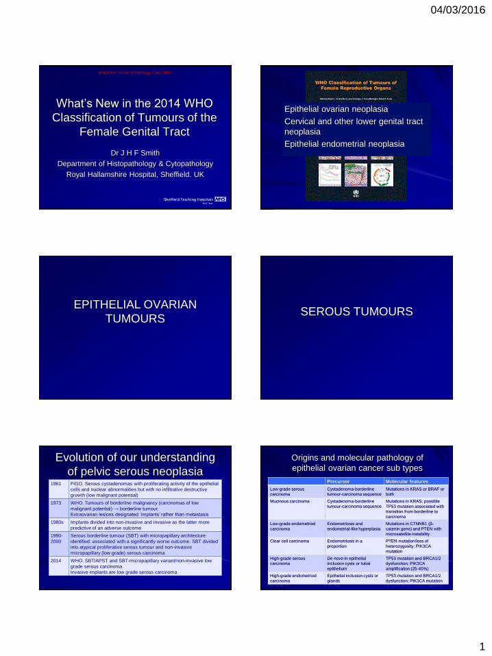
What’s New in the 2014 WHO
Classification of Tumours of the
Female Genital Tract
Dr J H F Smith
Department of Histopathology & Cytopathology
Royal Hallamshire Hospital, Sheffield. UK
Arab-Britsh School of Pathology Cairo 2014
Epithelial ovarian neoplasia
Cervical and other lower genital tract
neoplasia
Epithelial endometrial neoplasia
EPITHELIAL OVARIAN
TUMOURS SEROUS TUMOURS
Evolution of our understanding
of pelvic serous neoplasia 1961 FIGO. Serous cystadenomas with proliferating activity of the epithelial
cells and nuclear abnormalities but with no infiltrative destructive
growth (low malignant potential)
1973 WHO. Tumours of borderline malignancy (carcinomas of low
malignant potential) → borderline tumour.
Extraovarian lesions designated ‘implants’ rather than metastasis
1980s Implants divided into non-invasive and invasive as the latter more
predictive of an adverse outcome
1990-
2000
Serous borderline tumour (SBT) with micropapillary architecture
identified: associated with a significantly worse outcome. SBT divided
into atypical proliferative serous tumour and non-invasive
micropapillary (low grade) serous carcinoma
2014 WHO. SBT/APST and SBT-micropapillary variant/non-invasive low
grade serous carcinoma
Invasive implants are low grade serous carcinoma
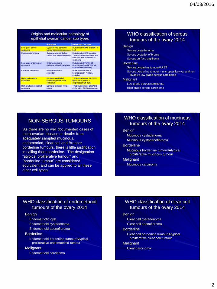
Origins and molecular pathology of
epithelial ovarian cancer sub types
Precursor Molecular features
Low-grade serous
carcinoma
Cystadenoma-borderline
tumour-carcinoma sequence
Mutations in KRAS or BRAF or
both
Mucinous carcinoma Cystadenoma-borderline
tumour-carcinoma sequence
Mutations in KRAS; possible
TP53 mutation associated with
transition from borderline to
carcinoma
Low-grade endometriod
carcinoma
Endometriosis and
endometrial-like hyperplasia
Mutations in CTNNB1 (β-
catenin gene) and PTEN with
microsatellite instability
Clear cell carcinoma Endometriosis in a
proportion
PTEN mutation/loss of
heterozygosity; PIK3CA
mutation
High-grade serous
carcinoma
De novo in epithelial
inclusion cysts or tubal
epithelium
TP53 mutation and BRCA1/2
dysfunction; PIK3CA
amplification (25-40%)
High-grade endometriod
carcinoma
Epithelial inclusion cysts or
glands
TP53 mutation and BRCA1/2
dysfunction; PIK3CA mutation
04/03/2016
2
Origins and molecular pathology of
epithelial ovarian cancer sub types
Precursor Molecular features
Low-grade serous
carcinoma
Cystadenoma-borderline
tumour-carcinoma sequence
Mutations in KRAS or BRAF or
both
Mucinous carcinoma Cystadenoma-borderline
tumour-carcinoma sequence
Mutations in KRAS; possible
TP53 mutation associated with
transition from borderline to
carcinoma
Low-grade endometriod
carcinoma
Endometriosis and
endometrial-like hyperplasia
Mutations in CTNNB1 (β-
catenin gene) and PTEN with
microsatellite instability
Clear cell carcinoma Endometriosis in a
proportion
PTEN mutation/loss of
heterozygosity; PIK3CA
mutation
High-grade serous
carcinoma
De novo in epithelial
inclusion cysts or tubal
epithelium
TP53 mutation and BRCA1/2
dysfunction; PIK3CA
amplification (25-40%)
High-grade endometriod
carcinoma
Epithelial inclusion cysts or
glands
TP53 mutation and BRCA1/2
dysfunction; PIK3CA mutation
WHO classification of serous
tumours of the ovary 2014
Benign
Serous cystadenoma
Serous cystadenofibroma
Serous surface papilloma
Borderline
Serous borderline tumour/APST
Serous borderline tumour – micropapillary variant/non-
invasive low grade serous carcinoma
Malignant
Low grade serous carcinoma
High grade serous carcinoma
NON-SEROUS TUMOURS
‘As there are no well documented cases of
extra-ovarian disease or deaths from
adequately sampled mucinous,
endometriod, clear cell and Brenner
borderline tumours, there is little justification
in calling them borderline. The designation
“atypical proliferative tumour” and
“borderline tumour” are considered
equivalent and can be applied to all these
other cell types.’
WHO classification of mucinous
tumours of the ovary 2014
Benign
Mucinous cystadenoma
Mucinous cystadenofibroma
Borderline
Mucinous borderline tumour/Atypical
proliferative mucinous tumour
Malignant
Mucinous carcinoma
WHO classification of endometrioid
tumours of the ovary 2014
Benign
Endometriotic cyst
Endometrioid cystadenoma
Endometrioid adenofibroma
Borderline
Endometrioid borderline tumour/Atypical
proliferative endometrioid tumour
Malignant
Endometrioid carcinoma
WHO classification of clear cell
tumours of the ovary 2014
Benign
Clear cell cystadenoma
Clear cell adenofibroma
Borderline
Clear cell borderline tumour/Atypical
proliferative clear cell tumour
Malignant
Clear carcinoma
04/03/2016
3
WHO classification of Brenner
tumours of the ovary 2014
Benign
– Brenner tumour
Borderline
– Borderline Brenner tumour/Atypical
proliferative Brenner tumour
Malignant
– Malignant Brenner tumour
NB. Transitional cell carcinoma of the ovary is a variant of
high grade serous carcinoma or endometrioid carcinoma
Takeuchi et al. Am J Surg Pathol 2013; 37: 1091:
WHO classification of seromucinous
tumours of the ovary 2014
Benign
Seromucinous cystadenoma
Seromucinous adenofibroma
Borderline
Seromucinous borderline tumour/Atypical
proliferative seromucinous tumour
Malignant
Seromucinous carcinoma
CERVICAL AND OTHER
LOWER GENITAL TRACT
NEOPLASIA
Cervical epithelial neoplasia
Broad agreement to replace CIN 1, CIN 2
and CIN 3 with LSIL and HSIL
Two tiered system more biologically and
clinically relevant and histologically
reproducible
Darragh et al J Loe Geni Tract Dis 2012; 16: 205
Stoler JAMA 2002; 287: 2140
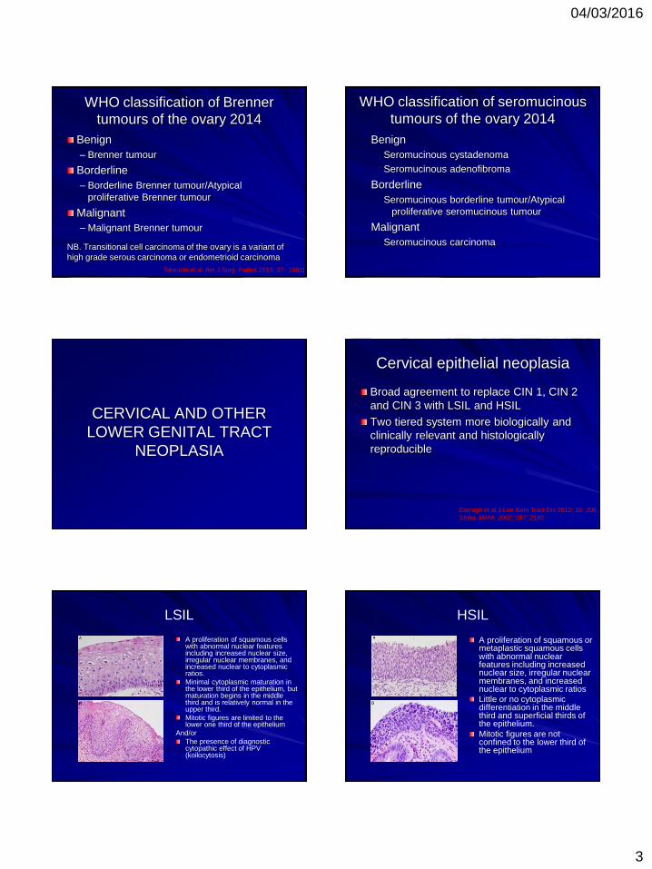
LSIL
A proliferation of squamous cells with abnormal nuclear features including increased nuclear size, irregular nuclear membranes, and increased nuclear to cytoplasmic ratios.
Minimal cytoplasmic maturation in the lower third of the epithelium, but maturation begins in the middle third and is relatively normal in the upper third.
Mitotic figures are limited to the lower one third of the epithelium
And/or
The presence of diagnostic cytopathic effect of HPV (koilocytosis)
HSIL
A proliferation of squamous or metaplastic squamous cells with abnormal nuclear features including increased nuclear size, irregular nuclear membranes, and increased nuclear to cytoplasmic ratios
Little or no cytoplasmic differentiation in the middle third and superficial thirds of the epithelium.
Mitotic figures are not confined to the lower third of the epithelium
04/03/2016
4
Abnormal Mitoses or Significant
Nuclear Atypia Abnormal mitoses and marked nuclear atypia more commonly seen in a high-grade lesions.
Lesions with the overall morphology of LSIL, but either marked nuclear atypia in the lower third of the epithelium or atypical mitoses at any level are considered to be consistent with HSIL
Positive p16 staining supports the diagnosis of HSIL
Thin SIL (Thin Dysplasia)
Immature intraepithelial lesions less than 10 cells thick.
If a lesion is unequivocal SIL with significant immature abnormal basal proliferation or mitosis above the basal cells, it is designated as HSIL.
If there is doubt about the nature of the proliferation (i.e. immature metaplasia versus SIL) then p16 staining can be used
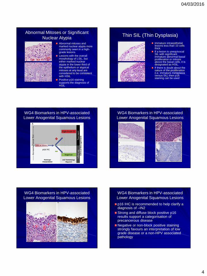
WG4 Biomarkers in HPV-associated
Lower Anogenital Squamous Lesions
WG4 Biomarkers in HPV-associated
Lower Anogenital Squamous Lesions
WG4 Biomarkers in HPV-associated
Lower Anogenital Squamous Lesions
WG4 Biomarkers in HPV-associated
Lower Anogenital Squamous Lesions
p16 IHC is recommended to help clarify a diagnosis of –IN2
Strong and diffuse block positive p16 results support a categorisation of precancerous disease
Negative or non-block positive staining strongly favours an interpretation of low grade disease or a non-HPV associated pathology

04/03/2016
5
WG4 Biomarkers in HPV-associated
Lower Anogenital Squamous Lesions
WG4 Biomarkers in HPV-associated
Lower Anogenital Squamous Lesions
WG4 Biomarkers in HPV-associated
Lower Anogenital Squamous Lesions
p16 is recommended for use as an adjudication
tool for cases in which there is a professional
disagreement in histology interpretation, with the
caveat that the differential diagnosis includes –
IN 2 or – IN 3
p16 IHC should not be used as a routine adjunct
to histological assessment of biopsy specimens
with morphological interpretations of negative, –
IN 1, and – IN 3
WG4 Biomarkers in HPV-associated
Lower Anogenital Squamous Lesions
Endocervical glandular
dysplasia/low grade CGIN Poorly reproducible diagnosis for which
criteria are not well defined
Minimal nuclear atypia with
hyperchromasia and slightly increased
mitoses or apoptotic bodies has been
suggested
Positive p16, high Ki-67 proliferation index
and absent hormone receptor staining
support diagnosis of AIS/HG-CGIN
WHO classification of squamous cell
tumours and precursors of the cervix 2014
Squamous cell tumours and precursors
Squamous intraepithelial lesions
Low grade squamous intraepithelial lesion (LSIL)
High grade squamous intraepithelial lesion (HSIL)
Squamous cell carcinoma
Keratinising, non-keratinising, papillary, basaloid,
warty, verrucous, squamotransitional,
lymphoepithelioma-like
Benign squamous cell lesions
Squamous metaplasia, condyloma acuminatum,
squamous papilloma, transitional metaplasia

04/03/2016
6
WHO Classification of Glandular
Tumours and Precursors 2014
Adenocarcinoma in situ
Adenocarcinoma
Endocervical adenocarcinoma, usual type
Mucinous carcinoma
Gastric type
Intestinal type
Signet ring type
Villoglandular carcinoma
Endometrioid carcinoma
Clear cell carcinoma
Serous carcinoma
Mesonephric carcinoma
Adenocarcinoma admixed with neuroendocrine carcinoma
VULVAL EPITHELIAL
NEOPLASIA
WHO Classification of tumours of the
vulva
Squamous cell tumours and precursors
– Squamous intraepithelial lesion
Low grade squamous intraepithelial lesion
High grade squamous intraepithelial lesion
Differentiated-type vulvar intraepithelial neoplasia
– Squamous cell carcinoma
– Basal cell carcinoma
Glandular tumours
– Paget’s disease
– Tumours of Bartholin and other anogenital glands
– Adenocarcinoma of other types
WHO Classification of VIN 2014
Low grade SIL (HPV only, VIN 1)
High grade SIL (usual type VIN 2/3)
Differentited type VIN
Paget’s disease
Two pathways to vulval
neoplasia HPV-related
Young women
Warty/basaloid
(undifferentiated) vulvar
intraepithelial neoplasia
Warty/basaloid carcinoma
Same HPV types as CIN
esp HPV 16
Mechanisms probably
similar
p16 surrogate marker
Two pathways to vulval
neoplasia Non-HPV-related
Older women
Associated with lichen
sclerosus
Differentiated (simplex) type
VIN
Often well differentiated
squamous cell carcinoma
but clinically aggressive
?p53 mutation important
04/03/2016
7
Does this matter?
Potential therapeutic relevance
– Imiquimod
– Other agents
Should we classify on the basis of HPV
expression?
Further molecular investigation of
differentiated type VIN required
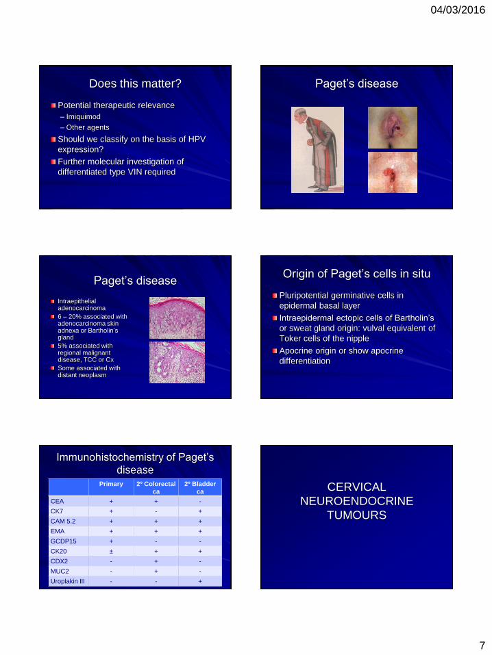
Paget’s disease
Paget’s disease
Intraepithelial adenocarcinoma
6 – 20% associated with adenocarcinoma skin adnexa or Bartholin’s gland
5% associated with regional malignant disease, TCC or Cx
Some associated with distant neoplasm
Origin of Paget’s cells in situ
Pluripotential germinative cells in
epidermal basal layer
Intraepidermal ectopic cells of Bartholin’s
or sweat gland origin: vulval equivalent of
Toker cells of the nipple
Apocrine origin or show apocrine
differentiation
Immunohistochemistry of Paget’s
disease
Primary 2º Colorectal
ca
2º Bladder
ca
CEA + + -
CK7 + - +
CAM 5.2 + + +
EMA + + +
GCDP15 + - -
CK20 ± + +
CDX2 - + -
MUC2 - + -
Uroplakin III - - +
CERVICAL
NEUROENDOCRINE
TUMOURS
04/03/2016
8
Terminology (neuro)endocrine
tumours of the cervix Carcinoid tumour
Carcinoid tumour with squamous cell carcinoma
Carcinoid tumour with adenocarcinoma
Argyrophil cell carcinoma
Apudoma
Poorly differentiated small cell carcinoid
Small cell tumour with neuroepithelial features
Nonendocrine carcinoid tumour
Endocrine carcinoma
intermediate cell type
Small cell undifferentiated
carcinoma
Oat cell carcinoma
Small cell carcinoma
Small cell neuroendocrine
carcinoma
Neuroendocrine carcinoma,
non-small cell type
Adenocarcinoma with
carcinoid features
Albores-Saavedra. Arch Path Lab Med 1997; 121: 34
Terminology (neuro)endocrine
tumours of the cervix
Typical (classical) carcinoid tumour
Atypical carcinoid tumour
Large cell neuroendocrine carcinoma
Small cell carcinoma
Albores-Saavedra. Arch Path Lab Med 1997; 121: 34
Terminology (neuro)endocrine
tumours of the cervix
Typical carcinoid
Atypical carcinoid
Large cell neuroendocrine carcinoma
Small cell carcinoma
Albores-Saavedra. Arch Path Lab Med 1997; 121: 34
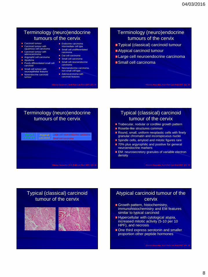
Typical (classical) carcinoid
tumour of the cervix Trabecular, nodular or cordlike growth pattern
Rosette-like structures common
Round, small, uniform neoplastic cells with finely granular chromatin and inconspicuous nuclei
Spindle cells, amyloid and mitotic figures rare
70% plus argyrophilic and positive for general neuroendocrine markers
EM: neurosecretory granules of variable electron density
Albores-Saavedra. Arch Path Lab Med 1997; 121: 34
Typical (classical) carcinoid
tumour of the cervix
Atypical carcinoid tumour of the
cervix Growth pattern, histochemistry, immunohistochemistry and EM features similar to typical carcinoid
Hypercellular with cytological atypia, increased mitotic activity (5-10 per 10 HPF), and necrosis
One third express serotonin and smaller proportion other peptide hormones
Albores-Saavedra. Arch Path Lab Med 1997; 121: 34

04/03/2016
9
Atypical carcinoid tumour of the
cervix
Large cell neuroendocrine
carcinoma of the cervix Organoid, trabecular or cordlike growth pattern with peripheral palisading and variable necrosis
Large neoplastic cells with abundant cytoplasm, vesicular nuclei and prominent nucleoli
Mitoses more than 10 per 10 HPF
Argyrophilic and positive for chromogranin or synaptophysin
Albores-Saavedra. Arch Path Lab Med 1997; 121: 34
Large cell neuroendocrine
carcinoma of the cervix
Small (oat) cell carcinoma of the
cervix Diffuse growth pattern or arranged in nests,
trabeculae and cords
Small round or fusiform cells with scant
cytoplasm
Hyperchromatic nuclei with finely granular
chromatin and absent of inconspicuous
cytoplasm
Peripheral palisading and perivascular
concentration of cells common
Immunohistochemistry not required for diagnosis Albores-Saavedra. Arch Path Lab Med 1997; 121: 34
WHO classification of neuroendocrine
tumours of the cervix 2014
Low grade neuroendocrine tumour
– Grade 1 carcinoid tumour
– Grade 2 atypical carcinoid tumour
High grade neuroendocrine carcinoma
– Grade 3 small cell neuroendocrine carcinoma
– Grade 4 large cell neuroendocrine carcinoma
Mirrors system used for gastrointestinal
and pancreatic tumours

04/03/2016
10
ENDOMETRIAL TUMOURS
Endometrioid carcinoma
precursors WHO 1994 Degree of architectural crowding
– Simple
– Complex
Nuclear alteration
– Non-atypical
– Atypical
Simple Non-atypical Hyperplasia
Variable gland size
Normal
gland/stroma ratio
No cytological atypia
Complex Non-atypical Hyperplasia
Architectural irregularity of the glands
Increased gland/stroma ratio
No cytological atypia
Complex Atypical Hyperplasia
Architectural irregularity and crowding of the glands
Increased gland/stroma ratio
Cytological atypia
Endometrioid carcinoma
precursors WHO 2014
Hyperplasia without atypia
Atypical hyperplasia/endometrioid
intraepithelial neoplasia
04/03/2016
11
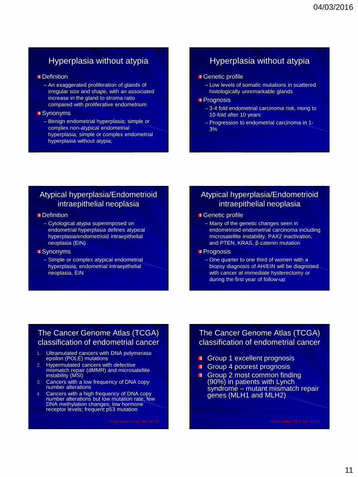
Hyperplasia without atypia
Definition
– An exaggerated proliferation of glands of
irregular size and shape, with an associated
increase in the gland to stroma ratio
compared with proliferative endometrium
Synonyms
– Benign endometrial hyperplasia; simple or
complex non-atypical endometrial
hyperplasia; simple or complex endometrial
hyperplasia without atypia;
Hyperplasia without atypia
Genetic profile
– Low levels of somatic mutations in scattered
histologically unremarkable glands
Prognosis
– 3-4 fold endometrial carcinoma risk, rising to
10-fold after 10 years
– Progression to endometrial carcinoma in 1-
3%
Atypical hyperplasia/Endometrioid
intraepithelial neoplasia
Definition
– Cytological atypia superimposed on
endometrial hyperplasia defines atypical
hyperplasia/endometrioid intraepithelial
neoplasia (EIN)
Synonyms
– Simple or complex atypical endometrial
hyperplasia; endometrial intraepithelial
neoplasia, EIN
Atypical hyperplasia/Endometrioid
intraepithelial neoplasia
Genetic profile
– Many of the genetic changes seen in
endometrioid endometrial carcinoma including
microsatellite instability, PAX2 inactivation,
and PTEN, KRAS, β-catenin mutation
Prognosis
– One quarter to one third of women with a
biopsy diagnosis of AH/EIN will be diagnosed
with cancer at immediate hysterectomy or
during the first year of follow-up
The Cancer Genome Atlas (TCGA)
classification of endometrial cancer
1. Ultramutated cancers with DNA polymerase epsilon (POLE) mutations
2. Hypermutated cancers with defective mismatch repair (dMMR) and microsatellite instability (MSI)
3. Cancers with a low frequency of DNA copy number alterations
4. Cancers with a high frequency of DNA copy number alterations but low mutation rate; few DNA methylation changes; low hormone receptor levels; frequent p53 mutation
TCGA. Nature 2013; 497: 67-73
The Cancer Genome Atlas (TCGA)
classification of endometrial cancer
Group 1 excellent prognosis
Group 4 poorest prognosis
Group 2 most common finding (90%) in patients with Lynch syndrome – mutant mismatch repair genes (MLH1 and MLH2)
TCGA. Nature 2013; 497: 67-73

04/03/2016
12
Any
Questions ?