where in the cell is your protein most likely found?
TRANSCRIPT

Where in the cell is your proteinmost likely found?

Where Are Proteins Located?• All proteins are synthesized in the cytoplasm.• Proteins with export signals can be directed to other
cellular locations:– cytoplasm, cytoplasmic membrane, outer membrane or
periplasm of Gram (-) bacteria, cell wall, or as secreted products in extracellular space
Gram PositiveCells
Gram NegativeCells
Insert Figure 1 from Gardy and Brinkman (2006)Methods for predicting bacterial protein subcellular localization.Nature Reviews Microbiology 4: 741-751.
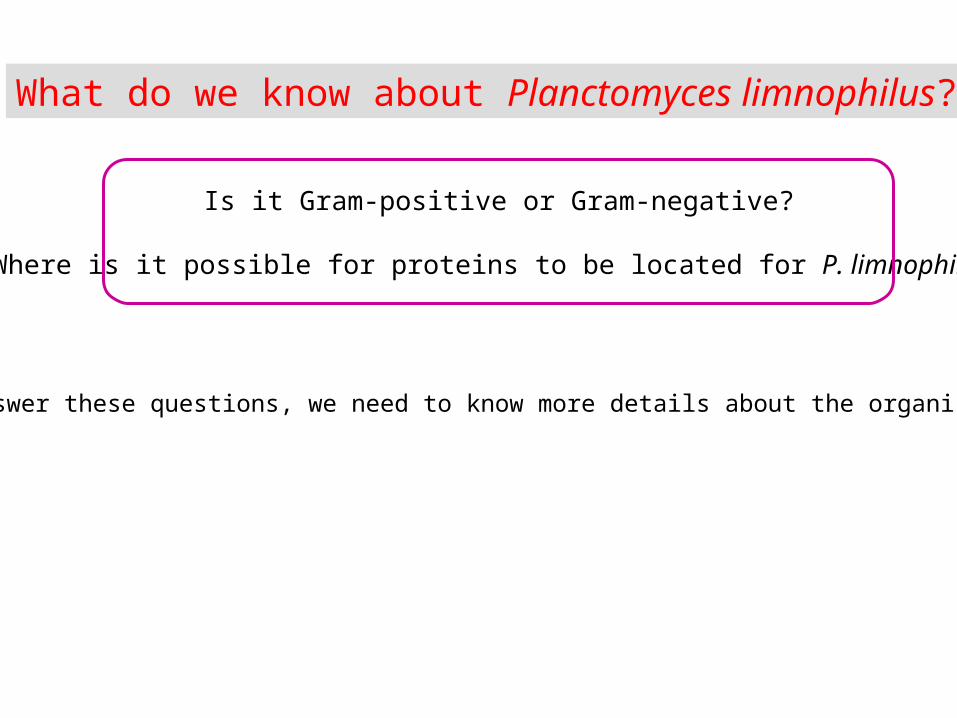
What do we know about Planctomyces limnophilus?
Is it Gram-positive or Gram-negative?
Where is it possible for proteins to be located for P. limnophilus?
To answer these questions, we need to know more details about the organism. . .

Recall: Planctomyces limnophilus DSM 3776
• budding, stalked bacterium isolated from the surface of a eutrophic freshwater lake in Holstein, Germany
Insert Figure 1D from Hirsch P and Müller M (1985)Planctomyces limnophilus sp. nov., a stalked and budding bacterium from freshwater.System. Appl. Microbiol. 6: 276-280.
Insert map of Germany fromwww.mapsofworld.com

Characteristics of Planctomyces
• form rosettes (star-like form) with cells connected by non-cellular, protein stalk
• When initially discovered were thought to be fungal conidia
Insert Figure 1 from Fuerst (1995)The planctomycetes: Emerging models for microbial ecology, evolution, and cell biology.Microbiology 141: 1493-1506.

Planctomyces possess internal,membrane-bound compartments
(blurs the boundary between prokaryotes & eukaryotes)
• some bound the nucleoid• Gemmata obscuriglobus
• some partition metabolic functions• Brocadia anammoxidans
• carries out anaerobic oxidation of NH3 to N2 within enclosed structure called an anammoxosome
Insert Figure 17-34b fromBrock Biology of Microorganisms 11/e
© 2006 Pearson Prentice Hall, Inc.Insert Figure 12.87 from
Brock Biology of Microorganisms 11/e© 2006 Pearson Prentice Hall, Inc.

• form red colonies
• do not form endospores
• mature cell shape & size• ovoid to spherical• 1.1 – 1.5 μm
• attach to surfaces using fibrous holdfast at end of long, rigid stalk composed of twisted fibrils
• stalk made of protein
• multiply by yeast-like budding
Planctomyces limnophilus DSM 3776young
bud
maturemothercell
nearlymaturebud
maturecell withshort stalk& fibrousholdfast
maturecell withlong stalk
Insert Figure 1 (panels A-D) fromHirsch P and Müller M (1985)Planctomyces limnophilus sp. nov., a stalked and budding bacterium from freshwater. System. Appl. Microbiol. 6: 276-280.

What other organisms use asymmetric cell division?
Budding yeast (eukaryote):Saccharomyces cereviseae
Budding bacterium:Planctomyces limnophilus
Insert Figure 1 (panels A-B) fromHirsch P and Müller M (1985)Planctomyces limnophilus sp. nov., a stalked and budding bacterium from freshwater. System. Appl. Microbiol. 6: 276-280.
Insert image of budding yeast cell

• surface appendageso stalk with holdfasto flagellumo pilio fimbriae
Cellular characteristics of Planctomyces
P. maris
• lack peptidoglycan in cell wallo Like mycoplasmas & clamydiaeo consequently stains Gram-negative
Does this imply P. limnophilis is Gram-negative? Why or why not?
o naturally resistant to penicillinInsert image fromASM Microbelibrary.org
Neisseria gonorrhoeae
Recall: Gram-negative cells are red
Insert Figure 12.86 fromBrock Biology of Microorganisms 11/e
© 2006 Pearson Prentice Hall, Inc.
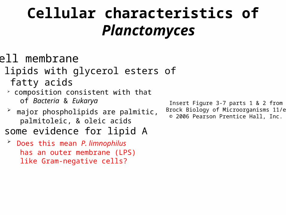
• cell membraneo lipids with glycerol esters of fatty acids
composition consistent with that of Bacteria & Eukarya major phospholipids are palmitic, palmitoleic, & oleic acids
• some evidence for lipid A Does this mean P. limnophilus has an outer membrane (LPS) like Gram-negative cells?
Cellular characteristics of Planctomyces
Insert Figure 3-7 parts 1 & 2 fromBrock Biology of Microorganisms 11/e
© 2006 Pearson Prentice Hall, Inc.

Cellular characteristics of Planctomyces
• Subunit composition of RNAP consistent with Bacteria
Ec – E. coli (Bacteria)Hs – Halobacterium salinarum (Archaea)Sa – Sulfolobus acidocaldarius (Archaea)Sc – Saccharomyces cerevisiae (Eukarya)
Insert Figure 11-19 fromBrock Biology of Microorganisms 11/e
© 2006 Pearson Prentice Hall, Inc.
Insert Figure 8-5C fromMicrobiology – An Evolving Science
© 2009 W.W. Norton & Company, Inc.

Why is this information important?
Fuerst (2005)Annu. Rev. Microbiol. 59: 299-328.
• At least 8% of the Rhodopirellula baltica proteome exhibits homology with eukaryotic genes (Glockner et al. 2003)
HGT or genes derived from the universal ancestor of all 3 domains?
Glockner et al. (2003)PNAS 100: 8298-8303.
• Planctomycetes have distinct cellular characteristics Absence of peptidoglycan but possible presence of lipid A in cell envelop
What does the structure of the cell envelop in P. limnophilus resemble? Gram-positive or Gram-negative?
Exhibit budding-like mechanism for cell division (like eukaryotes) Have internal membranes (compartmentalization like eukaryotes)
Is there an evolutionary relationship to origin of eukaryotic nucleus? Will sorting signals resemble those in bacteria or eukaryotes?
Is it Gram-positive or Gram-negative?
Where is it possible for proteins to be located for P. limnophilus?

How do we figure out where proteins are located?
Transmembrane Helices Hidden Markov Models (TMHMM)
Does my protein have transmembrane helices?
Signal Peptide (SignalP) Does my protein have a sequence of amino acids that
target it to a particular place in or outside the cell?
PSORT-B Where is my protein most likely located? The cytoplasm? The
membrane? The periplasm? The cell wall? The extracellular space?
Phobius Does my protein have transmembrane helices & signal peptides? Do
these results agree with TMHMM and SignalP?

Transmembrane Helices Hidden Markov Models (TMHMM)
• A Hidden Markov Model is a probabilistic model developed from observed sequences of proteins of a known function.
• TMHMM is a tool used to predict the presence of transmembrane helices in proteins. The results will indicate the segments of the protein that lie inside, outside or within the membrane.
Click link found in Lab Notebook

TMHMM Database Search
Enter “Protein Sequence” in FASTA format
“CLICK” **Make sure Javascript is enabled on your computer to read output
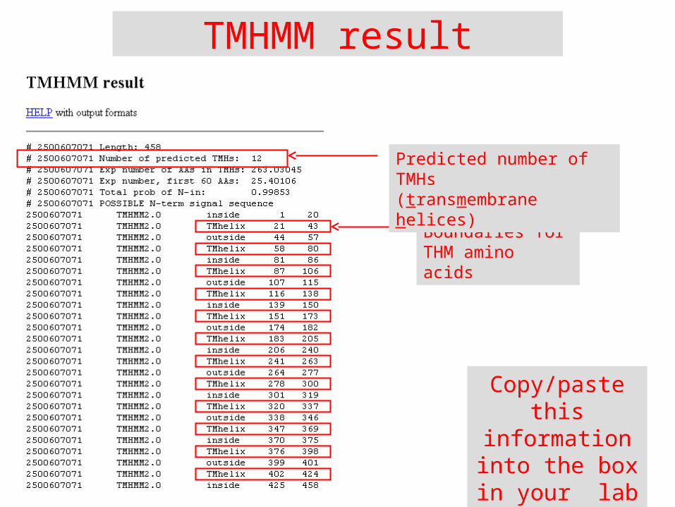
TMHMM result
Boundaries for THM amino acids
Predicted number of TMHs(transmembrane helices)
Copy/paste this information into the box in your
lab notebook

Interpreting the TMHMM plot
Residue numberX-axis: the amino acid number
Y-axis: the probability that the amino acid is located within the membrane, outside the cell, or in the cytoplasm
Ex: If probability >0.75, then result is significant. The maximum probability is 1, so the probability that amino acids #1-#20 are “inside” is 100%
Schematic that summarizes discrete regions within the protein; not probability.
Transmembrane
Inside (cytoplasm)
Outside (extracellular,
periplasm)
The 12predicted
TMHs
0.75

cytoplasm
membrane
By analyzing the probabilities shown on the plot, you can determine where segments within the protein are located.

Inserting the TMHMM plot into your notebook
Save image in GIF format to your computer and
insert into Lab Notebook

…and the comments
Summarize your analysis
of the TMHMM plot
in the box provided for “comments”.

Recording results in your Lab Notebook
OID 2500607071
Confirm you record the number of TMHs,with a text description
of the boundariesfor each TMH
Insert the TMHMM plot
Examine the plot & summarize results, with
a confidence rating based on the
probability score. Assess how its
structure is related to assigned function.

SignalP• A Signal Peptide (SignalP) is a series of amino acids in the polypeptide that directs the protein to its proper cellular location
• Ex: Single TMH at N-terminus of protein that gets cleaved by proteases once inserted into membrane
Click link found in Lab Notebook
Locating proteins in the cell using TargetP, SignalP, and related toolsOlof Emanuelsson, Søren Brunak, Gunnar von Heijne, Henrik NielsenNature Protocols 2, 953-971 (2007).
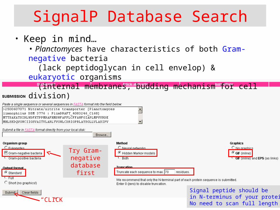
SignalP Database Search• Keep in mind…
• Planctomyces have characteristics of both Gram-negative bacteria (lack peptidoglycan in cell envelop) & eukaryotic organisms (internal membranes, budding mechanism for cell division)
“CLICK”
Signal peptide should bein N-terminus of your protein;No need to scan full length
Try Gram-negative
database first

Signal P (Gram - )
Look for Probability exceeding 0.50 threshold. If not, go back and select “eukaryotes” as an organism group
Represents Probability Position of Amino Acid
- Signal peptide cleaved by proteolytic enzymes - N-terminus of signal peptide- Hydrophobic Region (TMH)- C-terminus of signal peptide
0.75
0.50

SignalP Database Search
• If no significant results obtained searching the Gram-negative database, next try the eukaryotic database. . .
“CLICK”
Change this selection only

Signal P (Eukaryote)
If the probability is >0.50, then the results suggest that your gene encodes a signal peptide. Higher confidence in probability score if >0.75
0.75
What would you concludefor this protein?
- Signal peptide cleaved by proteolytic enzymes - N-terminus of signal peptide- Hydrophobic Region (TMH)- C-terminus of signal peptide
Possible protease cleavage site if probability > 0.75
0.50
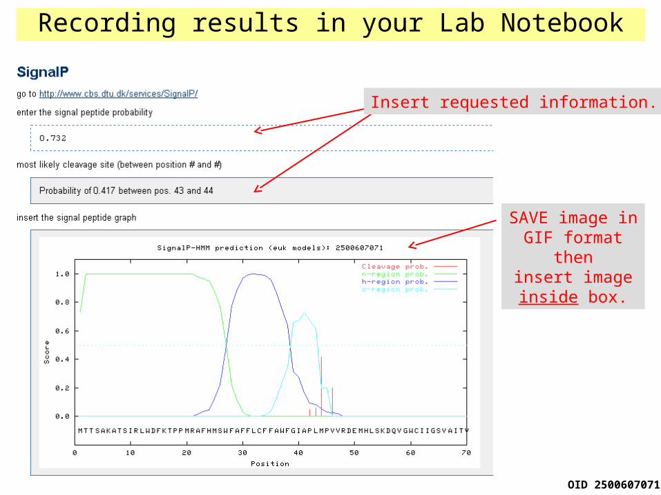
Recording results in your Lab Notebook
OID 2500607071
Insert requested information.
SAVE image inGIF format then
insert imageinside box.

PSORT-B
• Another useful tool in predicting bacterial protein localization
• The output is TEXTUAL, but the information still will be helpful
Click link found in Lab Notebook

Enter “Protein Sequence” in FASTA format
“CLICK”
Select “Negative” for Gram stain

Where this protein is predicted to be located in the cell
Enter in your Lab Notebook
Recording results in your Lab Notebook

Phobius• Graphical output
• Combination of transmembrane topology (TMHMM) and signal peptide predictor (SignalP)
“Click”
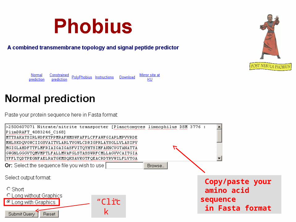
Copy/paste your amino acid sequence in Fasta format“Click”

Query Results
Graphicalsummary
Text listing predictedlocations of TMHs,intervening loops,and signal peptide
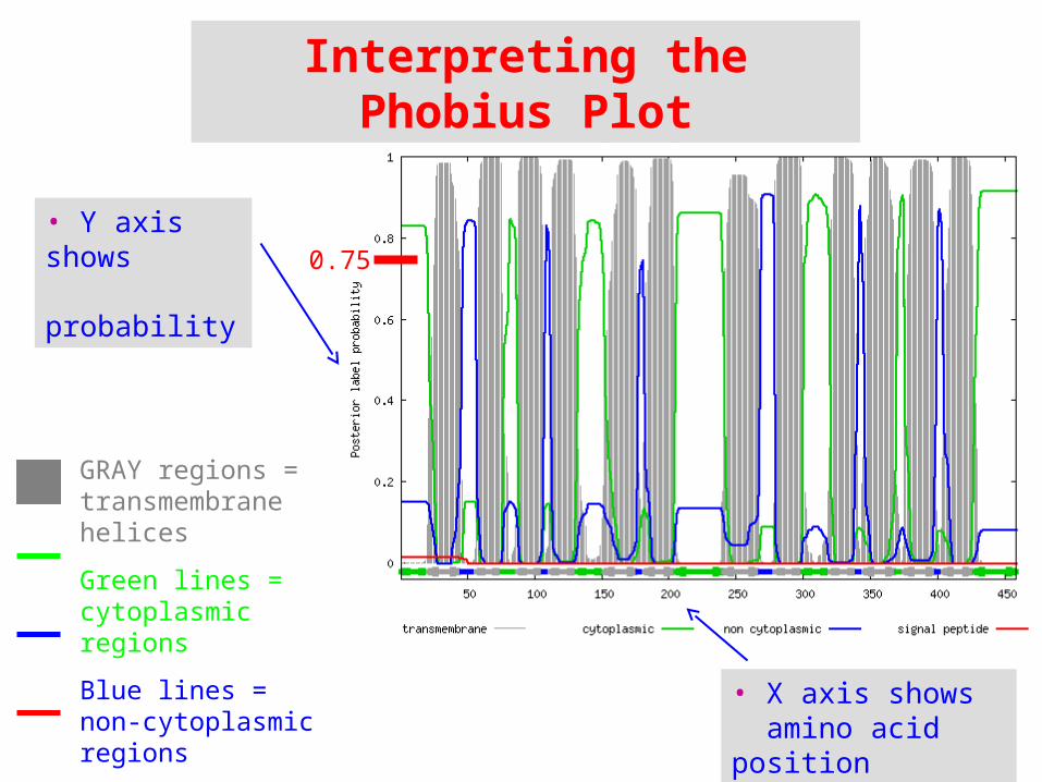
• Y axis shows probability
GRAY regions = transmembrane helices
Green lines = cytoplasmic regions
Blue lines =non-cytoplasmic regions
Red lines =signal peptides
Interpreting the Phobius Plot
• X axis shows amino acid position
0.75

cytoplasm
membrane
By analyzing the probabilities shown on the plot, you can determine where segments within the protein are located.

Recording results in your Lab Notebook
OID 2500607071
Add heading
Copy/paste text descriptioninto new box

Recording results in your Lab Notebook
OID 2500607071
SAVE image in PNGformat then insert intoNotebook inside box.

Finishing Up Cellular Localization ModuleNow that you’ve finished TMHMM, SignalP, PSORT, and Phobius, you shouldhave an idea about the cellular localization of the protein encoding by your gene.
Enter your conclusion about where you would expect tofind the protein under the Hypothesis section of this module
THINGS TO CONSIDER: • Did TMHMM indicate any transmembrane helices? If so, how many? • Did SignalP show evidence of a signal peptide at N-terminus? • Where did PSORT predict the protein was located in the cell? • Were Phobius results consistent with TMHMM and SignalP results?