whole-genome analysis of piscine reovirus (prv) shows prv represents a new genus in family
TRANSCRIPT
Kibenge et al. Virology Journal 2013, 10:230http://www.virologyj.com/content/10/1/230
RESEARCH Open Access
Whole-genome analysis of piscine reovirus (PRV)shows PRV represents a new genus in familyReoviridae and its genome segment S1 sequencesgroup it into two separate sub-genotypesMolly JT Kibenge1†, Tokinori Iwamoto1†, Yingwei Wang2, Alexandra Morton3, Marcos G Godoy4,5,6
and Frederick SB Kibenge1*
Abstract
Background: Piscine reovirus (PRV) is a newly discovered fish reovirus of anadromous and marine fish ubiquitousamong fish in Norwegian salmon farms, and likely the causative agent of heart and skeletal muscle inflammation(HSMI). HSMI is an increasingly economically significant disease in Atlantic salmon (Salmo salar) farms. Thenucleotide sequence data available for PRV are limited, and there is no genetic information on this virus outside ofNorway and none from wild fish.
Methods: RT-PCR amplification and sequencing were used to obtain the complete viral genome of PRV (10segments) from western Canada and Chile. The genetic diversity among the PRV strains and their relationship toNorwegian PRV isolates were determined by phylogenetic analyses and sequence identity comparisons.
Results: PRV is distantly related to members of the genera Orthoreovirus and Aquareovirus and an unambiguousnew genus within the family Reoviridae. The Canadian and Norwegian PRV strains are most divergent in thesegment S1 and S4 encoded proteins. Phylogenetic analysis of PRV S1 sequences, for which the largest number ofcomplete sequences from different “isolates” is available, grouped Norwegian PRV strains into a single genotype,Genotype I, with sub-genotypes, Ia and Ib. The Canadian PRV strains matched sub-genotype Ia and Chilean PRVstrains matched sub-genotype Ib.
Conclusions: PRV should be considered as a member of a new genus within the family Reoviridae with two majorNorwegian sub-genotypes. The Canadian PRV diverged from Norwegian sub-genotype Ia around 2007 ± 1, whereasthe Chilean PRV diverged from Norwegian sub-genotype Ib around 2008 ± 1.
BackgroundThe newly discovered piscine reovirus (PRV) belongsto the family Reoviridae, subfamily Spinareovirinae [1],probably in a new reovirus genus that is equally distantto the genera Orthoreovirus and Aquareovirus [2], al-though with 10 genome segments, PRV is like membersof the genus Orthoreovirus and unlike the genus Aqua-reovirus with 11 segments. The Orthoreovirus genus can
* Correspondence: [email protected]†Equal contributors1Department of Pathology and Microbiology, Atlantic Veterinary College,University of Prince Edward Island, 550 University Ave., Charlottetown, PEIC1A 4P3, CanadaFull list of author information is available at the end of the article
© 2013 Kibenge et al.; licensee BioMed CentraCommons Attribution License (http://creativecreproduction in any medium, provided the or
be divided into the fusogenic and non-fusogenic ortho-reoviruses based on the ability of the fusogenic ortho-reoviruses to induce cell-cell fusion during infectionresulting in syncytium formation [3] by virtue of posses-sion of a fusion-associated small transmembrane (FAST)protein [4]. Whereas the non-fusogenic orthoreoviruses,Mammalian Orthoreovirus (MRV), are not clinically sig-nificant [5], the fusogenic orthoreoviruses Nelson Bayvirus (NBV) [6] and Baboon Orthoreovirus (BRV) [7]that infect primates, Avian Orthoreovirus (ARV) [8] thatinfect birds, and Reptilian Orthoreovirus (RRV) [9] thatinfect reptiles, have been shown to cause significant andoften fatal disease. Most recently, PRV has been shown
l Ltd. This is an Open Access article distributed under the terms of the Creativeommons.org/licenses/by/2.0), which permits unrestricted use, distribution, andiginal work is properly cited.
Kibenge et al. Virology Journal 2013, 10:230 Page 2 of 20http://www.virologyj.com/content/10/1/230
to be more closely related with recognized orthoreo-viruses than with recognized aquareoviruses, and doesnot encode a FAST protein and is therefore non-fusogenic [10].PRV is associated with heart and skeletal muscle in-
flammation (HSMI) [2]; an emerging disease of marine-farmed Atlantic salmon [11], first recognized in 1999in western Norway [12] and subsequently in Scotland[13]. PRV has also been detected by real-time reversetranscription quantitative polymerase chain reaction(RT-qPCR) at a low prevalence in wild Atlantic salmon“S. salar” [2] and in certain marine fish species (Atlanticherring “Clupea harengus”, Capelin “Mallotus villosus”,Atlantic horse mackerel “Trachurus trachurus”, andGreat silver smelt “Argentina silus”) along the coast ofNorway [14]. PRV was also detected in 3% of anadro-mous trout (sea-trout) “Salmo trutta” tested, but not inanadromous Arctic char “Salvelinus alpinus” [15]. PRV isubiquitous in Norwegian salmon farms [16,17], but thereis a significant increase in the viral load and tissue distri-bution during outbreaks of HSMI [2,18]. The virus canbe propagated in the GF-1 cell line [19], derived from thetissue of orange-spotted grouper, Epinephelus coioides[20], and cardiac and skeletal muscle pathology typical ofHSMI can be reproduced in naïve Atlantic salmon by ex-perimental inoculation with the supernatant from cellculture passaged PRV [19]. Most recently, it has beenreported that serum enzymes creatine kinase and lactatedehydrogenase, associated with cardiac injury in humans[21], are significantly correlated with HSMI histopathologyin Atlantic salmon [22]. Other reports doubt the patho-genicity of PRV, describing PRV as an opportunistic virus[23,24] or non-pathogenic virus [15]. The virus has beendetected in marine-farmed Atlantic salmon in Chile[25,26]. There is anecdotal evidence that it is also presentin farmed Atlantic salmon and wild Pacific salmon inBritish Columbia-Canada [27], where 75% of 300 farmsalmon reportedly tested positive for PRV [27] but nosequence information was reported.The PRV genome comprises at least 10 dsRNA seg-
ments including three large (L), three medium (M), andfour small (S) size-class RNA genome segments [2]. Todate only two “isolates”, both from marine-farmed At-lantic salmon from Norway, have been sequenced on all10 genomic segments by high-throughput pyrosequen-cing of clinical samples: Reovirus sp. Salmo/GP-2010/NOR from HSMI [2], and CMS PRV from a CMSoutbreak [24]. However, only the Salmo/GP-2010/NORsequences are accessible from the GenBank Database(GenBank accession numbers GU994013-GU994022).The coding assignments of genomic segments S1 and S4initially reported to encode proteins with no identifiedhomologs in orthoreoviruses and aquareoviruses [2],were recently shown to be reversed such that S1 encodes
the major outer capsid protein, Outer clamp protein (σ3and VP7 in MRV and aquareoviruses, respectively), andS4 encodes the virus attachment protein, Outer fiberprotein (σ1 in MRV, which is absent in aquareoviruses)[10]. A further complication is that several sequences ofNorwegian PRV isolates were deposited in the GenBankdatabase as S4 sequences [18,28] but correspond to S1sequences [2, this study], and sequences of the remai-ning 9 genomic segments for these virus isolates havenot been reported.The sequence data available for PRV strains are lim-
ited, with no genetic information on this virus outside ofNorway, and none from wild fish despite the economicalimpact of HSMI on salmon aquaculture and the poten-tial for transmission of PRV to wild salmon populationsor from wild salmon to farmed salmon.The primary goal of the present study was to deter-
mine the genetic diversity among PRV strains detectedin tissue samples obtained from fish in western Canada,and in Chile, and their relationship to known NorwegianPRV sequences. We also attempted to sequence the com-plete genomes of three “isolates”, two Canadian and oneChilean to obtain more information about the taxonomicassignment of PRV.
Results and discussionAmplification and sequencing of cDNA of genomicsegments of PRV from fish samplesPiscine reovirus was readily detected by RT-qPCR duringtesting at the Atlantic Veterinary College laboratory infish tissue samples from western Canada, and at theETECMA diagnostic laboratory in fish tissue samplesfrom Chile (data not shown). Consistent with obser-vations elsewhere [2,18], PRV is ubiquitous in marine-farmed salmon. PRV was consistently detected in gilltissue, identifying the gills as suitable target tissue andlikely a primary transmission route for PRV. This is con-sistent with Orthoreovirus, which are commonly isolatedfrom enteric and respiratory tract tissues [1].Ten samples from western Canada with either low Ct
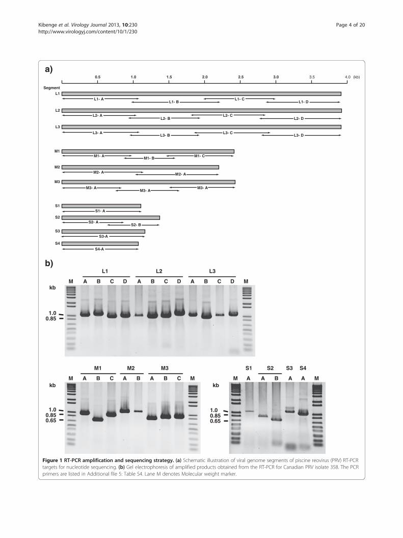
values or unique case histories, host species, and sam-pling times listed in (Additional file 1: Table S1a) wereselected for amplification and cloning of cDNA of viralgenome segments. Four additional samples for whichthe 3′ portion of genome segment L1 had been PCR-amplified during the original testing for PRV (Table 1),were included in the analysis of PRV sequences.Figure 1 shows the RT-PCR amplification and sequen-
cing strategy used for the PRV genome segments, basedon Canadian “isolate” 358. The new PRV nucleotidesequences (Additional file 2: Table S2) are availablethrough the GenBank database [29]. The complete PRVgenome (10 segments) was amplified from 2 of 10 sam-ples. Additional partial or full-length sequences were
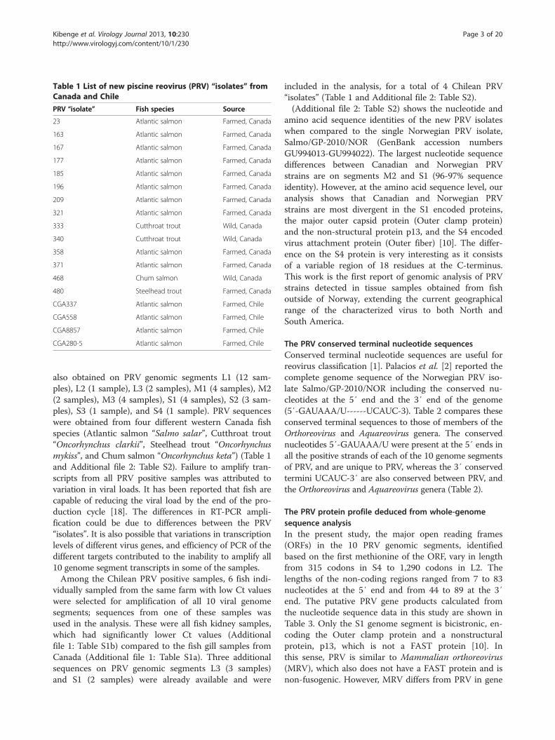
Table 1 List of new piscine reovirus (PRV) “isolates” fromCanada and Chile
PRV “isolate” Fish species Source
23 Atlantic salmon Farmed, Canada
163 Atlantic salmon Farmed, Canada
167 Atlantic salmon Farmed, Canada
177 Atlantic salmon Farmed, Canada
185 Atlantic salmon Farmed, Canada
196 Atlantic salmon Farmed, Canada
209 Atlantic salmon Farmed, Canada
321 Atlantic salmon Farmed, Canada
333 Cutthroat trout Wild, Canada
340 Cutthroat trout Wild, Canada
358 Atlantic salmon Farmed, Canada
371 Atlantic salmon Farmed, Canada
468 Chum salmon Wild, Canada
480 Steelhead trout Farmed, Canada
CGA337 Atlantic salmon Farmed, Chile
CGA558 Atlantic salmon Farmed, Chile
CGA8857 Atlantic salmon Farmed, Chile
CGA280-5 Atlantic salmon Farmed, Chile
Kibenge et al. Virology Journal 2013, 10:230 Page 3 of 20http://www.virologyj.com/content/10/1/230
also obtained on PRV genomic segments L1 (12 sam-ples), L2 (1 sample), L3 (2 samples), M1 (4 samples), M2(2 samples), M3 (4 samples), S1 (4 samples), S2 (3 sam-ples), S3 (1 sample), and S4 (1 sample). PRV sequenceswere obtained from four different western Canada fishspecies (Atlantic salmon “Salmo salar”, Cutthroat trout“Oncorhynchus clarkii”, Steelhead trout “Oncorhynchusmykiss”, and Chum salmon “Oncorhynchus keta”) (Table 1and Additional file 2: Table S2). Failure to amplify tran-scripts from all PRV positive samples was attributed tovariation in viral loads. It has been reported that fish arecapable of reducing the viral load by the end of the pro-duction cycle [18]. The differences in RT-PCR ampli-fication could be due to differences between the PRV“isolates”. It is also possible that variations in transcriptionlevels of different virus genes, and efficiency of PCR of thedifferent targets contributed to the inability to amplify all10 genome segment transcripts in some of the samples.Among the Chilean PRV positive samples, 6 fish indi-
vidually sampled from the same farm with low Ct valueswere selected for amplification of all 10 viral genomesegments; sequences from one of these samples wasused in the analysis. These were all fish kidney samples,which had significantly lower Ct values (Additionalfile 1: Table S1b) compared to the fish gill samples fromCanada (Additional file 1: Table S1a). Three additionalsequences on PRV genomic segments L3 (3 samples)and S1 (2 samples) were already available and were
included in the analysis, for a total of 4 Chilean PRV“isolates” (Table 1 and Additional file 2: Table S2).(Additional file 2: Table S2) shows the nucleotide and
amino acid sequence identities of the new PRV isolateswhen compared to the single Norwegian PRV isolate,Salmo/GP-2010/NOR (GenBank accession numbersGU994013-GU994022). The largest nucleotide sequencedifferences between Canadian and Norwegian PRVstrains are on segments M2 and S1 (96-97% sequenceidentity). However, at the amino acid sequence level, ouranalysis shows that Canadian and Norwegian PRVstrains are most divergent in the S1 encoded proteins,the major outer capsid protein (Outer clamp protein)and the non-structural protein p13, and the S4 encodedvirus attachment protein (Outer fiber) [10]. The differ-ence on the S4 protein is very interesting as it consistsof a variable region of 18 residues at the C-terminus.This work is the first report of genomic analysis of PRVstrains detected in tissue samples obtained from fishoutside of Norway, extending the current geographicalrange of the characterized virus to both North andSouth America.
The PRV conserved terminal nucleotide sequencesConserved terminal nucleotide sequences are useful forreovirus classification [1]. Palacios et al. [2] reported thecomplete genome sequence of the Norwegian PRV iso-late Salmo/GP-2010/NOR including the conserved nu-cleotides at the 5′ end and the 3′ end of the genome(5′-GAUAAA/U------UCAUC-3). Table 2 compares theseconserved terminal sequences to those of members of theOrthoreovirus and Aquareovirus genera. The conservednucleotides 5′-GAUAAA/U were present at the 5′ ends inall the positive strands of each of the 10 genome segmentsof PRV, and are unique to PRV, whereas the 3′ conservedtermini UCAUC-3′ are also conserved between PRV, andthe Orthoreovirus and Aquareovirus genera (Table 2).
The PRV protein profile deduced from whole-genomesequence analysisIn the present study, the major open reading frames(ORFs) in the 10 PRV genomic segments, identifiedbased on the first methionine of the ORF, vary in lengthfrom 315 codons in S4 to 1,290 codons in L2. Thelengths of the non-coding regions ranged from 7 to 83nucleotides at the 5′ end and from 44 to 89 at the 3′end. The putative PRV gene products calculated fromthe nucleotide sequence data in this study are shown inTable 3. Only the S1 genome segment is bicistronic, en-coding the Outer clamp protein and a nonstructuralprotein, p13, which is not a FAST protein [10]. Inthis sense, PRV is similar to Mammalian orthoreovirus(MRV), which also does not have a FAST protein and isnon-fusogenic. However, MRV differs from PRV in gene
M
M1 M2 M3
MM A B CA BCA B
M
L1
C DA B
L2
C DA B
L3
C DA B
MM
S1 S2 S3 S4
AA A AB
kb
kb kb
1.00.850.65
1.00.85
1.00.850.65
0.5 1.0 1.5 2.0 2.5 3.0 3.5 4.0 (kb)
M2- A M2- A
M2
M1- A M1- B M1- CM1
M3- AM3- A
M3- A
M3
S1- AS1
S3-AS3
S4-AS4
S2- AS2- B
S2
L1- AL1
L1- B L1- D
L3
L3- AL3- B L3- D
L2L2- A
L2- B L2- D
Segment
a)
b)
L1- C
L2- C
L3- C
Figure 1 RT-PCR amplification and sequencing strategy. (a) Schematic illustration of viral genome segments of piscine reovirus (PRV) RT-PCRtargets for nucleotide sequencing. (b) Gel electrophoresis of amplified products obtained from the RT-PCR for Canadian PRV isolate 358. The PCRprimers are listed in Additional file 5: Table S4. Lane M denotes Molecular weight marker.
Kibenge et al. Virology Journal 2013, 10:230 Page 4 of 20http://www.virologyj.com/content/10/1/230
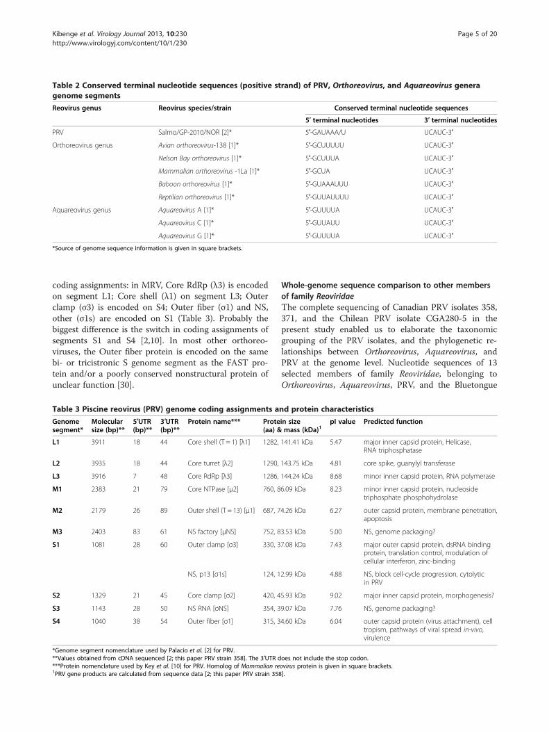
Table 2 Conserved terminal nucleotide sequences (positive strand) of PRV, Orthoreovirus, and Aquareovirus generagenome segments
Reovirus genus Reovirus species/strain Conserved terminal nucleotide sequences
5′ terminal nucleotides 3′ terminal nucleotides
PRV Salmo/GP-2010/NOR [2]* 5′-GAUAAA/U UCAUC-3′
Orthoreovirus genus Avian orthoreovirus-138 [1]* 5′-GCUUUUU UCAUC-3′
Nelson Bay orthoreovirus [1]* 5′-GCUUUA UCAUC-3′
Mammalian orthoreovirus -1La [1]* 5′-GCUA UCAUC-3′
Baboon orthoreovirus [1]* 5′-GUAAAUUU UCAUC-3′
Reptilian orthoreovirus [1]* 5′-GUUAUUUU UCAUC-3′
Aquareovirus genus Aquareovirus A [1]* 5′-GUUUUA UCAUC-3′
Aquareovirus C [1]* 5′-GUUAUU UCAUC-3′
Aquareovirus G [1]* 5′-GUUUUA UCAUC-3′
*Source of genome sequence information is given in square brackets.
Kibenge et al. Virology Journal 2013, 10:230 Page 5 of 20http://www.virologyj.com/content/10/1/230
coding assignments: in MRV, Core RdRp (λ3) is encodedon segment L1; Core shell (λ1) on segment L3; Outerclamp (σ3) is encoded on S4; Outer fiber (σ1) and NS,other (σ1s) are encoded on S1 (Table 3). Probably thebiggest difference is the switch in coding assignments ofsegments S1 and S4 [2,10]. In most other orthoreo-viruses, the Outer fiber protein is encoded on the samebi- or tricistronic S genome segment as the FAST pro-tein and/or a poorly conserved nonstructural protein ofunclear function [30].
Table 3 Piscine reovirus (PRV) genome coding assignments a
Genomesegment*
Molecularsize (bp)**
5′UTR(bp)**
3′UTR(bp)**
Protein name*** Prote(aa) &
L1 3911 18 44 Core shell (T = 1) [λ1] 1282,
L2 3935 18 44 Core turret [λ2] 1290,
L3 3916 7 48 Core RdRp [λ3] 1286,
M1 2383 21 79 Core NTPase [μ2] 760, 8
M2 2179 26 89 Outer shell (T = 13) [μ1] 687, 7
M3 2403 83 61 NS factory [μNS] 752, 8
S1 1081 28 60 Outer clamp [σ3] 330, 3
NS, p13 [σ1s] 124, 1
S2 1329 21 45 Core clamp [σ2] 420, 4
S3 1143 28 50 NS RNA [σNS] 354, 3
S4 1040 38 54 Outer fiber [σ1] 315, 3
*Genome segment nomenclature used by Palacio et al. [2] for PRV.**Values obtained from cDNA sequenced [2; this paper PRV strain 358]. The 3′UTR d***Protein nomenclature used by Key et al. [10] for PRV. Homolog of Mammalian re1PRV gene products are calculated from sequence data [2; this paper PRV strain 358
Whole-genome sequence comparison to other membersof family ReoviridaeThe complete sequencing of Canadian PRV isolates 358,371, and the Chilean PRV isolate CGA280-5 in thepresent study enabled us to elaborate the taxonomicgrouping of the PRV isolates, and the phylogenetic re-lationships between Orthoreovirus, Aquareovirus, andPRV at the genome level. Nucleotide sequences of 13selected members of family Reoviridae, belonging toOrthoreovirus, Aquareovirus, PRV, and the Bluetongue
nd protein characteristics
in sizemass (kDa)1
pI value Predicted function
141.41 kDa 5.47 major inner capsid protein, Helicase,RNA triphosphatase
143.75 kDa 4.81 core spike, guanylyl transferase
144.24 kDa 8.68 minor inner capsid protein, RNA polymerase
6.09 kDa 8.23 minor inner capsid protein, nucleosidetriphosphate phosphohydrolase
4.26 kDa 6.27 outer capsid protein, membrane penetration,apoptosis
3.53 kDa 5.00 NS, genome packaging?
7.08 kDa 7.43 major outer capsid protein, dsRNA bindingprotein, translation control, modulation ofcellular interferon, zinc-binding
2.99 kDa 4.88 NS, block cell-cycle progression, cytolyticin PRV
5.93 kDa 9.02 major inner capsid protein, morphogenesis?
9.07 kDa 7.76 NS, genome packaging?
4.60 kDa 6.04 outer capsid protein (virus attachment), celltropism, pathways of viral spread in-vivo,virulence
oes not include the stop codon.ovirus protein is given in square brackets.].
Kibenge et al. Virology Journal 2013, 10:230 Page 6 of 20http://www.virologyj.com/content/10/1/230
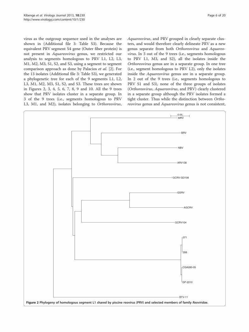
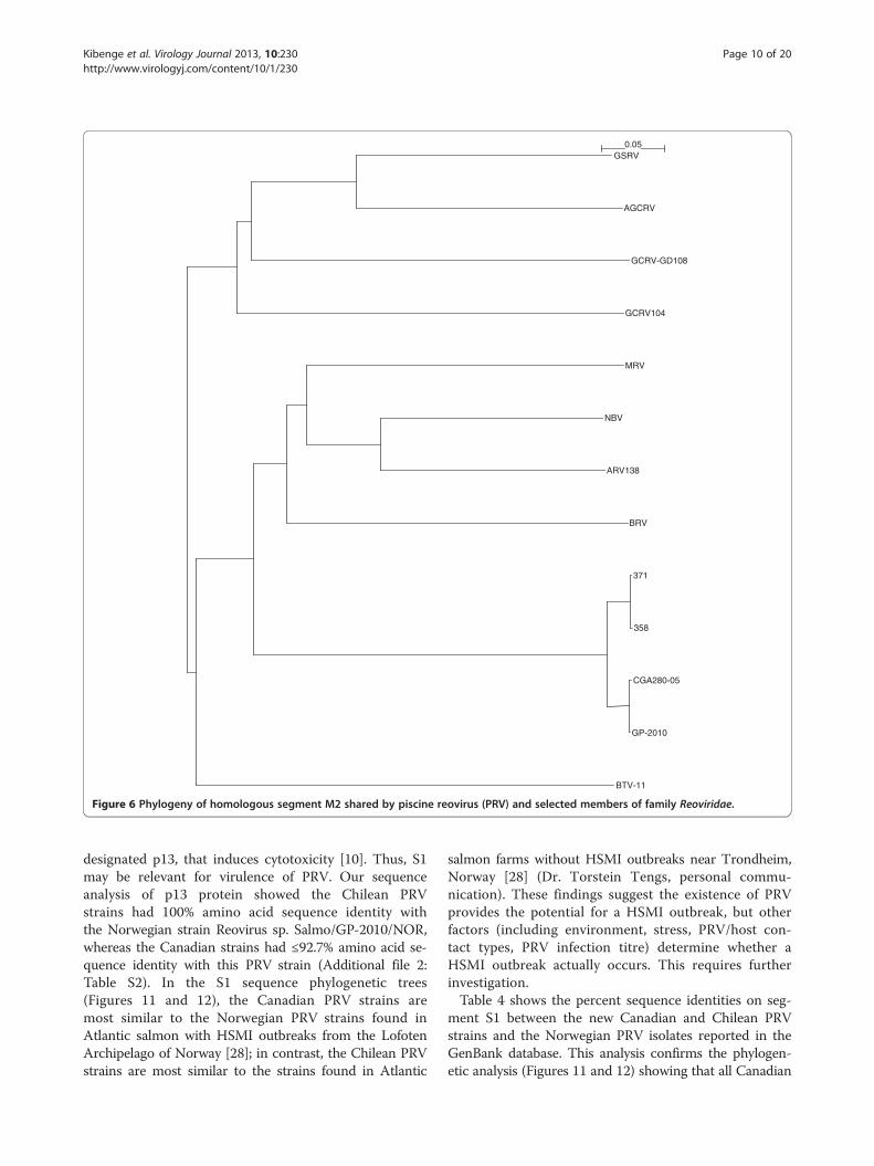
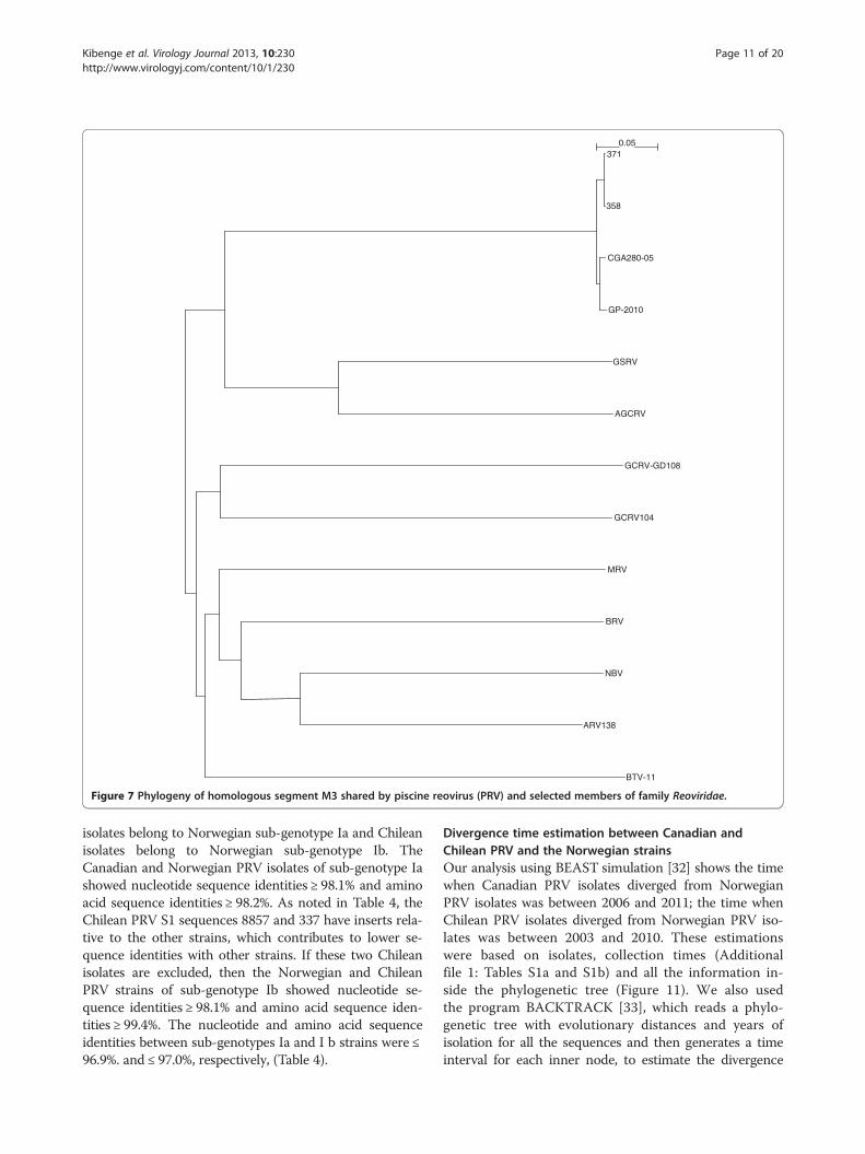
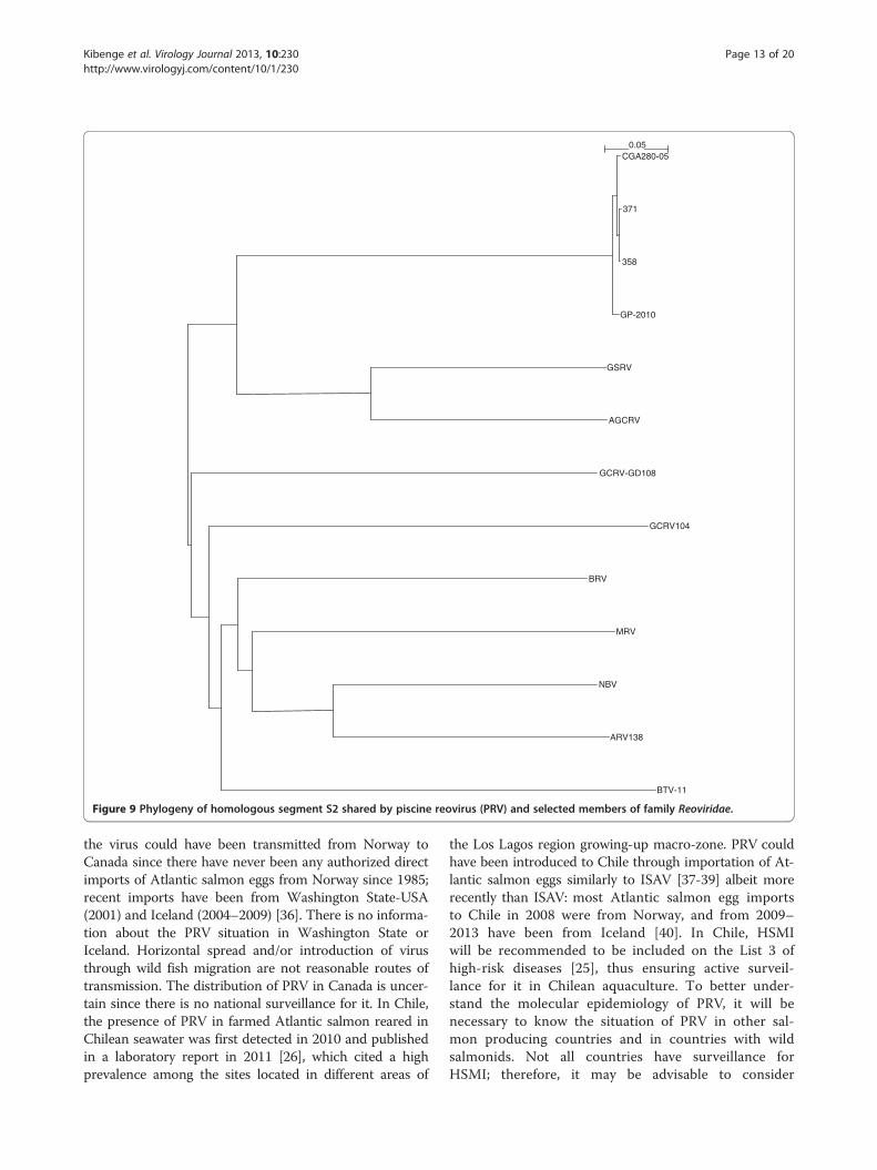
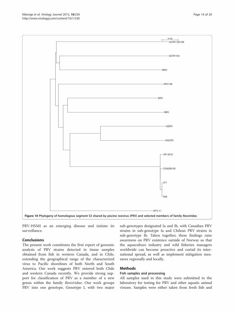
virus as the outgroup sequence used in the analyses areshown in (Additional file 3: Table S3). Because theequivalent PRV segment S4 gene (Outer fiber protein) isnot present in Aquareovirus genus, we restricted ouranalysis to segments homologous to PRV L1, L2, L3,M1, M2, M3, S1, S2, and S3, using a segment to segmentcomparison approach as done by Palacios et al. [2]. Forthe 13 isolates (Additional file 3: Table S3), we generateda phylogenetic tree for each of the 9 segments L1, L2,L3, M1, M2, M3, S1, S2, and S3. These trees are shownin Figures 2, 3, 4, 5, 6, 7, 8, 9 and 10. All the 9 treesshow that PRV isolates cluster in a separate group. In3 of the 9 trees (i.e., segments homologous to PRVL3, M1, and M2), isolates belonging to Orthoreovirus,
Figure 2 Phylogeny of homologous segment L1 shared by piscine reo
Aquareovirus, and PRV grouped in clearly separate clus-ters, and would therefore clearly delineate PRV as a newgenus separate from both Orthoreovirus and Aquareo-virus. In 3 out of the 9 trees (i.e., segments homologousto PRV L1, M3, and S2), all the isolates inside theOrthoreovirus genus are in a separate group. In one tree(i.e., segment homologous to PRV L2), only the isolatesinside the Aquareovirus genus are in a separate group.In 2 out of the 9 trees (i.e., segments homologous toPRV S1 and S3), none of the three groups of isolates(Orthoreovirus, Aquareovirus, and PRV) clearly clusteredin a separate group although the PRV isolates formed atight cluster. Thus while the distinction between Ortho-reovirus genus and Aquareovirus genus is not consistent,
BTV-11
GP-2010
CGA280-05
358
371
GCRV104
AGCRV
GSRV
GCRV-GD108
ARV138
NBV
BRV
MRV0.05
virus (PRV) and selected members of family Reoviridae.
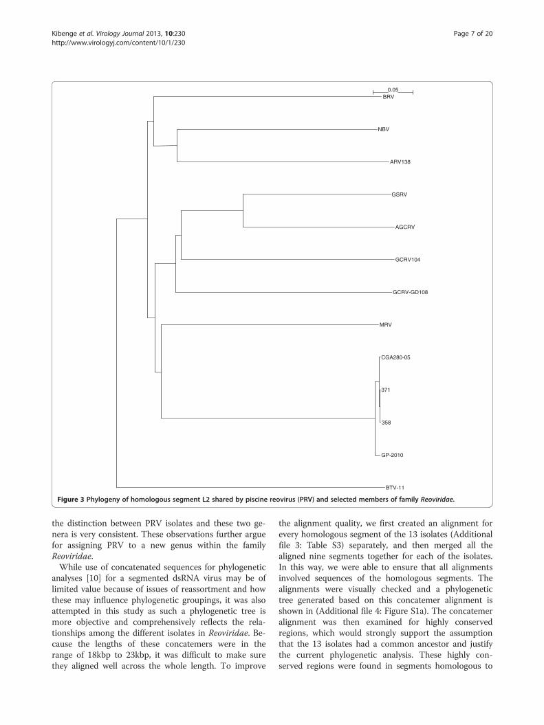
BTV-11
GP-2010
358
371
CGA280-05
MRV
GCRV-GD108
GCRV104
AGCRV
GSRV
ARV138
NBV
BRV0.05
Figure 3 Phylogeny of homologous segment L2 shared by piscine reovirus (PRV) and selected members of family Reoviridae.
Kibenge et al. Virology Journal 2013, 10:230 Page 7 of 20http://www.virologyj.com/content/10/1/230
the distinction between PRV isolates and these two ge-nera is very consistent. These observations further arguefor assigning PRV to a new genus within the familyReoviridae.While use of concatenated sequences for phylogenetic
analyses [10] for a segmented dsRNA virus may be oflimited value because of issues of reassortment and howthese may influence phylogenetic groupings, it was alsoattempted in this study as such a phylogenetic tree ismore objective and comprehensively reflects the rela-tionships among the different isolates in Reoviridae. Be-cause the lengths of these concatemers were in therange of 18kbp to 23kbp, it was difficult to make surethey aligned well across the whole length. To improve
the alignment quality, we first created an alignment forevery homologous segment of the 13 isolates (Additionalfile 3: Table S3) separately, and then merged all thealigned nine segments together for each of the isolates.In this way, we were able to ensure that all alignmentsinvolved sequences of the homologous segments. Thealignments were visually checked and a phylogenetictree generated based on this concatemer alignment isshown in (Additional file 4: Figure S1a). The concatemeralignment was then examined for highly conservedregions, which would strongly support the assumptionthat the 13 isolates had a common ancestor and justifythe current phylogenetic analysis. These highly con-served regions were found in segments homologous to
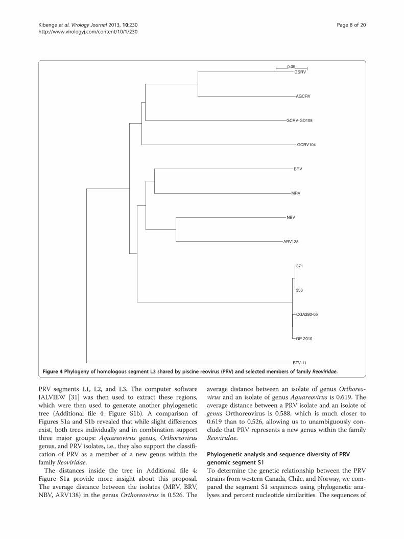
BTV-11
GP-2010
CGA280-05
358
371
ARV138
NBV
MRV
BRV
GCRV104
GCRV-GD108
AGCRV
GSRV0.05
Figure 4 Phylogeny of homologous segment L3 shared by piscine reovirus (PRV) and selected members of family Reoviridae.
Kibenge et al. Virology Journal 2013, 10:230 Page 8 of 20http://www.virologyj.com/content/10/1/230
PRV segments L1, L2, and L3. The computer softwareJALVIEW [31] was then used to extract these regions,which were then used to generate another phylogenetictree (Additional file 4: Figure S1b). A comparison ofFigures S1a and S1b revealed that while slight differencesexist, both trees individually and in combination supportthree major groups: Aquareovirus genus, Orthoreovirusgenus, and PRV isolates, i.e., they also support the classifi-cation of PRV as a member of a new genus within thefamily Reoviridae.The distances inside the tree in Additional file 4:
Figure S1a provide more insight about this proposal.The average distance between the isolates (MRV, BRV,NBV, ARV138) in the genus Orthoreovirus is 0.526. The
average distance between an isolate of genus Orthoreo-virus and an isolate of genus Aquareovirus is 0.619. Theaverage distance between a PRV isolate and an isolate ofgenus Orthoreovirus is 0.588, which is much closer to0.619 than to 0.526, allowing us to unambiguously con-clude that PRV represents a new genus within the familyReoviridae.
Phylogenetic analysis and sequence diversity of PRVgenomic segment S1To determine the genetic relationship between the PRVstrains from western Canada, Chile, and Norway, we com-pared the segment S1 sequences using phylogenetic ana-lyses and percent nucleotide similarities. The sequences of
BTV-11
GCRV104
GCRV-GD108
AGCRV
GSRV
GP-2010
CGA280-05
358
371
ARV138
NBV
MRV
BRV0.05
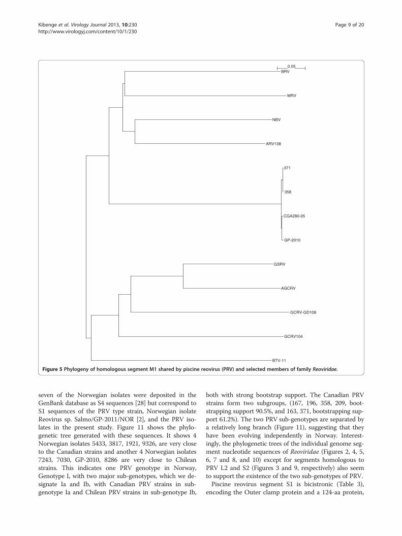
Figure 5 Phylogeny of homologous segment M1 shared by piscine reovirus (PRV) and selected members of family Reoviridae.
Kibenge et al. Virology Journal 2013, 10:230 Page 9 of 20http://www.virologyj.com/content/10/1/230
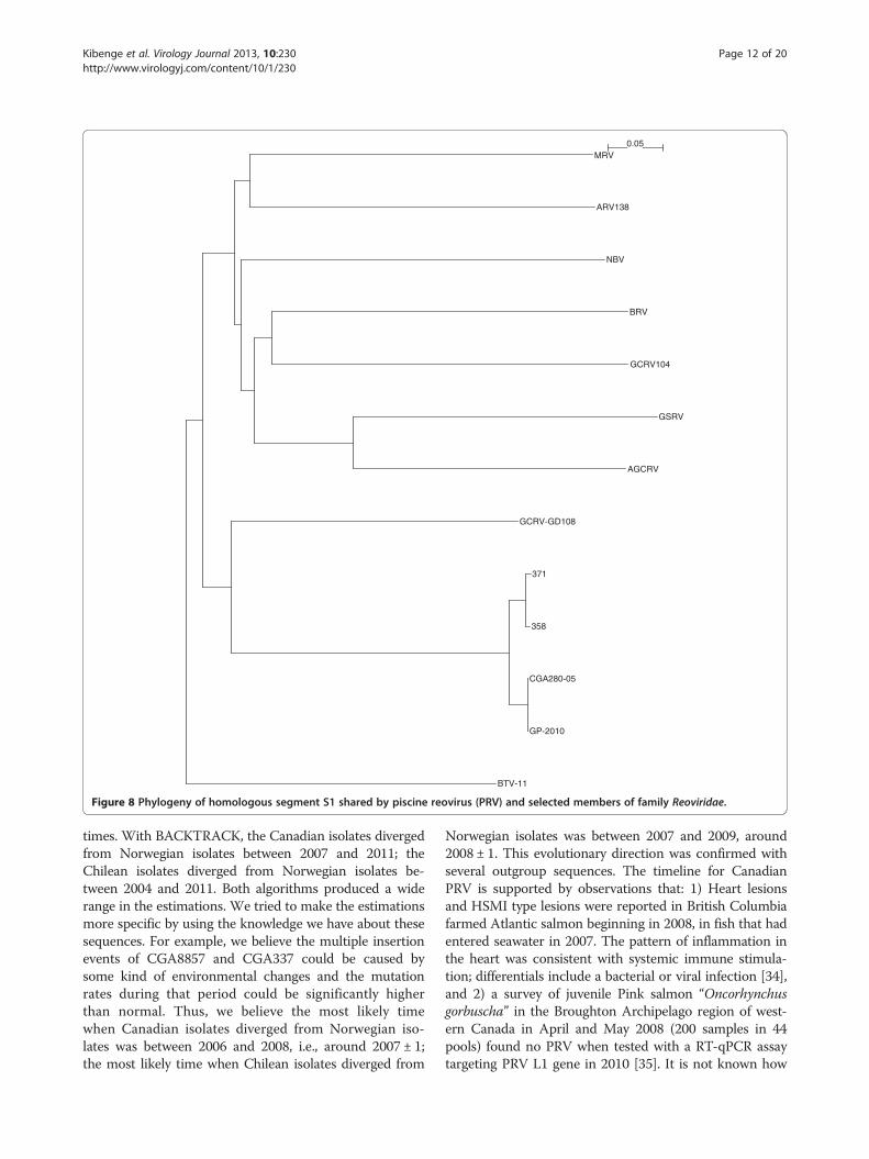
seven of the Norwegian isolates were deposited in theGenBank database as S4 sequences [28] but correspond toS1 sequences of the PRV type strain, Norwegian isolateReovirus sp. Salmo/GP-2011/NOR [2], and the PRV iso-lates in the present study. Figure 11 shows the phylo-genetic tree generated with these sequences. It shows 4Norwegian isolates 5433, 3817, 1921, 9326, are very closeto the Canadian strains and another 4 Norwegian isolates7243, 7030, GP-2010, 8286 are very close to Chileanstrains. This indicates one PRV genotype in Norway,Genotype I, with two major sub-genotypes, which we de-signate Ia and Ib, with Canadian PRV strains in sub-genotype Ia and Chilean PRV strains in sub-genotype Ib,
both with strong bootstrap support. The Canadian PRVstrains form two subgroups, (167, 196, 358, 209, boot-strapping support 90.5%, and 163, 371, bootstrapping sup-port 61.2%). The two PRV sub-genotypes are separated bya relatively long branch (Figure 11), suggesting that theyhave been evolving independently in Norway. Interest-ingly, the phylogenetic trees of the individual genome seg-ment nucleotide sequences of Reoviridae (Figures 2, 4, 5,6, 7 and 8, and 10) except for segments homologous toPRV L2 and S2 (Figures 3 and 9, respectively) also seemto support the existence of the two sub-genotypes of PRV.Piscine reovirus segment S1 is bicistronic (Table 3),
encoding the Outer clamp protein and a 124-aa protein,
BTV-11
GP-2010
CGA280-05
358
371
BRV
ARV138
NBV
MRV
GCRV104
GCRV-GD108
AGCRV
GSRV0.05
Figure 6 Phylogeny of homologous segment M2 shared by piscine reovirus (PRV) and selected members of family Reoviridae.
Kibenge et al. Virology Journal 2013, 10:230 Page 10 of 20http://www.virologyj.com/content/10/1/230
designated p13, that induces cytotoxicity [10]. Thus, S1may be relevant for virulence of PRV. Our sequenceanalysis of p13 protein showed the Chilean PRVstrains had 100% amino acid sequence identity withthe Norwegian strain Reovirus sp. Salmo/GP-2010/NOR,whereas the Canadian strains had ≤92.7% amino acid se-quence identity with this PRV strain (Additional file 2:Table S2). In the S1 sequence phylogenetic trees(Figures 11 and 12), the Canadian PRV strains aremost similar to the Norwegian PRV strains found inAtlantic salmon with HSMI outbreaks from the LofotenArchipelago of Norway [28]; in contrast, the Chilean PRVstrains are most similar to the strains found in Atlantic
salmon farms without HSMI outbreaks near Trondheim,Norway [28] (Dr. Torstein Tengs, personal commu-nication). These findings suggest the existence of PRVprovides the potential for a HSMI outbreak, but otherfactors (including environment, stress, PRV/host con-tact types, PRV infection titre) determine whether aHSMI outbreak actually occurs. This requires furtherinvestigation.Table 4 shows the percent sequence identities on seg-
ment S1 between the new Canadian and Chilean PRVstrains and the Norwegian PRV isolates reported in theGenBank database. This analysis confirms the phylogen-etic analysis (Figures 11 and 12) showing that all Canadian
BTV-11
ARV138
NBV
BRV
MRV
GCRV104
GCRV-GD108
AGCRV
GSRV
GP-2010
CGA280-05
358
3710.05
Figure 7 Phylogeny of homologous segment M3 shared by piscine reovirus (PRV) and selected members of family Reoviridae.
Kibenge et al. Virology Journal 2013, 10:230 Page 11 of 20http://www.virologyj.com/content/10/1/230
isolates belong to Norwegian sub-genotype Ia and Chileanisolates belong to Norwegian sub-genotype Ib. TheCanadian and Norwegian PRV isolates of sub-genotype Iashowed nucleotide sequence identities ≥ 98.1% and aminoacid sequence identities ≥ 98.2%. As noted in Table 4, theChilean PRV S1 sequences 8857 and 337 have inserts rela-tive to the other strains, which contributes to lower se-quence identities with other strains. If these two Chileanisolates are excluded, then the Norwegian and ChileanPRV strains of sub-genotype Ib showed nucleotide se-quence identities ≥ 98.1% and amino acid sequence iden-tities ≥ 99.4%. The nucleotide and amino acid sequenceidentities between sub-genotypes Ia and I b strains were ≤96.9%. and ≤ 97.0%, respectively, (Table 4).
Divergence time estimation between Canadian andChilean PRV and the Norwegian strainsOur analysis using BEAST simulation [32] shows the timewhen Canadian PRV isolates diverged from NorwegianPRV isolates was between 2006 and 2011; the time whenChilean PRV isolates diverged from Norwegian PRV iso-lates was between 2003 and 2010. These estimationswere based on isolates, collection times (Additionalfile 1: Tables S1a and S1b) and all the information in-side the phylogenetic tree (Figure 11). We also usedthe program BACKTRACK [33], which reads a phylo-genetic tree with evolutionary distances and years ofisolation for all the sequences and then generates a timeinterval for each inner node, to estimate the divergence
BTV-11
GP-2010
CGA280-05
358
371
GCRV-GD108
AGCRV
GSRV
GCRV104
BRV
NBV
ARV138
MRV0.05
Figure 8 Phylogeny of homologous segment S1 shared by piscine reovirus (PRV) and selected members of family Reoviridae.
Kibenge et al. Virology Journal 2013, 10:230 Page 12 of 20http://www.virologyj.com/content/10/1/230
times. With BACKTRACK, the Canadian isolates divergedfrom Norwegian isolates between 2007 and 2011; theChilean isolates diverged from Norwegian isolates be-tween 2004 and 2011. Both algorithms produced a widerange in the estimations. We tried to make the estimationsmore specific by using the knowledge we have about thesesequences. For example, we believe the multiple insertionevents of CGA8857 and CGA337 could be caused bysome kind of environmental changes and the mutationrates during that period could be significantly higherthan normal. Thus, we believe the most likely timewhen Canadian isolates diverged from Norwegian iso-lates was between 2006 and 2008, i.e., around 2007 ± 1;the most likely time when Chilean isolates diverged from
Norwegian isolates was between 2007 and 2009, around2008 ± 1. This evolutionary direction was confirmed withseveral outgroup sequences. The timeline for CanadianPRV is supported by observations that: 1) Heart lesionsand HSMI type lesions were reported in British Columbiafarmed Atlantic salmon beginning in 2008, in fish that hadentered seawater in 2007. The pattern of inflammation inthe heart was consistent with systemic immune stimula-tion; differentials include a bacterial or viral infection [34],and 2) a survey of juvenile Pink salmon “Oncorhynchusgorbuscha” in the Broughton Archipelago region of west-ern Canada in April and May 2008 (200 samples in 44pools) found no PRV when tested with a RT-qPCR assaytargeting PRV L1 gene in 2010 [35]. It is not known how
BTV-11
ARV138
NBV
MRV
BRV
GCRV104
GCRV-GD108
AGCRV
GSRV
GP-2010
358
371
CGA280-050.05
Figure 9 Phylogeny of homologous segment S2 shared by piscine reovirus (PRV) and selected members of family Reoviridae.
Kibenge et al. Virology Journal 2013, 10:230 Page 13 of 20http://www.virologyj.com/content/10/1/230
the virus could have been transmitted from Norway toCanada since there have never been any authorized directimports of Atlantic salmon eggs from Norway since 1985;recent imports have been from Washington State-USA(2001) and Iceland (2004–2009) [36]. There is no informa-tion about the PRV situation in Washington State orIceland. Horizontal spread and/or introduction of virusthrough wild fish migration are not reasonable routes oftransmission. The distribution of PRV in Canada is uncer-tain since there is no national surveillance for it. In Chile,the presence of PRV in farmed Atlantic salmon reared inChilean seawater was first detected in 2010 and publishedin a laboratory report in 2011 [26], which cited a highprevalence among the sites located in different areas of
the Los Lagos region growing-up macro-zone. PRV couldhave been introduced to Chile through importation of At-lantic salmon eggs similarly to ISAV [37-39] albeit morerecently than ISAV: most Atlantic salmon egg importsto Chile in 2008 were from Norway, and from 2009–2013 have been from Iceland [40]. In Chile, HSMIwill be recommended to be included on the List 3 ofhigh-risk diseases [25], thus ensuring active surveil-lance for it in Chilean aquaculture. To better under-stand the molecular epidemiology of PRV, it will benecessary to know the situation of PRV in other sal-mon producing countries and in countries with wildsalmonids. Not all countries have surveillance forHSMI; therefore, it may be advisable to consider
BTV-11
358
371
CGA280-05
GP-2010
AGCRV
GSRV
NBV
BRV
ARV138
MRV
GCRV104
GCRV-GD1080.05
Figure 10 Phylogeny of homologous segment S3 shared by piscine reovirus (PRV) and selected members of family Reoviridae.
Kibenge et al. Virology Journal 2013, 10:230 Page 14 of 20http://www.virologyj.com/content/10/1/230
PRV-HSMI as an emerging disease and initiate itssurveillance.
ConclusionsThe present work constitutes the first report of genomicanalysis of PRV strains detected in tissue samplesobtained from fish in western Canada, and in Chile,extending the geographical range of the characterizedvirus to Pacific shorelines of both North and SouthAmerica. Our work suggests PRV entered both Chileand western Canada recently. We provide strong sup-port for classification of PRV as a member of a newgenus within the family Reoviridae. Our work groupsPRV into one genotype, Genotype I, with two major
sub-genotypes designated Ia and Ib, with Canadian PRVstrains in sub-genotype Ia and Chilean PRV strains insub-genotype Ib. Taken together, these findings raiseawareness on PRV existence outside of Norway so thatthe aquaculture industry and wild fisheries managersworldwide can become proactive and curtail its inter-national spread, as well as implement mitigation mea-sures regionally and locally.
MethodsFish samples and processingAll samples used in this study were submitted to thelaboratory for testing for PRV and other aquatic animalviruses. Samples were either taken from fresh fish and
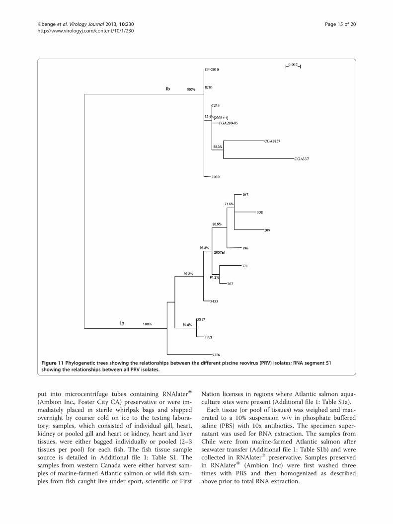
Figure 11 Phylogenetic trees showing the relationships between the different piscine reovirus (PRV) isolates; RNA segment S1showing the relationships between all PRV isolates.
Kibenge et al. Virology Journal 2013, 10:230 Page 15 of 20http://www.virologyj.com/content/10/1/230
put into microcentrifuge tubes containing RNAlaterW
(Ambion Inc., Foster City CA) preservative or were im-mediately placed in sterile whirlpak bags and shippedovernight by courier cold on ice to the testing labora-tory; samples, which consisted of individual gill, heart,kidney or pooled gill and heart or kidney, heart and livertissues, were either bagged individually or pooled (2–3tissues per pool) for each fish. The fish tissue samplesource is detailed in Additional file 1: Table S1. Thesamples from western Canada were either harvest sam-ples of marine-farmed Atlantic salmon or wild fish sam-ples from fish caught live under sport, scientific or First
Nation licenses in regions where Atlantic salmon aqua-culture sites were present (Additional file 1: Table S1a).Each tissue (or pool of tissues) was weighed and mac-
erated to a 10% suspension w/v in phosphate bufferedsaline (PBS) with 10x antibiotics. The specimen super-natant was used for RNA extraction. The samples fromChile were from marine-farmed Atlantic salmon afterseawater transfer (Additional file 1: Table S1b) and werecollected in RNAlaterW preservative. Samples preservedin RNAlaterW (Ambion Inc) were first washed threetimes with PBS and then homogenized as describedabove prior to total RNA extraction.
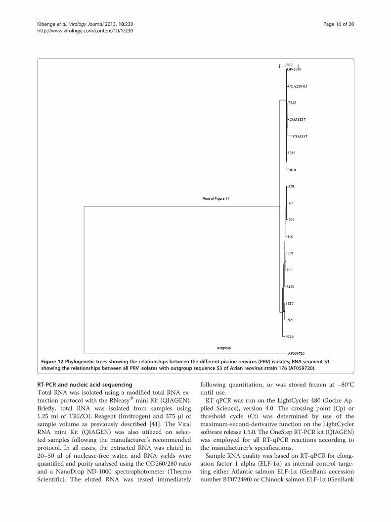
Figure 12 Phylogenetic trees showing the relationships between the different piscine reovirus (PRV) isolates; RNA segment S1showing the relationships between all PRV isolates with outgroup sequence S3 of Avian reovirus strain 176 (AF059720).
Kibenge et al. Virology Journal 2013, 10:230 Page 16 of 20http://www.virologyj.com/content/10/1/230
RT-PCR and nucleic acid sequencingTotal RNA was isolated using a modified total RNA ex-traction protocol with the RNeasyW mini Kit (QIAGEN).Briefly, total RNA was isolated from samples using1.25 ml of TRIZOL Reagent (Invitrogen) and 375 μl ofsample volume as previously described [41]. The ViralRNA mini Kit (QIAGEN) was also utilized on selec-ted samples following the manufacturer’s recommendedprotocol. In all cases, the extracted RNA was eluted in20–50 μl of nuclease-free water, and RNA yields werequantified and purity analysed using the OD260/280 ratioand a NanoDrop ND-1000 spectrophotometer (ThermoScientific). The eluted RNA was tested immediately
following quantitation, or was stored frozen at −80°Cuntil use.RT-qPCR was run on the LightCycler 480 (Roche Ap-
plied Science), version 4.0. The crossing point (Cp) orthreshold cycle (Ct) was determined by use of themaximum-second-derivative function on the LightCyclersoftware release 1.5.0. The OneStep RT-PCR kit (QIAGEN)was employed for all RT-qPCR reactions according tothe manufacturer’s specifications.Sample RNA quality was based on RT-qPCR for elong-
ation factor 1 alpha (ELF-1α) as internal control targe-ting either Atlantic salmon ELF-1α (GenBank accessionnumber BT072490) or Chinook salmon ELF-1α (GenBank
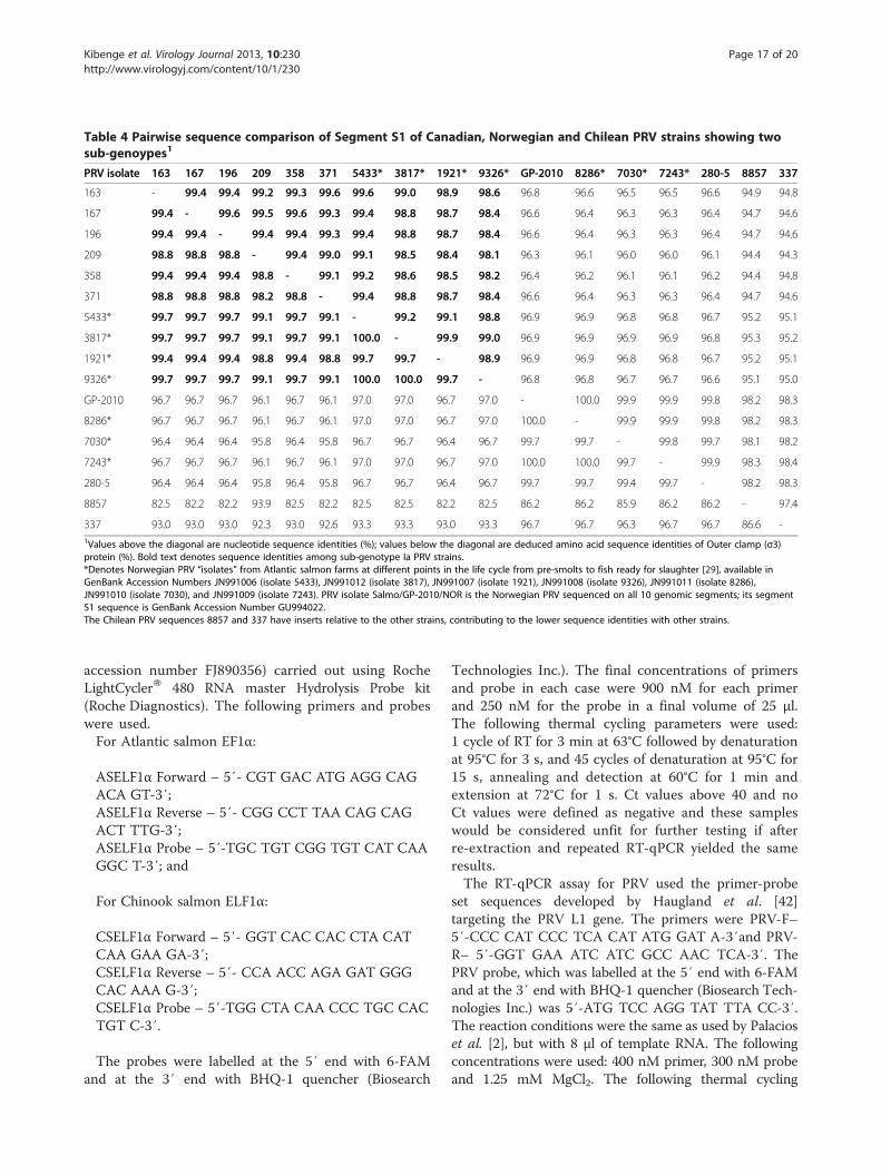
Table 4 Pairwise sequence comparison of Segment S1 of Canadian, Norwegian and Chilean PRV strains showing twosub-genoypes1
PRV isolate 163 167 196 209 358 371 5433* 3817* 1921* 9326* GP-2010 8286* 7030* 7243* 280-5 8857 337
163 - 99.4 99.4 99.2 99.3 99.6 99.6 99.0 98.9 98.6 96.8 96.6 96.5 96.5 96.6 94.9 94.8
167 99.4 - 99.6 99.5 99.6 99.3 99.4 98.8 98.7 98.4 96.6 96.4 96.3 96.3 96.4 94.7 94.6
196 99.4 99.4 - 99.4 99.4 99.3 99.4 98.8 98.7 98.4 96.6 96.4 96.3 96.3 96.4 94.7 94.6
209 98.8 98.8 98.8 - 99.4 99.0 99.1 98.5 98.4 98.1 96.3 96.1 96.0 96.0 96.1 94.4 94.3
358 99.4 99.4 99.4 98.8 - 99.1 99.2 98.6 98.5 98.2 96.4 96.2 96.1 96.1 96.2 94.4 94.8
371 98.8 98.8 98.8 98.2 98.8 - 99.4 98.8 98.7 98.4 96.6 96.4 96.3 96.3 96.4 94.7 94.6
5433* 99.7 99.7 99.7 99.1 99.7 99.1 - 99.2 99.1 98.8 96.9 96.9 96.8 96.8 96.7 95.2 95.1
3817* 99.7 99.7 99.7 99.1 99.7 99.1 100.0 - 99.9 99.0 96.9 96.9 96.9 96.9 96.8 95.3 95.2
1921* 99.4 99.4 99.4 98.8 99.4 98.8 99.7 99.7 - 98.9 96.9 96.9 96.8 96.8 96.7 95.2 95.1
9326* 99.7 99.7 99.7 99.1 99.7 99.1 100.0 100.0 99.7 - 96.8 96.8 96.7 96.7 96.6 95.1 95.0
GP-2010 96.7 96.7 96.7 96.1 96.7 96.1 97.0 97.0 96.7 97.0 - 100.0 99.9 99.9 99.8 98.2 98.3
8286* 96.7 96.7 96.7 96.1 96.7 96.1 97.0 97.0 96.7 97.0 100.0 - 99.9 99.9 99.8 98.2 98.3
7030* 96.4 96.4 96.4 95.8 96.4 95.8 96.7 96.7 96.4 96.7 99.7 99.7 - 99.8 99.7 98.1 98.2
7243* 96.7 96.7 96.7 96.1 96.7 96.1 97.0 97.0 96.7 97.0 100.0 100.0 99.7 - 99.9 98.3 98.4
280-5 96.4 96.4 96.4 95.8 96.4 95.8 96.7 96.7 96.4 96.7 99.7 99.7 99.4 99.7 - 98.2 98.3
8857 82.5 82.2 82.2 93.9 82.5 82.2 82.5 82.5 82.2 82.5 86.2 86.2 85.9 86.2 86.2 - 97.4
337 93.0 93.0 93.0 92.3 93.0 92.6 93.3 93.3 93.0 93.3 96.7 96.7 96.3 96.7 96.7 86.6 -1Values above the diagonal are nucleotide sequence identities (%); values below the diagonal are deduced amino acid sequence identities of Outer clamp (σ3)protein (%). Bold text denotes sequence identities among sub-genotype Ia PRV strains.*Denotes Norwegian PRV “isolates” from Atlantic salmon farms at different points in the life cycle from pre-smolts to fish ready for slaughter [29], available inGenBank Accession Numbers JN991006 (isolate 5433), JN991012 (isolate 3817), JN991007 (isolate 1921), JN991008 (isolate 9326), JN991011 (isolate 8286),JN991010 (isolate 7030), and JN991009 (isolate 7243). PRV isolate Salmo/GP-2010/NOR is the Norwegian PRV sequenced on all 10 genomic segments; its segmentS1 sequence is GenBank Accession Number GU994022.The Chilean PRV sequences 8857 and 337 have inserts relative to the other strains, contributing to the lower sequence identities with other strains.
Kibenge et al. Virology Journal 2013, 10:230 Page 17 of 20http://www.virologyj.com/content/10/1/230
accession number FJ890356) carried out using RocheLightCyclerW 480 RNA master Hydrolysis Probe kit(Roche Diagnostics). The following primers and probeswere used.For Atlantic salmon EF1α:
ASELF1α Forward – 5′- CGT GAC ATG AGG CAGACA GT-3′;ASELF1α Reverse – 5′- CGG CCT TAA CAG CAGACT TTG-3′;ASELF1α Probe – 5′-TGC TGT CGG TGT CAT CAAGGC T-3′; and
For Chinook salmon ELF1α:
CSELF1α Forward – 5′- GGT CAC CAC CTA CATCAA GAA GA-3′;CSELF1α Reverse – 5′- CCA ACC AGA GAT GGGCAC AAA G-3′;CSELF1α Probe – 5′-TGG CTA CAA CCC TGC CACTGT C-3′.
The probes were labelled at the 5′ end with 6-FAMand at the 3′ end with BHQ-1 quencher (Biosearch
Technologies Inc.). The final concentrations of primersand probe in each case were 900 nΜ for each primerand 250 nM for the probe in a final volume of 25 μl.The following thermal cycling parameters were used:1 cycle of RT for 3 min at 63°C followed by denaturationat 95°C for 3 s, and 45 cycles of denaturation at 95°C for15 s, annealing and detection at 60°C for 1 min andextension at 72°C for 1 s. Ct values above 40 and noCt values were defined as negative and these sampleswould be considered unfit for further testing if afterre-extraction and repeated RT-qPCR yielded the sameresults.The RT-qPCR assay for PRV used the primer-probe
set sequences developed by Haugland et al. [42]targeting the PRV L1 gene. The primers were PRV-F–5′-CCC CAT CCC TCA CAT ATG GAT A-3′and PRV-R– 5′-GGT GAA ATC ATC GCC AAC TCA-3′. ThePRV probe, which was labelled at the 5′ end with 6-FAMand at the 3′ end with BHQ-1 quencher (Biosearch Tech-nologies Inc.) was 5′-ATG TCC AGG TAT TTA CC-3′.The reaction conditions were the same as used by Palacioset al. [2], but with 8 μl of template RNA. The followingconcentrations were used: 400 nM primer, 300 nM probeand 1.25 mM MgCl2. The following thermal cycling
Kibenge et al. Virology Journal 2013, 10:230 Page 18 of 20http://www.virologyj.com/content/10/1/230
parameters were used: 1 cycle of RT for 30 min at 50°Cfollowed by denaturation at 94°C for 15 min, and 45 cyclesof denaturation at 94°C for 15 s, annealing at 54°C for 30 sand amplification and detection at 72°C for 15 s. Samplesto be considered positive had Ct values up to 40 and withan exponential curve; Ct values between 40.1 and 45 wereconsidered suspicious, and a sample was negative if therewas no Ct value.Because the laboratory did not have a PRV isolate
from cell culture to use as positive control, samples withpositive Ct values were further tested in classic RT-PCRtargeting the 3′ portion of genome segment L1 with thefollowing PCR primer pairs: PRV-L1 For1 – 5′-CACTCA CCA ATG ACC CAA ATG C-3′; PRV-L1 Rev1 –5′-TTG ACA GTC TGG CTA CTT CGG-3′ and/orPRV-L1 For2 – 5′-CTG AAC TGC TAG TTG AGGATG G-3′; PRV-L1 Rev2 – 5′-GCC AAT CCA AACAGA TTA GG-3′. These PCR primers and those usedto amplify the 10 genomic segments of PRV, listed in(Additional file 5: Table S4) were designed based on thepublished PRV sequences [2]. RT-PCR for the amplifica-tion of each viral genome segment was carried out byusing the OneStep RT-PCR kit (QIAGEN). Briefly, thereaction mixture contained 1 μl of total RNA, 4 μl of 5XQIAGEN OneStep RT-PCR buffer, 0.8 μl of dNTPs,0.5 μM (final concentration) of each primer pair, and0.8 μl of QIAGEN OneStep RT-PCR enzyme mix in afinal volume of 20 μl. Thermal cycling conditions wereas follows: an initial cycle of 50°C for 40 min and 95°Cfor 10 min; 40 cycles of 95°C for 30 s, 54°C for 30 sec,72°C for 70 s; and a final extension cycle of 72°C for10 min. Amplified products were analyzed by electro-phoresis on 1% agarose gel and purified using High PurePCR Product Purification Kit (Roche). The PCR pro-ducts were then either directly sequenced or they werecloned into the pCRII vector using a TOPO TA cloningkit (Invitrogen) in preparation for nucleotide sequencing.Plasmid DNA for sequencing was prepared as describedbefore [43]. DNA sequencing was performed as previ-ously described [44] by ACGT Corporation (Toronto,Ontario, Canada). DNA Sequencing was done either dir-ectly on RT-PCR products or on plasmid DNA containingthe cloned RT-PCR products obtained from reactionsusing total RNA from tissue samples.
Sequencing analysisSimilarity analysis was performed using BLAST pro-grams available via the National Center for Biotechnol-ogy Information [45] and the FASTA program packagefor personal computers [46]. Analysis to identify putativeORFs and their predicted amino acid sequences andother protein characteristics was conducted using theSequence Manipulation suite, version 2 [47].
Phylogenetic analysesThe Canadian and Chilean PRV sequences used in thephylogenetic analyses are described in (Additional file 2:Table S2). All the Norwegian PRV sequences were ob-tained from GenBank [29]. Sequences were processedusing ClustalX 2.1 [48]. The multiple sequence align-ment was manually verified and adjusted to reach highquality alignment. The phylogenetic trees were generatedwhen positions with gaps were excluded and correctionsfor multiple substitutions were used. Bootstrapping wasperformed for 1,000 times. In most cases, only the boot-strapping supports higher than 70% were noted. For someimportant branches, those bootstrapping values a littlelower than 70% were also noted. To verify the evolutiondirection, outgroup sequences were used to determine theroot of the phylogenetic trees.
Divergence time estimation in a rooted phylogenetic treeBEAST v1.7.5 [32] was used to estimate divergence time.To find the most suitable substitution model, we ranjModelTest 0.1.1 [49] against the aligned sequences. Theresult shows K80 model [50] is the most suitable model.Based on this result, a similar model, HKY85 model[51], was chosen in the BEAST simulation. We believethe mutation rates among lineages could be different,and the uncorrelated relaxed molecular clock waschosen. Five million simulation steps were performedand enough effective sample sizes (ESSs) were generated.We also used program BACKTRACK [33], which readsa phylogenetic tree with evolutionary distances and yearsof isolation for all the sequences and then generates atime interval for each inner node, to estimate the diver-gence times.
Additional files
Additional file 1: Title: Additional results on the piscine reovirus(PRV) positive samples from Canada and Chile. Description: Twotables showing RT-qPCR and conventional RT-PCR results of fish tissuesamples from Canada and Chile tested for PRV.
Additional file 2: List of new piscine reovirus (PRV) nucleotidesequences and their percent identity to Norwegian isolate Salmo/GP-2010/NOR.
Additional file 3: GenBank Accession numbers of genomesegments of selected members of family Reoviridae used inphylogenetic comparison of nucleotide sequences of individualgenome segments [52].
Additional file 4: Title: Phylogenetic trees showing therelationships between isolates in family Reoviridae at the genome-level. Description: (Figure S1a) Concatenated sequences of ninehomologous segments (segment L1, L2, L3, M1, M2, M3, S1, S2, S3)shared by piscine reovirus (PRV) and selected members of familyReoviridae, were used to generate a phylogenetic tree. (Figure S1b)Phylogeny of highly-conserved regions of concatemers in Figure S1a.
Additional file 5: Title: List of oligonucleotide primers used inamplification of piscine reovirus (PRV) genome segments.Description: Table listing oligonucleotide primers.
Kibenge et al. Virology Journal 2013, 10:230 Page 19 of 20http://www.virologyj.com/content/10/1/230
Competing interestsThe authors declare that they have no competing interests in this scientificwork.
Authors’ contributionsMJTK isolated total RNA from tissue samples, performed the RT-qPCR forELF-1α and PRV and developed and performed the classic RT-PCR for 3′portion of segment L1 of PRV, and helped to write the manuscript. TIdesigned the PRV PCR primers used to amplify transcripts of all the 10genome segments, performed the classic RT-PCR and cloned all PCRproducts for sequencing and helped to write the manuscript. YW performedall the phylogenetic analyses and helped to write the manuscript. AMprovided the Canadian samples for diagnostic testing and edited themanuscript. MGG coordinated the laboratory testing in Chile and edited themanuscript. FSBK coordinated all viral testing and DNA sequence analysisand helped to write the manuscript. All authors read and approved the finalmanuscript.
AcknowledgementsThis work was supported by the Virology Research Laboratory at the AtlanticVeterinary College, University of Prince Edward Island, Charlottetown, PE,Canada. Van City supported the work by Alexandra Morton. We thankDr. Richard Routledge for submitting the Cutthroat trout samples fordiagnostic testing.
Author details1Department of Pathology and Microbiology, Atlantic Veterinary College,University of Prince Edward Island, 550 University Ave., Charlottetown, PEIC1A 4P3, Canada. 2Department of Computer Science and InformationTechnology, University of Prince Edward Island, 550 University Ave.,Charlottetown, PEI C1A 4P3, Canada. 3Raincoast Research Society, Box 399,390 1st Street, Sointula, BC V0N 3E0, Canada. 4Centro de InvestigacionesBiológicas Aplicadas (CIBA), Diego de Almagro Norte 1013, No. 10, PuertoMontt, Chile. 5Universidad San Sebastián. Facultad de Ciencias, LagoPanguipulli 1390, Puerto Montt, Chile. 6ETECMA, Diego de Almagro Norte1013 No. 10, Sector Cardonal, Puerto Montt, X Región, Chile.
Received: 9 April 2013 Accepted: 5 July 2013Published: 11 July 2013
References1. Attoui H, Mertens PPC, Becnel J, Belaganahalli S, Bergoin M, Brussaard CP,
Chappell JD, Ciarlet M, del Vas M, et al: Family Reoviridae. In VirusTaxonomy: Ninth Report of the International Committee on Taxonomy ofViruses. Edited by King AMQ, Adams MJ, Carstens EB, Lefkowitz EJ.San Diego, CA: Academic Press; 2011:541–637.
2. Palacios G, Lovoll M, Tengs T, Hornig M, Hutchison S, et al: Heart andskeletal muscle inflammation of farmed salmon is associated withinfection with a novel reovirus. PLoS One 2010, 5:e11487.
3. Duncan R: Extensive sequence divergence and phylogeneticrelationships between the fusogenic and nonfusogenic orthoreoviruses:a species proposal. Virology 1999, 260:316.
4. Shmulevitz M, Duncan R: A new class of fusion-associated small trans-membrane (FAST) proteins encoded by the non-enveloped fusogenicreoviruses. EMBO J 2000, 19:902–912.
5. Sabin A: Reoviruses: a new group of respiratory and enteric virusesformerly classified as ECHO type 10 is described. Science 1959,130:1387–1389.
6. Chua K, Crameri G, Hyatt A, Yu M, Tompang M, et al: A previouslyunknown reovirus of bat origin is associated with an acute respiratorydisease in humans. Proc Nat Acad Sci USA 2007, 104:11424–11429.
7. Leland MM, Hubbard GB, Sentmore HT, Soike KF, Hilliard JK: Outbreak oforthoreovirus-induced meningoencephalomyelitis in baboons. CompMed 2000, 50:199–205.
8. van den Brand JM, Manvell R, Paul G, Kik MJ, Dorrestein GM: Reovirusinfections associated with high mortality in psittaciformes in TheNetherlands. Avian Pathol 2007, 36:293–299.
9. Lamirande EW, Nichols DK, Owens JW, Gaskin JM, Jacobson ER: Isolationand experimental transmission of a reovirus pathogenic in ratsnakes(Elaphe species). Virus Res 1999, 63:135–141.
10. Key T, Read J, Nibert ML, Duncan R: Piscine reovirus encodes acytotoxic, non-fusogenic, integral membrane protein and previouslyunrecognized virion outer-capsid proteins. J Gen Virol 2013,94:1039–1050.
11. Kibenge FSB, Godoy MG, Fast M, Workenhe S, Kibenge MJT:Countermeasures against viral diseases of farmed fish. Antiviral Res 2012,95:257–281.
12. Kongtorp RT, Kjerstad A, Taksdal T, Guttvik A, Falk K: Heart and skeletalmuscle inflammation in Atlantic salmon Salmo salar L: a new infectiousdisease. J Fish Dis 2004, 27:351–358.
13. Ferguson HW, Kongtorp RT, Taksdal T, Graham D, Falk K: An outbreak ofdisease resembling heart and skeletal muscle inflammation (HSMI) inScottish farmed salmon (Salmo salar L) with observations on myocardialregeneration. J Fish Dis 2005, 28:119–123.
14. Wiik-Nielsen CR, Løvoll M, Sandlund N, Faller R, Wiik-Nielsen J, Jensen BB:First detection of piscine reovirus (PRV) in marine fish species. Dis AquatOrg 2012, 97:255–258.
15. Garseth ÅH, Fritsvold C, Opheim M, Skjerve E, Biering E: Piscine reovirus(PRV) in wild Atlantic salmon, Salmo salar L., and sea-trout, Salmo truttaL., in Norway. J Fish Dis 2012. doi:10.1111/j.1365-2761.2012.01450.x.
16. National Veterinary Institute: In Farmed Fish Health Report 2007. Edited bySkjelstad H, Bornø G, Flesjå K, Hansen H, Nilsen H, Wasmuth MA, Hjeltnes B.Oslo: National Veterinary Institute; 2007:19.
17. Bornø G, Sviland C: The health situation in farmed salmonids 2010. InFarmed Fish Health Report 2010. Edited by Bornø G, Sviland C, Hellberg H.Oslo: National Veterinary Institute; 2010:36.
18. Løvoll M, Alarcón M, Jensen BB, Taksdal T, Kristoffersen AB, Tengs T:Quantification of piscine reovirus (PRV) at different stages of Atlanticsalmon Salmo salar production. Dis Aquat Org 2012, 99:7–12.
19. Mikalsen AB, Haugland O, Rode M, Solbakk IT, Evensen O: Atlantic salmonreovirus infection causes a CD8 T Cell myocarditis in Atlantic salmon(Salmo salar L.). PLoS One 2012, 7:e37269.
20. Chi SC, Hu WW, Lo BJ: Establishment and characterization of acontinuous cell line (GF-1) derived from grouper, Epinephelus coioides(Hamilton): a cell line susceptible to grouper nervous necrosis virus(GNNV). J Fish Dis 1999, 22:173–182.
21. Kemp M, Donovan J, Higham H, Hooper J: Biochemical markers ofmyocardial injury. Br J Anaesth 2004, 93:63–73.
22. Yousaf MN, Powell MD: The effects of heart and skeletal muscleinflammation and cardiomyopathy syndrome on creatine kinase andlactate dehydrogenase levels in Atlantic salmon (Salmo salar L.).Sci World J 2012, 741302:9.
23. Løvoll M, Wiik-Nielsen J, Grove S, Wiik-Nielsen CR, Kristoffersen AB,Faller F, et al: A novel totivirus and piscine reovirus (PRV) in Atlanticsalmon (Salmo salar) with cardiomyopathy syndrome (CMS). Virol J2010, 7:309.
24. Wiik-Nielsen J, Løvoll M, Fritsvold C, Kristoffersen AB, Haugland Ø, Hordvik I,Aamelfot M, Jirillo E, Koppang EO, Grove S: Characterization of myocardiallesions associated with cardiomyopathy syndrome in Atlantic salmon,Salmo salar L., using laser capture microdissection. J Fish Dis 2012,35:907–916.
25. Anonymous 2012: Salmon mortality levels rise in H1. http://fis.com/fis/worldnews/worldnews.asp?l=e&country=0&special=aquaculture&monthyear=&day=&id=54692&ndb=1&df=0 (Published August 17, 2012;Consulted August 20, 2012).
26. Bustos P, Rozas M, Bohle H, Iidefonso R, Sandoval A, Gaete A, Araya C,Grothusen H, Tapia E, Gallardo A, Rojas M, Lara M, Labra A, Gálvez C: Primerreporte de piscine reovirus en Salmon del Atlántico, Salmo salar,cultivado en Chile. ADL Diagnostic Chile Ltda. 2011, 7:1–4.
27. Feinberg J: Science divided on fish virus found in Cultus Lake trout.http://www.theprogress.com/news/163480656.html.
28. Løvoll M, Alarcón M, Jensen BB, Taksdal T, Kristoffersen AB, Tengs T:Quantification of piscine reovirus (PRV) at different stages of Atlanticsalmon Salmo salar production. Dis Aquat Org 2012, 99:7–12.supplementary material http://www.int-res.com/articles/suppl/d099p007_supp.pdf).
29. Benson DA, Cavanaugh M, Clark K, Karsch-Mizrachi I, Lipman DJ, Ostell J,Sayers EW: GenBank. Nucleic Acids Res 2013, 41(D1):D36–D42.
30. Yan X, Parent KN, Goodman RP, Tang J, Shou J, Nibert ML, Duncan R,Baker TS: Virion structure of baboon reovirus, a fusogenic orthoreovirusthat lacks an adhesion fiber. J Virol 2011, 85:7483–7495.
Kibenge et al. Virology Journal 2013, 10:230 Page 20 of 20http://www.virologyj.com/content/10/1/230
31. Waterhouse AM, Procter JB, Martin DMA, Clamp M, Barton GJ: JalviewVersion 2-a multiple sequence alignment editor and analysis workbench.Bioinformatics 2009, 25:1189–1191.
32. Drummond AJ, Suchard MA, Xie D, Rambaut A: Bayesian phylogeneticswith BEAUti and the BEAST 1.7. Mol Biol Evol 2012, 29:1969–1973.
33. Kibenge FSB, Kibenge MJ, Wang Y, Qian B, Hariharan S, McGeachy S:Mapping of putative virulence motifs on infectious salmon anaemiavirus surface glycoprotein genes. J Gen Virol 2007, 88:3100–3111.
34. The Honourable Bruce I: Cohen, Commissioner. Commission of Inquiry into theDecline of the Sockeye Salmon in the Fraser River, Final Report, October 2012,Exhibit #1549–310. Available at http://www.cohencommission.ca/en/Exhibits.php.
35. Saksida SM, Marty GD, Jones SRM, Manchester HA, Diamond CL, Bidulka J,St-Hilaire S: Parasites and hepatic lesions among pink salmon,Oncorhynchus gorbuscha (Walbaum), during early seawater residence.J Fish Dis 2012, 35:137–151.
36. DFO: Public reporting on aquaculture in the Pacific Region – salmon eggimports; 2012. http://www.pac.dfo-mpo.gc.ca/aquaculture/reporting-rapports/egg-oeuf-eng.htm (Published 2012-03-07; Consulted 2012-12-27).
37. Vike S, Nylund S, Nylund A: ISA virus in Chile: evidence of verticaltransmission. Arch Virol 2009, 154:1–8.
38. Kibenge FSB, Godoy MG, Wang Y, Kibenge MJT, Gherardelli V, Mansilla S,Lisperger A, Jarpa M, Larroquete G, Avendaño F, Lara M, Gallardo A:Infectious salmon anaemia virus (ISAV) isolated from the ISA diseaseoutbreaks in Chile diverged from ISAV isolates from Norway around1996 and was disseminated around 2005, based on surface glycoproteingene sequences. Virol J 2009, 6:88.
39. Plarre H, Nylund A, Karlsen M, Brevik Ø, Sæther PA: Evolution of infectioussalmon anaemia virus (ISA virus). Arch Virol 2012, 157:2309–2326.
40. Sernapesca: Estadística de importación de ovas por origen 2008 – 2013.http://www.sernapesca.cl/index.php?option=com_remository&Itemid=246&func=fileinfo&id=4696.
41. Workenhe ST, Kibenge MJT, Iwamoto T, Kibenge FSB: Absolutequantitation of infectious salmon anaemia virus using different real-timereverse transcription PCR chemistries. J Virol Methods 2008, 154:128–134.
42. Haugland Ø, Mikalsen AB, Nilsen P, Lindmo K, Thu BJ, Eliassen TM, Roos N,Rode M, Evensen O: Cardiomyopathy syndrome of Atlantic salmon(Salmo salar L.) is caused by a double-stranded RNA virus of theTotiviridae family. J Virol 2011, 85:5275–5286.
43. Kibenge FSB, Dybing JK, McKenna PK: Rapid procedure for large-scaleisolation of plasmid DNA. Biotechniques 1991, 11:65–67.
44. Kibenge FSB, Xu H, Kibenge MJT, Qian B, Joseph T: Characterization ofgene expression on genomic segment 7 of infectious salmon anaemiavirus. Virol J 2007, 4:34.
45. Altschul SF, Gish W, Miller W, Myers EW, Lipman DJ: Basic local alignmentsearch tool. J Mol Biol 1990, 215:403–410.
46. Pearson WR, Lipman DJ: Improved tools for biological sequencecomparison. Proc Nat Acad Sci USA 1988, 85:2444–2448.
47. Sequence Manipulation Suite version 2. http://www.ualberta.ca/~stothard/javascript/index.html.
48. Larkin MA, Blackshields G, Brown NP, Chenna R, McGettigan PA, McWilliam H,Valentin F, Wallace IM, Wilm A, Lopez R, Thompson JD, Gibson TJ, Higgins DG:Clustal W and Clustal X version 2.0. Bioinformatics 2007, 23:2947–2948.
49. Posada D: jModelTest: Phylogenetic Model Averaging. Mol Biol Evol 2008,25:1253–1256.
50. Kimura M: A simple method for estimating evolutionary rates of basesubstitutions through comparative studies of nucleotide sequences.J Mol Evol 1980, 16:111–120.
51. Hasegawa M, Kishino H, Yano T: Dating of human-ape splitting by amolecular clock of mitochondrial DNA. J Mol Evol 1985, 22:160–174.
52. Cheng L, Fang Q, Shah S, Atanasov IC, Zhou ZH: Subnanometer-resolutionstructures of the grass carp reovirus core and virion. J Mol Biol 2008,382:213–222.
doi:10.1186/1743-422X-10-230Cite this article as: Kibenge et al.: Whole-genome analysis of piscinereovirus (PRV) shows PRV represents a new genus in family Reoviridaeand its genome segment S1 sequences group it into two separate sub-genotypes. Virology Journal 2013 10:230.
Submit your next manuscript to BioMed Centraland take full advantage of:
• Convenient online submission
• Thorough peer review
• No space constraints or color figure charges
• Immediate publication on acceptance
• Inclusion in PubMed, CAS, Scopus and Google Scholar
• Research which is freely available for redistribution
Submit your manuscript at www.biomedcentral.com/submit