wif1 is a frequent target for aberrant...
TRANSCRIPT

WIF1 IS A FREQUENT TARGET FOR ABERRANT HYPERMETHYLATION IN CERVICAL SQUAMOUS CELL CARCINOMA
By
Amber Delmas
A THESIS PRESENTED TO THE GRADUATE SCHOOL OF THE UNIVERSITY OF FLORIDA IN PARTIAL FULFILLMENT
OF THE REQUIREMENTS FOR THE DEGREE OF MASTER OF SCIENCE
UNIVERSITY OF FLORIDA
2010
1

© 2010 Amber Delmas
2

ACKNOWLEDGMENTS
I am most thankful to both of my mentors, Mike and Kevin, for believing in me and constantly
stimulating my brain. Mike is an extremely intelligent individual who has continuously inspired
me to think outside of the box, while Kevin is one of the most dedicated and hardworking
individuals I know. Together you make the perfect boss.
To my committee members: Jim Resnick and Paul Gulig, thank you for your guidance and
support.
My deepest appreciation to my lab family: Carolina Pardo, Russell Darst, Nancy Nabilsi, Santhi
Pondugula, and Chris Hoffman. Thank you for putting up with my endless questions and
constant interruptions. I also wish to thank my extended lab family: Lisa Dyer for the mass of
cell culture work and Ryan Skehan for his wealth of computer knowledge.
I want to acknowledge the Moffitt Cancer Center and Erin Siegel in Tampa, Florida and wish to
thank Bridget Riggs for her hard work and dedication.
I am thankful to Joyce Conners and all of her hard work in assisting every Master’s student along
the road from acceptance to graduation.
I thank my Dad for trying to understand what exactly I’m researching and to my Mom for not
trying to have me explain what I do. I wish to express thanks to John Barli for allowing me the
opportunity to support myself through school, and to everyone else who has come in and out of
my life to get me where I am today.
3
TABLE OF CONTENTS page
ACKNOWLEDGMENTS ...............................................................................................................3
LIST OF FIGURES .........................................................................................................................6
LIST OF ABBREVIATIONS..........................................................................................................7
ABSTRACT...................................................................................................................................10
CHAPTER
1 BACKGROUND ....................................................................................................................12
Cervical Cancer ......................................................................................................................12 Human Papillomavirus ....................................................................................................13 Human Cell Culture.........................................................................................................15
Epigenetics..............................................................................................................................16 DNA Methylation............................................................................................................16 Histone Modifications .....................................................................................................17 Nuclesome Positioning....................................................................................................18
Epigenetics in Cancer .............................................................................................................20 Aberrant Methylation ......................................................................................................20 Chromatin Changes in Cancer.........................................................................................21
Wnt Pathway...........................................................................................................................23 Wnt Agonists and Antagonists ........................................................................................23 Wnt Receptors .................................................................................................................24 Canonical Pathway ..........................................................................................................24 Noncanonical Pathway ....................................................................................................26
2 MATERIALS AND METHODS ...........................................................................................28
Cervical Specimens ................................................................................................................28 Cell Culture and 5-aza-2'-deoxycytidine (5-azadC) Treatment..............................................28 RNA and DNA Isolation ........................................................................................................29 Sodium Bisulfite Modification ...............................................................................................30 Polymerase Chain Reaction and Pyrosequencing...................................................................31 Reverse Transcriptase PCR ....................................................................................................31 Methyltransferase Accessibility Protocol for Individual Templates (MAPit)........................32 Immunohistochemistry ...........................................................................................................33
3 RESULTS...............................................................................................................................35
WIF1 is Epigenetically Silenced in Cervical Cancer Cell Lines ............................................35 Aberrant Chromatin Footprint in CaSki and SiHa Cell Lines................................................39 WIF1 Promoter is Aberrantly Methylated in Squamous Cell Cervical Tumors.....................41 WIF1 Methylation is Not Correlated with WIF1 Protein Levels ...........................................43
4

4 DISCUSSION.........................................................................................................................46
LIST OF REFERENCES...............................................................................................................51
BIOGRAPHICAL SKETCH .........................................................................................................57
5

LIST OF FIGURES
Figure page 1-1 Normal Cervical Epithelium, CIN1 and CIN2-3 ...............................................................13
1-2 Diagram of Open and Closed Chromatin...........................................................................19
1-3 Diagram of Canonical Wnt Pathway .................................................................................25
3-1 RT-PCR of Cervical Cell Lines Before and After 5-azadC Treatment .............................36
3-2 WIF1 Promoter and Pyrograms of Cervical Cell Lines .....................................................37
3-3 Average Methylation of the WIF1 Promoter Before and After 5-azadC Treatment .........38
3-4 Chromatin Probing of CaSki and SiHa Cell Line..............................................................40
3-5 Average CpG methylation of Cervical SSC and Adjacent Normal Tissue at WIF1 .........42
3-6 Pyrograms of Pap Smear Samples at WIF1 .......................................................................42
3-7 WIF1 Immunohistochemistry of Normal Cervical Tissue and SSC..................................44
6

LIST OF ABBREVIATIONS
APC adenomatous polyposis coli
ATP adenosine-5'-triphosphate
CCD charged couple device
CK1 casein kinase 1
CLB cell lysis buffer
CIN cervical intraepithelial neoplasia
CIS carcinoma in situ
CpG cytosine guanine dinucleotide
CRB cell resuspension buffer
DAB diaminobenzidine
dg.dH20 degassed distilled water
dH20 distilled water
DKK Dickkopf
DNA deoxyribonucleic acid
DNMT deoxyribonucleic acid methyltransferase
dNTP deoxyribonucleotide triphosphate
Dvl Dishevelled
EGF epidermal growth factor
FBS fetal bovine serum
FFPE formalin-fixed paraffin-embedded
Fz or Fzd Frizzled receptor
GSK3β glycogen synthase kinase 3β
GTPase guanine triphosphotase
HAT histone acetyltransferase
7

HDAC histone deacetylase
HDM histone demethylase
HMGA2 high mobility group AT-hook 2
HMT histone methyltransferase
HPV human papillioma virus
hr hour(s)
IHC immunohistochemistry
JNK c-jun N-terminal kinases
LEF lymphoid enhancer factor
LRP lipoprotein receptor-related protein
MB methylation buffer
MAPit methyltransferase accessibility protocol for individual templates
MAZ MYC-associated zinc finger
min minute(s)
NF-kB nuclear factor-kappa B
NFR nucleosome-free region
NuRD nucleosome remodeling and deacetylase
Pap Papanicolau
PCR polymerase chain reaction
PPi pyrophosphate
Rb retinoblastoma protein
RNA ribonucleic acid
RT-PCR reverse transcriptase polymerase chain reaction
SCC squamous cell carcinoma
sec second(s)
8

sFRP secreted Frizzled receptor protein
SWI/SNF switch/sucrose non-fermentable
TCF T cell factor
TSG tumor suppressor gene
TSS transcription start site
UICC International Union Against Cancer
WD WIF1 domain
WIF1 Wnt inhibitor factor 1
WHO World Health Organization
9

Abstract of Thesis Presented to the Graduate School of the University of Florida in Partial Fulfillment of the
Requirements for the Degree of Master of Science
WIF1 IS A FREQUENT TARGET FOR ABERRANT HYPERMETHYLATION IN CERVICAL SQUAMOUS CELL CARCINOMA
By
Amber Delmas
August 2010
Chair: Michael Kladde Major: Medical Sciences
Aberrant activation of the Wnt/β-catenin signaling pathway is a prominent oncogenic
mechanism in numerous cancer types, and evidence indicates this pathway is of importance in
human cervical cancer. Wnt Inhibitory Factor 1 (WIF1) is one of several secreted antagonists
that bind to Wnt. Although it has been described as a tumor suppressor in various types of
cancer, regulation of the WIF1 gene has not been examined in human cervical cancer. Here we
show that WIF1 was unmethylated and expressed in normal cervical epithelium and aberrantly
methylated in high-grade, squamous cell carcinomas. Three of four cervical tumor cell lines
showed low or no WIF1 expression. In all three, expression increased when cells were cultured
with a DNA demethylating drug, indicating epigenetic silencing of WIF1. Interestingly,
differences were seen in chromatin structure between expressing and non-expressing cell lines as
shown by the chromatin footprinting technique, MAPit. The WIF1 promoter was aberrantly
methylated in ten of seventeen high-grade squamous cell cervical tumors, compared to paired
normal tissue. Immunohistochemistry of normal stratified squamous cervical epithelium revealed
that WIF1 expression was high in peribasal cells, but gradually diminished toward the superficial
epithelial layer. WIF1 protein was not detectable in tumors with high WIF1 methylation and was
absent in two of the seven unmethylated tumors. Thus, although WIF1 was hypermethylated in
10

most cervical cancer samples, other mechanisms may also contribute to its repression. Our
findings establish the WIF1 gene as commonly hypermethylated and a possible target for
epigenetic silencing in squamous cell carcinoma.
11

CHAPTER 1 BACKGROUND
Cervical Cancer
In 2009, the American Cancer Society estimated within a year approximately 4,070
women would die from cervical cancer in the United States. About 11,270 new cases of invasive
cervical cancer would be diagnosed, and four times as many women would have a non-invasive
cervical cancer [1]. Although cervical cancers can develop in the mucus-producing gland cells of
the endocervix, most form in the lining of the ectocervix, which is composed of stratified
squamous epithelium. The area defining the border between the endocervix and ectocervix is
most susceptible to dysplasia, and is termed the transformation zone (Figure 1-1A). Dysplasia is
characterized by four major pathological microscopic changes: unequally sized cells,
abnormally-shaped cells, an unusual number of cells that are currently dividing and
hyperchromatism (degeneration of cell nuclei or increased staining capacity) [2]. If molecular
changes occur in the basal cells where these cells originate from, they can promote the normal
tissue through increasing grades of dysplasia and if not caught, cervical carcinoma. The naming
and classification of cervical carcinoma precursor lesions has changed several times. Originally,
the World Health Organization (WHO) classified lesions as mild, moderate, and severe dysplasia
or carcinoma in situ (CIS). These precursor lesions were graded depending on the degree of
disruption of epithelial differentiation. These classifications were later standardized as cervical
intraepithelial neoplasia (CIN), with mild dysplasia designated CIN1 and severe as CIN3. The
most recent classification is the Bethesda System, which divides all cervical epithelial precursor
lesions into two groups [3]: low-grade squamous intraepithelial lesions that correspond to CIN1
(Figure 1-1B) and high-grade squamous intraepithelial lesions that include CIN2 and 3 (Figure
1-1C). These cellular changes can be detected by the Papanicolau test (Pap test) and treated to
12

prevent the development of cancer. Treatment consists of surgery in early stages or
chemotherapy and radiotherapy in advanced stages of the disease.
Figure 1-1. Normal Cervical Epithelium, CIN1 and CIN2-31 (A) Staining of the transformation zone showing both squamous and glandular epithelium on the surface and the underlying parenchyma. Nuclei are stained dark purple. (B) Squamous cells that have progressed to CIN1: moderate nuclear enlargement and rounded cells. (C) CIN 2-3 cells have large nuclei occupying the majority of the cell and increased staining.
Human Papillomavirus
Infection with oncogenic human papillomavirus (HPV) has been shown to be the primary
etiological factor for cervical cancer and its precursor lesions [5]. Papillomavirus is a small
deoxyribonucleic acid (DNA) virus that induces a variety of proliferative lesions. HPV infections
1 adapted with permission from Ref. 4. Lie, A.K. and G. Kristensen, Human papillomavirus E6/E7 mRNA testing as a predictive marker for cervical carcinoma. Expert Rev Mol Diagn, 2008. 8(4): p. 405-415.
13

are often transient, but persistent infections, especially with oncogenic HPV or high-risk types,
can further increase the likelihood of cervical dysplasia [6]. HPV-induced oncogenesis in
cervical carcinoma derives from the properties of the expressed viral gene products. Integration
of the viral circular DNA into the patient’s genome often disrupts the viral E2 gene, which leads
to deregulation and expression of the E6 and E7 viral proteins [7].
The E6 gene product is able to interact with many cellular proteins, but it primarily binds
with the human tumor suppressor p53 protein. E6 forms a complex with the transcription factor
AP1, binds to p53, and induces its degradation through the ubiquitin protein ligase pathway. The
p53 protein has many names, including "the guardian of the genome", due to its role in
maintenance of genome stability by preventing the transmission of DNA mutations that arising
due to cellular stress. Cells with dysfunctional p53 are unable to respond normally to DNA
damage and thus allow accumulation of genomic mutations [8, 9].
Viral E7 disrupts normal cell cycle regulation through its interaction with the tumor
suppressor retinoblastoma (Rb) protein. Under normal cell cycle regulation, Rb forms complexes
with the E2F-1 family of transcription factors thereby inhibiting their function. Viral E7 binds to
Rb thus disrupting the inhibitory complex resulting in the release of active E2F-1. Subsequently
E2F-1 binds and initiates transcription of target genes that facilitate the G1/S transition and begin
S phase. The E7 proteins of the high risk HPVs, such as HPV-16 and HPV-18, bind Rb with
10-fold higher affinity than do the E7 proteins of the low risk HPV types. The difference in
binding affinity correlates with the transforming potential of the different E7 proteins. HPV
infection alone is not sufficient to induce malignant transformation. Other significant cofactors
contribute to the multi-step process of tumor formation, such as individual genetic variations as
well as environmental factors; however, such cofactors are not important in the absence of HPV
14

[10]. The combined dysregulation of p53 and Rb allows cells with DNA damage to proceed into
S phase facilitating the accumulation of genetic mutations, which is the basis for carcinogenesis.
Human Cell Culture
Much of what is known about the correlation between cervical cancer and HPV infection
comes from cell culture studies [11]. The practice of tissue culture is commonly used to study all
types of cancer, including those of the cervix. Cell culture is a useful tool for the study of in vitro
animal cell biology because it allows access to a highly-controlled, easily-manipulated
environment. There are a number of cultured human cervical cell lines that are commonly used
to study cervical cancer. The cell lines used in this study are C-33A, CaSki, HeLa 229 and SiHa.
The C-33A cell line is one of a series of lines derived by N. Auersperg from cervical cancer
biopsies [12]. The line, when originally cultured, exhibited an epithelial morphology, and
karyological instability that was observed with continued passage. The line is pseudodiploid with
a modal chromosome number of forty-six, occurring in 70% of cells examined. The cells are
negative for human papillomavirus DNA and RNA. The CaSki line was established with cells
from a metastasis in the small bowel mesentery of a woman with epidermoid carcinoma of the
cervix. These cells are reported to contain integrated HPV-16 as well as sequences related to
HPV-18. HeLa 229 cells were created from an epithelial cervical adenocarcinoma that contains
HPV-18. They have a modal chromosome number of 82. HeLa 229 cells are essentially the same
as the original HeLa cell line; however, they are relatively resistant to infection by polioviruses.
The SiHa cell line was established from fragments of a primary tissue sample obtained after
surgery from a Japanese patient with grade-II squamous cell carcinoma that contains one to two
copies of HPV-16 per cell [12]. The HPV-positive cervical cancer cell lines express normal Rb
and low levels of wild-type p53 proteins, which are presumed to be altered in function as a
consequence of association with HPV E7 and E6 oncoproteins, respectively. In C-33A, the Rb
15

protein is present but abnormal in size. These cells also overexpress a p53 DNA binding mutant
with an arginine to cysteine substitution in the DNA binding domain [13]. Characterization of
these cell lines further supports that inactivation of the normal functions of the tumor suppressor
proteins Rb and p53 are important steps in human cervical carcinogenesis, either by genetic
mutation or through inactivation by the HPV E6 and E7 oncoproteins.
Epigenetics
Both genetic and epigenetic changes are components of tumorigenesis [14]. Epigenetics is
the study of the heritable changes in gene expression that occur independent of the DNA coding
sequence. Epigenetic mechanisms that modify chromatin structure can be divided into four main
categories: DNA methylation, covalent histone modifications, non-covalent mechanisms such as
nucleosome remodeling, and small, non-coding ribonucleic acids (RNAs) [15]. Collectively,
these modifications regulate chromatin accessibility and its compactness. Their interplay controls
how the genome manifests itself in different cell types and developmental stages. Inappropriate
alterations of components of these mechanisms are commonly found in diverse disease states,
including cancer.
DNA Methylation
In mammals, DNA methylation primarily occurs at the five position of the cytosine ring
when directly followed by guanine (CpG). The majority of the genome is CpG poor; however,
there are variable stretches of DNA that contain a high frequency of CpG dinucleotides within
the genome and are thus designated CpG islands [16]. CpG dinucleotides that are not located in
CpG islands are sparsely distributed, highly methylated, and generally found in regions of large
repetitive sequences such as retrotransposon elements and centromeric repeats [17]. CpG islands
are more commonly found in the 5' end of the gene proximal to the transcription start site (TSS)
and occupy approximately sixty percent of all human gene promoters [18]. The majority of CpG
16

islands are generally unmethylated and found in differentiated tissues. Promoters containing CpG
islands that become hypermethylated during development result in long-term transcriptional
silencing. Classic examples of such naturally occurring epigenetic silencing mechanisms are X-
chromosome inactivation and gene imprinting [19, 20].
In mammals, DNA methylation is catalyzed by two families of enzymes designated DNA
methyltransferases (DNMTs): DNMT1 and DNMT3. The threeknown membersof the DNMT3
family are DNMT3A, 3B and 3L. DNMT3A and DNMT3B can modify unmethylated and
hemimethylated CpG base pairs at the same rate, and are thus referred to as de novo
methyltransferases. Little is known about the regulation of de novo DNA methylation or which
proteins are involved. It is known that DNMT3L, although catalytically inactive, interacts with
DNMT3a and DNMT3b and the complexes co-localize in the nucleus [21]. DNMT3L may also
participate in transcriptional repression [22]. Maintenance methyltransferases maintain the
methylation patterns established by the de novo methyltransferase by methylating the nascent or
hemimethylated DNA strand following replication. DNMT1 is known as the maintenance
methyltranferase because it localizes to the replication foci and interacts with the proliferating
cell nuclear antigen to methylate the newly-synthesized DNA strand [23]. DNMT1 and DNMT3s
work together in a complex relationship to maintain existing and establish new cytosine
methylation in mammals.
Histone Modifications
The majority of the mammalian genome is normally packaged in a transcriptionally-
repressive chromatin state. This type of chromatin is heavily methylated and forms repeating
units of nucleosomes. Nucleosomes are made up of an octamer of four core histone proteins with
147 base pairs of DNA wrapped around the octamer. H3, H4, H2A and H2B are the core histone
proteins within the nucleosome [24]; however, several histone variants also exist. For instance, it
17
has been seen at active or poised genes that H2A and H3 are replaced by H2A.Z and H3.3
histone variants, respectively [25]. The histone amino-terminal tails are sites of frequent post-
translational covalent modifications including acetylation, methylation, ubiquitylation,
sumoylation and phosphorylation at specific residues [26]. These modifications regulate key
cellular processes such as transcription, replication and repair.
Nucleosome Positioning
Histone modifications work by either changing the accessibility of chromatin or by
recruiting and/or inhibiting binding by non-histone effector proteins. Families of enzymes
coordinate covalent histone modifications to precise residues. Histone acetyltransferases (HATs)
and histone methyltransferases (HMTs) add acetyl and methyl groups, respectively, while
histone deacetylases (HDACs) and histone demethylases (HDMs) remove such marks from the
histone tails. Interactions between the DNA methylation machinery and histone-modifying
enzymes occur at multiple levels to affect gene expression. DNA methylation itself can affect
histone methylation and acetylation through proteins such as methyl CpG binding protein 2
(MeCP2) recruiting HMTs and HDACs [27, 28]. Conversely, HMTs can direct DNA
methylation to specific genomic targets by directly recruiting DNMTs to stably silence genes
[29, 30], and by regulating the stability of DNMT-DNA interaction [31]. DNMTs can also
recruit HDACs and methyl-binding proteins to achieve gene silencing and chromatin
condensation [32, 33]. The histone modification mechanism of inheritance is still not fully
understood. Whether histone marks survive replication has yet to be answered, but it is thought
that chromatin states are metastable because they self perpetuate due to the network of DNMTs,
HMTs, and HDACs that are recruited by the heritable DNA methylation. Regardless, it is widely
accepted that the deacetylated and methylated state of the histone tails maintain nucleosomes in a
tightly-compacted and transcriptionally-silent state, though exceptions have been found [34].
18

Figure 1-2. Diagram of Open and Closed Chromatin2 Chromatin is shown in a closed state that is representative of the proposed 30 nm chromatin fiber (far right). This tight packing of nucleosomes is refractory to transcription because it impairs DNA binding by transcription factors. The cell is able to change the state of chromatin to allow access to DNA by a combination of nucleosome-modifying enzymes, DNA binding factors and nucleosome-remodeling activities. Open chromatin is represented as a beads-on-a-string configuration with a close up of two nucleosomes (far left). In open chromatin, variable lengths of linker DNA are observed between nucleosomes.
Only a small fraction of the genome, known as euchromatin, is transcriptionally active.
Both covalent and non-covalent mechanisms, such as nucleosome remodeling, regulate gene
expression by altering the accessibility of chromatin to transcription factors. Nucleosomes
around the promoter are more widely spaced than in heterochromatin and generally contain
heavily acetylated histones. It is generally thought that nucleosomal DNA resists transcriptional
activation, which requires the assembly of bulky transcription machinery onto the TSS. It has
long been hypothesized that nucleosomes need to be removed for active transcription [35], and
there are current models depicting this in yeast [36, 37] and mammalian systems [38, 39]. In the
human genome, it has been shown that nucleosomes immediately upstream of TSSs are depleted
upon activation of inducible genes. The adenosine-5'-triphosphate (ATP)-dependent chromatin
2 adapted from 44. Gilbert, N. and B. Ramsahoye, The relationship between chromatin structure and transcriptional
activity in mammalian genomes. Brief Funct Genomic Proteomic, 2005. 4(2): p. 129-42.
19

remodeling complex switch/sucrose non-fermentable (SWI/SNF) is thought to regulate promoter
nucleosome eviction by sliding and/or removing nucleosomes [40, 41]. Indeed, nucleosome-free
regions (NFRs) are found at the 5' and 3' ends of genes [42, 43]. Packaging of DNA into
chromatin by the formation of nucleosomes is vital to gene regulation and the organization of
such is only beginning to be understood.
Epigenetics in Cancer
Epigenetic alterations involve genome-wide losses and regional gains of DNA methylation
as well as altered patterns of histone modifications and the enzymes that regulate them; however,
the heritability of histone modifications is inconclusive since their mechanism of inheritance is
currently unknown [45]. These aberrant epigenetic events have been observed in cancer, but each
is currently being debated as to whether it is a foundation for or rather the consequence of
cancer.
Aberrant Methylation
The least understood of these epigenetic alterations is genome-wide loss of methylated
cytosine residues. Global DNA hypomethylation is most often observed within repetitive
sequences dispersed throughout the genome. Recently, hypomethylation has also been observed
within the body of genes [46]. Aberrant gene silencing is the more commonly studied cancer-
associated epigenetic event. Aberrant gene silencing is the abnormal transcriptional silencing of
genes by either direct methylation of the gene promoter or modifications to the chromatin
structure. The study of hypermethylation is generally focused on tumor suppressor genes (TSG).
TSGs are genes that normally inhibit cell growth. TSG proteins are found in many different
cellular pathways involving cell cycle checkpoint responses, detection and repair of DNA
damage, mitogenic signaling, differentiation and migration, and tumor angiogenesis [47]. In
most mammalian genes, the CpG islands are normally free of DNA methylation. In cancer cells,
20

however, various TSG promoters have densely methylated CpG islands resulting in a loss of
gene transcription [48]. This loss of transcription can act as one or possibly as both hits needed in
Knudson’s ‘two hit hypothesis’ [49]. Knudson’s hypothesis that two hits are required for the full
inactivation of a tumor-suppressor gene has been shown to be fundamentally correct in almost all
cases of human cancer, but it is now known that the hit is not always a genetic mutation. The
mechanism for aberrant methylation is largely unknown. It might be caused by dysregulation of
the DNMTs or other chromatin binding proteins. Despite its unknown cause, aberrant DNA
methylation is an important mechanism that is exploited by cancer.
Chromatin Changes in Cancer
Aberrant DNA methylation is one of a series of complex epigenetic silencing mechanism
that lead to alterations in gene expression through changes in chromatin structure. Experiments
showing that epigenetic gene silencing occurs in the absence of detectable CpG methylation
indicated that mechanisms other than DNA methylation modulate gene silencing [50, 51]. It is
now hypothesized that nucleosome remodeling works in concert with DNA methylation and
histone modifications to drive gene silencing. There are many possible mechanisms by which
this could take place in cancer. For example, the nucleosome remodeling and deacetylase
(NuRD) complex facilitates recruitment of the polycomb repressive complex 2 and DNMT3A to
target gene promoters leading to their permanent silencing by establishing a repressive chromatin
state [52]. Components of the NuRD complex have been linked directly to oncogenesis [53]. In
addition, it has been shown that alterations in the SWI/SNF complex are associated with cancer
development [54]. Interactions between the DNA methylation machinery and histone-modifying
enzymes further enhance the complexity of epigenetic regulation of gene expression. More is
known about the maintenance of tumor suppressor gene (TSG) methylation, but we are slowly
starting to understand how aberrant DNA methylation may be initiated.
21

Epigenetic alterations are important for the development and progression of malignant
diseases. In cervical cancer, alterations in DNA methylation are thought to be associated with
integration of viral DNA. The integrated viral DNA of Hepatitis B/C virus infections of the liver
and Epstein-Barr virus infections in stomach cancer are known to alter the DNA methylation
status of the host genome [55, 56]. HPV E7 protein has been reported to directly interact with
DNMT1 and stimulate its enzymatic activity [57]. This may be the cause of hypermethylation of
TSGs that has been seen in cervical carcinoma [58]. Reports that DNMT1 protein expression is
increased even in low-grade cervical intra-epithelial neoplasias compared with normal squamous
epithelium, and further increased in higher-grade cervical intra-epithelial neoplasias and
squamous cell carcinomas of the uterine cervix, indicate that this may act as an initiating event in
cervical carcinogenesis [59]. The regulation of DNA methyltransferases may be altered by viral
oncogenes through interactions with Rb-associated proteins [60]. Furthermore, E6, E7, and their
interactions with host proteins drive the cell cycle to ignore normal DNA regulation checkpoints,
which leads to genomic instability [61]. These virally-induced alterations in host gene structure
contribute to the molecular pathogenesis of HPV-associated cancers.
As the role of epigenetics in cancer becomes clearer and the interrelationships between
DNA methylation and chromatin components are increasingly understood, the possibility of
epigenetic approaches to cancer prevention, detection and therapy arise. A number of epigenetic
alterations occur during all stages of cervical carcinogenesis. From a diagnostic point of view,
because epigenetic abnormalities occur very early in the carcinogenic process they can
potentially be exploited as molecular markers for early detection. Assessment of
hypermethylated genes in primary tumors could potentially serve as a prognostic factor or as a
means of predicting response to radiation, chemotherapy or transcriptional agents. The focus of
22
this project is to determine if Wnt inhibitory factor, an antagonist of the Wnt pathway, exhibits
epigenetic aberrations in cervical cancer that may be exploited for future cancer therapies.
Wnt Pathway
The Wnt extracellular signaling pathway, wingless in Drosophila, is a highly-conserved
signal transduction pathway that mediates a wide range of biological processes. From hydra to
humans, Wnt signals control multiple aspects of development, including the proliferation, fate
specification, polarity and migration of cells [62, 63]. There are at least two distinct pathways of
Wnt signaling. The first is the Wnt/β-catenin pathway named for the molecule that activates it,
Wnt. The β-catenin pathway of Wnt signaling is also commonly referred to as the ‘canonical’
pathway. The second is the non-canonical or β-catenin-independent pathway. Two decades of
research have given us a firm understanding of the Wnt pathway. In addition, activating
mutations in components of the Wnt signaling pathway have been found to play a role in
oncogenesis [64, 65].
Wnt Agonists and Antagonists
There are various agonists and antagonists that act on the Wnt signaling pathway. To
date, nineteen Wnt ligands have been found in mammals. They are cysteine-rich proteins of
approximately 350-400 amino acids and contain an N-terminal signal peptide for secretion. Wnts
are glycosylated and lipid modified in the endoplasmic reticulum, and subsequently escorted by
Wntless from the Golgi to the plasma membrane for secretion. After secretion, mature Wnts
either bind to heparin sulfate proteoglycans and lipoprotein particles, or form multimers with
each other. Wnt antagonists can be divided into two functional classes [66]: the secreted
Frizzled-related protein (sFRP) class and the Dickkopf class. Members of the sFRP class include
the sFRP family, Wnt inhibitory protein (WIF1) and Cerberus all of which bind directly to Wnts,
thereby altering Wnt’s ability to bind to one of its receptor complexes. Only sFRPs are able to
23

bind both Wnts and the Wnt receptor Frizzled. The Dickkopf (DKK) class of inhibitors binds
directly to the LRP5/6 co-receptor of the Wnt receptor complex (Figure 1-3B).
Wnt Receptors
The Wnt receptor complex that activates the canonical pathway contains two
components: the family of seven-pass transmembrane Frizzled (Fz or Fzd) receptors and a co-
receptor from the low-density lipoprotein receptor-related protein 5/6 family (LRP5 or LRP6).
There are ten Fz genes in mammals which likely have functional redundancy and have varying
capabilities to activate Wnt signaling when co-overexpressed with Wnt and LRP5/6 [67]. Either
one of two single-span transmembrane proteins, LRP5 or 6, are also needed. In vitro studies
support the model that Wnt induces the formation of Fz-LRP5/6 complex [68]; however, this has
yet to be clearly shown in vivo. A particular Wnt may activate canonical and/or noncanonical
pathways depending on the receptor complement [69], though it remains unclear whether the
noncanonical Wnt pathway also requires LRP5/LRP6.
Canonical Pathway
The Wnt/β-catenin pathway, also referred to as the ‘canonical’ pathway, is activated when
a Wnt ligand binds a member of the Fz receptor and the co-receptor LRP5 or 6 (Figure 1-3A).
Formation of the Wnt-Fz-LRP complex recruits the scaffolding protein Dishevelled (Dvl),
resulting in LRP phosphorylation and activation which recruits Axin. This leads to inhibition of
Axin-mediated β-catenin degradation. Without constant degradation, β-catenin accumulates in
the cytoplasm and translocates to the nucleus. In the nucleus, β-catenin complexes with the
family of T cell factor/lymphoid enhancer factors (TCF/LEF). β-catenin binding to TCF/LEF
proteins provides a transcription activation domain. As a result of this complex, Wnt target gene
expression is activated [65].
24

Figure 1-3. Diagram of the Canonical Wnt Pathway 3 (A) Wnts are the primary agonists and form a complex with LRP5/6 and Fz to activate signaling. In the presence of Wnt ligand, a receptor complex forms between Fz and LRP5/6. Dvl recruitment by Fz leads to LRP5/6 phosphorylation and Axin recruitment. This disrupts Axin-mediated phosphorylation/degradation of β-catenin, allowing β-catenin to accumulate in the nucleus where it serves as a coactivator for TCF to activate Wnt-responsive genes. (B) WIF1 and sFRP antagonists bind directly to secreted Wnts and/or Fz binds directly to secreted sFRP. DKK antagonist proteins bind LRP5/6 to prevent Fz-LRP6 complex formation. In the absence of Wnt, cytoplasmic β-catenin forms a multiprotein complex and is phosphorylated by CK1 (blue) and subsequently by GSK3 (yellow). Phosphorylated β-catenin is recognized by the ligase β-Trcp, which targets β-catenin for proteasomal degradation. Wnt target genes are repressed by TCF-TLE/Groucho and HDAC.
In the absence of Wnt (Figure 1-3B), cytoplasmic β-catenin interacts with the adenomatous
polyposis coli gene product (APC) as well as Axin scaffold proteins and is a substrate for casein
kinase 1 (CK1) and glycogen synthase kinase 3β (GSK3β). CK1 and GSK3β sequentially
phosphorylate the amino terminal region of β-catenin, resulting in β-catenin ubiquitination by
β-Trcp and proteasomal degradation. Constant elimination of β-catenin prevents it from reaching
3 Modified from Ref. 65. MacDonald, B.T., K. Tamai, and X. He, Wnt/beta-catenin signaling: components, mechanisms, and diseases. Dev Cell, 2009. 17(1): p. 9-26.
25

the nucleus; thereby, allowing TCF/LEF to stay bound to and repress Wnt target genes.
TCF/LEF factors, when bound to DNA at Wnt-responsive genes, are able to interact with other
factors, such as Groucho and histone deacetylase, to repress transcription [70].
Noncanonical Pathway
Noncanonical Wnt signaling pathways are less understood, but appear to function in a
β-catenin-independent manner to regulate processes such as convergent extension during
vertebrate gastrulation. In vertebrates, the noncanonical pathways have been termed the
Wnt/Calcium and Wnt/c-Jun N-terminal kinases (JNK) pathways. In brief, activation of the
Wnt/Calcium pathway involves Wnt binding to a Frizzled receptor, leading to the release of
intracellular calcium and the activation of enzymes such as Ca2+/Calmodulin-Dependent Protein
Kinase II and Protein Kinase C. The Wnt/JNK pathways appear to similarly use Frizzled
receptors, Dishevelled, JNK and Rho family guanosine triphosphatases (GTPases). Recent data
have also implicated components of noncanonical Wnt pathway in diverse forms of human
cancer [71].
There are numerous components of the Wnt network, from the plasma membrane to the
cell nucleus. While an understanding of the overall organization of the network is beginning to
emerge, much is still unknown. Wnt signals ultimately promote cell proliferation and tissue
expansion. Numerous Tcf target genes have been identified in diverse biological systems and
include genes involved in cancer, such as cMyc and cyclin D1.
The Wnt signaling pathway is frequently altered in cancer. While no direct mutations of
Wnt have been found in cancer, several of the downstream molecules in the Wnt signaling
pathway (i.e., β-catenin, APC, Axin) are dysregulated in a variety of human tumors. Probably the
most characterized of them are inactivating, germline-mutations in APC found in colorectal
cancer. Activation of Wnt signaling is believed to play a role in the progression and pathogenesis
26
of cervical cancer. It has been shown that β-catenin is increased within the cytoplasm and
nucleus in invasive cervical carcinomas. In addition, in vivo activation of the Wnt pathway is
sufficient to induce transformation of HPV-immortalized human keratinocytes [72].
We focused specifically on Wnt inhibitory protein (WIF1), an antagonist of the Wnt
pathway. WIF1 has a unique N-terminal signal sequence, WIF domain (WD), and five epidermal
growth factor (EGF)-like repeats which mediate Wnt binding to WIF1 [73]. WIF1 mainly
functions as a secreted inhibitor of Wnt signaling by binding to Wnt proteins and thus competing
with the binding of Wnt proteins to the frizzled receptor. WIF1 expression is downregulated in
prostate, breast, lung and bladder tumors [74], suggesting that WIF1 is a potential TSG. Loss of
WIF1 expression in tumors may lead to unrestricted binding of Wnt ligands to the frizzled
receptor, followed by enhanced transcription of target genes in the Wnt pathway. Several groups
have correlated decreased WIF1 expression with aberrant promoter hypermethylation. While the
WIF1 gene is a target for epigenetic silencing in some tumor types and its expression is
downregulated in several cancers, it is currently unknown if this gene is subject to epigenetic
silencing in cervical cancer. To determine if WIF1 is epigenetically silenced in cervical cancer,
we have analyzed the expression and DNA methylation status of the WIF1 promoter in a panel of
cultured human cervical tumor cell lines and surgically-obtained human cervical tumors. Our
results demonstrate that WIF1 hypermethylation is a frequent event in cervical cancer and lend
strength to the idea that dysregulation of Wnt signaling is an important contributor of human
cervical tumorigenesis.
27

CHAPTER 2 MATERIALS AND METHODS
Cervical Specimen
All human tissue samples were obtained from patients under protocols approved by
Institutional Review Boards at the Moffitt Cancer Center (Tampa, FL). Tumor tissue (n = 22)
and adjacent matched normal stratified epithelium (n = 22) were obtained from archived
formalin-fixed paraffin-embedded (FFPE) blocks from the tumor repository. The archived tissue
came from women diagnosed with squamous cell cervical cancer and having a surgical
procedure between 1993 and 1999 at the Moffitt Cancer Center. Cases were restricted to those
that did not receive radiation treatment before surgery. Histological diagnosis, tumor stage and
grade were reviewed by a pathologist at Moffitt Cancer Center according to the WHO and the
international union against cancer (UICC) classification of tumors.
The Pap samples (n = 8) are scrapings of the cervix obtained from patients from four sites:
the local Health Department, Tampa General Hospital, Lifetime Cancer Screening and the
Moffitt Cancer Center. Routine gynecological examinations of healthy, disease-free women were
conducted. Exfoliated cells were placed in 20 mL of PreserveCyt solution, mixed and separated
into 4 ml aliquots and stored at −80oC. One milliliter of the solution was transferred to a new
tube with a pipette and pelleted. To resuspend the pellet, 200 µl of phosphate-buffered saline
(PBS) was added, and DNA was extracted using the QIAamp DNA extraction kit (Qiagen,
Valencia, CA). Sample #3 is the only one that is HPV negative. The other seven samples were
found to have one or more HPV types: 1, 16, 51, 58, 59 and 62.
Cell Culture and 5-azadC Treatment
The cervical tumor cell lines C-33A, CaSki, HeLa 229 and SiHa were purchased from the
American Type Culture Collection and cultured according to their specifications. In short, HeLa,
28

SiHa and C-33 lines were grown in Eagle's Minimal Essential medium (Mediatech, Manassas,
VA) with 10% fetal bovine serum (FBS) (Invitrogen Life Technologies, Grand Island, NY) and
penicillin-streptomycin (Mediatech). The CaSki line was grown in Roswell Park Memorial
Institute 1640 medium (Meditech) with 10% FBS. All cell lines were cultured and maintained in
monolayers at 37°C in a humidified incubator under 95% O2 and 5% CO2. For the drug
treatment, cells were seeded, allowed to recover overnight, then 5-azadC (Sigma-Aldrich, St.
Louis, MO) was added to complete growth media at a final concentration of 5 µM. Fresh drug
was added every 24 hr for 5 d. After treatment, the cells were washed with PBS and allowed to
recover in their respective medium for 2 d prior to collection.
RNA and DNA Isolation
One 10 cm plate of confluent cells cultured with and without 5-azadC was harvested for
RNA and DNA extraction with 1 mL of TRizol (Invitrogen) reagent for 5 min at room
temperature. After phenol/chloroform extraction and phase separation, RNA was digested
according to the manufacturer’s instructions for on-column DNase I digestion with RNase-free
DNase I (Qiagen) and RNAeasy Kit (Qiagen). The DNA was isolated according to
manufacturer’s instructions from the organic phase of the RNA isolation. The 260/280 nm
absorbance ratio and sample concentrations of the RNA and DNA was determined using the
NanoDrop ND-8000 (Thermo Scientific, Wilmington, DE).
All genomic DNA isolated from patient samples was conducted at the Moffitt Cancer
Center by either centrifugation of 1-4 ml of Thin Prep solutions into a pellet that was
resuspended in PBS (Pap samples) or by sectioning tissues from paraffin blocks into small pieces
and deparaffinized by xylene extraction. The genomic DNA was then extracted using QIAmp
mini DNA Kit (Qiagen) according to manufacturer’s protocol. The concentration of the extracted
DNA was then determined by the NanoDrop1000 Spectrophotometer (Thermo Scientific).
29
Sodium Bisulfite Modification
At the Moffitt Cancer Center, sodium bisulfite conversion of 1 µg of patient tissue-
derived DNA was carried out with the EZ DNA Methylation-Direct kit (Zymo Research
Corporation, Orange, CA), following the manufacturer's protocol. Bisulfite-modified DNA was
resuspended in 30 µl of water and stored at −80°C in six 5 µl aliquots to avoid several
freeze/thaws. Cell line DNA was bisulfite treated using a specific in-laboratory protocol that
consistently yields a 99.8% efficiency of cytosine conversion [75]. The protocol is: the day
before bisulfite treatment, distilled water was degassed (dg.dH2O) by boiling ~200 mL of
distilled H2O (dH2O) for 20 min in a glass beaker, and carefully poured into a 125 mL bottle
until it was completely full (above the lip). The cap was screwed on the bottle tightly and the
dg.dH2O was cooled overnight on the bench top. Immediately prior to bisulfite treatment, 3 N
NaOH and 100 mM hydroquinone were prepared by dissolving each chemical in dg.dH2O. Total
genomic DNA (1-2 µg) in 20 µl 0.1X TE was pipetted into a 0.65 ml micro-centrifuge tube (that
fits in a thermocycler) containing 10 µl of denaturation buffer (6.5 μL dg.dH2O, 3.0 μL 3N
NaOH, and 0.5 μL 0.5 M EDTA, pH 8.0). Saturated sodium metabisulfite solution was made as
follows: to a 20 ml-capacity glass scintillation vial containing a small stir bar, 0.1 ml of 100 mM
hydroquinone, 5 g sodium metabisulfite (Sigma, previously aliquoted into air-tight vials), and
7 ml dg.dH2O were added, and the solution was immediately stirred. Then, 1.0 ml of 3 N NaOH
was quickly added. The solution was adjusted to pH 5.0 at room temperature with additional 3 N
NaOH and preheated to 50°C in a water bath. A saturated solution was employed to promote
sulfonation of cytosine, the first step of bisulfite deamination. Samples of genomic DNA in
denaturation solution were heated at 98°C for 5 min with the tube caps open in a thermocycler.
With one minute remaining in the denaturation step, the preheated sodium metabisulfite solution
30

was transferred to a stir-plate and stirred gently. After 5 min denaturation, 0.2 ml of sodium
metabisulfite was rapidly added to each sample while in the thermocycler. Samples were capped,
vortexed and incubated for 6 hr in a covered water bath at 50°C. Following deamination, the
DNA was desulfonated and cleaned using the EZ Bisulfite DNA Clean-up kit (Zymo Research
Corporation) according to manufacturer protocol and eluted in 10 µl of M-Elution Buffer.
Polymerase Chain Reaction (PCR) and Pyrosequencing
Following bisulfite modification, a 20 µl PCR with a final concentration of 1× Coral
Buffer, 250 nM deoxyribonucleotide triphosphates (dNTPs), 250 nM forward and reverse
primer, 1 U of HotStarTaq Plus (Qiagen) and 2 µl of bisulfite-treated DNA was incubated in the
thermocycler as follows: 95°C for 5 min, once; 94°C for 30 sec, 59°C for 30 sec, 72°C for
30 sec, repeated 45 cycles. The specific primers used were (Forward, F) 5'-GTA GGT TTT TTG
GTA TTT AGG T -3' and (Reverse, R) 5'-CAT ACT ACT CAA AAC CTC CT-3'. A second
nested PCR was preformed using 2 µl of the original PCR product in the same reaction mixture.
Amplification was checked by agarose gel electrophoresis. A series of controls were used to
validate the assay: no template, no sequencing primer, sequencing primer only, sequencing
primer with reverse primer only, beads only and a blank well. Also, in vitro methylated bisulfite-
treated DNA (Zymo Research Corporation) was used as a methylation positive control for all
pyrosequencing runs. All samples and controls for each reaction were then analyzed using the
PyroMark MD system (Biotage, Charlotte, NC). Methylation density was quantified using the
PyroMark Pyro Q-CpG 1.0.9 software.
Reverse Transcriptase PCR (RT-PCR)
Total RNA was used in first-strand complementary DNA (cDNA) synthesis reactions
using GoScript Reverse Transcriptase System and random hexamer primers (Promega, Madison,
WI). WIF1 transcript levels were subsequently analyzed by PCR. The cDNA WIF1-specific
31
primers used were (F) 5'-CCG AAA TGG AGG CTT TTG TA-3' and (R) 5'-TGG TTG AGC
AGT TTG CTT TG-3'. They were designed to span a gap between exons to ensure amplification
of cDNA and not genomic DNA. β-actin served as a control for RNA integrity and was amplified
using primers (F) 5'-CCC TGG CAC CCA GCA C-3' and (R) 5'-GCC GAT CCA CAC GGA
GTA C -3'. PCR thermocycler conditions for WIF1 were 95°C 5 min, once; 94°C for 30 sec,
59°C for 30 sec, 72°C for 30 sec, repeated 45 cycles. PCR conditions for β-actin mRNA were the
same as for WIF1 transcript except that an annealing temperature of 60°C was used and PCR was
conducted for 20 cycles. A PCR reaction substituting dH2O for cDNA was conducted as a
negative control.
Methyltransferase Accessibility Protocol for Individual Templates (MAPit)
Nuclei isolation and DNMT probing of nuclei from CaSki, HeLa and SiHa cells were
isolated from ~4-7 x 10⁶ cells at 4°C under buffer conditions which preserve the integrity of
nuclei and chromatin structure. After harvesting, cells were washed twice with PBS and
resuspended in cell resuspension buffer (CRB: 20 mM HEPES, pH 7.5, 70 mM NaCl, 0.25 mM
EDTA, 0.5 mM EGTA, 0.5% glycerol (v/v), 10 mM DTT, 0.25 mM phenylmethylsulfonyl
fluoride). After pelleting by centrifugation at 1000 × g, cells were resuspended in cell lysis buffer
(CLB: 1× CRB plus 0.19% (v/v) NP-40) for ten minutes on ice. Nuclei were then washed twice
with CRB, and a 1-2µL aliquot was stained with 4% (w/v) trypan blue and visualized by light
microscopy to confirm their integrity. Nuclei were finally resuspended in methylation buffer
(MB; 1× CRB plus 160 µM S-adenosylmethionine). After pre-warming nuclei to 37°C for 5 min,
60 U and 100 U of M.CviPI were added for 15 min at 37°C. Methylation reactions were stopped
by adding two volumes of methylation stop buffer (MSB; 100 mM NaCl, 10 mM EDTA, pH 8.0,
1% SDS (w/v)), and then incubated overnight with 100 µg/ml proteinase K at 50°C. DNA was
32

isolated by extraction with phenol:chloroform:isoamyl alcohol (25:24:1) and concentrated by
ethanol precipitation.
After isolation, DNA was treated with sodium bisulfite as described above. PCR primers
designed to amplify a 712 basepair segment of the promoter were designed. Primer sequences
were (F) 5'- ATT ATT ATT ATT ATT ATT AGY ATT TAG TT-3' and (R) 5'- CAA RCA CAA
AAA AAT RCT CCA AA -3'. Three 20 µl mixtures with a final concentration of 1× Coral
Buffer, 250 nM dNTPs, 250 nM forward and reverse primers, 275 nM MgCl2, 1 U of HotStarTaq
Plus (Qiagen) and 4 µl of bisulfite-treated DNA were incubated in the thermocycler as follows:
5 min at 95°C, once; 95°C for 30 sec, 53°C for 30 sec, 72°C for 2 min, repeated for 40 cycles.
Following PCR, products were digested with restriction enzymes R.HindIII and R.XhoI. After
digestion the products were pooled and resolved by an agarose gel electrophoresis. Subsequently,
the bands were excised and gel purified using the QiaexII Gel Extraction kit (Qiagen). The
digested, purified PCR products were then ligated at 16°C overnight into a R.HindIII- and
R.XhoI-digested BlueScript SK minus plasmid (Stratagene, Santa Clara, CA) with T4 ligase
(New England Biolabs, Ipswich, MA). Recombinants were identified by colony PCR with Apex
Taq (Genesee Scientific, San Diego, CA) in the same reaction mixture as above. The inserts were
sequenced at the University of Florida Interdisciplinary Center for Biotechnology Research DNA
sequencing core laboratory using a vector-encoded primer (M13-Rev).
Immunohistochemistry
Two tissue microarrays were assembled at Moffitt with 4 µm sections of FFPE tissue
sectioned from each cervical tumor with matching normal adjacent tissue. After sections were
deparaffinized by xylene and rehydrated through graded alcohols, the samples were blocked
using the Avidin/Biotin Blocking Kit (Vector Laboratories, Burlingame, CA) following the
33

manufacturer’s recommended protocol. Antigen retrieval by steam heat for 20 min with 10 mM
sodium citrate, pH 6.0 was conducted prior to overnight incubation with the rabbit anti-C-
terminus primary antibody diluted 1:50 (Abcam, Cambridge, MA). Samples were then washed
and incubated with biotinylated-goat anti-rabbit antibody from the Rabbit IgG ABC kit (Vector
Laboratories). Detection of the antigen was carried out by avidin-biotin complex following
standard procedures. The tissue microarray was developed with peroxidase and
3,3'-diaminobenzidine tetrahydrochloride (reddish-brown reaction product). Sections were then
counterstained with haematoxylin to stain the nuclei purple and mounted in an aqueous mounting
medium (Fisher). A control section was performed where the primary antibodies was replaced by
irrelevant rabbit monoclonal antibody of the same isotype and at the same concentration as the
specific primary antibody. Analysis was performed under 100× magnification on a Leica
DM6000B microscope (Leica Microsystems, Bannockburn, IL), and images were taken with a
Retiga SRV Fast 1394 digital camera (QImaging, Surrey, British Columbia, Canada).
34

CHAPTER 3 RESULTS
WIF1 is Epigenetically Silenced in Cervical Cancer Cell Lines
To determine if the WIF1 gene is targeted for epigenetic silencing in cervical cancer, we
cultured a panel of four cervical cancer lines (C-33A, CaSki, HeLa, and SiHa) in the presence or
absence of the DNA demethylating drug 5-azadC. Using RT-PCR we observed in the untreated
cells that C-33A, CaSki, and HeLa cell lines expressed varying amounts of WIF1 transcript
while SiHa did not. However, when cultured in the presence of 5 mM 5-azadC, the CaSki, HeLa,
and SiHa lines displayed elevated levels of WIF1 transcript (Figure 3-1). These data suggest that
WIF1 expression may be epigenetically repressed by DNA methylation in CaSki, HeLa and SiHa
cells.
The varied reactions of the cell lines to 5-azadC were of note. The SiHa cell line continued
to divide during 5-azadC treatment, while the HeLa cell remained quiescent and quickly divided
during the two-day recovery period in the absence of drug. Similarly, CaSki cell numbers
remained constant during 5-azadC treatment, but slowly divided afterward. In contrast some
C-33A cells died during 5-azadC treatment, and there was little cell division during the two-day
recovery period. The difference in response to the drug treatment appeared to correlate with the
presence of the HPV genome and inversely correlate with the methylation status at this locus,
which may or may not be of significance.
To determine if the difference in WIF1 expression levels was correlated with promoter
DNA hypermethylation, we analyzed the methylation status of WIF1 by pyrosequencing.
Pyrosequencing is essentially ‘sequencing by synthesis’ to quantitatively analyze cytosine
35

Figure 3-1. RT-PCR of Cervical Cell Lines Before and After 5-azadC Treatment. Cultures of the cervical tumor cell lines C-33A, CaSki, HeLa and SiHa were untreated (–) or treated with 5 µM 5-azadC (+) for 5 d (fresh medium and drug were added every 24 hr). Following this treatment, total RNA and genomic DNA were prepared from the cells. RT-PCR analysis of WIF1 expression (top panel) and the housekeeping gene β-actin as a control for RNA integrity (bottom panel).
methylation at individual CpG dinucleotides after PCR amplification of a bisulfite-converted
sequence. Briefly, bisulfite conversion of unmethylated cytosines in CpG dinucleotides results in
conversion of C to U while methylated cytosines remain as C. Pyrosequencing requires PCR of a
segment of the gene of interest from bisulfite-converted genomic DNA using primers that
amplify in a methylation-independent manner by avoiding areas with CpG present. In addition,
one primer is designed with a 5' biotin tag. PCR amplicons are subsequently purified using
streptavidin-coated beads, and then denatured to single-stranded DNA. A sequencing primer is
annealed to the captured strand and added to a 96 well reaction plate containing DNA
polymerase and dNTPs. As nucleotides are incorporated onto the 3' end of the oligonucleotide
primer, pyrophosphate (PPi) is given off as a byproduct. The reaction mix also contains ATP
sulfurylase which quantitatively converts PPi to ATP in the presence of adenosine 5'
phosphosulfate. This ATP drives a luciferase-mediated conversion of luciferin to oxyluciferin
that generates visible light in amounts that are proportional to the ATP concentration. The light
36
produced in the luciferase-catalyzed reaction is detected by a charge coupled device (CCD)
camera and is recorded by the pyrosequencer. Each light signal is proportional to the number of
nucleotides incorporated, and thus DNA methylation is quantified at each CpG dinucleotide.
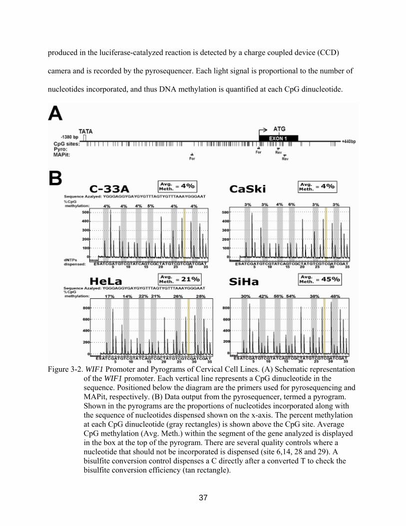
Figure 3-2. WIF1 Promoter and Pyrograms of Cervical Cell Lines. (A) Schematic representation
of the WIF1 promoter. Each vertical line represents a CpG dinucleotide in the sequence. Positioned below the diagram are the primers used for pyrosequencing and MAPit, respectively. (B) Data output from the pyrosequencer, termed a pyrogram. Shown in the pyrograms are the proportions of nucleotides incorporated along with the sequence of nucleotides dispensed shown on the x-axis. The percent methylation at each CpG dinucleotide (gray rectangles) is shown above the CpG site. Average CpG methylation (Avg. Meth.) within the segment of the gene analyzed is displayed in the box at the top of the pyrogram. There are several quality controls where a nucleotide that should not be incorporated is dispensed (site 6,14, 28 and 29). A bisulfite conversion control dispenses a C directly after a converted T to check the bisulfite conversion efficiency (tan rectangle).
37

We designed pyrosequencing primers to amplify a 164 basepair region of the WIF1
promoter. This assay quantitatively measures the first seven CpGs following the transcriptional
start site using the forward PCR primer as the sequencing primer (Figure 3-2A). When
pyrosequencing was conducted on bisulfite-modified genomic DNA harvested from the four
cervical cancer cell lines, we observed that the cell lines that express WIF1 in the absence of
5-azadC (C-33A and CaSki), exhibited very low levels of DNA methylation (<3%). Conversely,
cell lines which expressed little to no WIF1 in the absence of 5-azadC, HeLa and SiHa cells,
exhibited high and moderate levels of methylation, respectively (21% and 45%) (Figure 3-2B).
Genomic DNA harvested from HeLa and SiHa cells cultured with 5-azadC displayed a 10-15%
decrease in average methylation, while C-33A and CaSki showed little change (Figure 3-3).
These data demonstrate that the expression of WIF1 is correlated to the methylation status of its
promoter in cervical cell lines.
Figure 3-3. Average Methylation of the WIF1 Promoter Before and After 5-azadC Treatment. Cultures of the cervical tumor cell lines C-33A, CaSki, HeLa and SiHa were untreated (white bars) or treated with 5 µM 5-azadC (black bars). Shown are the averages and standard deviation for the six CpGs assayed by pyrosequencing as outlined in Materials and Methods.
38
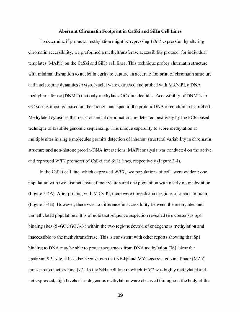
Aberrant Chromatin Footprint in CaSki and SiHa Cell Lines
To determine if promoter methylation might be repressing WIF1 expression by altering
chromatin accessibility, we preformed a methyltransferase accessibility protocol for individual
templates (MAPit) on the CaSki and SiHa cell lines. This technique probes chromatin structure
with minimal disruption to nuclei integrity to capture an accurate footprint of chromatin structure
and nucleosome dynamics in vivo. Nuclei were extracted and probed with M.CviPI, a DNA
methyltransferase (DNMT) that only methylates GC dinucleotides. Accessibility of DNMTs to
GC sites is impaired based on the strength and span of the protein-DNA interaction to be probed.
Methylated cytosines that resist chemical deamination are detected positively by the PCR-based
technique of bisulfite genomic sequencing. This unique capability to score methylation at
multiple sites in single molecules permits detection of inherent structural variability in chromatin
structure and non-histone protein-DNA interactions. MAPit analysis was conducted on the active
and repressed WIF1 promoter of CaSki and SiHa lines, respectively (Figure 3-4).
In the CaSki cell line, which expressed WIF1, two populations of cells were evident: one
population with two distinct areas of methylation and one population with nearly no methylation
(Figure 3-4A). After probing with M.CviPI, there were three distinct regions of open chromatin
(Figure 3-4B). However, there was no difference in accessibility between the methylated and
unmethylated populations. It is of note that sequence inspection revealed two consensus Sp1
binding sites (5'-GGCGGG-3') within the two regions devoid of endogenous methylation and
inaccessible to the methyltransferase. This is consistent with other reports showing that Sp1
binding to DNA may be able to protect sequences from DNA methylation [76]. Near the
upstream SP1 site, it has also been shown that NF-kβ and MYC-associated zinc finger (MAZ)
transcription factors bind [77]. In the SiHa cell line in which WIF1 was highly methylated and
not expressed, high levels of endogenous methylation were observed throughout the body of the
39

promoter and accessibility to the methyltransferase seemed to vary more, thus suggesting
dynamic chromatin reorganization (Figure 3-4C).
Figure 3-4. Chromatin Probing of CaSki and SiHa Cell Line. Nuclei from both cells lines were isolated and chromatin structure was probed as stated in the Materials and Methods. Endogenously methylated CpG (CG) sites are depicted with black circles and unmethylated CpG with white circles. All GC sites are denoted by triangles, and those that were methylated by M.CviPI probe are red. Regions that were not protected against methylation are indicated by rectangles spanning at least two contiguous GC sites (i.e., one methylated site does constitute accessibility). Methylated GCG (Gm5CG) sites are indicated in gray and cannot be discerned from endogenous methylation or M.CviPI modified sites. The bisulfite conversion efficiency (C.E.) for each molecule is shown at the right. Transcription factor binding sites for Sp1 (blue) and both NF-κB and MYC-associated zinc finger (MAZ) (tan) are indicated.
40

WIF1 Promoter is Aberrantly Methylated in Squamous Cell Cervical Tumors
To determine if promoter methylation of the WIF1 gene occurs in primary cervical tumors,
we used pyrosequencing to analyze the methylation of WIF1 in patient-derived cervical
squamous cell carcinoma samples. Matched adjacent normal cervical stratified epithelium and
Pap smear samples from different patients were also analyzed. Cervical tumors and normal
tissues were obtained from the Moffitt Cancer Center Tissue Bank. DNA was extracted, treated
with sodium bisulfite and PCR amplified with the WIF1 pyrosequencing primers. PCR
amplicons where then analyzed with the PyroMark MD and Pyro Q-CpG software. All cases
samples were re-analyzed with repeated PCR and pyrosequencing reactions to validate the initial
pyrosequencing findings. Subsets of bisulfite-treated samples were also analyzed multiple times
by bisulfite genomic sequencing, with analogous results (data not shown).
To ascertain if aberrant methylation of the WIF1 promoter occurs in cervical cancer, we
pyrosequenced 22 high-grade squamous cell carcinomas. Of these 22 tumors, only 17 produced
PCR amplicons of sufficient quality for pyrosequencing. We grouped these specimens into three
categories based on their average methylation of six CpG sites as followed: little or no
methylation (0-9%), moderate methylation (10-19%) and heavily methylated (>20%). Of the 17
tumor samples, 6 were not methylated, 3 had moderate methylation, and 8 were heavily
methylated (Figure 3-5).
We also analyzed a set of 8 Pap smear samples that had been previously genotyped and 7
of which were found to be HPV positive. We found no significant methylation, <5%, in all 8 of
the Pap smear samples (Figure 3-6). In addition we analyzed the paired adjacent
normal tissue from each tumor sample. Unfortunately, several of theses samples failed to
41

Figure 3-5. Average CpG methylation of Cervical SSC and Adjacent Normal Tissue at WIF1.
The tumors and adjacent matching normal tissues were given arbitrary numbers. Both the PCR and the pyrosequencing were run in duplicate, and the average methylation status of the WIF1 promoter is shown along with the standard deviation for each sample. Black bars represent the methylation of the tumor samples and the white bars show the methylation of the paired normal stratified epithelium.
Figure 3-6. Pyrograms of Pap Smear Samples at WIF1. The percent methylation at each CpG
dinucleotide (gray rectangles) is shown above the CpG site. Average CpG methylation (Avg. Meth.) within the segment of the gene analyzed is displayed in the box at the top of the pyrograms. Seven additional pap smear samples were analyzed with similar results (data not shown).
yield usable amounts of DNA, and we were not able to assess WIF1 promoter methylation.
However, the 9 matched normal samples that were analyzed all had little or no methylation
(<5%) (Figure 3-5). These data show that this region of the WIF1 promoter is essentially
42

methylation-free in normal cervical tissue and supports the notion that the DNA methylation that
we observed in the tumor samples resulted from the disease and was not part of the normal
biology of cervical tissue.
WIF1 Methylation is not Correlated with WIF1 Protein Levels
To assess if the methylation status of the WIF1 promoter correlated with the expression
of WIF1 protein, we conducted immunohistochemical staining on a tissue microarray including
22 tumors and their adjacent normal tissue. Immunohistochemistry (IHC) is a method for
demonstrating the presence and location of proteins in the context of intact tissue. IHC staining is
accomplished with antibodies that recognize the target protein. The antibody-antigen interaction
is visualized using chromogenic detection, in which an enzyme conjugated to the antibody
cleaves a substrate to produce a colored precipitate, in this case brownish-red, at the location of
the protein.
Immunostaining of normal tissue with anti-WIF1 antibody indicated that the protein was
expressed in the stratified squamous epithelium of the ectocervix. No staining of the underlying
cervical parenchyma (Figure 3-7 A1) was observed, except within the endometrial cell layer of
blood vessels, which stained intensely with anti-WIF1 antibody. Close examination of the
staining indicated a gradient of WIF1 expression within epithelial tissue (Figure 3-7A).
Specifically, nearly no staining was observed in the basal epithelium or underlying basement
membrane (Figure 3-7A, zones 1 and 2). However, strong staining was evident within the
peribasal epithelial layer (Figure 3-7A, zone 3). We observed notable extracellular staining
within the layers of differentiated non-keratinized epithelium with diminished staining in cells at
the apical surface of the ectocervix (Figure 3-7A, zone 5). A negative control for background
staining was incubated with only rabbit IgG and demonstrated that there was no background
staining of the WIF1 antibody within the cervical epithelium (Figure 3-7 B).
43
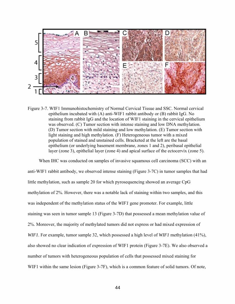
Figure 3-7. WIF1 Immunohistochemistry of Normal Cervical Tissue and SSC. Normal cervical epithelium incubated with (A) anti-WIF1 rabbit antibody or (B) rabbit IgG. No staining from rabbit IgG and the location of WIF1 staining in the cervical epithelium was observed. (C) Tumor section with intense staining and low DNA methylation. (D) Tumor section with mild staining and low methylation. (E) Tumor section with light staining and high methylation. (F) Heterogeneous tumor with a mixed population of stained and unstained cells. Bracketed at the left are the basal epithelium (or underlying basement membrane, zones 1 and 2), peribasal epithelial layer (zone 3), epithelial layer (zone 4) and apical surface of the ectocervix (zone 5).
When IHC was conducted on samples of invasive squamous cell carcinoma (SCC) with an
anti-WIF1 rabbit antibody, we observed intense staining (Figure 3-7C) in tumor samples that had
little methylation, such as sample 20 for which pyrosequencing showed an average CpG
methylation of 2%. However, there was a notable lack of staining within two samples, and this
was independent of the methylation status of the WIF1 gene promoter. For example, little
staining was seen in tumor sample 13 (Figure 3-7D) that possessed a mean methylation value of
2%. Moreover, the majority of methylated tumors did not express or had mixed expression of
WIF1. For example, tumor sample 32, which possessed a high level of WIF1 methylation (41%),
also showed no clear indication of expression of WIF1 protein (Figure 3-7E). We also observed a
number of tumors with heterogeneous population of cells that possessed mixed staining for
WIF1 within the same lesion (Figure 3-7F), which is a common feature of solid tumors. Of note,
44

we observed more intense WIF1 staining in less de-differentiated SCC lesions. These studies
show that low or absent WIF1 expression is commonly observed in invasive SCC of the cervix,
but that this tumor feature may occur independent of WIF1 promoter methylation status. These
findings also suggest that mechanism(s) other then promoter hypermethylation may serve to
inhibit WIF1 expression. We conclude that hypermethylation of the WIF1 gene is a common
event in cervical squamous cell carcinoma but that promoter methylation is not the only
necessary cause of its silencing.
45

CHAPTER 4 DISCUSSION
The rationale for examining DNA methylation at the WIF1 promoter in cervical cancer
was based on three primary factors. First, exploitation of the Wnt pathway is a common
mechanism of tumorigenesis. Uncontrolled β-catenin signaling, either from disruption of
function of its regulatory proteins (i.e., Axin, APC, WIF1) or genetic mutations that prevent β-
catenin degradation, leads to excessive proliferation that predisposes cells to tumorigenesis [65].
Second, WIF1 expression has been shown to be downregulated in a variety of cancer types, such
as bladder, breast, lung and prostate [74]. WIF1 downregulation is an epigenetic event correlated
to promoter hypermethylation in many of these cancers. Third, while some studies have
examined the epigenetic regulation of WIF1 and other genes encoding molecular components of
the Wnt signaling pathway, no research has examined WIF1 silencing in cervical carcinomas.
Using both cervical cancer cell lines and primary formalin-fixed/paraffin-embedded
squamous cell carcinomas samples, we established that the WIF1 gene is commonly subjected to
DNA hypermethylation in cervical tumors. In vitro studies in cervical cell lines indicated that
WIF1 expression correlated with promoter methylation. Furthermore, we showed that the DNA
demethylating agent 5-azadC increased WIF1 expression. In an effort to show that WIF1
expression is epigenetically silenced in primary tissue, we conducted pyrosequencing and
immunostaining on cervical tumor samples. We found that the WIF1 promoter is unmethylated
and WIF1 protein is expressed in the peribasal and the less differentiated epithelial layers of
normal cervical epithelium. In contrast, within high-grade squamous cell carcinomas, WIF1
protein expression was often low to nonexistent. This loss of WIF1 protein expression correlated
with promoter hypermethylation in several of the tumors; however, loss of WIF1 protein
expression in two of the tumors could not be attributed to WIF1 promoter hypermethylation. This
46

suggests that WIF1 is epigenetically silenced in cervical cancer, but other mechanisms in
addition to promoter hypermethylation may be involved.
Lack of complete concordance between WIF1 promoter hypermethylation and lack of
protein expression could be due to several factors. First, pyrosequencing analyses revealed the
six CpG sites in the WIF1 promoter in CaSki cells were unmethylated; however, transcript levels
increased after treatment with 5-azaC as assayed by RT-PCR. These findings suggest that the
WIF1 gene is epigenetically silenced in these cells, but by methylation of CpG sites other then
the six downstream of the TSS we assayed by pyrosequencing. This is supported by MAPit
methylation footprinting experiments, which showed two areas of endogenous methylation
further upstream of the TSS in CaSki cells. PCR using the MAPit primers was attempted on
several of the tumor samples without success. This failure is likely because FFPE- and bisulfite-
treated DNA is very fragmented and thus unable to yield large PCR products. One method to
examine if the tumor samples had similar patches of methylation to the CaSki cell line further
upstream of the TSS would be to create primers specific for the areas of interest that yield
smaller PCR products.
Pyrosequencing of small bisulfite-converted amplicons at the two upstream methylated
regions is needed to test the extent to which hypermethylation correlates with WIF1
downregulation in the primary tumor samples. Methylation of either of these upstream regions
correlating with the loss of WIF1 expression as determined by IHC would lend more support to
the hypothesis that the WIF1 gene is directly epigenetically silenced in cervical cancer.
HPV may also play a role in the epigenetic silencing of the WIF1 promoter in cervical
cancer. This is based on the observation that the three cell lines (CaSki, SiHa and HeLa) that
displayed varying levels of WIF1 epigenetic silencing were all derived from HPV-infected
47

specimens. By contrast, the cell line derived from a non-HPV-infected specimen did not appear
to be epigenetically silenced. HPV viral proteins can directly effect DNA methylation by binding
to and recruiting DNMTs [57]. Knockdown of expression of the viral E6 and E7 genes may help
to understand if the virus plays a role in WIF1 hypermethylation. Furthermore, HPV genotyping
of the patient cervical tumors should be done to look for a correlation between WIF1 expression
and the presence of HPV.
MAPit footprinting of the CaSki and SiHa cell lines may yield insight into how WIF1
silencing might occur regardless of promoter methylation. In the WIF1-expressing CaSki cells,
we observed a region of protection against methylation by M.CviPI just upstream of the
transcriptional start site. We hypothesized that this area would be nucleosome free and hence
accessible to the DNMT, as has been observed at other transcriptionally-active promoters. The
size of the inaccessible region is consistent with a nucleosome (~147 base pairs) that occupies a
variety of positions. The variable location of the protected region on each molecule seems
inconsistent with protection of the region against methylation by a site-specific transcription
factor bound to DNA, such as SP1. It is possible that SP1 or another transcription factor is
forming a ternary complex with a nucleosome that occupies several positions.
Interestingly, two different populations of molecules with distinct endogenous DNA
methylation patterns were observed in the CaSki cell line. In one group, nearly no endogenous
methylation was observed, whereas the other group had two distinct areas of endogenous
methylation straddling the upstream SP1 site. Despite these two patterns of methylation by
endogenous enzymes, there appears to be little difference in accessibility to M.CviPI between
them. This suggests that whatever is bound to the DNMT accessible area binds regardless of the
methylation status of the gene. It should be noted, however, that we have only examined a
48
limited number of molecules from one experiment and there exists the possibility the molecules
shown were selectively sequenced due to PCR or cloning bias. Similarly, the heavily methylated
WIF1 promoter in SiHa cells showed clear regions of M.CviPI accessibility but with greater
variability (Figure 7C). Often, areas of heavily methylated DNA are inaccessible to the
methyltransferase and unable to provide clear footprints. This suggests that, whereas the WIF1
promoter is heavily methylated in the SiHa cell line, it is not in a fully compacted
heterochromatin state.
Further experiments are needed to clarify what is binding to the area upstream of the WIF1
TSS. Knockdown experiments of the various transcription factors, particularly SP1, in the cell
line followed by MAPit might help to clarify what is binding to the DNA. It would be of interest
to see if knockdown of SP1 alters WIF1 endogenous methylation or chromatin accessibility,
particularly in the highly methylated SiHa cell line. However, preferential association of NF-κB
or MYC-associated with the WIF1 promoter in Caski, but not in SiHa, cells may explain the
difference in expression between the cell lines. Chromatin immunoprecipitation experiments
probing for histones might help to create a clearer picture of regulation of the WIF1 promoter
and establish if there is a bias in the MAPit footprint. Better understanding of the WIF1 promoter
region and its associated proteins could potentially lead to use as a cervical cancer drug therapy.
In addition to technical limitations that may have precluded detection of DNA methylation
in cervical tumors lacking WIF1 protein expression, it is also possible that non-epigenetic factors
are also responsible for loss of WIF1 expression in these tumors. For example, chromosome
rearrangements involving 12q13-15, where WIF1 is located, are frequent occurrences among
tumors. While no DNA mutations of WIF1 have been reported, one group demonstrated salivary
gland pleomorphic adenoma expressing an high mobility group AT-hook 2 (HMGA2)/WIF1
49
fusion transcript, which resulted in re-expression of wild-type HMGA2 transcripts and very low
levels of WIF1 expression [78]. An alternative explanation for the lack of correlation between
WIF1 promoter methylation and protein expression is that WIF1 transcription may require an
additional activator which could be inhibited or after chromosome rearrangement the gene may
be excised. These are a few alternative mechanisms aside from epigenetic silencing that could
result in downregulation of WIF1 protein in cervical cancer.
A common aim of TSG research is to evaluate the possibility of a gene being used as either
a diagnostic or prognostic tool. We showed that the WIF1 gene is aberrantly methylated in high-
grade squamous cell carcinomas of the cervix, which makes it a potential prognostic marker.
However, we failed to obtain any low-grade or mildly dysplastic cervical squamous cells for
analysis within the confines of the study. Examining the status of WIF1 expression at pre-
cancerous stages or within low-grade carcinomas may help determine when during tumor
progression aberrant methylation of the WIF1 promoter occurs, and if hypermethylation is an
indicator of high-grade squamous cell carcinoma. There exists a possibility that a correlation
between expression of WIF1 and the pathological state or grade of the tumor could be made to
provide insight into WIF1 being a possible biomarker for diagnosis.
It is clear that cancer cells undergo global changes in DNA methylation that lead to
chromatin alterations involving the whole epigenome and that entire pathways relevant to cell
division are subject to epigenetic dysregulation. As the role of epigenetics in cancer becomes
clearer new approaches to cancer prevention, detection, and therapy will begin to emerge.
50

LIST OF REFERENCES
1. Detailed Guide: Cervical Cancer. What are the key statistics about cervical cancer? Available from: http://www.cancer.org/docroot/CRI/CRI_2_3x.asp?rnav=cridg&dt=8.
2. Schoell, W.M.J., M.F. Janicek, and R. Mirhashemi, Epidemiology and biology of
cervical cancer. Seminars in Surgical Oncology, 1999. 16(3): p. 203-11. 3. Kurman, R.J., et al., From Papanicolaou to Bethesda: the rationale for a new cervical
cytologic classification. Obstet Gynecol, 1991. 77(5): p. 779-82. 4. Lie, A.K. and G. Kristensen, Human papillomavirus E6/E7 mRNA testing as a predictive
marker for cervical carcinoma. Expert Rev Mol Diagn, 2008. 8(4): p. 405-15. 5. Walboomers, J.M.M., et al., Human papillomavirus is a necessary cause of invasive
cervical cancer worldwide. J Pathol, 1999. 189(1): p. 12-9. 6. Ho, G.Y.F., et al., Persistent Genital Human Papillomavirus Infection as a Risk Factor for
Persistent Cervical Dysplasia. J Natl Cancer Institute, 1995. 87(18): p. 1365-71. 7. Choo, K.B., C.C. Pan, and S.H. Han, Integration of human papillomavirus type-16 into
cellular DNA of cervical carcinoma - Preferential Deletion of the E2 gene and invariable retention of the long control region and the E6/E7 Open Reading Frames. Virology, 1987. 161(1): p. 259-61.
8. Di Leonardo, A., et al., DNA damage triggers a prolonged p53-dependent G1 arrest and
long-term induction of Cip1 in normal human fibroblasts. Genes Dev, 1994. 8(21): p. 2540-51.
9. Smith, M.L., et al., Involvement of the p53 tumor suppressor in repair of U.V.-type DNA
damage. Oncogene, 1995. 10(6): p. 1053-9. 10. Subramanya, D. and P.D. Grivas, HPV and cervical cancer: updates on an established
relationship. Postgrad Med, 2008. 120(4): p. 7-13. 11. Gissmann, L., Human papillomaviruses and genital cancer. Semin Cancer Biol, 1992.
3(5): p. 253-61. 12. American Type Culture Collection., Catalogue of strains II, American Type Culture
Collection.: Rockville, Md.,. p. v. 13. Scheffner, M., et al., The state of the p53 and retinoblastoma genes in human cervical
carcinoma cell lines. Proc Natl Acad Sci USA, 1991. 88(13): p. 5523-7. 14. Jones, P.A. and S.B. Baylin, The epigenomics of cancer. Cell, 2007. 128(4): p. 683-92.
51

15. Jones, P.A., Overview of cancer epigenetics. Semin Hematol, 2005. 42(3 Suppl 2): p. S3-
8. 16. Cooper, D.N. and M. Krawczak, Cytosine methylation and the fate of CpG dinucleotides
in vertebrate genomes. Hum Genet, 1989. 83(2): p. 181-8. 17. Bird, A., DNA methylation patterns and epigenetic memory. Genes Dev, 2002. 16(1): p.
6-21. 18. Larsen, F., et al., CpG islands as gene markers in the human genome. Genomics, 1992.
13(4): p. 1095-107. 19. Mohandas, T., R.S. Sparkes, and L.J. Shapiro, Reactivation of an inactive human X
chromosome: evidence for X inactivation by DNA methylation. Science, 1981. 211(4480): p. 393-6.
20. Li, E., C. Beard, and R. Jaenisch, Role for DNA methylation in genomic imprinting.
Nature, 1993. 366(6453): p. 362-5. 21. Suetake, I., et al., DNMT3L stimulates the DNA methylation activity of Dnmt3a and
Dnmt3b through a direct interaction. J Biol Chem, 2004. 279(26): p. 27816-23. 22. Otani, J., et al., Structural basis for recognition of H3K4 methylation status by the DNA
methyltransferase 3A ATRX-DNMT3-DNMT3L domain. EMBO Rep, 2009. 10(11): p. 1235-41.
23. Hermann, A., H. Gowher, and A. Jeltsch, Biochemistry and biology of mammalian DNA
methyltransferases. Cell Mol Life Sci, 2004. 61(19-20): p. 2571-87. 24. Luger, K., et al., Crystal structure of the nucleosome core particle at 2.8 Å resolution.
Nature, 1997. 389(6648): p. 251-60. 25. Sarma, K. and D. Reinberg, Histone variants meet their match. Nat Rev Mol Cell Biol,
2005. 6(2): p. 139-49. 26. Kouzarides, T., Chromatin modifications and their function. Cell, 2007. 128(4): p. 693-
705. 27. Fuks, F., et al., The methyl-CpG-binding protein MeCP2 links DNA methylation to
histone methylation. J Biol Chem, 2003. 278(6): p. 4035-40. 28. Nan, X., et al., Transcriptional repression by the methyl-CpG-binding protein MeCP2
involves a histone deacetylase complex. Nature, 1998. 393(6683): p. 386-9.
52

29. Tachibana, M., et al., G9a/GLP complexes independently mediate H3K9 and DNA methylation to silence transcription. EMBO J, 2008. 27(20): p. 2681-90.
30. Zhao, Q., et al., PRMT5-mediated methylation of histone H4R3 recruits DNMT3A,
coupling histone and DNA methylation in gene silencing. Nat Struct Mol Biol, 2009. 16(3): p. 304-11.
31. Esteve, P.O., et al., Regulation of DNMT1 stability through SET7-mediated lysine
methylation in mammalian cells. Proc Natl Acad Sci USA, 2009. 106(13): p. 5076-81. 32. Jones, P.L., et al., Methylated DNA and MeCP2 recruit histone deacetylase to repress
transcription. Nat Genet, 1998. 19(2): p. 187-91. 33. Robertson, K.D., et al., DNMT1 forms a complex with Rb, E2F1 and HDAC1 and
represses transcription from E2F-responsive promoters. Nat Genet, 2000. 25(3): p. 338-42.
34. Wang, Z., et al., Genome-wide mapping of HATs and HDACs reveals distinct functions
in active and inactive genes. Cell, 2009. 138(5): p. 1019-31. 35. Lorch, Y., J.W. LaPointe, and R.D. Kornberg, Nucleosomes inhibit the initiation of
transcription but allow chain elongation with the displacement of histones. Cell, 1987. 49(2): p. 203-10.
36. Bernstein, B.E., et al., Global nucleosome occupancy in yeast. Genome Biol, 2004. 5(9):
p. R62. 37. Jiang, C. and B.F. Pugh, Nucleosome positioning and gene regulation: advances through
genomics. Nat Rev Genet, 2009. 10(3): p. 161-72. 38. Ozsolak, F., et al., High-throughput mapping of the chromatin structure of human
promoters. Nat Biotechnol, 2007. 25(2): p. 244-8. 39. Heintzman, N.D., et al., Distinct and predictive chromatin signatures of transcriptional
promoters and enhancers in the human genome. Nat Genet, 2007. 39(3): p. 311-8. 40. Boeger, H., J. Griesenbeck, and R.D. Kornberg, Nucleosome retention and the stochastic
nature of promoter chromatin remodeling for transcription. Cell, 2008. 133(4): p. 716-26. 41. Dechassa, M.L., et al., SWI/SNF has intrinsic nucleosome disassembly activity that is
dependent on adjacent nucleosomes. Mol Cell, 2010. 38(4): p. 590-602. 42. Shivaswamy, S., et al., Dynamic remodeling of individual nucleosomes across a
eukaryotic genome in response to transcriptional perturbation. PLoS Biol, 2008. 6(3): p. e65.
53
43. Schones, D.E., et al., Dynamic regulation of nucleosome positioning in the human genome. Cell, 2008. 132(5): p. 887-98.
44. Gilbert, N. and B. Ramsahoye, The relationship between chromatin structure and
transcriptional activity in mammalian genomes. Brief Funct Genomic Proteomic, 2005. 4(2): p. 129-42.
45. Ptashne, M., On the use of the word 'epigenetic'. Curr Biol, 2007. 17(7): p. R233-6. 46. Zemach, A., et al., Genome-wide evolutionary analysis of eukaryotic DNA methylation.
Science, 2010. 328(5980): p. 916-9. 47. Sherr, C.J., Principles of tumor suppression. Cell, 2004. 116(2): p. 235-46. 48. Jones, P.A. and S.B. Baylin, The fundamental role of epigenetic events in cancer. Nat
Rev Genet, 2002. 3(6): p. 415-428. 49. Garinis, G.A., et al., DNA hypermethylation: when tumour suppressor genes go silent.
Hum Genet, 2002. 111(2): p. 115-27. 50. Boyes, J. and A. Bird, Repression of genes by DNA methylation depends on CpG density
and promoter strength: evidence for involvement of a methyl-CpG binding protein. EMBO J, 1992. 11(1): p. 327-33.
51. Wolffe, A.P., Chromatin remodeling: why it is important in cancer. Oncogene, 2001.
20(24): p. 2988-90. 52. Morey, L., et al., MBD3, a component of the NuRD complex, facilitates chromatin
alteration and deposition of epigenetic marks. Mol Cell Biol, 2008. 28(19): p. 5912-23. 53. Ramirez, J. and J. Hagman, The Mi-2/NuRD complex: a critical epigenetic regulator of
hematopoietic development, differentiation and cancer. Epigenetics, 2009. 4(8): p. 532-6. 54. Reisman, D., S. Glaros, and E.A. Thompson, The SWI/SNF complex and cancer.
Oncogene, 2009. 28(14): p. 1653-68. 55. Etoh, T., et al., Increased DNA methyltransferase 1 (DNMT1) protein expression
correlates significantly with poorer tumor differentiation and frequent DNA hypermethylation of multiple CpG islands in gastric cancers. Am J Pathol, 2004. 164(2): p. 689-99.
56. Muller, K., H. Heller, and W. Doerfler, Foreign DNA integration. Genome-wide
perturbations of methylation and transcription in the recipient genomes. J Biol Chem, 2001. 276(17): p. 14271-8.
54

57. Burgers, W.A., et al., Viral oncoproteins target the DNA methyltransferases. Oncogene, 2007. 26(11): p. 1650-5.
58. Dong, S.M., et al., Promoter hypermethylation of multiple genes in carcinoma of the
uterine cervix. Clin Cancer Res, 2001. 7(7): p. 1982-6. 59. Sawada, M., et al., Increased expression of DNA methyltransferase 1 (DNMT1) protein
in uterine cervix squamous cell carcinoma and its precursor lesion. Cancer Lett, 2007. 251(2): p. 211-9.
60. Bigey, P., et al., Transcriptional regulation of the human DNA Methyltransferase
(DNMT1) gene. Gene, 2000. 242(1-2): p. 407-18. 61. Ghittoni, R., et al., The biological properties of E6 and E7 oncoproteins from human
papillomaviruses. Virus Genes, 2010. 40(1): p. 1-13. 62. Cadigan, K.M. and R. Nusse, Wnt signaling: a common theme in animal development.
Genes Dev, 1997. 11(24): p. 3286-305. 63. Hobmayer, B., et al., WNT signalling molecules act in axis formation in the diploblastic
metazoan Hydra. Nature, 2000. 407(6801): p. 186-9. 64. Reya, T. and H. Clevers, Wnt signalling in stem cells and cancer. Nature, 2005.
434(7035): p. 843-50. 65. MacDonald, B.T., K. Tamai, and X. He, Wnt/beta-catenin signaling: components,
mechanisms, and diseases. Dev Cell, 2009. 17(1): p. 9-26. 66. Kawano, Y. and R. Kypta, Secreted antagonists of the Wnt signalling pathway. J Cell Sci,
2003. 116(Pt 13): p. 2627-34. 67. Cong, F., L. Schweizer, and H. Varmus, Wnt signals across the plasma membrane to
activate the β-catenin pathway by forming oligomers containing its receptors, Frizzled and LRP. Development, 2004. 131(20): p. 5103-15.
68. He, X., et al., LDL receptor-related proteins 5 and 6 in Wnt/β-catenin signaling: arrows
point the way. Development, 2004. 131(8): p. 1663-77. 69. van Amerongen, R., A. Mikels, and R. Nusse, Alternative Wnt signaling is initiated by
distinct receptors. Sci Signal, 2008. 1(35): p. re9. 70. Arce, L., K.T. Pate, and M.L. Waterman, Groucho binds two conserved regions of LEF-1
for HDAC-dependent repression. BMC Cancer, 2009. 9: p. 159. 71. Jessen, J.R., Noncanonical Wnt signaling in tumor progression and metastasis. Zebrafish,
2009. 6(1): p. 21-8.
55

72. Uren, A., et al., Activation of the canonical Wnt pathway during genital keratinocyte
transformation: a model for cervical cancer progression. Cancer Res, 2005. 65(14): p. 6199-206.
73. Hsieh, J.C., et al., A new secreted protein that binds to Wnt proteins and inhibits their
activities. Nature, 1999. 398(6726): p. 431-6. 74. Wissmann, C., et al., WIF1, a component of the Wnt pathway, is down-regulated in
prostate, breast, lung, and bladder cancer. J Pathol, 2003. 201(2): p. 204-12. 75. Warnecke, P.M., et al., Identification and resolution of artifacts in bisulfite sequencing.
Methods, 2002. 27(2): p. 101-7. 76. Macleod, D., et al., Sp1 sites in the mouse aprt gene promoter are required to prevent
methylation of the CpG island. Genes Dev, 1994. 8(19): p. 2282-92. 77. Reguart, N., et al., Cloning and characterization of the promoter of human Wnt inhibitory
factor-1. Biochem Biophys Res Commun, 2004. 323(1): p. 229-34. 78. Persson, F., et al., High-resolution genomic profiling of adenomas and carcinomas of the
salivary glands reveals amplification, rearrangement, and fusion of HMGA2. Genes Chromosomes Cancer, 2009. 48(1): p. 69-82.
56

BIOGRAPHICAL SKETCH
Amber Lyn Delmas was born in Stuart, Florida. Through dual enrollment during high
school, she attended Indian River Community College. She received an Associates of Arts
degree in science the summer after graduating from high school in 2003. She then attended the
University of Florida and earned a Bachelor of Science in microbiology and cell science in 2006.
After graduation, she worked as a laboratory technician in the Department of Biochemistry and
Molecular Biology at the University of Florida for two years. In August 2010, she received a
Master of Science in medical sciences from the University of Florida.
57