wildlife science long-term monitoring of circulating
TRANSCRIPT
1431
*Correspondence to: Funasaka, N.: [email protected], Yoshioka, M.: [email protected]#These authors contributed equally to this work.©2018 The Japanese Society of Veterinary Science
This is an open-access article distributed under the terms of the Creative Commons Attribution Non-Commercial No Derivatives (by-nc-nd) License. (CC-BY-NC-ND 4.0: https://creativecommons.org/licenses/by-nc-nd/4.0/)
FULL PAPERWildlife Science
Long-term monitoring of circulating progesterone and its relationship to peripheral white blood cells in female false killer whales Pseudorca crassidensNoriko FUNASAKA1)#*, Motoi YOSHIOKA1)#*, Keiichi UEDA2), Haruka KOGA2), Makio YANAGISAWA2), Sotaro KOGA2) and Kouji TOKUTAKE2)
1)Cetacean Research Center, Graduate School of Bioresources, Mie University, 1577 Kurimamachiya, Tsu, Mie 514-8507, Japan
2)Okinawa Churashima Foundation, 888 Ishikawa, Motobu, Kunigami, Okinawa 905-0206, Japan
ABSTRACT. Long-term monitoring of circulating progesterone levels in three captive female false killer whales, Pseudorca crassidens, was conducted to characterize their reproductive events and to reveal the relationship between their estrous cycles or pregnancies and peripheral white blood cell (WBC) counts. Blood samples were collected at 2–3-day intervals or on a weekly-to-monthly basis for up to 10 years, from 2006 to 2017. In two mature females (initial body lengths of 4.22 and 4.07 m), some cyclic progesterone elevations were detected during the study period; the estimated mean (± SE) estrous cycle length was 40.5 ± 0.7 days (n=12). The seasonality of ovulation, estimated from the elevation of progesterone levels, varied among individuals or years, and ovulation did not occur every year. The third female (3.26 m) showed progesterone elevations, despite irregular cycles after sexual maturity, and became pregnant. The progesterone levels during pregnancy ranged from 7.3 to 42.2 ng/ml, and the gestation period lasted for 14 months until parturition. The mean WBC counts during estrous cycles were the lowest before the progesterone levels began to increase and then gradually increased toward the luteal phase. The WBC counts were significantly higher during pregnancy than before and were particularly high in early pregnancy. To the best of our knowledge, this is the first report of the relationship between the estrous cycle or pregnancy and WBC counts in cetaceans.
KEY WORDS: estrous cycle, false killer whale, pregnancy, progesterone, white blood cell
False killer whales Pseudorca crassidens are a member of the family Delphinidae and are considered a wide-ranging open-ocean species inhabiting tropical and warm-temperate regions [23]. These are maintained in several aquariums of the world, including those in Japan. The impact of human-related activities on this species is currently unknown, and the species is categorized as “Data Deficient” in the current International Union for Conservation of Nature (IUCN) Red List [37]. The Hawaiian population, in particular, appears to consist of no more than a few hundred individuals; therefore, in 2010, the National Marine Fisheries Service proposed that the Hawaiian insular false killer whales be listed as endangered under the US Endangered Species Act [9]. The management of wild populations and development of controlled breeding programs for captive false killer whales depend on a thorough knowledge of their reproduction, including data regarding the age of sexual maturity, reproductive season, estrous cycle, and gestation period. However, little is known about the basic reproductive biology and physiology of false killer whales. Most of the information currently available on the reproduction of false killer whales has been derived from the examination of gonads obtained from stranded individuals or from fisheries [10, 16, 25]. It has been estimated that female false killer whales likely reach sexual maturity at 8–11 years of age and at a length of 3.4–3.8 m, with an approximate 15-month gestation period [10, 16]. The lengths of their calving seasons range from several months to a year, as estimated from the number of scars or corpus albicans in the ovaries [25, 27].
The circulating levels of male and female gonadal steroid hormones provide valuable information for evaluating the reproductive activity of small captive cetaceans [19], such as common bottlenose dolphins Tursiops truncatus [20, 32, 35, 36, 41], killer whales Orcinus orca [30], white whales Delphinapterus leucas [31], Pacific white-sided dolphins Lagenorhynchus obliquidens [33], and
Received: 13 February 2018Accepted: 5 July 2018Published online in J-STAGE: 18 July 2018
J. Vet. Med. Sci. 80(9): 1431–1437, 2018doi: 10.1292/jvms.18-0075
N. FUNASAKA ET AL.
1432doi: 10.1292/jvms.18-0075
finless porpoises Neophocaena asiaeorientalis [12]. Atkinson et al. [1] reported changes in plasma progesterone levels in captive female false killer whales (two mature and one immature), suggesting that progesterone is a good indicator for assessing seasonal ovarian activity in mature females. Although blood samples were collected weekly from non-pregnant individuals, their ovulation patterns (spontaneous or induced ovulation) and estrous cycle lengths, as well as hormonal profiles during pregnancy, were not reported.
Progesterone, one of the ovarian steroid hormones, is associated with a number of physiological changes in female mammals, including humans. A complex relationship between the reproductive steroid hormone levels and immune system has been described [24], and the susceptibility of the uterus to infection reportedly increases during the luteal phase of the estrous cycle in rabbits [2] and cows [28]. Ovarian hormones influence bacterial adhesion to the mucosa, which is the first step of infection [38]. Few reports are available regarding hematological changes and estrous cycles in farm animals. For example, the circulating white blood cell (WBC) counts in cows and mares might be modulated by their ovarian hormones [3, 4]. It is well known that leukocytosis occurs during pregnancy in humans [5, 11] and cynomolgus monkeys, Macaca fascicularis [40], although the mechanism behind this remains unclear. To date, no reports have been published on this relationship in cetacean species.
The objectives of our study were to quantify the reproductive events in female false killer whales and to investigate the relationship between their estrous cycles or pregnancies and WBC counts.
MATERIALS AND METHODS
Experimental ethicsThis study was conducted in accordance with the Rules of Animal Experiment reviewed by the Animal Research Committee,
Mie University. The false killer whales in the aquarium were treated properly during the entire experimental period, and as a member aquarium of the Japanese Association of Zoos and Aquariums, we acted in compliance with the “Law Concerning the Protection and Control of Animals” and “Care and Feeding Standards for Display Animals” established by the Ministry of the Environment in Japan. All samples were obtained from unrestrained animals using routine husbandry training. All sampling procedures, animal husbandry, and management were performed under the careful supervision of veterinarians at the aquarium.
Animals and sample collectionThree female false killer whales (F1, F2, and F3) kept at the Ocean Expo Park (Okinawa, Japan; 26° N latitude, 127° E
longitude) were used in this study. At the beginning of the study, these whales had body lengths of 4.22, 4.07, and 3.26 m, and estimated ages of 31, 13, and 7 years, respectively. They were kept in outdoor pools that were exposed to natural outdoor lighting, and the water temperatures were maintained at approximately 23–28°C. They received approximately 20 kg/day of a diet comprised of capelins, mackerels, flying fishes, surf smelts, and squids. F1 and F2 were kept separately from the males for the entire study period; F3 was kept with a sexually mature male for three months, from February to May 2016.
Blood samples were obtained for routine health assessments of the subject whales. They were collected at 2–3-day intervals or on a weekly-to-monthly basis from July 2006 to October 2011 for F1 (64 months, 308 samples), from January 2007 to April 2011 for F2 (52 months, 182 samples), and from April 2007 to June 2017 for F3 (123 months, 208 samples). All females were sufficiently trained in the husbandry method of blood sampling from tail fluke veins. Approximately 10 ml of blood was drawn in the morning and divided into two specimens for analyzing progesterone levels and WBC counts. After centrifugation (3,000 rpm, 15 min), the plasma or serum samples were frozen at −30°C until hormonal analyses. The whole blood samples were used for WBC analysis promptly after sampling.
Progesterone immunoassay and white blood cell countsThe levels of plasma or serum progesterone were measured using commercially available enzyme immunoassay kits after
diluting the samples twice (Progesterone Ultra Kit, #402410, standard range: 0.1–2 ng/ml, Neogen; Lansing, MI, U.S.A.). The samples with high progesterone levels (>4 ng/ml) were reanalyzed using an enzyme immunoassay kit with a higher standard range (Progesterone Kit, #402310, standard range: 0.4–40 ng/ml, Neogen). The low progesterone levels in the samples obtained from F3 from November 2011 to the end of the study period were determined using a fluorescence immunoassay method with a fully-automated microfluidic immunoassay system (Mini Vidas SV-5010, SYSMEX bioMerieux; Tokyo, Japan), because the additional Progesterone Ultra Kits were not available in Japan at the time of our study. All procedures were performed according to the manufacturer’s instructions. The intra- and inter-assay coefficients of variation were less than 10% for all three methods. The WBC counts were analyzed with an automated hematology analyzer (KX-21NV, Sysmex; Kobe, Japan).
Data analysisValues are expressed as mean ± SE unless otherwise stated. The baseline progesterone levels were calculated for each female
using an iterative process, whereby the values greater than the mean plus two standard deviations (SDs) were excluded, and the remaining mean value was considered the baseline for the animal [13, 18]. In delphinid species, an average baseline progesterone level of 0.3 ng/ml (<1 ng/ml) was previously reported based on a radioimmunoassay [20, 39], and the levels above 3 ng/ml for an individual female are considered presumptive evidence of luteal activity [35]. Therefore, the values tenfold greater than the baseline were considered suggestive of the presence of a corpus luteum (CL) in the ovary. The estimation of the length of estrous cycle and analysis of the relationship between estrous cycles and WBC counts were limited to the females from which the blood
PROGESTERONE AND LEUKOCYTES IN CETACEANS
1433doi: 10.1292/jvms.18-0075
was sampled at least once per week. For the WBC analysis, we omitted the data that was collected during the period of antibiotic/antifungal administration at the veterinarian’s discretion, as there was a potential for substantial fluctuations in WBC parameters.
The differences in the average levels of circulating progesterone and WBC counts during the estrous cycle and pregnancy were tested using the one-way analysis of variance (ANOVA) followed by the Tukey-Kramer test. The differences in the average WBC counts between before and during pregnancy were tested using Welch’s t-test. The statistical analyses were performed using the Excel add-in software, Ekuseru-Toukei 2012 (Social Survey Research Information Co., Tokyo, Japan).
RESULTS
Changes in circulating progesteroneFluctuations in circulating progesterone levels in the three females throughout the study period are shown in Figs. 1 and 2. The
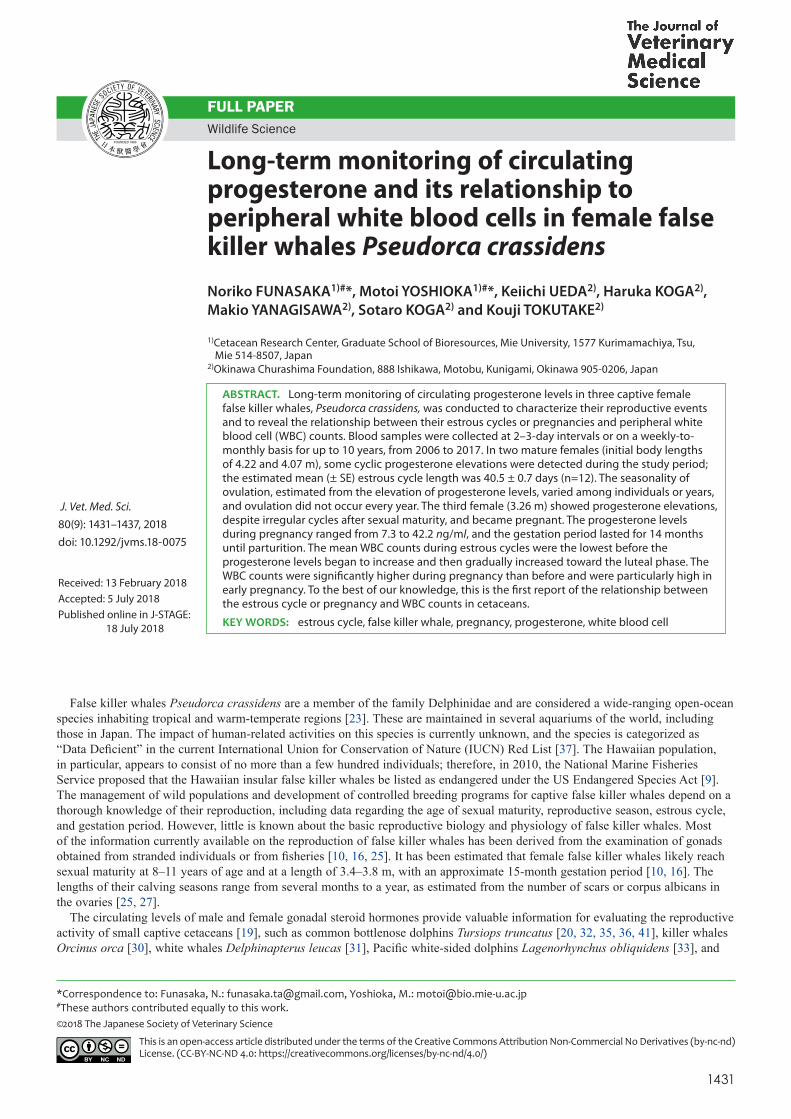
calculated baseline progesterone levels for F1, F2, and F3 were 0.3, 0.2, and 0.2 ng/ml, respectively. The levels of progesterone in F1 and F2 ranged from <0.1 to 23.1 ng/ml and <0.1 to 35.7 ng/ml, respectively. Clearly defined cyclic elevations in progesterone levels were observed in F1 and F2. In the summer of 2007, F1 experienced two cyclic rises in progesterone levels that were indicative of the luteal phase post-ovulation. In F2, four to nine cyclic rises were observed throughout the year, except for the period from the latter half of 2009 to 2011. Progesterone elevation above the baseline levels, but under the threshold suggesting the presence of CL, was observed in the summer of 2009 in F1 and in the spring-to-summer period of 2009 in F2 (Fig. 1).
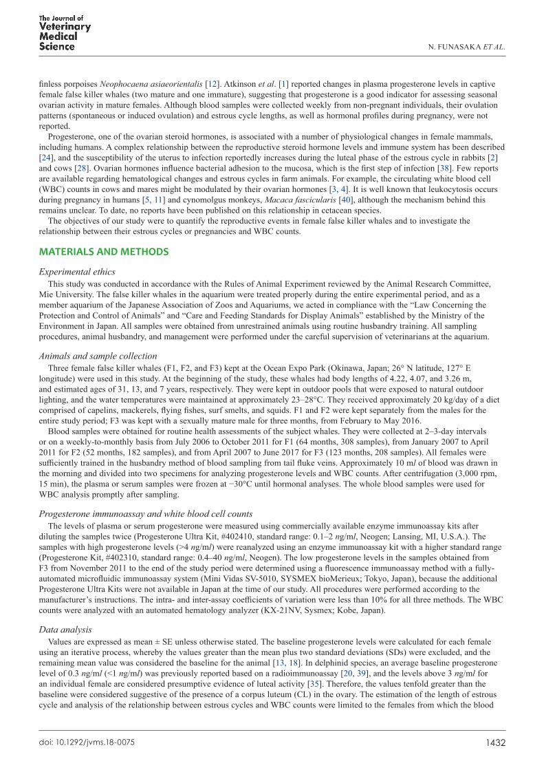
For F3, all progesterone levels were the baseline values until spring 2009 (0.2 ± 0.02 ng/ml, n=58; Fig. 2). The first progesterone elevation above the baseline value occurred in April 2009 (1.2 ng/ml), and then the levels increased again in June 2009 (2.2 ng/ml), February and March 2010 (2.2 and 2.1 ng/ml), and July 2012 (2.0 ng/ml). In F3, the circulating progesterone levels reached a characteristic level of maturity (4.8 ng/ml) in April 2014. After three cyclic rises in progesterone levels in the spring-to-summer period of 2015 and one rise in March 2016, extremely high progesterone levels were observed in F3 from April 12, 2016 to May 22, 2017 (Fig. 2). F3 delivered a female offspring on May 23, 2017.
Estrous cycle and pregnancy characteristicsThe length of estrous cycle (i.e., the interval between the peak progesterone levels in each estrous cycle) was 40.5 ± 0.7 days in
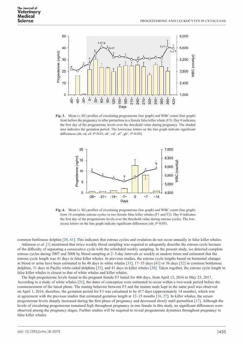
the two mature females (F1: 42 days, n=1; F2: 35–42 days, n=11). The progesterone levels during pregnancy in F3 ranged from 7.3 to 42.2 ng/ml (mean: 23.5 ± 0.9 ng/ml, n=53), with high progesterone levels suggesting the presence of a CL being observed for 406 days. The progesterone levels tended to be higher in early pregnancy, but no significant changes were observed in the levels throughout the pregnancy (P>0.05; Figs. 2 and 3).
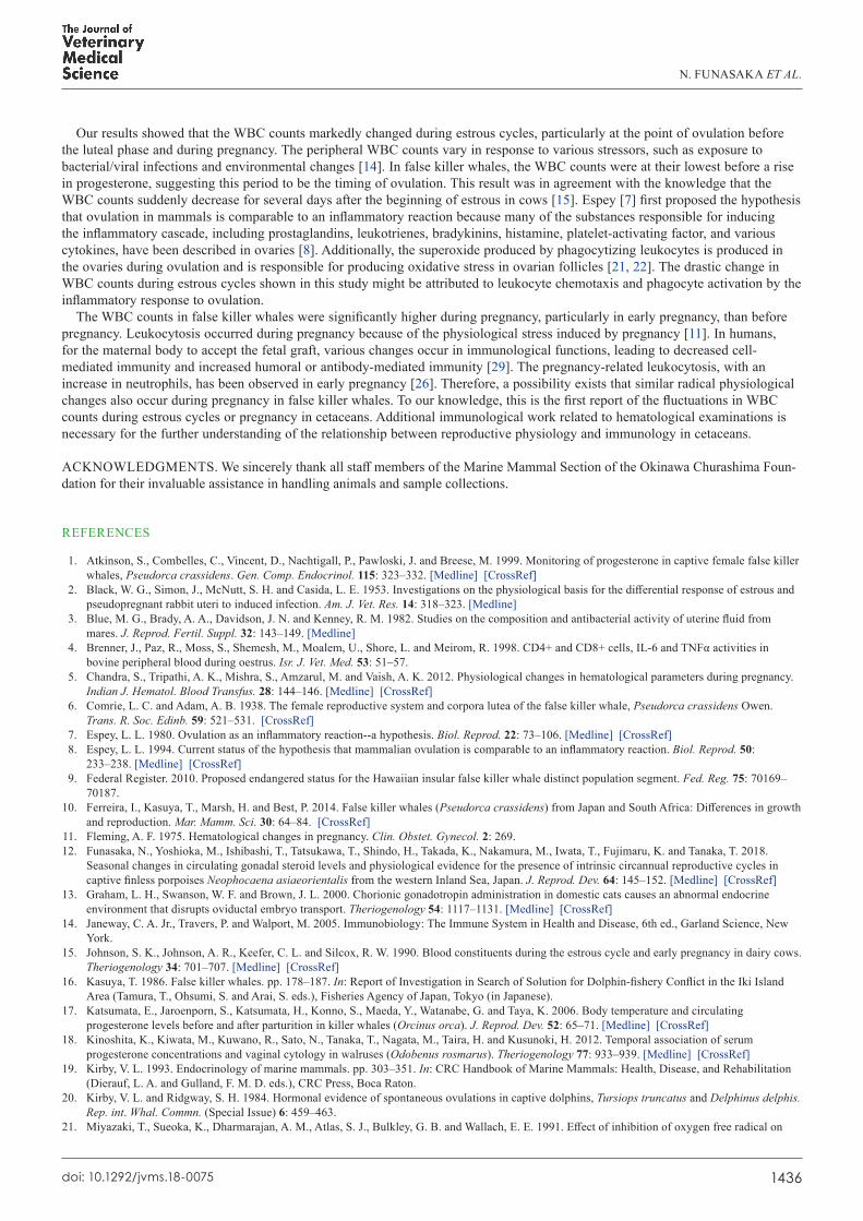
Relationship between estrous cycles or pregnancy and WBC countsThe profiles of mean progesterone levels and WBC counts from 14 complete estrous cycles in F1 and F2 whales are shown in
Fig. 4. The F3 data were excluded from this analysis because the blood samples were collected typically only once or twice per monthly interval. The WBC counts were the lowest before an initial rise in progesterone, and increased gradually, even after the progesterone levels decreased (P<0.05). The mean WBC counts were significantly higher during pregnancy (5,586 ± 103 count/µl, n=56) than before pregnancy (4,257 ± 129 count/µl, n=7) (P<0.01). The WBC counts were particularly high in early pregnancy, followed by a gradual decline until another peak appeared just before parturition (P<0.01; Fig. 3).
DISCUSSION
In this study, changes in circulating progesterone levels were monitored in three false killer whales over an extended period of years. All progesterone levels observed in F3 were below 1 ng/ml until the spring of 2009, indicating sexual immaturity in this young female [19]. Additionally, before beginning this study, no measurements of blood progesterone levels exceeded 1 ng/ml (data not shown). The progesterone levels in F3 were elevated above the baseline values from the spring of 2009 until the summer of 2012, but then reached a characteristic level of maturity in April 2014. Because of the long blood sampling intervals, we must approach a discussion regarding the timing of sexual maturity with caution; however, F3 was assumed to be pubertal until 2014. According to the data collected from the wild populations of this species around Iki Island, Japan, mature female body lengths measured between 3.4 and 3.8 m typically correspond to the ages of 8–11 years, based on the presumed annual deposition of growth layers in dentine and cementum [10, 16]. Moreover, Purves and Pilleri [27] reported that sexual maturity is reached when females measure 3.7–4.3 m in length, which is attained at 8–10 years of age. These data were based on the body length and histological data obtained from a stranded false killer whale population in the eastern North Atlantic. The body length and estimated age of F3 were 3.74 m at 9 years (2009) and 3.86 m at 14 years (2014), which corresponded well with the sexual maturity data reported in previous studies [10, 16, 27]. Additionally, the cyclic rises in progesterone levels that were indicative of ovarian activity were detected in both F1, who produced offspring in 2000, and F2, who had an estimated age of 13 years at the beginning of this study, suggesting that both individuals had reached sexual maturity prior to this study.
The cyclic rises in progesterone levels were detected in the three females when they were not kept with males, suggesting that ovulation occurs spontaneously in the false killer whale, as described by Atkinson et al. [1]. The breeding season of mammalian species in warm climates typically extends over several months or a year, and some previous reports have shown similar results in false killer whales using morphological and histological methods [6, 16, 25, 34]. The seasonality of ovarian activity varied
N. FUNASAKA ET AL.
1434doi: 10.1292/jvms.18-0075
among individuals: F1 and F3 showed ovarian activity in spring to summer, whereas ovaries in F2 appeared to have a year-round activity. Kasuya [16] described that the false killer whales around Iki Island, Japan, could breed in all seasons with peak copulation occurring from December to January. These differences in seasonality might be due to the smaller sample size of our study, as false killer whales in Japanese waters are most likely year-round breeders on the population level. No progesterone rise was detected in 2008, 2010, and 2011 for F1 and in 2010 for F2; this was indicative of anestrous periods and have also been reported in the
Fig. 2. Circulating progesterone levels in a female false killer whale (F3) during the study period. See Fig. 1 for an explanation of the different line types. The shaded area represents the period when the female F3 was housed with a mature male. The double horizontal lines and star indicate the gestation period and parturition, respectively.
Fig. 1. Circulating progesterone levels in two female false killer whales (F1 and F2) during the study period. Horizontal dotted lines in the lower part of each graph indicate the threshold suggesting the presence of a corpus luteum (CL) in each female. The solid lines and solid lines with double-headed arrows in the upper part of each graph indicate the period of anestrous and estrous cycles, respectively. The oblique lines indicate the period of possible estrous cycles, where the progesterone levels were above the baseline but equal to or less than the threshold value, for each female.
PROGESTERONE AND LEUKOCYTES IN CETACEANS
1435doi: 10.1292/jvms.18-0075
common bottlenose dolphin [20, 41]. This indicates that estrous cycles and ovulation do not occur annually in false killer whales.Atkinson et al. [1] mentioned that twice weekly blood sampling was required to adequately describe the estrous cycle because
of the difficulty of separating a consecutive cycle with the scheduled weekly sampling. In the present study, we detected complete estrous cycles during 2007 and 2008 by blood sampling at 2–3-day intervals or weekly at random times and estimated that the estrous cycle length was 41 days in false killer whales. In previous studies, the estrous cycle lengths based on hormonal changes in blood or urine have been estimated to be 48 days in white whales [31], 17–35 days [41] or 36 days [32] in common bottlenose dolphins, 31 days in Pacific white-sided dolphins [33], and 41 days in killer whales [30]. Taken together, the estrous cycle length in false killer whales is closest to that of white whales and killer whales.
The high progesterone levels found in the pregnant female F3 lasted for 406 days, from April 12, 2016 to May 23, 2017. According to a study of white whales [31], the dates of conception were estimated to occur within a two-week period before the commencement of the luteal phase. The mating behavior between F3 and the mature male kept in the same pool was observed on April 1, 2016; therefore, the gestation period for F3 was calculated to be 417 days (approximately 14 months), which was in agreement with the previous studies that estimated gestation length at 12–15 months [16, 27]. In killer whales, the serum progesterone levels sharply increased during the first phase of pregnancy and decreased slowly until parturition [17]. Although the levels of circulating progesterone remained high throughout pregnancy in one female in this study, no significant differences were observed among the pregnancy stages. Further studies will be required to reveal progesterone dynamics throughout pregnancy in false killer whales.
Fig. 3. Mean (± SE) profiles of circulating progesterone (bar graph) and WBC count (line graph) from before the pregnancy to after parturition in a female false killer whale (F3). Day 0 indicates the first day of the progesterone levels over the threshold value during pregnancy. The shaded area indicates the gestation period. The lowercase letters on the line graph indicate significant differences (ab, cd, ef: P<0.01; ab’, cd’, ef’, gh’: P<0.05).
Fig. 4. Mean (± SE) profiles of circulating progesterone (bar graph) and WBC count (line graph) from 14 complete estrous cycles in two female false killer whales (F1 and F2). Day 0 indicates the first day of the progesterone levels over the threshold value during estrous cycles. The low-ercase letters on the line graph indicate significant differences (ab: P<0.05).
N. FUNASAKA ET AL.
1436doi: 10.1292/jvms.18-0075
Our results showed that the WBC counts markedly changed during estrous cycles, particularly at the point of ovulation before the luteal phase and during pregnancy. The peripheral WBC counts vary in response to various stressors, such as exposure to bacterial/viral infections and environmental changes [14]. In false killer whales, the WBC counts were at their lowest before a rise in progesterone, suggesting this period to be the timing of ovulation. This result was in agreement with the knowledge that the WBC counts suddenly decrease for several days after the beginning of estrous in cows [15]. Espey [7] first proposed the hypothesis that ovulation in mammals is comparable to an inflammatory reaction because many of the substances responsible for inducing the inflammatory cascade, including prostaglandins, leukotrienes, bradykinins, histamine, platelet-activating factor, and various cytokines, have been described in ovaries [8]. Additionally, the superoxide produced by phagocytizing leukocytes is produced in the ovaries during ovulation and is responsible for producing oxidative stress in ovarian follicles [21, 22]. The drastic change in WBC counts during estrous cycles shown in this study might be attributed to leukocyte chemotaxis and phagocyte activation by the inflammatory response to ovulation.
The WBC counts in false killer whales were significantly higher during pregnancy, particularly in early pregnancy, than before pregnancy. Leukocytosis occurred during pregnancy because of the physiological stress induced by pregnancy [11]. In humans, for the maternal body to accept the fetal graft, various changes occur in immunological functions, leading to decreased cell-mediated immunity and increased humoral or antibody-mediated immunity [29]. The pregnancy-related leukocytosis, with an increase in neutrophils, has been observed in early pregnancy [26]. Therefore, a possibility exists that similar radical physiological changes also occur during pregnancy in false killer whales. To our knowledge, this is the first report of the fluctuations in WBC counts during estrous cycles or pregnancy in cetaceans. Additional immunological work related to hematological examinations is necessary for the further understanding of the relationship between reproductive physiology and immunology in cetaceans.
ACKNOWLEDGMENTS. We sincerely thank all staff members of the Marine Mammal Section of the Okinawa Churashima Foun-dation for their invaluable assistance in handling animals and sample collections.
REFERENCES
1. Atkinson, S., Combelles, C., Vincent, D., Nachtigall, P., Pawloski, J. and Breese, M. 1999. Monitoring of progesterone in captive female false killer whales, Pseudorca crassidens. Gen. Comp. Endocrinol. 115: 323–332. [Medline] [CrossRef]
2. Black, W. G., Simon, J., McNutt, S. H. and Casida, L. E. 1953. Investigations on the physiological basis for the differential response of estrous and pseudopregnant rabbit uteri to induced infection. Am. J. Vet. Res. 14: 318–323. [Medline]
3. Blue, M. G., Brady, A. A., Davidson, J. N. and Kenney, R. M. 1982. Studies on the composition and antibacterial activity of uterine fluid from mares. J. Reprod. Fertil. Suppl. 32: 143–149. [Medline]
4. Brenner, J., Paz, R., Moss, S., Shemesh, M., Moalem, U., Shore, L. and Meirom, R. 1998. CD4+ and CD8+ cells, IL-6 and TNFα activities in bovine peripheral blood during oestrus. Isr. J. Vet. Med. 53: 51–57.
5. Chandra, S., Tripathi, A. K., Mishra, S., Amzarul, M. and Vaish, A. K. 2012. Physiological changes in hematological parameters during pregnancy. Indian J. Hematol. Blood Transfus. 28: 144–146. [Medline] [CrossRef]
6. Comrie, L. C. and Adam, A. B. 1938. The female reproductive system and corpora lutea of the false killer whale, Pseudorca crassidens Owen. Trans. R. Soc. Edinb. 59: 521–531. [CrossRef]
7. Espey, L. L. 1980. Ovulation as an inflammatory reaction--a hypothesis. Biol. Reprod. 22: 73–106. [Medline] [CrossRef] 8. Espey, L. L. 1994. Current status of the hypothesis that mammalian ovulation is comparable to an inflammatory reaction. Biol. Reprod. 50:
233–238. [Medline] [CrossRef] 9. Federal Register. 2010. Proposed endangered status for the Hawaiian insular false killer whale distinct population segment. Fed. Reg. 75: 70169–
70187. 10. Ferreira, I., Kasuya, T., Marsh, H. and Best, P. 2014. False killer whales (Pseudorca crassidens) from Japan and South Africa: Differences in growth
and reproduction. Mar. Mamm. Sci. 30: 64–84. [CrossRef] 11. Fleming, A. F. 1975. Hematological changes in pregnancy. Clin. Obstet. Gynecol. 2: 269. 12. Funasaka, N., Yoshioka, M., Ishibashi, T., Tatsukawa, T., Shindo, H., Takada, K., Nakamura, M., Iwata, T., Fujimaru, K. and Tanaka, T. 2018.
Seasonal changes in circulating gonadal steroid levels and physiological evidence for the presence of intrinsic circannual reproductive cycles in captive finless porpoises Neophocaena asiaeorientalis from the western Inland Sea, Japan. J. Reprod. Dev. 64: 145–152. [Medline] [CrossRef]
13. Graham, L. H., Swanson, W. F. and Brown, J. L. 2000. Chorionic gonadotropin administration in domestic cats causes an abnormal endocrine environment that disrupts oviductal embryo transport. Theriogenology 54: 1117–1131. [Medline] [CrossRef]
14. Janeway, C. A. Jr., Travers, P. and Walport, M. 2005. Immunobiology: The Immune System in Health and Disease, 6th ed., Garland Science, New York.
15. Johnson, S. K., Johnson, A. R., Keefer, C. L. and Silcox, R. W. 1990. Blood constituents during the estrous cycle and early pregnancy in dairy cows. Theriogenology 34: 701–707. [Medline] [CrossRef]
16. Kasuya, T. 1986. False killer whales. pp. 178–187. In: Report of Investigation in Search of Solution for Dolphin-fishery Conflict in the Iki Island Area (Tamura, T., Ohsumi, S. and Arai, S. eds.), Fisheries Agency of Japan, Tokyo (in Japanese).
17. Katsumata, E., Jaroenporn, S., Katsumata, H., Konno, S., Maeda, Y., Watanabe, G. and Taya, K. 2006. Body temperature and circulating progesterone levels before and after parturition in killer whales (Orcinus orca). J. Reprod. Dev. 52: 65–71. [Medline] [CrossRef]
18. Kinoshita, K., Kiwata, M., Kuwano, R., Sato, N., Tanaka, T., Nagata, M., Taira, H. and Kusunoki, H. 2012. Temporal association of serum progesterone concentrations and vaginal cytology in walruses (Odobenus rosmarus). Theriogenology 77: 933–939. [Medline] [CrossRef]
19. Kirby, V. L. 1993. Endocrinology of marine mammals. pp. 303–351. In: CRC Handbook of Marine Mammals: Health, Disease, and Rehabilitation (Dierauf, L. A. and Gulland, F. M. D. eds.), CRC Press, Boca Raton.
20. Kirby, V. L. and Ridgway, S. H. 1984. Hormonal evidence of spontaneous ovulations in captive dolphins, Tursiops truncatus and Delphinus delphis. Rep. int. Whal. Commn. (Special Issue) 6: 459–463.
21. Miyazaki, T., Sueoka, K., Dharmarajan, A. M., Atlas, S. J., Bulkley, G. B. and Wallach, E. E. 1991. Effect of inhibition of oxygen free radical on
PROGESTERONE AND LEUKOCYTES IN CETACEANS
1437doi: 10.1292/jvms.18-0075
ovulation and progesterone production by the in-vitro perfused rabbit ovary. J. Reprod. Fertil. 91: 207–212. [Medline] [CrossRef] 22. Nariai, K., Tsubota, A., Ishikawa, M., Eguchi, K., Toyoda, Y., Shidara, M., Oyaizu, K., Yuasa, M. and Fujise, K. 2006. The role of superoxide in
ovarian follicle during ovulation. pp. 267–268. In: Abstracts of the Annual Meetings of the Sei-i-kwai, Tokyo (in Japanese). 23. Odell, D. K. and McClune, K. M. 1999. False killer whale Pseudorca crassidens (Owen, 1846). pp. 213–244. In: Handbook of Marine Mammals, 6,
The Second Book of Dolphins and the Porpoises (Ridgway, S. H. and Harrison, R. eds.), Academic Press, London. 24. Olsen, N. J. and Kovacs, W. J. 1996. Gonadal steroids and immunity. Endocr. Rev. 17: 369–384. [Medline] 25. Perrin, W. F. and Reilly, S. B. 1984. Reproductive parameters of dolphins and small whales of the family Delphinidae. Rep. Int. Whaling Commn.
(Special Issue) 6: 97–133. 26. Pramanik, S. S., Pramanik, T., Mondal, S. C. and Chanda, R. 2007. Number, maturity and phagocytic activity of neutrophils in the three trimesters
of pregnancy. East. Mediterr. Health J. 13: 862–867. [Medline] 27. Purves, P. E. and Pilleri, G. 1978. The functional anatomy and general biology of Pseudorca crassidens (Owen) with a review of hydrodynamics
and acoustics in Cetacea. Invest. Cetacea 9: 67–230. 28. Rawson, L. E. A., Lamming, G. E. and Fry, R. M. 1953. The relationship between ovarian hormones and uterine infection. Vet. Rec. 65: 335–340. 29. Redman, C. W. G., Sargent, I. L. and Roberts, J. M. 2009. Immunology of normal pregnancy and preeclampsia. pp. 129–144. In: Chesley’s
Hypertensive Disorders in Pregnancy, 3rd ed. (Lindheimer, M. D., Cunningham, F. G. and Roberts, J. M. eds.), Elsevier, Amsterdam. 30. Robeck, T. R., Steinman, K. J., Gearhart, S., Reidarson, T. R., McBain, J. F. and Monfort, S. L. 2004. Reproductive physiology and development of
artificial insemination technology in killer whales (Orcinus orca). Biol. Reprod. 71: 650–660. [Medline] [CrossRef] 31. Robeck, T. R., Monfort, S. L., Calle, P. P., Dunn, J. L., Jensen, E., Boehm, J. R., Young, S. and Clark, S. T. 2005. Reproduction, growth and
development in captive beluga (Delphinapterus leucas). Zoo Biol. 24: 29–49. [CrossRef] 32. Robeck, T. R., Steinman, K. J., Yoshioka, M., Jensen, E., O’Brien, J. K., Katsumata, E., Gili, C., McBain, J. F., Sweeney, J. and Monfort, S. L.
2005. Estrous cycle characterisation and artificial insemination using frozen-thawed spermatozoa in the bottlenose dolphin (Tursiops truncatus). Reproduction 129: 659–674. [Medline] [CrossRef]
33. Robeck, T. R., Steinman, K. J., Greenwell, M., Ramirez, K., Van Bonn, W., Yoshioka, M., Katsumata, E., Dalton, L., Osborn, S. and O’Brien, J. K. 2009. Seasonality, estrous cycle characterization, estrus synchronization, semen cryopreservation, and artificial insemination in the Pacific white-sided dolphin (Lagenorhynchus obliquidens). Reproduction 138: 391–405. [Medline] [CrossRef]
34. Ross, G. J. B. 1984. The smaller cetaceans of the south east coast of southern Africa. Ann. Cape Prov. Mus. Nat. Hist. 15: 173–410. 35. Sawyer-Steffan, J. E., Kirby, V. L. and Gilmartin, W. G. 1983. Progesterone and estrogens in the pregnant and nonpregnant dolphin, Tursiops
truncatus, and the effects of induced ovulation. Biol. Reprod. 28: 897–901. [Medline] [CrossRef] 36. Schroeder, J. P. 1990. Breeding bottlenose dolphins in captivity. pp. 435–446. In: The Bottlenose Dolphin (Leatherwood, S. and Reeves, R. R. eds.),
Academic Press, San Diego. 37. Taylor, B. L., Baird, R., Barlow, J., Dawson, S. M., Ford, J., Mead, J. G., Notarbartolo di Sciara, G., Wade, P. and Pitman, R. L. 2008. Pseudorca
crassidens. The IUCN Red List of Threatened Species, e.T18596A8495147. http://dx.doi.org/10.2305/IUCN.UK.2008.RLTS.T18596A8495147.en [accessed on December 14, 2017].
38. Watson, E. D. 1988. Effect of ovarian steroids on migration of uterine lumenal neutrophils and on chemokinetic factors in uterine secretions from mares. Equine Vet. J. 20: 368–370. [Medline] [CrossRef]
39. Wells, R. S. 1984. Reproductive behavior and hormonal correlates in Hawaiian spinner dolphins Stenella longirostris. Rep. int. Whal. Comm. 6: 465–472.
40. Yoshida, T., Ohtoh, K., Cho, F., Honjo, S. and Goto, N. 1988. [Discriminant analyses for pregnancy-related changes in hematological and serum biochemical values in cynomolgus monkeys (Macaca fascicularis)]. Jikken Dobutsu 37: 257–262 (in Japanese). [Medline]
41. Yoshioka, M., Mohri, E., Tobayama, T., Aida, K. and Hanyu, I. 1986. Annual changes in serum reproductive hormone levels in the captive female bottle-nosed dolphins. Nippon Suisan Gakkaishi 52: 1939–1946. [CrossRef]