wilmsʼ tumor gene - siesonline cilloni.pdf · 6: maurer u, weidmann e, karakas t, hoelzer d,...
TRANSCRIPT

WILMS’ TUMOR GENE
Ø WT1 was identified as a tumor suppressor gene but in the setting of leukemia it functions as an oncogene
Ø WT1 is expressed at low levels in normal tissues and in normal haematopoietic cells
Ø WT1 is overexpressed in many types of hematological malignancies
Ø WT1 is a marker of MRD
1. Inoue K, Sugiyama H, Ogawa H, Nakagawa M, Yamagami T, Miwa H, Kita K, Hiraoka A, Masaoka T, Nasu K, et al. WT1 as a new prognos@c factor and a new marker for the detec@on of minimal residual disease in acute leukemia. Blood. 1994 Nov 1;84(9):3071-‐9.
2. Inoue K, Ogawa H, Yamagami T, Soma T, Tani Y, Tatekawa T, Oji Y, Tamaki H, Kyo T, Dohy H, Hiraoka A, Masaoka T, Kishimoto T, Sugiyama H. Long-term follow-up of minimal residual disease in leukemia patients by monitoring WT1 (Wilms tumor gene) expression levels. Blood. 1996 Sep 15;88(6):2267-78.
3: Tamaki H, Ogawa H, Inoue K, Soma T, Yamagami T, Miyake S, Oka Y, Oji Y,Tatekawa T, Tsuboi A, Tagawa S, Kitani T, Aozasa K, Kishimoto T, Sugiyama H, Miwa H, Kita K. Increased expression of the Wilms tumor gene (WT1) at relapse in acute leukemia. Blood. 1996 Dec 1;88(11):4396-8. No abstract available.
5: Inoue K, Ogawa H, Sonoda Y, Kimura T, Sakabe H, Oka Y, Miyake S,
Tamaki H, Oji Y, Yamagami T, Tatekawa T, Soma T, Kishimoto T, Sugiyama H. Aberrant overexpression of the Wilms tumor gene (WT1) in human leukemia. Blood. 1997 Feb 15;89(4):1405-12.

1. Menssen HD, Renkl HJ, Rodeck U, Maurer J, Notter M, Schwartz S, Reinhardt R,Thiel E. Presence of Wilms' tumor gene (wt1) transcripts and the WT1 nuclear protein in the majority of human acute leukemias. Leukemia. 1995 Jun;9(6):1060-7.
2. 2: King-Underwood L, Renshaw J, Pritchard-Jones K. Mutations in the Wilms' tumor gene WT1 in leukemias. Blood. 1996 Mar 15;87(6):2171-9.
3. 3: Schmid D, Heinze G, Linnerth B, Tisljar K, Kusec R, Geissler K, Sillaber C,Laczika K, Mitterbauer M, Zöchbauer S, Mannhalter C, Haas OA, Lechner K, Jäger U, Gaiger A. Prognostic significance of WT1 gene expression at diagnosis in adult de novo acute myeloid leukemia. Leukemia. 1997 May;11(5):639-43.
4. 4: Bergmann L, Maurer U, Weidmann E. Wilms tumor gene expression in acute myeloid leukemias. Leuk Lymphoma. 1997 May;25(5-6):435-43. Review.
5. 5: Bergmann L, Miething C, Maurer U, Brieger J, Karakas T, Weidmann E, Hoelzer D. High levels of Wilms' tumor gene (wt1) mRNA in acute myeloid leukemias areassociated with a worse long-term outcome. Blood. 1997 Aug 1;90(3):1217-25.
6. 6: Maurer U, Weidmann E, Karakas T, Hoelzer D, Bergmann L. Wilms tumor gene (wt1) mRNA is equally expressed in blast cells from acutemyeloid leukemia and normal CD34+ progenitors. Blood. 1997 Nov 15;90(10):4230-2.
7. 7: Menssen HD, Renkl HJ, Rieder H, Bartelt S, Schmidt A, Notter M, Thiel E. Distinction of eosinophilic leukaemia from idiopathic hypereosinophilic syndrome by analysis of Wilms' tumour gene expression. Br J Haematol. 1998 May;101(2):325-34.
8. 8: Gaiger A, Schmid D, Heinze G, Linnerth B, Greinix H, Kalhs P, Tisljar K,Priglinger S, Laczika K, Mitterbauer M, Novak M, Mitterbauer G, Mannhalter C,Haas OA, Lechner K, Jäger U. Detection of the WT1 transcript by RT-PCR in complete remission has no prognostic relevance in de novo acute myeloid leukemia. Leukemia. 1998 Dec;12(12):1886-94.
9. 9: Kreuzer KA, Saborowski A, Lupberger J, Appelt C, Na IK, le Coutre P, Schmidt CA. Fluorescent 5'-exonuclease assay for the absolute quantification of Wilms' tumour gene (WT1) mRNA: implications for monitoring human leukaemias .Br J Haematol. 2001 Aug;114(2):313-8.
10. 10 Siehl JM, Thiel E, Leben R, Reinwald M, Knauf W, Menssen HD. Quantitative real-time RT-PCR detects elevated Wilms tumor gene (WT1) expression in autologous blood stem cell preparations (PBSCs) from acute myeloid leukaemia (AML) patients indicating contamination with leukemic blasts.Bone Marrow Transplant. 2002 Mar;29(5):379-81.
11. 11: Trka J, Kalinová M, Hrusák O, Zuna J, Krejcí O, Madzo J, Sedlácek P, Vávra V, Michalová K, Jarosová M, Starý J; For Czech Paediatric Haematology Working Group. Real-time quantitative PCR detection of WT1 gene expression in children with AML:prognostic significance, correlation with disease status and residual diseasedetection by flow cytometry. Leukemia. 2002 Jul;16(7):1381-9.
12. 12: Menssen HD, Siehl JM, Thiel E. Wilms tumor gene (WT1) expression as a panleukemic marker. Int J Hematol. 2002 Aug;76(2):103-9. Review.1
13. 13: Cilloni D, Gottardi E, De Micheli D, Serra A, Volpe G, Messa F, Rege-Cambrin G, Guerrasio A, Divona M, Lo Coco F, Saglio G. Quantitative assessment of WT1 expression by real time quantitative PCR may be a useful tool for monitoring minimal residual disease in acute leukemia patients. Leukemia. 2002 Oct;16(10):2115-21.
Until few years ago there were contrasting data in literature
Different procedures?


Standardization of Real Time procedure for WT1 detection
Turin"London"Manchester"Naples"Prague"Olomouc"Rotterdam"Nijmegen"Aarhus"Munich "Barcelona"Lille"
"
ü 9 published and in house WT1 sets of primers and probe were tested"
ü Plasmid containing the full length WT1 sequence was provided by Ipsogen (Marseille, France)"
ü Standard curves: plasmid dilutions"ü Normal and diagnostic BM and PB samples""ü The influence of different instruments and reagents was established"
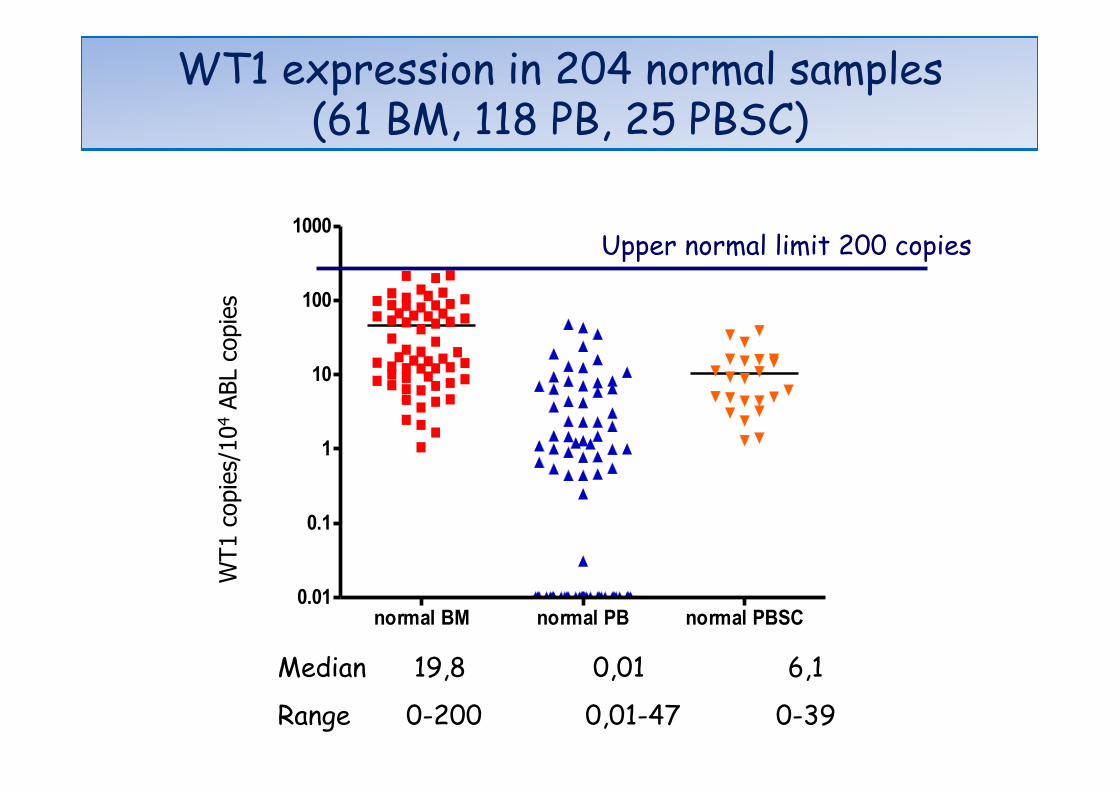
normal BM normal PB normal PBSC0.01
0.1
1
10
100
1000W
T1
copi
es/1
0.00
0 A
BL
copi
es
Median 19,8 0,01 6,1
Range 0-200 0,01-47 0-39
WT1
cop
ies/
104
ABL
copi
es
Upper normal limit 200 copies
WT1 expression in 204 normal samples (61 BM, 118 PB, 25 PBSC)

BM PB0.01
0.1
1
10
100
1000
10000
100000
1000000
1.0×1007
WT1 expression in 729 samples from AML at diagnosis
(collected by the European Leukemia Net)
(588 BM, 141 PB)
11% 12% WT1
cop
ies/
104 A
BL c
opie
s

• No significant difference in WT1 expression at diagnosis by stra@fying the pa@ents according to: – cytogene@c risk groups (except for APL pa@ents who show significantly higher WT1 values)
– muta@ons of NPM1 or FLT3
favorable intermediate adverse10
100
1000
10000
100000
1000000W
T1 c
opie
s/10
000
AB
L co
pies

• 114 patients evaluated at diagnosis and during follow-up
Ø All the patients included have been previously characterized by cytogenetic and molecular analysis
Ø Clinical data available
Ø All the patients were treated with intensive anthracycline and ARA-C
Ø 91/114 (80%) showed WT1 copies > 20.000/104
ABL at diagnosis (2 logs higher than normal controls)
AML patients during follow-up (ELN study)

Kinetics of WT1 response following induction therapy predicts risk of subsequent relapse
Analysis in 91/114 cases with baseline WT1 > 2 x 10⁴ copies/ 104 ABL copies!!!
Greater reduction in WT1 decreases risk of relapse!HR 0.54 (0.36-0.83) p=0.004!
p=0,004
Cilloni et al. JCO 2009

Does log reduction add to the risk score?
• Regression analysis showed that “log reduction” is an independent predictor of relapse
• adjusted for age: HR 0.54 (0.35-0.83) p=0.05 • adjusted for WBC: HR 0.54 (0.35-0.81) p=0.003 • adjusted for cytogenetics: HR 0.63 (0.41-0.98)
p=0.04
• Log reduction remains prognostic even when adjusted for age, WBC, cytogenetics individually

The achievement of normal WT1 values after induction chemotherapy is predictive of relapse
Cilloni et al. JCO 2009

The achievement of normal WT1 values after consolidation chemotherapy is predictive of relapse
Cilloni et al. JCO 2009
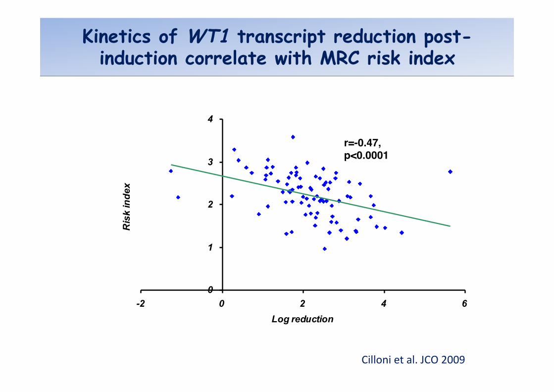
Kinetics of WT1 transcript reduction post-induction correlate with MRC risk index
0
1
2
3
4
-2 0 2 4 6Log reduction
Ris
k in
dex
r=-0.47, !p<0.0001!
Cilloni et al. JCO 2009

Prospec@c study
WT1 at diagnosis and during follow up in AML patients ( 18-60 years ) enrolled in the GIMEMA study treated with the same chemotherapeutical scheme

Is PB better than BM?

WT1 assessment in PB samples
• 82 AML patients prospectively studied • WT1 evaluation in Pb samples
– Diagnosis, – after induction and consolidation treatment – During follow-up
• Patients treated with GIMEMA protocols
Cilloni et al. Haematologica 2008 ; 93:921
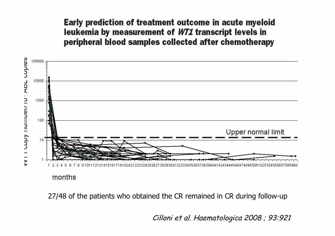
Cilloni et al. Haematologica 2008 ; 93:921
27/48 of the patients who obtained the CR remained in CR during follow-up
Cilloni et al. Haematologica 2008 ; 93:921
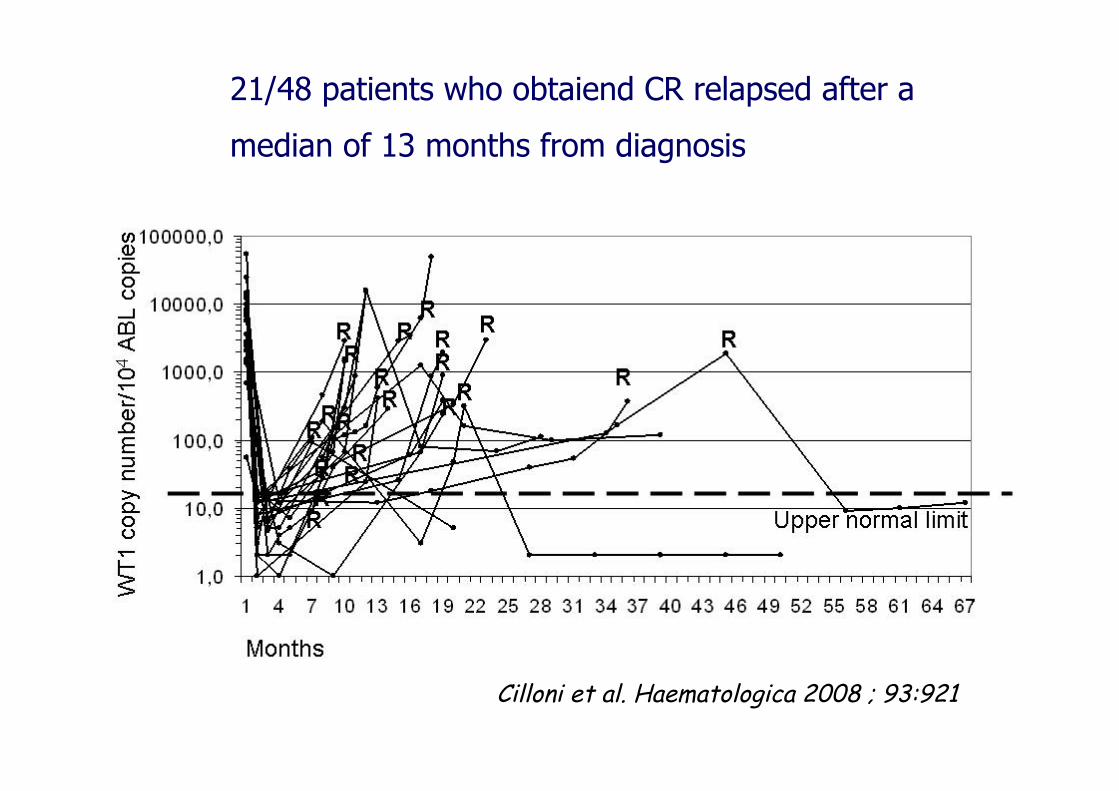
21/48 patients who obtaiend CR relapsed after a
median of 13 months from diagnosis

Cilloni et al. Haematologica 2008 ; 93:921
23 CR patients with WT1 above the normal upper limit relapsed after a median of 7 months from diagnosis (range 6-44)

WT1 transcript after chemotherapy in patients with normal values at diagnosis (<200 copies/104 ABL)
diagnosis post induction0
102030405060708090
100110120
WT1
cop
ies/
10.0
00 A
BL
copi
es
p=0,85!

Prognos@c significance of early peripheral blast clearance as assessed by WT1 transcript reduc@on during the first days of standard induc@on therapy (day 1 and 5) in 57 adult pa@ents with AML
Adapted from Haematologica 2010; 95: 833

Adapted from Gianfaldoni et al. Haematologica 2010; 95: 833
Early reduc@on of WT1 in PB ( day 1 and 5) predicts DFS and OS
MRD in the seang of bone marrow transplanta@on (BMT)


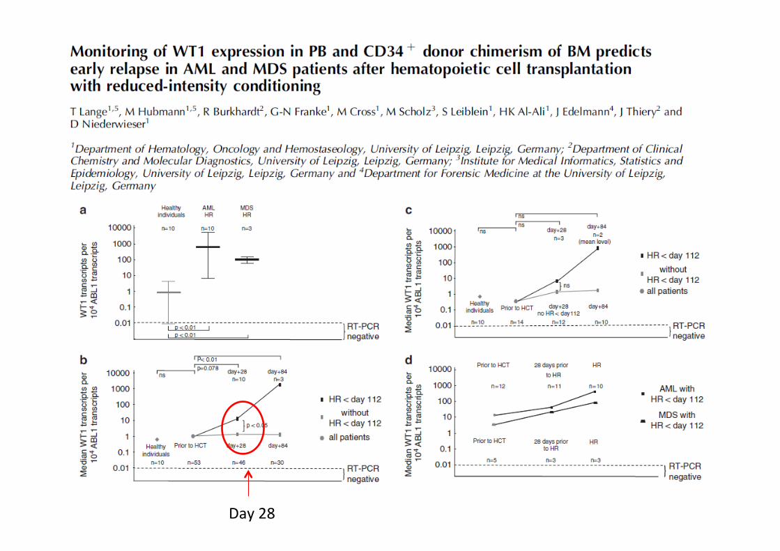
Day 28

Which is the best time point to evaluate WT1 after allogeneic BM transplant?
§ Lack of studies aimed at investigating the best time point after transplant
§ Early detection generate false positive?
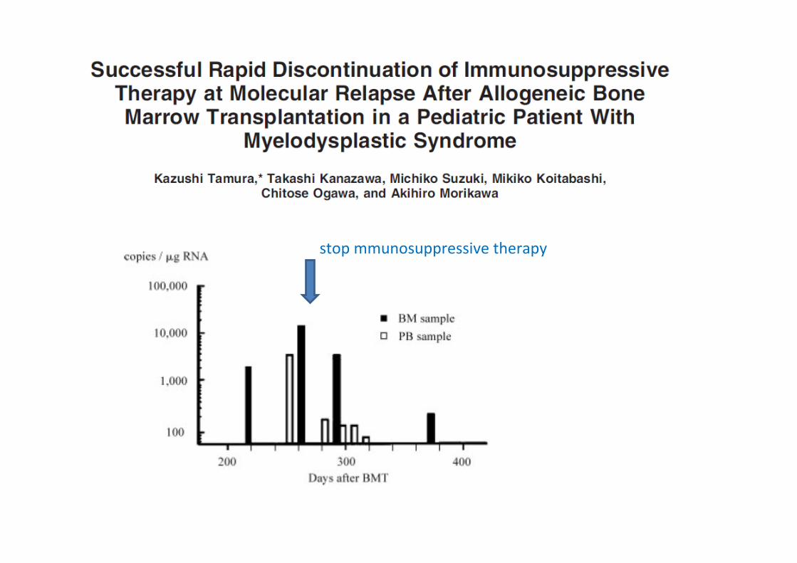
stop mmunosuppressive therapy

MRD in the seang of allogeneic bone marrow transplanta@on (BMT)
Ø To assess the disease status at transplant: prognos@c value
Ø To iden@fy the reappearance of the leukemic clone: possibity of early interven@on

CONCLUSION These data suggest that pre-‐HCT MRD is associated with increased risk of relapse and death aber myeloabla@ve HCT for AML in first morphologic CR, even aber controlling for other risk factors
Impact of Pretransplanta8on Minimal Residual Disease, As Detected by Mul8parametric Flow Cytometry, on Outcome of Myeloabla8ve Hematopoie8c Cell Transplanta8on for Acute Myeloid Leukemia
PATIENTS: 99 consecu@ve pa@ents receiving myeloabla@ve HCT for AML in first morphologic remission MRD detec5on: Ten-‐color mul@parametric flow cytometry (MFC)
Walter et al. JCO 2011

Pechè WT1 è un marcatore precoce di cellula “leucemica” ?


0
2000
4000
6000
8000
10000
12000
14000
16000
COS COS transfected COS transfected treatedwith Imatinib
WT1
cop
y nu
mbe
r/106
β2
mic
rogl
obul
in c
opie
s
Espressione di WT1 in cellule COS dopo trasfezione con BCR-ABL

Ras val 12 CTRL
WT1
0
2000
40006000
8000
10000
1200014000
16000
18000
RAS CTRL
WT1
cop
ies/
104 A
BL
copi
es
Espressione di WT1 in cellule 293T dopo trasfezione con Ras Val12
Actin

È possibile aumentare la specificità e sensibilità?

WT1 isoforms

• Rapporto KTS+/KTS- fisiologico: 1-1,5 • Nelle leucemie il rapporto è sbilanciato
KTS+
KTS+
KTS+
KTS+
KTS+
KTS-
KTS-
KTS-
- -
0
20
40
60
80
100
120
esordio remissione esordio remissione
paziente 1 paziente 2
KTS-KTS+
Follow-up di 2 pazienti

§ WT1 is overexpressed in the majority of AML patients
§ High WT1 level after induction chemotherapy is a negative prognostic factor
§ Increasing of WT1 during follow-up predicts subsequent relapse (even after transplant )
§ WT1 ca be monitored in PB whit high frequency
Conclusions