with thioredoxin from escherichia coli in mice
TRANSCRIPT

Release of two-cell block by reduction of protein disulfidewith thioredoxin from Escherichia coli in mice
S. Natsuyama, Y. Noda, K. Narimoto, Y. Umaoka and T. MoriDepartment of Gynaecology and Obstetrics, Faculty of Medicine, Kyoto University, Kyoto 606,
Japan
Summary. The development of mouse pronuclear-stage embryos in media containingvarious concentrations of thioredoxin was monitored and the influence of anti-thioredoxin immunoglobulin G (IgG) and heat-treated thioredoxin on the thioredoxin\x=req-\induced effects was evaluated. A significant increase in the number of four-cell embryos(76\m=.\3%)and blastocysts (37\m=.\3%)was observed when embryos were cultured in themedium containing 50\g=m\gthioredoxin ml\m=-\1 compared with the rates (55\m=.\8and 3\m=.\8%,respectively) in the basic medium. The number of blastocysts increased significantlyto a maximum of 70\m=.\2%at 500 pg ml\m=-\1. The biological activity of thioredoxin wasevident after dialysis, but was markedly impaired by the addition of anti-thioredoxinIgG to the culture medium. Treatment at 60\s=deg\Cfor 5 min did not affect the enzymaticand biological activity of thioredoxin. More severe heat treatment (121\s=deg\Cfor 30 min)attenuated the enzymatic activity to 40% of its initial value and reduced the biologicalactivity (number of blastocysts, from 77\m=.\8to 51\m=.\6%).
These results indicate that the effect of thioredoxin on the two-cell block is due tothe thioredoxin molecule itself, and suggest that disulfide formation within or betweenproteins resulting from oxidative stress is one of the major causes of the two-cell block.
Keywords: thioredoxin; embryo; mouse
IntroductionMammalian preimplantation embryos exhibit retarded growth and developmental blockage invitro (Wright & Bondioli, 1981; Fisher, 1987). For example, in mice, embryos of outbred strainsexhibit the developmental block at the two-cell stage in vitro (Whitten, 1957; Yanagimachi &Chang, 1964; Whittingham, 1975). In humans, eggs fertilized in vitro show retarded growth at thefour-cell and more advanced stages and few embryos grow to the blastocyst stage (Fishel, 1986).These phenomena are thought to be caused by deficiencies of the conventional culture systemcompared with the in vivo environment; neither growth retardation nor developmental blockage areobserved in vivo.
Oxygen concentration in vitro (i.e. the atmosphere) is about 3-10 times higher than that in vivo(i.e. oviduct or uterus) (Bishop, 1956; Mastroianni & Jones, 1965; Maas et ai, 1976). Low oxygentension of culture media has been reported to promote embryo development in many species(Whitten, 1970). We have shown that the two-cell block in mice is released by adding Superoxidedismutase (SOD), a scavenger of Superoxide anión radicals, to the culture medium (Noda et ai,1991). An additive effect of low oxygen and SOD on mouse embryo development has been demon¬strated (Umaoka et ai, 1992). Generation of reactive oxygen species in blocked embryos(Nasr-Esfahani et ai, 1990a) and overcoming the two-cell block by apotransferrin and ironchelators, which suppress the Haber-Weiss reaction (Nasr-Esfahani et ai, 1990b), have also beenreported. Considering these results together, it is possible that the two-cell block in mice is due todamage to the embryos by oxygen radicals.

It is known that oxygen radicals inactivate various enzymes and damage membranes and DNA(Halliwell & Gutteridge, 1989). The sulfhydryl group of proteins, readily oxidized under oxidativestress (Brigelius, 1985), may be damaged by oxygen radicals resulting in blocked embryo develop¬ment in vitro. Recently, thioredoxin (Laurent et ai, 1964), a small heat-resistant enzyme (molecularmass of about 12 kDa), which promotes the redox reaction of sulfhydryl groups of proteins, was
demonstrated to have a defensive role against oxidative stress (Holmgren, 1985). In this study theeffects of thioredoxin on mouse embryo development were evaluated and the specificity of theenzyme was established by maintaining mouse embryo cultures in media containing thioredoxin,anti-thioredoxin IgG or heat-treated thioredoxin.
Materials and Methods
Chemicals and enzymesThioredoxin (from Escherichia coli) was purchased from Promega Co. (Madison, WI, USA). Before use, the
thioredoxin preparation was dialysed extensively against distilled water followed by lyophilization. Pregnant mares
serum gonadotrophin (PMSG) and human chorionic gonadotrophin (hCG) were purchased from Teikoku Zoki Co.(Tokyo, Japan). Bovine serum albumin (BSA), hyaluronidase (bovine) and insulin (bovine) were purchased fromSigma Chemical Co. (MO, USA). The other chemicals were of reagent grade and purchased from Nacalai Tesque Co.(Kyoto, Japan).
Preparation of anti-thioredoxin antiserum and extraction of the IgG fractionThe anti-thioredoxin antiserum was prepared by injecting the immunogen (0-5 mg ml
"
'), with Freund's completeadjuvant, into the dorsal skin of female rabbits six times at intervals of 2 weeks. The rabbits were bled 7 days after thelast injection. The IgG fractions of anti-thioredoxin antiserum ( -Trx-IgG) and normal rabbit serum (NRS-IgG) wererecovered from a protein- Sepharose 4B column (Duhamel et ai, 1979). The IgG preparations were dialysed againstdistilled water and lyophilized.
Reactivity of -Trx-IgG was examined by the method of Ouchterlony & Nilson (1978). Thioredoxin, BSA, humanalbumin, mouse liver homogenate and -Trx-IgG were added to the diffusion well and incubated at 4°C overnight.The formation of precipitation bands was examined.
Heat treatment of thioredoxinThioredoxin was dissolved in deionized water at 1 mg ml"1 and the solution was divided into three parts, which
were then heat-treated (i) in a hot waterbath at 60°C for 5 min, (ii) at 100"C for 30 min, or (iii) in an autoclave at 121 °Cfor 30 min. The heat-treated thioredoxin specimens were freeze-dried and stored.
Enzyme activity of the heat-treated thioredoxin was assayed by a turbidimetric assay using dithiothreitol (DTT)and insulin (Holmgren, 1979). The heat-treated thioredoxin samples were dissolved in phosphate buffer (0-1 mol 1
"
' )and then added to the reaction cuvette (volume 1 -2 ml) at a final concentration of 60 pg ml
~
' (5 pmol 1). Reactionmedia contained 0-13 mmol insulin 1" ', 0-33 mmol DTT 1" ', 2 mmol EDTA 1
"
' and 0-1 mol phosphate buffer 1"
'.Increase in absorbance at 650 nm (A650) caused by splitting of insulin molecules was recorded using a ShimadzuUV-260 spectrophotometer. Thioredoxin activity was evaluated using the maximum increase in A650 in one minute.Standard curves were made from original samples of thioredoxin at 0, 2-5, 5, 7-5 and 10 pmol 1
"
'.
Embryo collection and culture conditionsTUCK (outbred) mice were purchased from A. Tuck and Son Ltd (Battlesbridge, UK). Female mice, aged 4-5
weeks, were injected i.p. with PMSG (5 iu) and hCG (5 iu) at an interval of 48 h to induce superovulation. They were
then mated with 12-week-old male mice of the same strain. Vaginal plug formation was confirmed on the next
morning (day 1 of pregnancy). Female mice were killed by cervical dislocation, and pronuclear-stage embryos were
collected from the ampulla of oviducts by the scratching method 17 h after hCG injection. After the removal ofcumulus cells with 0-1 % hyaluronidase, all the embryos were pooled in an 80 pi spot of Dulbecco's phosphate saline,and 10-18 embryos with normal morphology were placed into each experimental spot of medium at random and thencultured at 37°C under 5% C02 in air. Degenerated embryos were excluded.
The basic medium for embryo culture was Biggers-Whitten-Whittingham (BWW) solution (Biggers el al, 1971)supplemented with 0-3% BSA. An 80 pi spot of medium was placed in each well of the four-well multidish (Nunc Co.,Denmark) and covered with mineral oil. Embryos were observed every 24 h under an Olympus IMT-2 microscopewith a Nomarski differential interferometer. The culture efficacy in the following experiments was evaluated bydetermining the proportion of embryos reaching two-cell (day 2), four-cell (day 3) and blastocyst (day 5) stages.

Experiment 1: embryo culture in the thioredoxin-supplemented mediumPronuclear-stage embryos were cultured after adding various concentrations (10, 50, 100, 500 and 1000 pg ml-1)
of thioredoxin to the basic medium. As a control, pronuclear embryos were cultured in the basic medium. Embryocultures in each thioredoxin-supplemented medium were started at the same time.
Experiment 2: inhibition of thioredoxin-induced effects by -Trx-IgGPronuclear-stage embryos were cultured in the following media: group 1, basic medium containing thioredoxin
(30pgml~\ 2-5 pmol l"1); group 2, basic medium containing thioredoxin (30 pgmP1) and NRS-IgG (500pgml_1);group 3, basic medium containing thioredoxin (30pgml_1) and -Trx-IgG (500pgml_1); group 4, basic mediumcontaining NRS-IgG (500 pg ml" '); group 5, basic medium containing -Trx-IgG (500 pg ml" '); and group 6, basicmedium. Embryo cultures in the six groups were started at the same time.
Experiment 3: influence of heat treatment on thioredoxin-induced effectsPronuclear-stage embryos were cultured after adding heat-treated thioredoxin at 500pgml~' (41-7 pmol l"1)
to the basic medium. As a control, pronuclear-stage embryos were cultured in the basic medium containing untreatedthioredoxin (500 pg ml" '). Embryo cultures in each medium were started at the same time.
Statistical analysisThe results of each experiment were analysed by the 2 test.
Results
Reactivity of -Trx-IgG was examined by Ouchterlony's method. A single precipitation band wasobserved between -Trx-IgG (80 pg) and thioredoxin (1 pg) (Fig. 1). No precipitation band wasobserved against BSA, human albumin and mouse liver homogenate at various amounts (01, 1, 10and 100 pg).
Fig. 1. Precipitation pattern between the immunoglobulin fraction of anti-thioredoxin rabbitantiserum ( -Trx-IgG) and thioredoxin. A single precipitation band is observed between a-Trx-IgG (A: 80 pg) and thioredoxin (C: 1 pg). No precipitation bands are found between -Trx-IgG(80 pg) and BSA (B: 1 pg), human albumin (D: 1 pg), or mouse liver homogenate (E: 1 pg).

The enzyme activity of heat-treated thioredoxin solution remained high after 5 min at 60°C(Table 1). Heat treatment at 100°C for 30 min and at 121 °C for 30 min decreased enzymatic activityto 80% and 40% of that of untreated thioredoxin, respectively.
Table 1. Turbidimetric assay of thioredoxin
Enzymatic activitySpecimens ( A650 nm min" ')Thioredoxin (pmol ml"')
0 00002-5 006750 00937-5 0149
100 0178Heat-treated thioredoxin (50 pmol ml" ')
60°C, 5 min 0088100°C, 30min 0075121°C, 30 min 0039
Effects of thioredoxin on embryo developmentA significant increase in the numbers of embryos at the four-cell (day 3) (76-3%) and blastocyst
(day 5) (37-3%) stages was observed when the culture media contained 50 pg thioredoxin ml-1(4-2 pmol 1" '), compared with culture in the basic medium (55-8%, 3-8%). When the concentrationwas increased, the frequency of blastocysts also increased and then decreased gradually at higherconcentrations (Table 2).
Table 2. Effects of thioredoxin on mouse embryo development
Number ofNumber (%)b of embryos developed to
Number of embryos two-cell four-cell blastocystConditions trials examined" stage stage stage
Basic medium+ thioredoxin(pgrnl"')
10 4 60 50(83-3) 31(51-7) 9(150)50 4 59 52(88-1) 45(76-3)* 22(37-3)**
100 4 59 54(91-5) 44(74-6)* 35(59-3)**500 4 57 49(860) 48(84-2)** 40(70-2)**
1000 4 55 46(83-6) 43(78-2)* 34(61-8)**5000 4 57 52(91-2) 46(80-7)** 31(54-4)**
Basic medium 4 52 46(88-5) 29(55-8) 2 (3-8)(control)
"Pronuclear-stage embryos collected 17 h after administration of hCG. 12-16 embryoswere used per experiment.
bPercentages of embryos at each stage represent cumulative totals.*P < 005; **P < 001, compared with the control value.
Inhibition of thioredoxin-induced effects by -Trx-IgGFlocculate precipitates were observed 12 h after the addition of -Trx-IgG to the medium
containing thioredoxin, but not otherwise.

A significantly higher incidence of blastocysts was obtained by culture with thioredoxin alone(28-1%, group 1) or with thioredoxin and 500 pg NRS-IgG ml"1 (28-8%, group 2), compared withthat (41%) noted in the basic medium (group 6). However, the incidence of blastocysts in themedium containing thioredoxin and -Trx-IgG in group 3 was low (10-7%) and comparable withthat found in the basic medium. The addition of NRS-IgG or -Trx-IgG alone to the basic mediumdid not significantly affect the incidence of blastocysts (groups 4 or 5) (Table 3).
Table 3. Inhibition of thioredoxin-induced effects by a-thioredoxin-IgG in mice
Number (%)b of embryos developed toNumber of-Number of embryos two-cell four-cell blastocyst
Conditions trials examined" stage stage stage
BWW + Trx 4 57 51(89-5) 40(70-2)* 16(28-1)**BWW + Trx 4 56 51(981) 27(51-9) 15(28-8)**
+ NRS-IgGBWW + Trx 4 52 53(94-6) 15(26-8) 6(10-7)
+ a-Trx-IgGBWW 4 60 59(98-3) 22(36-6) 1 (1-7)
+ NRS-IgGBWW 4 63 62(98-4) 23(36-5) 4 (6-3)
+ a-Trx-IgGBWW (control) 4 49 45(91-8) 23(46-9) 2 (41)
"Pronuclear-stage embryos collected 17 h after administration of hCG. 10-17 embryos were
used per experiment.'Percentages of embryos at each stage represent cumulative totals.BWW: Biggers-Whitten-Whittingham solution.Trx: Thioredoxin from Escherichia coli at 30 pg ml" '.NRS-IgG: IgG fraction of normal rabbit serum at 500 pg ml" '. -Trx-IgG: IgG fraction of anti-thioredoxin antiserum at 500 pg ml" '.*P < 005; **P < 001, compared with the control values.
Influence of heat treatment on thioredoxin-induced effects
A significant increase in the incidence of both the four-cell and blastocyst stages was observed inthe culture media containing each heat-treated thioredoxin or untreated thioredoxin, comparedwith the incidence in the basic medium. The embryo promoting effect of thioredoxin was notinfluenced significantly by heat treatment at 60°C for 5 min or at 100°C for 30 min, but was
impaired by heat treatment at I21°C for 30 min (Table 4).
Discussion
In this study, we demonstrated that thioredoxin overcomes the two-cell block in mice and promotesthe development of mouse embryos in vitro. The biological activity of thioredoxin was not lostafter dialysis, but was markedly impaired after the addition of -Trx-IgG to the culture medium,indicating that the effect of the thioredoxin preparation on the two-cell block phenomenon is due tothe thioredoxin molecule itself. Heat treatment at 60°C for 5 min did not diminish the enzymaticand biological activity of thioredoxin, showing the extraordinary heat stability of this enzyme(Laurent et al, 1964; Holmgren, 1985). Heat treatment at 121°C for 30 min reduced the enzymaticactivity to 40% of initial values and decreased the biological activity even more, suggesting that thepromotion of embryo development by thioredoxin is a phenomenon dependent on the enzymeactivity of thioredoxin.
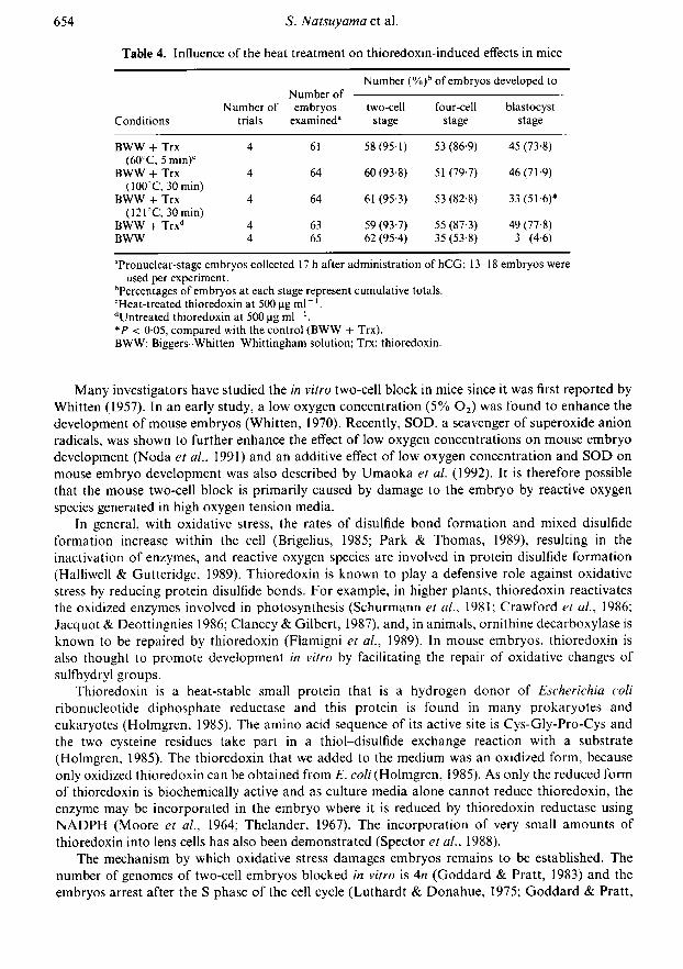
Table 4. Influence of the heat treatment on thioredoxin-induced effects in mice
Conditions
Number ofNumber of embryos
trials examined"
Number (%)b of embryos developed to
two-cellstage
four-cellstage
blastocyststage
BWW + Trx(60°C, 5 min)'
BWW + Trx(100°C, 30 min)
BWW + Trx(121°C, 30 min)
BWW + Trx"BWW
61
64
64
6365
58(95-1)60 (93-8)61 (95 3)
59 (93-7)62 (95-4)
53 (86-9)51 (79-7)53 (82-8)55 (87-3)35 (53-8)
45 (73-8)46(71-9)
33(51-6)*49 (77-8)
3 (4-6)
"Pronuclear-stage embryos collected 17 h after administration of hCG; 13-18 embryos wereused per experiment.
'Percentages of embryos at each stage represent cumulative totals.cHeat-treated thioredoxin at 500pgml_1.dUntreated thioredoxin at 500 pg ml" '.*P < 005, compared with the control (BWW + Trx).BWW: Biggers-Whitten-Whittingham solution; Trx: thioredoxin.
Many investigators have studied the in vitro two-cell block in mice since it was first reported byWhitten (1957). In an early study, a low oxygen concentration (5% 02) was found to enhance thedevelopment of mouse embryos (Whitten, 1970). Recently, SOD, a scavenger of Superoxide aniónradicals, was shown to further enhance the effect of low oxygen concentrations on mouse embryodevelopment (Noda et ai, 1991) and an additive effect of low oxygen concentration and SOD on
mouse embryo development was also described by Umaoka et al. (1992). It is therefore possiblethat the mouse two-cell block is primarily caused by damage to the embryo by reactive oxygenspecies generated in high oxygen tension media.
In general, with oxidative stress, the rates of disulfide bond formation and mixed disulfideformation increase within the cell (Brigelius, 1985; Park & Thomas, 1989), resulting in theinactivation of enzymes, and reactive oxygen species are involved in protein disulfide formation(Halliwell & Gutteridge, 1989). Thioredoxin is known to play a defensive role against oxidativestress by reducing protein disulfide bonds. For example, in higher plants, thioredoxin reactivatesthe oxidized enzymes involved in photosynthesis (Schurmann et ai, 1981; Crawford et ai, 1986;Jacquot & Deottingnies 1986; Clancey & Gilbert, 1987), and, in animals, ornithine decarboxylase isknown to be repaired by thioredoxin (Flamigni et ai, 1989). In mouse embryos, thioredoxin isalso thought to promote development in vitro by facilitating the repair of oxidative changes ofsulfhydryl groups.
Thioredoxin is a heat-stable small protein that is a hydrogen donor of Escherichia coliribonucleotide diphosphate reductase and this protein is found in many prokaryotes andeukaryotes (Holmgren, 1985). The amino acid sequence of its active site is Cys-Gly-Pro-Cys andthe two cysteine residues take part in a thiol-disulfide exchange reaction with a substrate(Holmgren, 1985). The thioredoxin that we added to the medium was an oxidized form, becauseonly oxidized thioredoxin can be obtained from E. coli (Holmgren, 1985). As only the reduced formof thioredoxin is biochemically active and as culture media alone cannot reduce thioredoxin, theenzyme may be incorporated in the embryo where it is reduced by thioredoxin reductase usingNADPH (Moore et al., 1964; Thelander, 1967). The incorporation of very small amounts ofthioredoxin into lens cells has also been demonstrated (Spector et ai, 1988).
The mechanism by which oxidative stress damages embryos remains to be established. Thenumber of genomes of two-cell embryos blocked in vitro is An (Goddard & Pratt, 1983) and theembryos arrest after the S phase of the cell cycle (Luthardt & Donahue, 1975; Goddard & Pratt,

1983), suggesting that a system that regulates the cell cycle from the S to M phase or a factorinvolved in cell division is damaged in the two-cell embryos blocked in vitro. The release of mousetwo-cell block by thioredoxin suggests that the sulfhydryl group of proteins involved in cell divisionor cell cycle regulation is one of the targets of oxidative stress. For example, tubulin may be a
target, because the sulfhydryl group in the cystine residue of tubulin, which is a component of themicrotubules in the mitotic apparatus, is involved in the polymerization of tubulin to form themicrotubules (Kuriyama & Sakai, 1974).
In conclusion, we have demonstrated that thioredoxin releases mouse two-cell block andpromotes the in vitro development of mouse embryos, and that the effects were induced by thethioredoxin molecule itself. Taking into consideration the fact that low oxygen concentration andSOD also promote development of embryos, we suggest that embryos must be carefully protectedfrom oxidative stress in vitro. The precise mechanism of action of thioredoxin remains to bedetermined.
This study was supported by Grants in Aid for General Scientific Research by the Ministry ofEducation, Science and Culture (No. 01480391) and for the Scientific Research on Priority Area(No. 0164004 and No. 02222104).
References
Biggers, J.D., Whitten, W.K. & Whittingham, D.G.(1971) The culture of mouse embryos in vitro. InMethods in Mammalian Embryology, pp. 86-116. Ed.J. C. Daniel, Jr. Freeman Co., San Francisco.
Bishop, D.W. (1956) Oxygen concentration in the rabbitgenital tract. In Proceedings of the Third InternationalCongress of Animal Reproduction, Physiology, pp.53-58. Ed. D.W. Bishop. Brown, Knight & TruscottLtd, Cambridge, UK.
Brigelius, R. (1985) Mixed disulfide. In Oxidative Stress,pp. 247-279. Ed. H. Sies. Academic Press, London.
Clancey, C.J. & Gilbert, H.F. (1987) Thiol/disulfideexchange in the thioredoxin-catalized reductiveactivation of spinach chloroplast fructose-1,6-bis-phosphatase. Kinetics and thermodynamics. JournalofBiological Chemistry 262, 13 545-13 549.
Crawford, N.A., Yee, B.C., Hutcheson, S.W., Wolosiuk,R.A. & Buchanan, B.B. (1986) Enzyme regulationof C4 photosynthesis: purification, properties andactivities of thioredoxin from C4 and C3 plants.Archives of Biochemistry and Biophysics 244, 1-15.
Duhamel, R.C., Schur, P.H., Brendel, . & Meezan, E.(1979) pH gradient elution of human IgGl, IgG2and IgG4 from proteinA-sepharose. Journal ofImmunology Methods 31, 211-217.
Fishel, S. (1986) Growth of the human conceptus invitro. In In Vitro Fertilization, pp. 107-126. Eds S.Fishel & E. M. Simonds. IRL Press, Oxford.
Fisher, B. (1987) Developmental retardation in culturedpreimplantation rabbit embryos. Journal of Repro¬duction and Fertility. 79, 115-123.
Flamigni, F., Marmiroli, S., Caldarera, CM. &Guarnieri, C. (1989) Involvement of thiol transferase-and thioredoxin-dependent systems in the protectionof'essential' thiol groups of ornithine decarboxylase.Biochemical Journal 259, 111-115.
Goddard, M.J. & Pratt, H.P.M. (1983) Control ofevents during early cleavage of the mouse embryo: an
analysis of the 2-cell block. Journal of Embryologyand Experimental Morphology 73, 111—133.
Halliwell, . & Gutteridge, J.M.C. (1989) Protectionagainst oxidants in biological systems: the Superoxidetheory of oxygen toxicity. In Free Radicals inBiology and Medicine (2nd edn), pp. 86-187. OxfordUniversity Press, New York.
Holmgren, A. (1979) Thioredoxin catalyses the reductionof insulin disulfides by dithiothreitol and dihydro-lipoamide. Journal of Biological Chemistry. 254,9627-9632.
Holmgren, A. (1985) Thioredoxin. Annual Reviews ofBiochemistry. 54, 237-271.
Jacquot, J.P. & Decottingnies, P. (1986) Furtherevidence for a role of sulfhydryls in the thioredoxindependent activation of corn NADP-malate de-hydrogenase. Use of cysteine free mutant of E. colithioredoxin. FEBS Letters 209, 87-91.
Kuriyama, R. & Sakai, H. (1974) Role of tubulin-SHgroups in polymerization to microtubules: func-tional-SH groups in tubulin for polymerization.Journal of Biochemistry 76, 651-654.
Laurent, T.C., Moore, E.C. & Reichard, P. (1964)Enzymatic synthesis of deoxyribonucleotides: IV.isolation and characterization of thioredoxin, thehydrogen donor from Escherichia coli . Journal ofBiological Chemistry 239, 3436-3444.
Luthardt, F.W. & Donahue, R.P. (1975) DNA synthesisin developing two-cell mouse embryo. DevelopmentalBiology 44,210-216.
Maas, D.H.A., Storey, B.T. & Mastroianni, L., Jr(1976) Oxygen tension in the oviduct of the rhesusmonkey (Macaca mulatta). Fertility and Sterility 27,1312-1317.
Mastroianni, L., Jr & Jones, R. (1965) Oxygen tension inthe rabbit fallopian tube. Journal ofReproduction andFertility 9,99-102.
Moore, E.C, Reichard, P. & Thelander, L. (1964)Enzymatic synthesis of deoxyribonucleotides: V. pur¬ification and properties of thioredoxin reductasefrom Escherichia coli . Journal of BiologicalChemistry 239, 3445-3452.
Nasr-Esfahani, M.H., Aitken, J.R. & Johnson, M.H.(1990a) Hydrogen peroxide levels in mouse oocytesand early cleavage stage embryos developed in vitroor in vivo. Development 109, 501-507.
Nasr-Esfahani, M.H., Johnson, M.H. & Aitken, J.R.(1990b) The effect of iron and iron chelators onthe in-vitro block to development of the mouse pre¬implantation embryo: BAT6 a new medium forimproved culture of mouse embryos in vitro. HumanReproduction 5, 997-1003.
Noda, Y., Matsumoto, II.. Umaoka, Y., Tatsunii. K.,Kishi, J. & Mori, T. (1991) Involvement of super-oxide radicals in the mouse 2-cell block. MolecularReproduction and Development 28, 356-360.
Ouchterlony, O. & Nillson, L.A. (1978) Immunodiffusionand immunoelectrophoresis. In Handbook of Exper¬imental Immunology, pp. 19.1-19.44. Ed. D. M. Weir.Blackwell Scientific Publications, London.
Park, E.M. & Thomas, J.A. (1989) Reduction of proteinmixed disulfides (dethiolation) by Escherichia colithioredoxin: a study with glycogen phosphorylase band creatine kinase. Archives of Biochemistry andBiophysics 272, 25-31.
Schurmann, P., Maeda, K. & Tsugita, A. (1981) Isomersin thioredoxins of spinach chloroplasts. EuropeanJournal ofBiochemistry 116, 37^t5.
Spector, ., Yan, G.Z., Huang, R.R.C, McDermott,M.J., Gascoyne, P.R.C. & Pigiet, V. (1988) The effect
of H202 upon thioredoxin-enriched lens epithelialcells. Journal of Biological Chemistry 263, 4984^990.
Thelander, L. (1967) Thioredoxin reductase: characteriz¬ation ofa homogeneous preparation from Escherichiacoli . Journal of Biological Chemistry 242, 852-859.
Umaoka, Y., Noda, Y., Narimoto, K. & Mori, T. (1992)Effects of oxygen toxicity on early developmentof mouse embryos. Molecular Reproduction andDevelopment 31,28-33.
Whitten, W.K. (1957) Culture of tubai ova. Nature 179,1081-1082.
Whitten, W.K. (1970) Nutrient requirements for theculture of preimplantation embryos in vitro. InAdvances in the Biosciences 6, pp. 129-141. Ed. G.Raspe. Pergamon Press, Oxford.
Whittingham, D.G. (1975) Fertilization, early develop¬ment and storage of mammalian ova in vitro. In TheEarly Development of Mammals, pp. 1-24. Eds M.Balls & A. E. Wild. Cambridge University Press,Cambridge.
Wright, R.J., Jr & Bondioli, K.R. (1981) Aspects of invitro fertilization and embryo culture in domesticanimals. Journal of Animal Science 53, 702-728.
Yanagimachi, R. & Chang, M.C. (1964) In vitro fertiliz¬ation of golden hamster ova. Journal of ExperimentalZoology 156, 361-376.
Received 22 April 1991