withaferin a: a novel therapeutic approach for …
TRANSCRIPT

WITHAFERIN A: A NOVEL THERAPEUTIC APPROACH FOR MALIGANT BRAIN
TUMORS
By
Patrick Thomas Grogan
Submitted to the graduate degree program in Pharmacology, Toxicology, and Therapeutics and
to the Graduate Faculty of the University of Kansas Medical Center in partial fulfillment of the
requirements for the degree of Doctor of Philosophy.
_____________________________
Committee Chair: Thomas Pazdernik, Ph.D.
_____________________________
Mentor: Mark Cohen, M.D., F.A.C.S.
_____________________________
Qi Chen, Ph.D.
_____________________________
Partha Kasturi, Ph.D.
_____________________________
Timothy Fields, M.D., Ph.D.
Date defended: May 19, 2014
Copyright 2014
Patrick Thomas Grogan
ii
The Dissertation Committee for Patrick Thomas Grogan certifies that this is the approved
version of the following dissertation:
WITHAFERIN A: A NOVEL THERAPEUTIC APPROACH FOR MALIGANT BRAIN
TUMORS
_____________________________
Committee Chair: Thomas Pazdernik, Ph.D.
Date approved: May 19, 2014

iii
Abstract
High-grade gliomas, including the astrocytoma glioblastoma multiforme (GBM), are the
most common adult primary malignant brain tumor. The mean post-diagnosis survival time of
patients with GBM is approximately 14 months and has improved only minimally over the last
several decades given a lack of novel and effective therapeutic strategies or interventions.
Similar issues persist for other forms of brain cancer, notably medulloblastomas (MB) in the
pediatric patient population. Given our inability to extend survival and enhance quality of life
adequately in these brain tumor patients, there is a critical need for novel chemotherapeutic
agents in the treatment of GBM and MB that may work as monotherapy agents or in synergistic
combinations with current interventions.
In this work, the role of the natural product withaferin A (WA), a steroidal lactone with
intriguing cytotoxic properties, was studied alone or in combination with currently approved
anti-cancer agents (temozolomide, radiation therapy, and proteosome inhibitors) against GBM
and MB brain tumors. It was shown that WA could produce G2/M cell cycle arrest and apoptosis
with inhibitory modulation of the Akt/mammalian target of rapamycin (mTOR) pathway in
GBM. Similarly, WA inhibited Wnt/β-catenin signaling through degradation of transcription
factor (TCF)/lymphoid enhancer-binding factor (LEF) family members in MB. Overall, exposure
to WA was associated with generalized N-acetyl-L-cysteine-repressible cellular oxidation, thiol
reactivity, and alterations in the heat shock protein (HSP) 90 chaperone axis. WA failed to alter
intrinsic HSP90 activity but reduced the association between HSP90 and co-chaperone Cdc37.
These findings were expanded to demonstrate WA-mediated potentiation of cytotoxicity with
concurrent proteasomal inhibition through an accumulation of aberrant proteins. WA also
increased tumor cell radiosensitivity through disruption of normal DNA damage recognition and

iv
repair. While WA failed to significantly enhance the cytotoxicity of temozolomide (TMZ), it
demonstrated the ability to re-sensitize TMZ-resistant GBM through reduction in O6-
methylguanine-DNA methyltransferase (MGMT).
This study identifies novel utility for the cytotoxic steroid lactone WA in the treatment of
the malignant brain tumors GBM and MB through its alterations of oncogenic cellular signaling
pathways, protein homeostasis, and the DNA-damage response mechanism. As such, WA
represents a promising experimental therapeutic that warrants further translational exploration.

v
Dedication
In memory of John R. Ohlfest, Ph.D.:
Dr. Ohlfest was my friend and a truly great mentor at the University of Minnesota and was taken
at far too young an age by the very disease he was working to cure. His drive and passion to
develop novel and innovative ideas to better the prognosis of cancer patients still serves as a
source of inspiration for me, and his underlying desire to help people at their most vulnerable
time will always be provide me with the motivation to keep trying.
John, you are missed.
vi
Acknowledgements
To my mentor, Dr. Mark Cohen, I am sincerely grateful for your support and the opportunity that
you have provided me over the last five years. Your research program is one of the main reasons
I came to KUMC and then moved to the University of Michigan. I thank you for the trust that
you have provided me to develop into an independent researcher and learn through both my
successes and failures and your willingness to allow me to continue with you at the time of your
move. You have been a wonderful example of a physician-scientist for me to follow and from
whom I could learn. I appreciate all of your help and advice as I develop as a researcher and
hopefully an eventual physician.
To my committee chairperson, Dr. Thomas Pazdernik, I am indebted to you for your willingness
to first participate on my committee and then take over as chairperson upon Dr. Cohen’s
transition to the University of Michigan. Your input as a committee member and both a graduate
and medical teacher have proven to be invaluable in my development and progression. Your
medical school lectures undoubtedly improved and advanced my clinical comprehension and
certainly that of many of my peers and was a key foundation for my graduate work. I thank you
as well for the time you’ve invested walking me through the administrative steps of graduate
school to ensure that I maintained course.
To my committee member and MD/PhD Program director, Dr. Timothy Fields, I sincerely thank
you for the opportunities and guidance that you have provided me to at KUMC. You were also
one of the primary reasons that I chose to attend KUMC and am very fortunate that you allowed
vii
me that chance. I appreciate all of our discussions on science and life and truly take something
from each.
To my present committee members, Dr. Qi Chen and Dr. Partha Kasturi, and my past member,
Dr. Bao-Ting Zhu, your input and support allowed me to get to where I am. I am grateful for the
significant efforts that you have put into mentoring me and helping me move forward. I would
also like to thank Drs. Kenneth McCarson and Bruno Hagenbuch for their willingness to serve
on my comprehensive examination committee.
To current and former members of the Cohen laboratory or other individuals working within the
physical laboratory, including Dr. Chitra Subramanian, Mr. Joseph Bazzill, Dr. Ridhwi Mukerji,
Dr. Abbas Samadi, Mr. Piero Protti, Ms. Eileen Brandes, Dr. Michael Sim, Dr. Roy Lirov, Dr.
Stephanie Cohen, Dr. Erica Person, Mrs. Jennifer Buseman, and Dr. Xuan Zhang, I thank you for
all of your help – especially with the small, unappreciated help and tasks – that made for a fun
and productive environment that I enjoyed coming to every day. I am especially grateful to Dr.
Chitra Subramanian for her kindness and mentorship within the laboratory, taking the time to
teach me new techniques and working together as a great team on a number of projects.
To the past and present administration of the MD/PhD program, Mrs. Janice Fletcher, Dr. Joseph
Bast, and Dr. Brenda Rongish, I sincerely appreciate your support during my graduate phase
progression. I’d like to especially state my gratitude for Mrs. Fletcher who has put in countless
extra hours to make sure that I stay in good standing with my location change. I would be

viii
completely lost without all of your assistance and guidance on a myriad of too many items to list.
You have been such a valuable asset to both me and the program as a whole.
To the administration of the graduate program in the Department of Pharmacology, Toxicology,
and Therapeutics, namely Dr. Bruno Hagenbuch and Mr. Cody Tully, I appreciate your
assistance and guidance, especially in light of my location change.
To now-Dr. Shane Stecklein, I thank you for your help, advice, and guidance with elements of
the radiation sensitization chapter of this work.
To Dr. Barbara Timmermann and her lab members Huaping Zhang and Robert Gallagher, I
thank you for providing purified withanolides and extracts to evaluate.
To Dr. Yoichi Osawa, Dr. Yoshihiro Morishima, and Dr. William Pratt, I thank you for your
advice and help with elements of the HSP90 experimentation.
To my previous mentors (who still remain mentors to this day), Dr. Jann Sarkaria, Dr. John
Ohlfest, and Dr. Michael Olin, the opportunities that you provided me and the trust you had in
me have helped to shape who I am today. Your passion is admirable, and I thank you for the
examples you have provided.
To my parents, Thomas and Debra Grogan, and my brother, Michael Grogan, thank you for your
love and support. I would not be to this point without your guidance, advice, assistance, and

ix
motivation. To my girlfriend and best friend, Sarah Kay VanOosten, thank you for taking this
journey with me and all the patience that you have shown. I cannot thank you enough for your
love and support. I am proud of all that you have and will accomplish on your own path. To the
VanOosten family, especially Ann and Jim VanOosten, I am forever grateful for your kindness,
support, and understanding. To my feathered buddy Astro (named after the presumed normal cell
of origin for GBM), thanks for always being full of energy and happy to see me. I love you all.
To my other best friend, Paul Schwingler, thanks for always being there and even spending time
rotating through the lab. You are truly dependable and one-of-a-kind.
To my friends in the MD/PhD Program and medical school, thank you being there. And to
members of other labs at the University of Michigan for general advice, assistance, and
friendship, notably Joe Nguyen, Lukasz Ochyl, Molly Kozminsky, and Dr. Luis Villa Diaz, I
thank you.
To various funding sources for making this work possible including: the National Institutes of
Health (NIH-COBRE P20 RR015563 P.I. B. Timmermann), the Institute for Advancing Medical
Innovation (PI: MS Cohen), two University of Kansas Cancer Center Summer Student Training
Program grants (PT Grogan), an Alex’s Lemonade Stand Foundation POST award to initiate
medulloblastoma work that could be expanded on for the dissertation (PT Grogan), the
Departments of Surgery at the University of Kansas Medical Center and University of Michigan
(MS Cohen), and a University of Michigan Comprehensive Cancer Center CCSG Development
award (PI: MS Cohen).

x
To anyone else who I have inevitably forgotten or could not mention, thank you for your
contribution, small or large.

xi
Table of Contents
Title page………………………………………………………………………………………..i
Acceptance Page………………………………………………………………………………..ii
Abstract………………………………………...………………………………………………iii
Dedication………………………………………………………………………….………..….v
Acknowledgements…………………………………………………………………...………..vi
Table of Contents………………………………………………………………………………xi
List of Abbreviations…………………………………...……..………………………………xx
List of Figures…………………………………………………………………………….….xxiv
List of Appendices………………………………….……………………………………….xxxiv
Chapter 1
Background and Introduction
1.1 Brain tumors…………………………………………………………………………...2
1.1.1 Glioblastoma multiforme………………………………………………………2
1.1.2 Blood-brain-barrier……………………………………………………………..4
1.1.3 Medulloblastoma……………………………………………………………….5
1.2 Proposed targets of withaferin A……………………………………………………....6
1.2.1 Oncogenic pathway signaling inhibition……………………………………….6
1.2.1a MAPK………………………………………………………………...10
1.2.1b PI3K/Akt/mTOR……………………………………………..……….13
xii
1.2.1c Wnt/β-catenin…………………………………………………………16
1.2.2 Proteotoxicity…………………………………………………………………19
1.2.3 DNA-damage response modulation..................................................................24
1.3 Specific Aims…………………………………………………………………………30
1.4 Statement of Purpose……………………………………………………………….....32
Chapter 2
Cytotoxicity of withaferin A in glioblastomas involves induction of an oxidative stress-mediated
heat shock response while altering Akt/mTOR and MAPK signaling pathways
2.1 Abstract……………………………………………………………………………….36
2.2 Introduction…………………………………………………………………………...37
2.3 Materials and methods………………………………………………………………..38
2.3.1 Cell culture and general reagents……………………………………………..38
2.3.2 Cell proliferation and viability assays………………………………………...39
2.3.3 Cell cycle analysis…………………………………………………………….40
2.3.4 Analysis of cell death…………………………………………………………40
2.3.5 Western blotting………………………………………………………………41
2.3.6 Detection of reactive oxygen species…………………………………………42
2.3.7 Statistical analysis…………………………………………………………….43
2.4 Results
2.4.1 WA reduces cell proliferation and viability in GBM cells…………………...43
2.4.2 WA induces G2/M cell cycle arrest in GBM cells in a dose-dependent

xiii
manner………………………………………………………………………..44
2.4.3 WA induces GBM cell death………………………………………………...49
2.4.4 WA alters normal protein expression and activation in the Akt/mTOR
and MAPK pathways…………………………………………………….......52
2.4.5 WA elevates pro-oxidant potential in GBM cells and induces a cellular
oxidative stress response……………………………………………………..56
2.4.6 Pre-treatment with a thiol-antioxidant protects GBM cells from the anti-
proliferative and cytotoxic effects of WA…………………………………...59
2.5 Discussion……………………………………………………………………………63
Chapter 3
Oxidative cytotoxic agent withaferin A resensitizes temozolomide-resistant glioblastomas via
MGMT depletion and induces apoptosis through Akt/mTOR pathway inhibitory modulation
3.1 Abstract……………………………………………………………………………...69
3.2 Introduction…………………………………………………………………………70
3.3 Materials and methods………………………………………………………………72
3.3.1 Cell culture and general reagents……………………………………………72
3.3.2 MTS assay…………………………………………………………………...72
3.3.3 CellTiter-Glo luminescent assay…………………………………………….73
3.3.4 Cell cycle analysis…………………………………………………………...74
3.3.5 Apoptosis studies……………………………………………………………74
3.3.6 Immunoblotting……………………………………………………………..75

xiv
3.3.7 Evaluation of reactive oxygen species………………………………………75
3.3.8 Statistical analysis…………………………………………………………...76
3.4 Results………………………………………………………………………………76
3.4.1 Characterization of TMZ-resistant cell lines………………………………..77
3.4.2 Diminished cell proliferation and viability following WA exposure……….77
3.4.3 Induction of G2/M cell cycle arrest by withaferin A in a dose-
dependent manner…………………………………………………………..80
3.4.4 WA induces cell death through both the intrinsic and extrinsic
apoptotic pathways…………………………………………………………83
3.4.5 WA modulates the Akt/mTOR and MAPK pathways……………………..86
3.4.6 WA elevates oxidative status and induces a heat shock stress response
in TMZ-resistant cells……………………………………………………...90
3.4.7 WA resensitizes TMZ-resistant GBM cells to TMZ through MGMT
depletion…………………………………………………………………...94
3.5 Discussion………………………………………………………………………….98
Chapter 4
Withaferin A is a novel inhibitor of the Wnt/β-catenin signaling pathway in medulloblasoma
through proteasome-mediated degradation of TCF/LEF
4.1 Abstract……………………………………………………………………………108
4.2 Introduction……………………………………………………………………….109
4.3 Materials and methods…………………………………………………………….112

xv
4.3.1 Cell culture and general reagents………………………………………….112
4.3.2 MTS assay…………………………………………………………………113
4.3.3 CellTiter Glo assay………………………………………………………...113
4.3.4 Cell cycle analysis…………………………………………………………114
4.3.5 Apoptosis evaluation………………………………………………………114
4.3.6 Immunoblotting……………………………………………………………114
4.3.7 Reactive oxygen species measurement…………………………………….117
4.3.8 TOP/FOP FLASH reporter assay………………………………………….117
4.3.9 Transfections………………………………………………………………118
4.3.10 Real-time polymerase chain reaction……………………………………...118
4.3.11 Co-immunoprecipitation…………………………………………………...119
4.3.12 Immunocytochemistry……………………………………………………..120
4.3.13 Data and statistical analysis………………………………………………..121
4.4 Results……………………………………………………………………………..121
4.4.1 WA reduces viability and proliferation of MB……………………………121
4.4.2 WA prevents cell proliferation through G2/M cell cycle arrest…………...122
4.4.3 WA induces dose-dependent cell death through apoptosis………………..125
4.4.4 WA promotes an NAC-repressible elevation in cellular oxidation……….126
4.4.5 WA induces inhibitory phosphorylation of GSK-3β independent of
alterations in Akt/mTOR pathway signaling………………………………130
4.4.6 WA inhibits the Wnt/β-catenin signaling pathway………………………..134
4.4.7 WA disrupts Wnt signaling through depletion of TCF/LEF transcription
factors and without blocking nuclear translocation of β-catenin………….137

xvi
4.4.8 WA-mediated degradation of TCF/LEF proteins is mediated by the
proteasome………………………………………………………………...142
4.4.9 TCF/LEF proteins require functional HSP90 for stability and WA
disrupts the HSP90/Cdc37 interaction in MB…………………………….142
4.5 Discussion…………………………………………………………………………145
Chapter 5
Withaferin A promotes global proteasome-mediated protein degradation through oxidation and
inhibition of the HSP90 chaperone axis and is cytotoxically potentiated by inhibition of the
proteasome
5.1 Abstract……………………………………………………………………………159
5.2 Introduction……………………………………………………………………….160
5.3 Materials and methods…………………………………………………………….163
5.3.1 Cell culture and general reagents………………………………………….163
5.3.2 MTS assay………………………………………………………………....164
5.3.3 CellTiter Glo assay………………………………………………………...164
5.3.4 Evaluation of cell death…………………………………………………...165
5.3.5 Immunoblotting……………………………………………………………165
5.3.6 Immunocytochemistry……………………………………………………..167
5.3.7 Steroid-binding assay……………………………………………………...168
5.3.8 Co-immunoprecipitation…………………………………………………...169
5.3.9 Reactive oxygen species measurement…………………………………….170

xvii
5.3.10 Determination of glutathione levels………………………………………..170
5.3.11 Transfections……………………………………………………………….171
5.3.12 Data and statistical analysis………………………………………………...171
5.4 Results……………………………………………………………………………...172
5.4.1 WA alters the HSP90/HSP70 balance to favor HSP70 upregulation
and molecular ubiquitination……………………………………………….172
5.4.2 WA inhibits the HSP90 axis through disruption of the HSP90/Cdc37
interaction…………………………………………………………………..175
5.4.3 Cytotoxicity of WA is driven by thiol-reactivity and cellular oxidation…..181
5.4.4 WA-mediated protein degradation requires the proteasome……………….185
5.4.5 Proteasomal inhibition potentiates the cytotoxicity of WA………………..189
5.5 Discussion…………………………………………………………………………..193
Chapter 6
Withaferin A disrupts the cellular DNA-damage response to promote radiosensitization
6.1 Abstract…………………………………………………………………………….203
6.2 Introduction………………………………………………………………………...204
6.3 Materials and methods……………………………………………………………..206
6.3.1 Cell culture and reagents…………………………………………………...206
6.3.2 Clonogenic assay…………………………………………………………...207
6.3.3 Cell cycle analysis…………………………………………………………..208
6.3.4 Immunoblotting…………………………………………………………….208
xviii
6.3.5 Immunocytochemistry……………………………………………………...209
6.3.6 Comet assay………………………………………………………………...210
6.3.7 DR-GFP reporter assay……………………………………………………..211
6.3.8 Data and statistical analysis………………………………………………...211
6.4 Results……………………………………………………………………………...212
6.4.1 WA potentiates the cytotoxicity of radiation therapy……………………...212
6.4.2 Combination WA and RT favors reduced cell cycling but not enhanced
apoptosis or autophagy…………………………………………………….215
6.4.3 WA induces depletion of key proteins in HR, NHEJ, and MMR…………220
6.4.4 WA alters radiation-induced DNA damage recognition…………………..224
6.4.5 WA blocks dsDNA damage repair………………………………………...228
6.5 Discussion………………………………………………………………………….232
Chapter 7
Summary, Significance, and Future Directions
7.1 Summary and significance…………………………………………………………245
7.1.1 Introduction………………………………………………………………..245
7.1.2 Chapter 2…………………………………………………………………..246
7.1.3 Chapter 3…………………………………………………………………..247
7.1.4 Chapter 4…………………………………………………………………..248
7.1.5 Chapter 5…………………………………………………………………..250
7.1.6 Chapter 6…………………………………………………………………..251

xix
7.2 Future directions…………………………………………………………………..253
References………………………………………………………………………………...259
Appendix I: Citations of published papers during graduate training……………………..275
Appendix II: License agreements for published papers and copyrighted materials……...278
xx
List of Abbreviations
17-AAG 17-N-allylamino-17-demethoxygeldanamycin
5-FU 5-fluorouracil
AA Ascorbic acid
AMPK 5' AMP-activated protein kinase
AP-1 Activator protein 1
APC Adenomatous polyposis coli protein
ATM Ataxia telangiectasia mutated
ATP Adenosine triphosphate
ATR ATM and Rad3-related
ATCC American Type Culture Collection
BCR-ABL Breakpoint cluster region protein-Abelson murine leukemia viral
oncogene (product of Philadelphia chromosome)
BCNU Carmustine
BRCA1 Breast cancer type 1 susceptibility protein
BRCA2 Breast cancer type 2 susceptibility protein
BSO DL-buthionine-(S,R)-sulfoximine
C Celsius (degrees)
c-Met Hepatocyte growth factor receptor
Cdc37 Cell division cycle protein 37
Cdk4 Cyclin-dependent kinase 4
Cdk6 Cyclin-dependent kinase 6
cDNA Complementary DNA
Chk1 Checkpoint kinase 1
Chk2 Checkpoint kinase 2
CLL Chronic lymphocytic leukemia
CNS Central nervous system
co-IP Co-immunoprecipitation
CO2 Carbon dioxide
CtBP C-terminal binding protein
CYP450 Cytochrome P450
DAPI 4',6-diamidino-2-phenylindole
DDR DNA-damage response
DMEM Dulbecco’s modified Eagle’s medium
DMSO Dimethylsulfoxide
DNA Deoxyribonucleic acid
DNA-PKcs DNA-dependent protein kinase, catalytic subunit (DNA-PK)
DSB Double-strand DNA break

xxi
dsDNA Double-strand DNA
DTT Dithiothreitol
EDTA Ethylenediaminetetraacetic acid
EGFR Epidermal growth factor receptor
EGTA Ethylene glycol tetraacetic acid
ER Estrogen receptor
ERK Mitogen-activated protein kinase 1/extracellular signal-regulated
kinase
FBS Fetal bovine serum
FDA U.S. Food and Drug Administration
FiGR Mouse monoclonal antibody to glucocorticoid receptor
FITC Fluorescein isothiocyanate
FLAG FLAG-tag DYKDDDDK octapeptide
G0/G1 Gap 0/Gap 1
G2/M Gap 2/mitosis
GAPDH Glyceraldehyde 3-phosphate dehydrogenase
GBM Glioblastoma multiforme
GFP Green fluorescent protein
GR Glucocorticoid receptor
Grp94 Heat shock protein 90kDa beta member 1
GSH Glutathione
GSK-3β Glycogen synthase kinase-3β
Gy Gray
h hour
H2A.X H2A histone family, member X
H2O2 Hydrogen peroxide
H3 Tritium
HA Hemagglutinin
HDAC Histone deacetylase
HEPES 4-(2-hydroxyethyl)-1-piperazineethanesulfonic acid
Her2/neu/ERBB2 Human epidermal growth factor receptor 2
Hop Hsp70-Hsp90 Organizing Protein
HPLC High-performance liquid chromatography
HR Homologous recombination
HRP Horseradish peroxidase
HSF1 Heat shock factor 1
HSP27 Heat shock protein 27
HSP32 Heat shock protein 32/heme oxygenase 1
HSP40 Heat shock protein 40
HSP70 Heat shock protein 70

xxii
HSP90 Heat shock protein 90
IC50 Half maximal inhibitory concentration
ICC Immunocytochemistry
IP Immunoprecipitation
IR Ionizing radiation
JAK Janus kinase
JNK c-Jun N-terminal kinase
Ku70 X-ray repair cross-complementing 6 (XRCC6)
Ku80 X-ray repair cross-complementing 5 (XRCC5)
LC3B Microtubule-associated proteins 1A/1B light chain 3B
LEF1 Lymphoid enhancer-binding factor 1
m minute
MAPK Mitogen-activated protein kinase
MB Medulloblastoma
MEK Mitogen-activated protein kinase kinase
MEM Minimum essential medium
MGMT O6-methylguanine-DNA methyltransferase
MLH1 MutL homolog 1
MMR Mismatch repair
MRN complex Mre11, Rad50, p95/NBS1
mRNA Messenger RNA
MSH2 MutS homolog 2
MSH6 MutS homolog 6
mTOR Mammalian target of rapamycin
MTS 3-(4,5-dimethylthiazol-2-yl)-5-(3-carboxymethoxyphenyl)-2-(4-
sulfophenyl)-2H-tetrazolium
Na3VO4 Sodium orthovanadate
NAC N-acetyl-L-cysteine
NFκB Nuclear factor kappa-light-chain-enhancer of activated B cells
NHEJ Non-homologous end-joining
OTSA101-DTPA-90Y Yttrium90-labeled Frizzled-10 monoclonal antibody OTSA101
p21/cip1 Cyclin-dependent kinase inhibitor 1
p23 Prostaglandin E synthase 3
p27/kip1 Cyclin-dependent kinase inhibitor 1B
p70 S6K p70 S6 kinase
p95/NBS1 Nibrin/Nijmegen breakage syndrome 1
PARP Poly(ADP-ribose) polymerase
PBS Phosphate-buffered saline
PI Propidium iodide
PI3K PI3 kinase

xxiii
PMSF Phenylmethylsulfonyl fluoride
PTEN Phosphatase and tensin homolog
Raf-1 RAF proto-oncogene serine/threonine-protein kinase (c-Raf; MAP
kinase kinase kinase; rapidly accelerated fibrosarcoma protein)
Ras Rat sarcoma protein
Rb Retinoblastoma protein
RIPA Radioimmunoprecipitation assay
RNA Ribonucleic acid
ROS Reactive oxygen species
rpm Revolutions per minute
RT Radiation therapy
RT-PCR Real-time polymerase chain reaction
S Synthesis
SDS Sodium dodecyl sulfate
SDS–PAGE Sodium dodecyl sulfate–polyacrylamide gel electrophoresis
Ser Serine
SHH Sonic hedgehog
SOD Superoxide dismutase
SSB Single-strand DNA break
ssDNA Single-strand DNA
STAT Signal transducer and activator of transcription
TCF1 Transcription factor 1
TCF3 Transcription factor 3
TCF4 Transcription factor 4
Thr Threonine
TMZ Temozolomide
Trap1 TNF receptor-associated protein 1
TSC1 Hamartin/Tuberous sclerosis 1
TSC2 Tuberin/Tuberous sclerosis 2
VEGF Vascular endothelial growth factor
WA Withaferin A
Wnt Wingless
WT Wild-type
XLF XRCC4-like factor/Non-homologous end-joining factor 1

xxiv
List of Figures
Figure 1-1: Structure of withaferin A………………………………………………………7
Figure 1-2A: Simplified schematic of the MAPK/ERK signaling pathway…………………11
Figure 1-2B: Detailed schematic of the MAPK/ERK signaling pathway…………………...11
Figure 1-3: Schematic of the PI3K/Akt/mTOR signaling pathway………………………..14
Figure 1-4: Schematic representation of the Wnt signaling pathway……………………...17
Figure 1-5: HSP90/HSP70 protein homeostasis schematic………………………………..22
Figure 1-6: Schematic of double strand DNA break repair by HR and NHEJ……………26
Figure 1-7A: Simplified schematic of proteins involved in HR and NHEJ…………………28
Figure 1-7B: Simplified schematic demonstrating ATM phosphorylation-mediated
signal transduction cascade……………………………………………………28
Figure 2-1: Dose-dependent anti-proliferative effect of WA on GBM cells……………….45
Figure 2-2A: WA induces G2/M cell cycle arrest in GBM………………………………….47
Figure 2-2B: WA increases expression of cyclin B1 in GBM……………………………….47
Figure 2-3A: WA induces cell death through apoptosis in GBM assessed by flow
cytometry……………………………………………………………………...50
Figure 2-3B: Confirmation of WA-mediated apoptosis in GBM molecularly by
procaspase and PARP cleavage……………………………………………….50
Figure 2-4: Effects of WA on the Akt/mTOR and MAPK signaling pathways in GBM…53
Figure 2-5A: Generation of peroxide-type reactive oxygen species with exposure to
WA in GBM…………………………………………………………………..57
Figure 2-5B: Mitochondrial accumulation of superoxide radicals with WA treatment in

xxv
GBM…………………………………………………………………………..57
Figure 2-5C: Dose-dependent molecular heat shock response to WA with induction of
HSP32 and HSP70 in GBM…………………………………………………..57
Figure 2-5D: Time-dependent molecular heat shock response to WA with induction of
HSP32 and HSP70 in GBM…………………………………………………..57
Figure 2-6A: NAC effectively prevented the WA-mediated decrease in viability by the
ATP-quantifying CellTiter-Glo assay in GBM………………………………61
Figure 2-6B: NAC effectively prevented WA-mediated G2/M cell cycle arrest in GBM
assessed by propidium iodide staining………………………………………..61
Figure 2-6C: NAC effectively prevented WA-mediated cell death by propidium
iodide/annexin V dual staining in GBM……………………………………...61
Figure 2-6D: NAC eliminated molecular protein changes corresponding with
` WA-mediated cytotoxicity in GBM………………………………………….61
Figure 3-1A: Characterization of TMZ-resistant cells U251TMZ, U87TMZ, T98G,
and U138 compared to parental U251 and U87 cells………………………...78
Figure 3-1B: Dose-dependent anti-proliferative effect of WA on TMZ-resistant GBM
cells…………………………………………………………………………...78
Figure 3-2A: WA induced G2/M cell cycle arrest in TMZ-resistant GBM………………...81
Figure 3-2B: Histograms demonstrated cell cycle distribution for U251TMZ and
U87TMZ cells at WA concentrations yielding maximal G2/M arrest and
lower………………………………………………………………………….81
Figure 3-2C: WA increased expression of cyclin B1 in TMZ-resistant GBM……………..81
Figure 3-3A: WA induced cell death through apoptosis in GBM assessed by flow
cytometry…………………………………………………………………….84
Figure 3-3B: Dot plots demonstrating propidium iodide and annexin V-FITC staining
following WA treatment show representative examples from U87TMZ
and U251TMZ cells………………………………………………………….84

xxvi
Figure 3-3C: Confirmation of WA-mediated apoptosis in TMZ-resistant GBM
molecularly by procaspase and PARP cleavage……………………………..84
Figure 3-3D: WA-mediated apoptosis was driven by both the intrinsic and extrinsic
apoptotic pathways…………………………………………………………..84
Figure 3-4A: Effects of WA on the Akt/mTOR and MAPK signaling pathways in TMZ-
resistant GBM………………………………………………………………..87
Figure 3-4B: WA-mediated caspase 3 and PARP cleavage was elevated with
simultaneous MEK inhibition………………………..………………………87
Figure 3-5A: Generation of peroxide-type reactive oxygen species with exposure to
WA in TMZ-resistant GBM…………………………………………………92
Figure 3-5B: NAC-repressible dose-dependent molecular heat shock response to WA
with induction of HSP32 and HSP70 in TMZ-resistant GBM………………92
Figure 3-5C: NAC effectively prevented the WA-mediated decrease in viability by the
ATP-quantifying CellTiter-Glo assay in TMZ-resistant GBM……………...92
Figure 3-5D: NAC effectively prevented WA-mediated cell death by propidium
iodide/annexin V dual staining in TMZ-resistant GBM…………………….92
Figure 3-6A: WA treatment reduced protein levels of MGMT in TMZ-resistant
U251TMZ, T98G, and U138 cell lines……………………………………...95
Figure 3-6B: Pretreatment with WA re-sensitized MGMT-expressing U251TMZ
and T98G cells to TMZ in a dose-dependent manner………………………95
Figure 3-6C: Pretreatment with WA potentiated and/or synergized with TMZ to
induce further depletion of MGMT in MGMT-expressing lines……………95
Supplemental Figure 3-1: Evaluation of total and phosphorylated AMPKα and
TSC2 in response to WA……………………………………103
Supplemental Figure 3-2A: Assessment of combination WA and TMZ therapy in
TMZ-sensitive GBM cell viability…………………………..105

xxvii
Supplemental Figure 3-2B: Evaluation of markers of oxidation during combination
therapy of WA and TMZ in TMZ-resistant cells……………105
Figure 4-1A: Dose-dependent anti-proliferative effect of WA on MB cells………………123
Figure 4-1B: WA induces G2/M cell cycle arrest in DAOY and ONS76 MB cells………123
Figure 4-1C: Representative histograms of maximal G2/M arrest in DAOY and
ONS76 MB cells…………………………………………………………….123
Figure 4-1D: WA increases expression of cyclin B1 in DAOY and ONS76 MB cells……123
Figure 4-1E: WA induces cell death in DAOY and ONS76 MB cells assessed by flow
cytometry……………………………………………………………………123
Figure 4-1F: Dot plots demonstrating propidium iodide and annexin V-FITC staining
following WA treatment show representative examples from DAOY and
ONS76 MB cells…………………………………………………………….123
Figure 4-1G: Confirmation of WA-mediated apoptosis in DAOY and ONS76 MB cells
molecularly by procaspase and PARP cleavage…………………………….123
Figure 4-2A: Generation of peroxide-type reactive oxygen species with exposure to
WA in DAOY and ONS76 MB cells………………………………………..127
Figure 4-2B: Dose-dependent molecular heat shock response to WA with induction of
HSP32 and HSP70 in DAOY and ONS76 MB cells………………………..127
Figure 4-2C: NAC eliminated molecular protein changes corresponding with
WA-mediated cytotoxicity and the heat shock response in DAOY and
ONS76 MB cells…………………………………………………………….127
Figure 4-2D: NAC effectively prevented the WA-mediated decrease in viability by the
ATP-quantifying CellTiter-Glo assay in DAOY and ONS76 MB cells…….127
Figure 4-2E: NAC effectively prevented WA-mediated cell death by propidium
iodide/annexin V dual staining in DAOY and ONS76 MB cells…………...127
Figure 4-3A: Akt/mTOR signaling in MB cells lines DAOY, ONS76, D425, and
D283 demonstrated variable downstream responses to WA exposure……..131

xxviii
Figure 4-3B: Pharmacological inhibition of PI3K was conducted prior to WA treatment
to evaluate the responsibility of residual p-Akt for phosphorylating
GSK-3β in DAOY and ONS76 MB cells…………………………………..131
Figure 4-4A: WA reduced activity of the TOP FLASH luciferase reporter assay for
Wnt signaling in DAOY cells……………………………………………...135
Figure 4-4B: Protein levels of known Wnt signaling target genes were reduced in
DAOY and ONS76 MB cells following exposure to WA………………....135
Figure 4-4C: mRNA levels of Wnt signaling target genes survivin and cyclin D1 were
decreased following exposure to WA……………………………………...135
Figure 4-5A: Evaluation of nuclear translocation of β-catenin in MB cells following
WA treatment……………………………………………………………....139
Figure 4-5B: Immunocytochemistry of β-catenin in DAOY cells following WA
treatment……………………………………………………………………139
Figure 4-5C: Total levels of TCF/LEF transcription factors TCF1, TCF3, TCF4, and
LEF1 were depleted by WA in a dose-dependent manner in DAOY and
ONS76 MB cells……………………………………………………………139
Figure 4-5D: Immunocytochemistry of TCF1 in DAOY MB cells after WA……………139
Figure 4-5E: Immunocytochemistry of LEF1 in DAOY MB cells after WA……………139
Figure 4-5F: Co-immunoprecipitation experiments revealed decreased β-catenin
association with TCF/LEF members after treatment with WA……………139
Figure 4-6A: Degradation of TCF/LEF proteins in DAOY and ONS76 MB cells was
mediated by the proteasome………………………………………………..143
Figure 4-6B: Co-immunoprecipitation experiments in DAOY cells demonstrated that
WA reduced the association between HSP90 and Cdc37…………………143
Figure 4-6C: Treatment of DAOY cells with 17-AAG demonstrated dose-dependent
decreases in total levels of TCF1, TCF3, TCF4, and LEF1 but not
β-catenin……………………………………………………………………143

xxix
Supplemental Figure 4-1A: WA induces G2/M cell cycle arrest in D425 and D283
MB cells……………………………………………………152
Supplemental Figure 4-1B: WA increases expression of cyclin B1 in D425 MB cells….152
Supplemental Figure 4-1C: WA induces cell death in D425 and D283 MB cells
assessed by flow cytometry………………………………..152
Supplemental Figure 4-1D: Confirmation of WA-mediated apoptotic processes in
D425 and D283 MB cells molecularly by procaspase and
PARP cleavage……………………………………………..152
Supplemental Figure 4-2A: Generation of peroxide-type reactive oxygen species with
exposure to WA in D425 and D283 MB cells……………..154
Supplemental Figure 4-2B: Dose-dependent molecular heat shock response to WA
with induction of HSP32 and HSP70 in D425 and D283
MB cells……………………………………………………154
Supplemental Figure 4-2C: NAC eliminated molecular protein changes corresponding
with WA-mediated cytotoxicity and the heat shock
response in D425 MB cells………………………………...154
Supplemental Figure 4-2D: NAC effectively prevented the WA-mediated decrease in
viability by the ATP-quantifying CellTiter-Glo assay in
D425 and D283 MB cells………………………………….154
Supplemental Figure 4-2E: NAC effectively prevented WA-mediated cell death by
propidium iodide/annexin V dual staining in D425 and
D283 MB cells……………………………………………..154
Supplemental Figure 4-3A: Protein levels of known Wnt signaling target genes were
reduced in D425 and D283 MB cells following exposure
to WA……………………………………………………...156
Supplemental Figure 4-3B: Immunocytochemistry of β-catenin in ONS76 cells
following WA treatment…………………………………..156
Supplemental Figure 4-3C: Total levels of TCF/LEF transcription factors TCF1,

xxx
TCF3, TCF4, and LEF1 were depleted by WA in a
dose-dependent manner in D425 and D283 MB cells……..156
Supplemental Figure 4-3D: Immunocytochemistry of TCF1 in ONS76 MB cells after
WA………………………………………………………...156
Figure 5-1A: Immunocytochemistry revealed the dose-dependent shift in the
HSP90/HSP70 balance toward HSP70 in U87 and HeLa cells after WA
treatment…………………………………………………………………..173
Figure 5-1B: Western blotting in U87 and HeLa cells demonstrated minimal changes
in total HSP90 levels but dose-dependent increases in HSP70 and HSP40
after WA treatment………………………………………………………..173
Figure 5-1C: Dose-dependent increases of ubiquitin staining with WA treatment were
observed in both HeLa and U87…………………………………………..173
Figure 5-2A: WA treatment of U87 and HeLa caused protein depletion of known
HSP90 clients but failed to decrease levels of non-HSP90 client
proteins……………………………………………………………………177
Figure 5-2B: HSP90 function was measured through the steroid binding ability of GR
following exposure to cellular lysate treated with WA or 17-AAG……...177
Figure 5-2C: Co-immunoprecipitation experiments in HeLa cells showed that WA
reduced the association between HSP90 and Cdc37 and Cdc37-
dependent protein Cdk6 in a dose-dependent manner……………………177
Figure 5-2D: Co-immunoprecipitation experiments in HeLa cells showed that WA
reduced the association between HSP90 and Cdc37 and Cdc37-
dependent protein Cdk6 in a time-dependent manner……………………177
Figure 5-2E: Parental HeLa cells demonstrated 66-fold increased susceptibility to
17-AAG treatment compared to a developed resistant line………………177
Figure 5-2F: 17-AAG resistant HeLa cells demonstrate 1.7-fold less susceptibility to
WA treatment compared to a parental line……………………………….177
Figure 5-2G: Both WA and 17-AAG decreased total levels of HSP90 client proteins
EGFR, Akt, and Cdk4 and increased the apoptotic cleavage of PARP in

xxxi
parental HeLa cells, but only WA demonstrated these findings in the
resistant line………………………………………………………………177
Figure 5-3A: WA-generated peroxide-type radicals in U87 cells were completely
abrogated with NAC but only slightly reduced with AA………………...183
Figure 5-3B: WA treatment reduced GSH levels in both HeLa and U87……………...183
Figure 5-3C: WA-induced reduction of viability was completely eliminated with
NAC but not AA pretreatment in HeLa and U87 cells…………………..183
Figure 5-3D: Proteins aggregates of HSP90 were dose-dependently increased by WA
in the triton insoluble protein fraction and reduced with NAC
pretreatment……………………………………………………………...183
Figure 5-3E: Proteins aggregates of HSP90 were dose-dependently increased by WA
in the triton insoluble protein fraction and enhanced with BSO
pretreatment……………………………………………………………...183
Figure 5-4A: Degradation of HSP90 client proteins in U87 and HeLa cells was
mediated by the proteasome and in part by lysosomal degradation……..187
Figure 5-4B: WA depleted endogenous c-myc in 293T cells but not
c-mycT58A/S62A
-FLAG, a variant with mutations to disrupt proteasomal
degradation………………………………………………………………187
Figure 5-5A: MG132 pretreatment potentiated the anti-proliferative response to WA
in an MTS assay………………………………………………………….190
Figure 5-5B: MG132 pretreatment potentiated the cytotoxic response to WA by flow
cytometry cell death analysis…………………………………………….190
Figure 5-5C: Representative example of the MG132 and WA combination therapy
cell death flow cytometry results in HeLa cells………………………….190
Figure 5-5D: MG132 pretreatment potentiated the cytotoxic response to WA by
evaluation of caspase 3 and PARP cleavage………………………….....190
Figure 5-5E: Combined treatment with MG132 and WA resulted in accumulation of
ubiquitinated proteins in HeLa and U87 cells…………………………...190

xxxii
Figure 6-1: WA potentiated or otherwise enhance the anti-proliferative effects of
RT in both U87 and HeLa cells by a clonogenic assay………………….213
Figure 6-2A: Combination of WA and RT produced enhanced depletion in the
percentage of S-phase cells at 24 h and maintained the depletion after
72 h in U87 and HeLa cells……………………………………………...217
Figure 6-2B: Evaluation of cell cycle progression and inhibition proteins with WA
and RT combination therapy in U87 and HeLa cells……………………217
Figure 6-2C: Treatment with RT diminished total levels of total Rb protein and
phosphorylation at Ser780, Ser795, and Ser807/811……………………217
Figure 6-2D: Evaluation of apoptosis protein PARP and autophagy protein LC3B
protein with WA and RT combination therapy in U87 and HeLa cells…217
Figure 6-3: WA reduced total levels of DNA-damage response (DDR) proteins
involved in damage recognition, signaling, and repair in U87 and
HeLa cells………………………………………………………………..222
Figure 6-4A: WA pretreatment disrupted the normal signal transduction response
with ATM and Chk1 following RT in U87 and HeLa cells……………..226
Figure 6-4B: WA demonstrated differential modulation in the recognition of DNA
damage evaluated by immunocytochemistry for p-H2A.X in U87 and
HeLa cells………………………………………………………………..226
Figure 6-4C: WA demonstrated differential modulation in the recognition of DNA
damage evaluated by Western blotting for p-H2A.X in U87 and HeLa
cells………………………………………………………………………226
Figure 6-5A: Combination of WA and RT significantly enhanced DNA damage in
U87 cells compared to either agent alone……………………………….229
Figure 6-5B: Representative examples of comets from all treatment groups in U87
cells………………………………………………………………………229
Figure 6-5C: WA demonstrated significant dose-dependent reduction in HR function
in both HeLa and U87 cells by flow cytometry utilizing a GFP reporter

xxxiii
assay……………………………………………………………………..229
Figure 6-5D: Representative examples of flow cytometry results for HeLa and U87
in the HR reporter assay…………………………………………………229
Supplemental Figure 6-1: Complete cell cycle analysis following WA and RT
treatment in U87 and HeLa cells………………………...240
Supplemental Figure 6-2: WA demonstrated significant reduction in HR function
in HeLa cells by microscopy via a GFP reporter assay
(representative examples)………………………………..242

xxxiv
List of Appendices
Appendix I: Citations of published papers during graduate training……………………275
Appendix II: License agreements for published papers and copyrighted materials…….278

1
Chapter 1
Background and Introduction

2
1.1 Brain tumors
1.1.1 Glioblastoma mutiforme
According to the National Cancer Institute, there were an estimated 22,070 new cases of
primary brain tumors with 12,920 deaths from such tumors in 2009 in the United States. High
grade gliomas, such as anaplastic astrocytoma and glioblastoma (GBM), account for
approximately 38% of primary brain tumors, and, unfortunately, few definitive observations
have been reported for potential environmental, occupational, or genetic causes of these tumors.
The cell of origin for GBMs remains poorly defined, but studies have shown the most likely
target cells to be astrocytes, neural stem cells, and/or oligodendrocyte precursor cells (Jiang and
Uhrbom, 2012). Given the fact that these tumors occur in the central nervous system (CNS),
treatment options have remained limited. Following optimal surgical debulking, radiation
therapy (RT) is able to increase mean survival time from approximately six months to only one
year (Gillingham and Yamashita, 1975; Onoyama et al., 1976; Sheline, 1977). Even ideal
utilization of stereotactic radiosurgery known as Gamma Knife has only been shown to improve
survival by approximately 2.3 months (Pouratian et al., 2009). Of the few treatment options
available and approved for high grade malignant gliomas, surgical debulking followed by
radiation therapy and concurrent chemotherapy with the methylating agent temozolomide (TMZ)
represents the most effective option but still only yields a two-year survival rate in GBM patients
of 26.5% (compared to 10.4% for radiation-only patients, the previous standard) (Stupp et al.,
2002; DeAngelis, 2005; Stupp et al., 2005; Taphoorn et al., 2005; Chamberlain et al., 2007).

3
While TMZ confers survival benefit to the glioma patient population as a whole and was
approved for use in 2005 by the U.S. Food and Drug Administration (FDA), its extent of efficacy
is largely influenced by the methylation state and expression status of the MGMT (O6-
methylguanine-DNA methyltransferase) gene promoter, which occurs in 35-45% of these tumors
(Paz et al., 2004; Hegi et al., 2005). In patients whose tumors have an unmethylated promoter,
the MGMT protein is expressed and repairs the initial DNA lesions caused by TMZ, essentially
eliminating any anti-cancer effects of the drug (Hansen et al., 2007; Kitange et al., 2009). These
statistics suggest that well over 55-65% of glioma patients do not display tumor phenotypes
favorable for treatment with TMZ, and given the lack of adequate preliminary screening
techniques for MGMT status, many of these patients end up receiving a therapy that will
ultimately have no or minimally significant impact on disease progression. This clinical reality
exposes the critical need for improved diagnostic and therapeutic approaches for patients with
gliomas.
Although not typically considered first-line standard of care, the Gliadel wafer represents
an FDA-approved localized therapy that combines a biodegradable polymer backbone with the
alkylating agent carmustine (BCNU) (Buonerba et al., 2011). Following surgical debulking, the
wafer is set into the resection cavity to allow for a maintained release of drug to residual cancer
cells at the margin of the resection. Average post-diagnosis survival was shown to be increased
by two months with the Gliadel wafer, which, while significant, remains a poor outcome for
these patients (Westphal et al., 2003). Lastly, in 2009, the anti-angiogenic agent bevacizumab, a
monoclonal antibody against vascular endothelial growth factor (VEGF)-A to prevent the
recruitment of additional vasculature to the site of the tumor, was approved by the FDA for use
in patients with malignant gliomas (Serwer and James, 2012). This option is typically reserved

4
for patients with recurrent lesions who have failed or otherwise become resistant to TMZ and RT
and has demonstrated the ability to extend progression-free survival in Phase II trials (Desjardins
et al., 2008; Friedman et al., 2009; Zhang et al., 2012a); Phase III studies are ongoing (Serwer
and James, 2012).
1.1.2 Blood-brain-barrier
Currently, the mean post-diagnosis survival time of patients with GBM and other
aggressive high grade brain malignancies is approximately 14 months and has improved only
minimally over the last several decades. This low survival duration following treatment is partly
due to the lack of long-term efficacy of current treatment and partly a result of the highly
infiltrative nature of these tumors, resulting in residual invasive tumor cells after surgical
resection, high rates of recurrence, and poor clinical outcomes (Giese et al., 2003). Various
groups are attempting to identify novel treatment modalities that better target these infiltrative
lesions, including identification of chemotherapeutic agents possessing structures more likely to
pass through the blood-brain-barrier (BBB), novel delivery mechanisms that circumvent current
limitations, and enhancement of the anti-cancer properties of the immune system through
immunotherapy (Alam et al., 2010; Serwer and James, 2012; Hamilton and Sibson, 2013). The
brain displays a unique perivascular environment as a result of this selectively permeable BBB
that limits the movement of chemicals and endogenous molecules from the blood into the brain
parenchyma and thereby acts in a protective nature. However, this same protective mechanism
under normal conditions establishes a therapeutic challenge in the case of a primary or metastatic
brain lesion. In fact, while many inhibitors of pathways known to be important in the progression

5
of brain tumor growth already exist, the unimpressive results of their use in clinical trials can
almost always be attributed, at least in part, to pharmacokinetic and biodistribution shortcomings
(Serwer and James, 2012). While the architecture of the BBB is largely disrupted in advanced
cases, such a finding becomes largely irrelevant in the context of the poor prognosis
subsequently observed at that point (Zhang et al., 1992). Additionally, treatment of such lesions
where the BBB has been compromised, while sometimes locally successful due to disruption of
the barrier in this location, often fails due to recurrence from peripheral and/or infiltrative
portions of the tumor where the BBB is still intact (Agarwal et al., 2011). A large portion of the
success achieved with TMZ in brain tumors is due to its ability to cross the BBB (Patel et al.,
2003).
1.1.3 Medulloblastoma
One major challenge faced in the treatment of cancer, particularly brain tumors with
CNS-directed chemotherapy and radiotherapy, is the damage known to be caused to normal brain
and peripheral tissue, resulting in acute and chronic cognitive impairment known as post-
chemotherapy cognitive impairment or “chemobrain” (Butler et al., 2006; Nokia et al., 2012).
Consideration of long-term complications is particularly important in the treatment of children
who are affected by these diseases. The most common brain tumor in children is
medulloblastoma (MB), a high grade lesion that typically arises in the posterior fossa and
represents 25-30% of primary brain tumors in children but can arise in individuals of all ages
(Khatua et al., 2012). MBs are categorized in several unique molecular subgroups based on
tumor genetics, cell histology, growth, and clinical outcome including sonic hedgehog (SHH),

6
Wingless (Wnt), group 3, and group 4 (Northcott et al., 2012). As with glioma, the cell of origin
for MB has remained elusive, but studies have suggested that different subgroups may develop
from different cancer stem cell precursors including granule neuronal precursor cells in the SHH
subgroup and dorsal brainstem precursor cells in the Wnt subgroup (Gilbertson and Ellison,
2008; McCarthy, 2011). Although the overall 5-year survival rate for MBs in both children and
adolescents is around 70%, differing somewhat between subgroups, therapeutic challenges
remain to simultaneously address both treating the tumor and ensuring a high long-term quality
of life in the pediatric patient population (Smoll, 2012). Current standards of care includes
surgical resection followed by craniospinal radiation and DNA-damaging chemotherapeutic
agents such as vincristine, lomustine, cyclophosphamide, carboplatin, and cisplatin (Khatua et
al., 2012). However, because of significant morbidities associated with these agents, it has been
reported that long-term survivors of childhood MB suffer from endocrine, cognitive, and
neurological complications with significant deficits in daily psychosocial functioning including
driving, employment, independent living, and personal relationships when compared to their age-
matched peers (Boman et al., 2009; Frange et al., 2009; Robinson et al., 2012).
1.2 Proposed targets of withaferin A
1.2.1 Oncogenic pathway signaling inhibition
A recent screening of over 200 compounds derived from natural sources with promising
anti-cancer potential at the University of Kansas Lawrence identified a 28-carbon steroidal
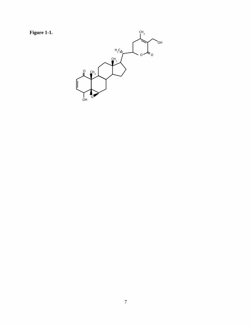
lactone, withaferin A (WA), with intriguing cytotoxic properties (Figure 1-1). Interest in WA has
7
Figure 1-1.
H3C
CH3
O OH
O
CH3
OH
CH3
O O

8
Figure 1-1. Structure of withaferin A.

9
increased within the last ten years as demonstrated by increased publication on its anti-cancer
and anti-inflammatory properties (Vanden Berghe et al., 2012; Vyas and Singh, 2014). In these
publications, WA is reported to target or otherwise modulate a plethora of different proteins and
signaling pathways including Akt, Notch-1, heat shock protein (HSP) 90, nuclear factor kappa-
light-chain-enhancer of activated B cells (NFκB), activator protein 1 (AP-1), estrogen receptor
(ER), RET, and p38 among others (Mohan et al., 2004; Singh et al., 2007; Mandal et al., 2008;
Oh et al., 2008; Stan et al., 2008; Oh and Kwon, 2009; Shah et al., 2009; Koduru et al., 2010;
Samadi et al., 2010a; Yu et al., 2010). Recently, our group and others have demonstrated a pro-
oxidant potential of WA in non-brain tumor cells (Malik et al., 2007; Widodo et al., 2010; Hahm
et al., 2011; Mayola et al., 2011; Grogan et al., 2013) and its ability to bind to certain thiol
residues (Yu et al., 2010; Thaiparambil et al., 2011; Santagata et al., 2012). Despite these
findings, the specific mechanism of WA’s diverse and widespread effects remained elusive,
especially in brain tumors where work has been largely absent. Regardless of the ultimate
mechanism, development of WA as an anti-cancer agent has been suggested to provide a
potentially unique approach compared to most standard therapies because, in contrast to
disrupting a single molecular pathway or interaction like the integrity of DNA as with TMZ, WA
demonstrates the ability to modulate several oncogenic pathways simultaneously (Vanden
Berghe et al., 2012; Vyas and Singh, 2014). Many cancers, including GBM and MB, often
display overactivation and/or dysregulation of multiple oncogenic signaling cascades, including
the mitogen-activated protein kinase (MAPK) and PI3 kinase (PI3K)/Akt/mammalian target of
rapamycin (mTOR) pathways, establishing therapeutic challenges given the heterogeneity of the
activation and the great level of proliferative signaling (Gilbertson et al., 2006; Wen and Kesari,
2008; Mohan et al., 2012).

10
1.2.1a Mitogen-activated protein kinase pathway
The MAPK/ERK signaling pathway involves a chain of proteins in the cell that
communicates a signal from a receptor on the cell surface to the nucleus where promotion of
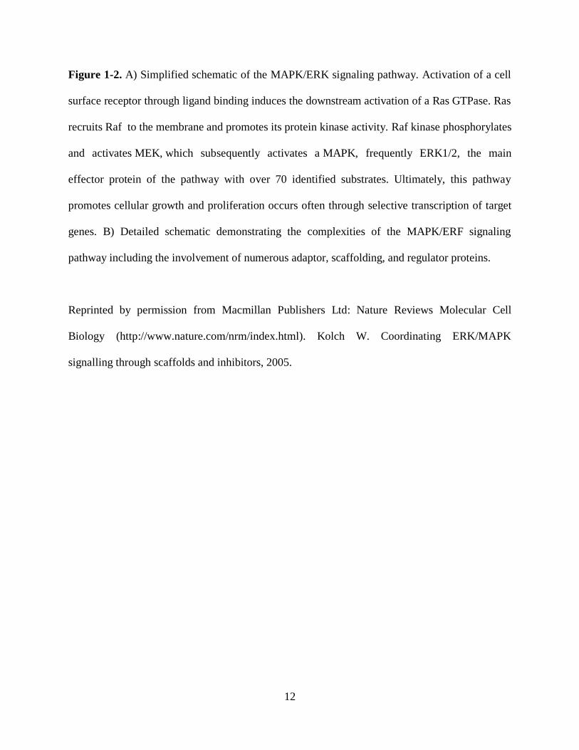
growth and proliferation occurs through selective transcription (Figure 1-2A and 1-2B) (Chang
and Karin, 2001). The generation of an activated membrane receptor complex activates rat
sarcoma protein (Ras), which subsequently promotes the protein kinase activity of
rapidly accelerated fibrosarcoma protein (Raf). Raf kinase phosphorylates and activates MEK
(MAPKK), which subsequently activates a MAPK, frequently extracellular signal-regulated
kinase (ERK1/2) (Avruch et al., 2001). c-Jun N-terminal kinase (JNK) and p38 MAPKs are more
responsive to stress signals. Disruption of this pathway at any point can lead to a failure of signal
transduction and a slowing of cell growth. Inversely, various cancers often demonstrate
activating mutations of proteins in the cascade like Ras and Raf or elevated activation of a
surface receptor that promotes pathway signal transduction (Dhillon et al., 2007; Krakstad and
Chekenya, 2010). GBMs predominately favor the latter, with 40-60% of cases demonstrating
amplification of the tyrosine kinase epidermal growth factor receptor (EGFR) and 40% of those
displaying activating EGFR mutations (Krakstad and Chekenya, 2010). Indeed, targeted
therapeutics against EGFR are currently in clinical trials for GBM, but, despite some promise,
they have not been approved as standard therapeutic agents (Krakstad and Chekenya, 2010).
Trials in pediatric CNS tumors patients have also been ongoing but little has been published to
date (MacDonald et al., 2014). These studies suggest that inhibition of MAPK signaling in brain
tumors may yield improved therapeutic benefits. WA has been shown to affect this signaling

11
Figure 1-2.
12
Figure 1-2. A) Simplified schematic of the MAPK/ERK signaling pathway. Activation of a cell
surface receptor through ligand binding induces the downstream activation of a Ras GTPase. Ras
recruits Raf to the membrane and promotes its protein kinase activity. Raf kinase phosphorylates
and activates MEK, which subsequently activates a MAPK, frequently ERK1/2, the main
effector protein of the pathway with over 70 identified substrates. Ultimately, this pathway
promotes cellular growth and proliferation occurs often through selective transcription of target
genes. B) Detailed schematic demonstrating the complexities of the MAPK/ERF signaling
pathway including the involvement of numerous adaptor, scaffolding, and regulator proteins.
Reprinted by permission from Macmillan Publishers Ltd: Nature Reviews Molecular Cell
Biology (http://www.nature.com/nrm/index.html). Kolch W. Coordinating ERK/MAPK
signalling through scaffolds and inhibitors, 2005.
13
pathway in non-brain tumor models. In leukemic cells and squamous cell carcinoma, WA was
shown to deplete total levels of Raf-1 but induce phosphorylation of ERK1/2 and the isoforms
p38 and JNK (Mandal et al., 2008; Samadi et al., 2010b), with activation of p38 associated with
induction of apoptosis (Mandal et al., 2008).
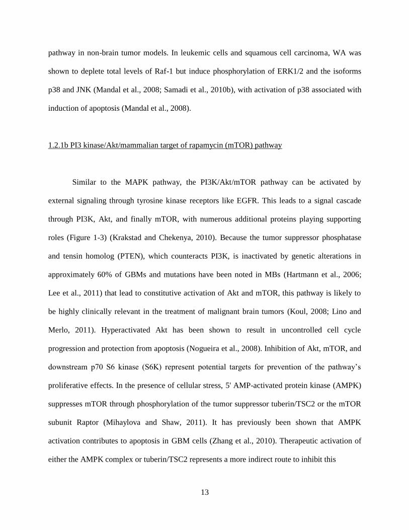
1.2.1b PI3 kinase/Akt/mammalian target of rapamycin (mTOR) pathway
Similar to the MAPK pathway, the PI3K/Akt/mTOR pathway can be activated by
external signaling through tyrosine kinase receptors like EGFR. This leads to a signal cascade
through PI3K, Akt, and finally mTOR, with numerous additional proteins playing supporting
roles (Figure 1-3) (Krakstad and Chekenya, 2010). Because the tumor suppressor phosphatase
and tensin homolog (PTEN), which counteracts PI3K, is inactivated by genetic alterations in
approximately 60% of GBMs and mutations have been noted in MBs (Hartmann et al., 2006;
Lee et al., 2011) that lead to constitutive activation of Akt and mTOR, this pathway is likely to
be highly clinically relevant in the treatment of malignant brain tumors (Koul, 2008; Lino and
Merlo, 2011). Hyperactivated Akt has been shown to result in uncontrolled cell cycle
progression and protection from apoptosis (Nogueira et al., 2008). Inhibition of Akt, mTOR, and
downstream p70 S6 kinase (S6K) represent potential targets for prevention of the pathway’s
proliferative effects. In the presence of cellular stress, 5' AMP-activated protein kinase (AMPK)
suppresses mTOR through phosphorylation of the tumor suppressor tuberin/TSC2 or the mTOR
subunit Raptor (Mihaylova and Shaw, 2011). It has previously been shown that AMPK
activation contributes to apoptosis in GBM cells (Zhang et al., 2010). Therapeutic activation of
either the AMPK complex or tuberin/TSC2 represents a more indirect route to inhibit this
14
Figure 1-3.

15
Figure 1-3. Schematic of the PI3K/Akt/mTOR signaling pathway. PI3K is primarily activated by
extracellular signaling through tyrosine kinase receptors at the cell surface but can also
associated with Ras. PI3K activity is negatively regulated by PTEN. PI3K ultimately activates a
signaling cascade including Akt and mTOR with signaling through phosphorylation. mTOR
increases protein synthesis and cell proliferation through inhibitory phosphorylation of
translational repressor 4E-BP1 and activating phosphorylation of p70 S6K. Reduction of
pathway activity is mediated by stress indicator AMPK and tumor suppressors TSC1/2.
Reprinted and adapted by permission from Macmillan Publishers Ltd: Nature Reviews Drug
Discovery (http://www.nature.com/nrd/index.html). Faivre S, et al. Current development of
mTOR inhibitors as anticancer agents, 2006.

16
pathway. Current early phase clinical trials in GBM are utilizing agents to inhibit the function of
EGFR, Akt, PI3K, and mTOR in this pathway (Krakstad and Chekenya, 2010). Although
multiple groups have demonstrated decreased levels of total and phosphorylated Akt in response
to WA treatment in several non-brain tumor cancer models (Oh et al., 2008; Oh and Kwon,
2009; Samadi et al., 2010a; Samadi et al., 2010b), no previous reports described its effects on
other proteins in the PI3K/Akt/mTOR signaling pathway despite this early promise.
1.2.1c Wnt/β-catenin signaling pathway
MBs are categorized in several unique molecular subgroups including SHH, Wnt, group
3, and group 4 based on a number of unique tumor traits (Northcott et al., 2012). The Wnt
subgroup of MB patients is defined by overactivation of the canonical Wnt/β-catenin signaling
pathway, often through overexpression of β-catenin or inactivating mutations of adenomatous
polyposis coli protein (APC) and Axin1/2 thereby acting independently of extracellular signaling
of Wnt through the frizzled surface receptor and downstream disheveled protein (Figure 1-4)
(Kim et al., 2011). This leads to excessive nuclear accumulation of β-catenin, which associates
with transcription factors of the transcription factor (TCF)/lymphoid enhancer-binding factor
(LEF) family to promote cell migratory and proliferative activity of the pathway at the
transcriptional level (Taylor et al., 2012). As a result, this pathway has been suggested as a
potential therapeutic target in MB. Only poly(ADP-ribose) polymerase (PARP) inhibition,
described to destabilize β-catenin through Axin, is under trial for MB (Waaler et al., 2012).
The Akt/mTOR pathway establishes cross-talk with the Wnt/β-catenin pathway through
glycogen synthase kinase-3β (GSK-3β) such that activation of Akt produces inhibitory

17
Figure 1-4.

18
Figure 1-4. Schematic representation of the Wnt signaling pathway in (a) the absence of Wnt
ligand and (b) the presence of Wnt ligand. Without Wnt ligand, β-catenin is sequestered by a
complex that includes GSK-3β, Axin, and APC which targets it for proteasomal degradation.
TCF/LEF transcription factors in the nucleus remain associated with repressor proteins Groucho
to prevent target protein transcription. In the presence of Wnt ligand binding a receptor complex
that includes Frizzled and low-density lipoprotein receptor-related protein (LRP), the functions
of dishevelled and CK1 ultimately lead to diminished GSK-3β and a failure to form the
destruction complex. This leads to accumulation of β-catenin with its eventual translocation into
the nucleus where it associates with TCF/LEF members to activate gene transcription. Activity
of GSK-3β can also be reduced with cross-talk from the Akt/mTOR pathway in which Akt
phosphorylates GSK-3β at Ser9 to inactivate it.
Reprinted by permission from Macmillan Publishers Ltd: Nature Reviews Drug Discovery
(http://www.nature.com/nrd/index.html). Barker N and Clevers H. Mining the Wnt pathway for
cancer therapeutics, 2006.

19
phosphorylation of GSK-3β, a reduction in the phosphorylation of β-catenin, and enhanced
stability of β-catenin. As such, inhibition of PI3K/Akt signaling has been shown to inhibit the
Wnt/β-catenin pathway in MB (Baryawno et al., 2010). Given the previously described
alterations in Akt induced by WA exposure, it was hypothesized that WA could act as a novel
inhibitor of the Wnt pathway in MB by a similar mechanism. Until this study, WA had not
previously been explored as a Wnt/β-catenin signaling inhibitor nor examined against MB.
1.2.2 Proteotoxicity
Malignant transformation to cancer often results in deregulation of normal homeostatic
processes that is subsequently associated with increased cellular stress, including oxidative,
replicative, metabolic, genomic, and proteotoxic stress (Luo et al., 2009). Cancer cell adaptation
to these stressors is therefore necessary for survival, resulting in dependence on non-oncogene
proteins and mechanisms that would otherwise play a less vital role in a non-malignant cell.
Pharmacological induction of oxidative stress has emerged as an intriguing means by which to
mount an anti-cancer effect. It is shown that pro-oxidant intervention can alter the redox
homeostasis of cancer cells, already stressed by exposure to constitutively high levels of reactive
oxygen species (ROS) not observed in normal cells, by shifting the balance to a state of
cytotoxicity (Laurent et al., 2005; Cabello et al., 2007). In fact, several investigational
chemotherapeutics with redox potential have recently been explored in preclinical and clinical
settings (Wondrak, 2009; Tew and Townsend, 2011). Such an oxidative stress mechanism has
also been demonstrated to effectively induce the proteotoxic stress of protein
unfolding/misfolding, resulting in a downstream secondary cytotoxicity that likely preferentially

20
affects these already susceptible cancer cells (Xu et al., 2011; Qiao et al., 2012). Previous reports
of others have demonstrated the pro-oxidant potential of WA in multiple cancer models (Malik et
al., 2007; Widodo et al., 2010; Hahm et al., 2011; Mayola et al., 2011; Grogan et al., 2013).
While this has largely remained a general finding, mechanistic evaluation demonstrated that
overexpression of Cu,Zn-superoxide dismutase (SOD) resulted in a partial protective effect
against WA treatment in breast cancer cells (Hahm et al., 2011). The same study suggested that
WA-mediated ROS generation and toxicity was a result of mitochondrial respiration inhibition.
While the WA exhibits both thiol reactivity and the ability to induce ROS, it has been
suggested that the nature of such activity occurs with some protein specificity. It has been
described that WA is capable of binding to the C-terminus of the chaperone protein HSP90 and
altering its activity (Yu et al., 2010), although evidence of the latter is largely indirect and
downstream. HSP90 is an well-conserved protein, making up ~1-3% of total cellular protein in
any given cell, directly responsible for interacting with and maintaining the stability and function
of over 400 key cellular proteins (Barrott and Haystead, 2013). Inhibition of HSP90 results in the
depletion of client proteins critical to the proliferation and ultimate survival of cancer cells such
as pro-growth signaling proteins, tyrosine kinases, cell cycle regulators, and steroid receptors
(Zhang and Burrows, 2004; Powers and Workman, 2006; Sidera and Patsavoudi, 2014).
Inhibition of this activity is cytotoxic. Cancer cells demonstrate enhanced responsiveness to
HSP90 inhibition compared to normal tissue, and this has been suggested to be a result of
enhanced reliance on HSP90 for stabilization of oncoproteins, localization in stressful
microenvironments, and selective accumulation of inhibitors in cancer cells (Sidera and
Patsavoudi, 2014). To date, 17 HSP90 inhibitors, all N-terminal inhibitors of ATPase activity
such as 17-allylamino-17-demethoxygeldanamycin (17-AAG) (Workman, 2003), have been

21
brought to clinical trial, and while some have shown therapeutic promise despite formulation
challenges, none have been approved for standard use against any cancer (Heath et al., 2005;
Heath et al., 2008; Solit et al., 2008; Pacey et al., 2012). It has been noted that WA may function
to indirectly modulate the HSP90 chaperone axis through disruption of the HSP90/ cell division
cycle protein 37 (Cdc37) interaction (Yu et al., 2010; Grover et al., 2011; Gu et al., 2014). Cdc37
functions as a co-chaperone to HSP90 and plays a vital role in localizing target kinases of the
kinome with HSP90 for folding (Karnitz and Felts, 2007).
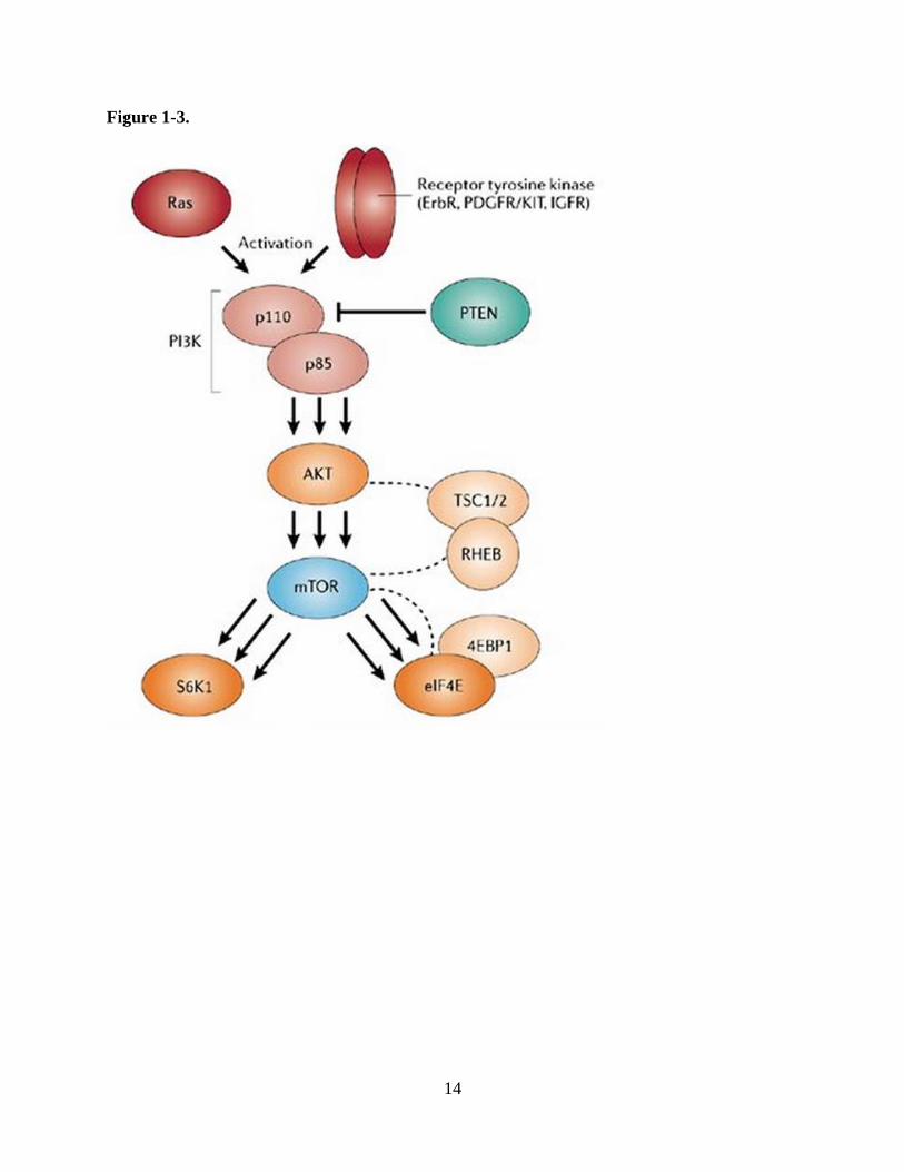
It has been long-noted that oxidative and other stressor damage to proteins triggers their
subsequent degradation (Stadtman, 1986). The major chaperones involved in protein quality
control decisions are HSP70 and HSP90, which act together in a multi-chaperone complex to
regulate the function, trafficking, and turnover of a wide variety of signaling proteins. However,
despite being an oversimplification of the complexities of protein chaperoning, these two
components of the machinery play opposite yet essential roles with HSP70 promoting
ubiquitination followed by proteosomal degradation and HSP90 stabilizing proteins against
degradation and instead promoting their refolding (Figure 1-5) (Pratt et al., 2010). This
mechanism is supported by studies demonstrating that treatment with HSP90 inhibitors promotes
the degradation of client proteins via the ubiquitin-proteasome pathway (Whitesell et al., 1994;
Sepp-Lorenzino et al., 1995), and over-expression of HSP70 decreases the level of dysfunctional
or misfolded proteins and improves viability in cellular models of certain neurodegenerative
diseases characterized by the aberrant protein accumulation such as Parkinson’s disease,
Huntington’s disease, and spinal and bulbar muscular atrophy (Jana et al., 2000; Bailey et al.,
2002; Klucken et al., 2004). Indeed, recent reports showed enhanced protein ubiquitination in the
presence of WA (Bargagna-Mohan et al., 2006; Yang et al., 2012) and elevated total levels of
22
Figure 1-5.

23
Figure 1-5. In general, chaperone HSP90 promotes protein refolding while HSP70 favors protein
degradation to maintain protein homeostasis and prevent the accumulation of aberrant proteins.
Various stressors, including oxidation, increases the frequency of protein misfolding. Inhibition
of HSP90 shifts protein maintenance toward HSP70-mediated proteasomal degradation and vice
versa. Simultaneous inhibition of the HSP90 and HSP70 axes, especially in the presence of a
stressor, promotes cellular cytotoxicity through accumulation of many aberrant proteins, given
an elimination of both homeostatic pathways.

24
HSP70 following treatment (Grogan et al., 2013). While several reports indicate that WA at least
partially inhibits the proteasome itself, which can lead to an accumulation of ubiquitinated
proteins (Yang et al., 2007; Yang et al., 2012), this finding remains unclear in the context of
other data demonstrating that pretreatment with proteasome inhibitor MG132 completely
reverses WA-mediated degradation of certain proteins (Zhang et al., 2011b) and that the doses
used to achieve the effect were significantly above pharmacologically-relevant levels.
Given reports of WA-mediated alterations in proteins and oncogenic signaling pathways
potentially through oxidation and modulation of the HSP90 chaperone axis, ultimately resulting
in protein depletion and proteotoxicity, it was hypothesized that combinational therapy with WA
and an inhibitor of the proteasome would successfully synergize by eliminating the
cytoprotective effect of homeostatic aberrant protein degradation and allowing the accumulation
of these dysfunctional molecules. Indeed, inhibition of HSP70 or the proteasome in the presence
of traditional N-terminal HSP90 inhibitors promotes a synergistic effect (Minnnaugh et al., 2004;
Ma et al., 2014). In this work, we further define WA’s role in modulating the HSP90 axis and
evaluate its efficacy in combination with a proteasome inhibitor.
1.2.3 DNA-damage response modulation
Maintenance of genomic integrity is an essential process for cell homeostasis. While
cancer cells are transformed to malignant status through interruption and manipulation of this
integrity, excessive disruption of the genome may lead to lethality. The DNA damage response
(DDR) promotes accurate transmission of genomes in dividing cells by reversing the extrinsic
and intrinsic DNA damage and is required for cell survival during replication (Harper and

25
Elledge, 2007). Radiation and genotoxic drugs represent a large proportion of anti-cancer agents
used in the clinic for years, but DNA damage repair mechanisms are often associated with
chemo- and radio-resistance. These agents promote DNA single- and double-strand breaks
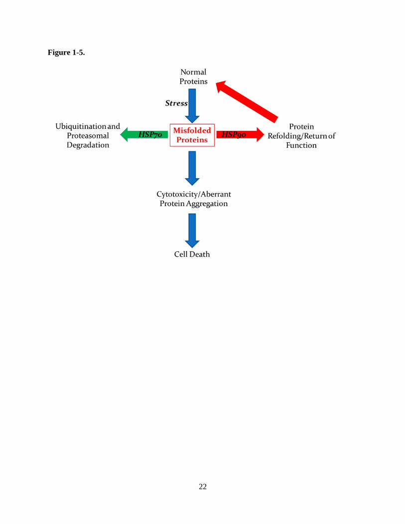
(SSBs; DSBs) that induce cellular cytotoxicity when unrepaired. DSBs are regarded as the
primary lesion of ionizing radiation and require repair through homologous recombination (HR),
which maintains fidelity of the sequence at the break site, or non-homologous end-joining
(NHEJ), which simply anneals two broken ends together (Figure 1-6) (Mladenov et al., 2013).
The overall response to DNA damage is regarded as an immensely complex coordination of
damage recognition and repair with simultaneous signal transduction to halt cell cycling during
the repair (Figures 1-7A and 1-7B). If such efforts fail, cells enter a sustained period of
senescence or simply die (Lamarche et al., 2010; Baskar et al., 2012).
To increase the efficacy of these DNA-damaging treatments and promote the failure of
repair, inhibitors of the major components of the DDR such as ATM (ataxia telangiectasia
mutated), ATR (ATM and Rad3-related), DNA-PKcs (DNA-dependent protein kinase, catalytic
subunit), Chk1 (checkpoint protein 1), and Chk2 (checkpoint protein 2) among others have been
used or have attracted interest in the drug development realm (Furgason and Bahassi, 2012).
These inhibitors have demonstrated to sensitize cancer cells to the genotoxic standard therapies.
Unfortunately, few of these therapeutic options have emerged in the treatment of brain tumors
and limited compounds have been taken to clinical trials largely due to translational challenges
including trial funding and poor drug pharmacokinetics.
Given the observation that numerous pathways are altered in response to WA, perhaps in
part due to wide-scale oxidation or inhibition of the HSP90 axis, WA’s mechanism would be
expected to overlap with those involved in the response to other therapeutic agents. Disruption of
26
Figure 1-6.

27
Figure 1-6. Double strand DNA breaks are repaired by either HR or NHEJ. In HR, lesions are
detected by the MRN complex which subsequently recruits additional repair proteins that utilize
the homologous templeate to copy and restore the original genetic code that was damaged. In
NHEJ, lesions are identified by the Ku70/Ku80 heterodimer with a known role of the MRN
complex as well which functions to recruit additional proteins to form the DNA-PK complex.
This complex promotes the ligation of two broken ends independent of original DNA sequence
and is therefore error-prone.
Reprinted by permission from Macmillan Publishers Ltd: Nature Reviews Molecular Cell
Biology (http://www.nature.com/nrm/index.html). Misteli T and Soutoglou E. The emerging role
of nuclear architecture in DNA repair and genome maintenance, 2009.

28
Figure 1-7.
29
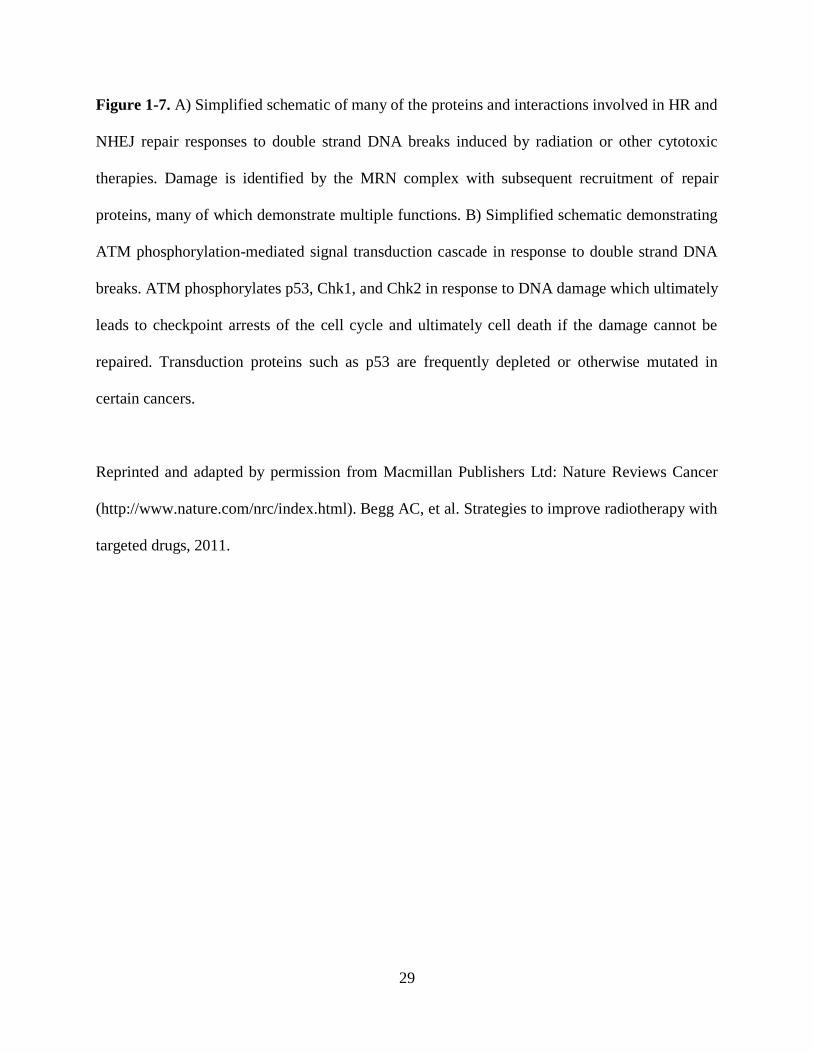
Figure 1-7. A) Simplified schematic of many of the proteins and interactions involved in HR and
NHEJ repair responses to double strand DNA breaks induced by radiation or other cytotoxic
therapies. Damage is identified by the MRN complex with subsequent recruitment of repair
proteins, many of which demonstrate multiple functions. B) Simplified schematic demonstrating
ATM phosphorylation-mediated signal transduction cascade in response to double strand DNA
breaks. ATM phosphorylates p53, Chk1, and Chk2 in response to DNA damage which ultimately
leads to checkpoint arrests of the cell cycle and ultimately cell death if the damage cannot be
repaired. Transduction proteins such as p53 are frequently depleted or otherwise mutated in
certain cancers.
Reprinted and adapted by permission from Macmillan Publishers Ltd: Nature Reviews Cancer
(http://www.nature.com/nrc/index.html). Begg AC, et al. Strategies to improve radiotherapy with
targeted drugs, 2011.

30
proteins important in the recognition and repair of DNA damage would be expected to sensitize
glioma cells to the first-line standard therapy of TMZ and RT, both genotoxic agents. Indeed,
direct inhibition of HSP90 to destabilize a number of client proteins has previously been shown
to act as a radiosensitizer (Dote et al., 2006; Noguchi et al., 2006; Kabakov et al., 2010). Several
preliminary studies have suggested that WA functions as a radiosensitizer in multiple cancer
models. Utilizing a knockout model to alter the normal function of HR and NHEJ, one study
reported that WA may inhibit NHEJ (Devi et al., 2008). Other studies have correlated
combination of WA and radiation to enhanced cellular oxidation (Yang et al., 2011b; Yang et al.,
2011a), however, the underlying mechanism producing WA-induced sensitization to DNA
damage has not been identified. We hypothesized that WA would disrupt the normal DDR and
have explored that in this work as a means of radiosensitization and also re-sensitization of GBM
cells to TMZ through alterations in MGMT, a protein involved in the removal of TMZ-mediated
DNA alkylation.
1.3 Specific Aims
The objective of this study was to evaluate the potential of WA either as a stand-alone
chemotherapeutic in the treatment of chemo-sensitive and -resistant brain tumors including GBM
and MB, or as an agent whose combination with current standard approaches could be
synergized or potentiated. As a single agent therapy, the effects of WA were evaluated on several
major proliferative pathways including the MAPK, PI3K/Akt/mTOR, and Wnt/β-catenin
signaling pathways as well as its ability to generate intracellular stress through thiol reactivity
and the creation of ROS. Further, given the induction of DNA DSBs in tumor cells treated with
31
TMZ or RT, interference with recognition and/or repair of this damage by the tumor cells
indicated a potential broad mechanism through which WA could potentiate the cytotoxicity of
DNA-damaging therapy. To this regard, proteins of the DNA damage response mechanism were
evaluated in the context of WA treatment and functional manifestations of DNA damage
recognition and repair were measured. Additionally, protein levels of repair mechanisms specific
to TMZ including MGMT and mismatch repair were explored.
It was hypothesized that WA would act as a potent cytotoxic agent against brain tumors
and potentiate the activity of several standard therapies through disruption of homeostatic
cellular processes necessary in the response to those agents. The specific aims designed to
explore the underlying mechanisms and validate this hypothesis are as follows:
1) Determination of the key anti-cancer mechanistic properties of WA in oncogenic
signal transduction pathways in GBM and MB in vitro.
a. Ability of WA to induce cell cycle arrest and apoptosis.
b. Effects of WA on several major proliferative pathways including the MAPK,
PI3K/Akt/mTOR, and Wnt/β-catenin signaling pathways.
c. Ability of WA to generate intracellular stress through thiol reactivity and the
production of ROS and the response of the cell to this stress.
2) Evaluation of the proteotoxic mechanism of WA and its therapeutic implications in
vitro.
a. The effects of WA on cellular oxidation and the underlying implications.
b. WA-mediated modulation of the HSP90/HSP70 protein chaperone.

32
c. Therapeutic implications of combination therapy with WA and proteasome
inhibitors.
3) Exploration of the effects of WA on the DDR mechanism in vitro.
a. Exploration of WA’s effects on the DNA damage recognition and repair
responses.
b. Evaluation of the efficacy of WA in combination with radiation and TMZ.
c. Determination the ability of WA to alter mechanisms of resistance to provide
TMZ resensitization in resistant cell lines.
This research demonstrates several means by which the novel natural compound WA was
studied in the context brain tumors to further understand its properties so as to better evaluate its
potential as a new treatment for this disease. Completion of these studies, as outlined in the
subsequent chapters, provides excellent evidence for the benefit of WA as a novel
chemotherapeutic strategy for brain tumors, including GBM and MB with great translational
potential both as a monotherapy and in combination with standard agent. Ultimately, the goal of
this proposal was to promote progression of novel pharmaceutical development to improve brain
tumor patient survival and quality of life.
1.4 Statement of Purpose
The purpose of the conducted research was to identify, define, and utilize the cytotoxic
anti-cancer properties and clinical utility of the steroidal lactone WA in high-grade brain tumors

33
through its alterations of oncogenic cellular signaling pathways, protein homeostasis, and the
DNA-damage response mechanism.
Briefly, the following chapters identify and describe key cellular changes in brain tumor
cells following exposure to WA. In Chapter 2 (Specific aims 1a-1c), WA was screened as a
potential cytotoxic agent against GBM brain tumor cells and was identified to exhibit anti-
proliferative activity through induction of G2/M cell cycle arrest and ultimately apoptosis. These
findings were associated with modulation of the Akt/mTOR and MAPK pathways and induction
of thiol-mediated oxidation. In Chapter 3 (Specific aims 1a-1c and 3b-3c), TMZ-resistant GBM
cells were shown to demonstrate enhanced responsiveness to WA as a monotherapy with a
similar responsiveness to WA as sensitive cells. This study expanded on the inhibitory nature of
the effect on the Akt/mTOR pathway but showed that alterations in the MAPK are a
compensatory response to the compound. Additionally, WA pretreatment was shown to deplete
MGMT and functionally re-sensitize these cells to TMZ. Chapter 4 (Specific aims 1a-1c) further
demonstrates the cytotoxicity of WA in brain tumors with a model of MB. A comparable pattern
of general efficacy was observed as in GBM. WA was demonstrated to inhibit Wnt/β-catenin
signaling through proteasome-mediated degradation of the TCF/LEF transcription factors
without significant alterations in the total levels nor localization of β-catenin. These findings
were correlated with TCF/LEF depletion following HSP90 inhibition and WA-mediated
alterations in HSP90 co-chaperone association. In Chapter 5 (Specific aims 2a-2c), mechanisms
of action for the observed manifestations of WA treatment were explored. Specifically, WA
induced reduction of many HSP90 client proteins and disrupted the interaction between HSP90
and Cdc37 as well as Cdc37-dependent proteins without directly inhibiting intrinsic HSP90
activity. Additionally, WA induced an NAC-repressible oxidation that was enhanced with

34
glutathione depletion. Overall, WA produced protein degradation through the proteasome and
cytotoxicity that could be enhanced through the build-up of aberrant proteins with pretreatment
of a proteasome inhibitor. Finally, Chapter 6 (Specific aims 3a-3b) demonstrates the
radiosensitizing properties of WA through disruption of the DDR mechanisms and enhanced
arrest of the cell cycle. WA depleted proteins involved in DNA damage recognition, signaling,
and ultimately repair through HR and NHEJ. This was functionally associated with altered
phosphorylation of H2A.X and enhanced DSB with combination of WA and RT.

35
Chapter 2
Cytotoxicity of withaferin A in glioblastomas involves
induction of an oxidative stress-mediated heat shock
response while altering Akt/mTOR and MAPK signaling
pathways
Reprinted with permission from Springer Science + Business Media:
Grogan, P.T., et al., Cytotoxicity of withaferin A in glioblastomas involves induction of an
oxidative stress-mediated heat shock response while altering Akt/mTOR and MAPK signaling
pathways. Invest New Drugs, 2013. 31(3): p. 545-557.

36
2.1 Abstract
Withaferin A (WA), a steroidal lactone derived from the plant Vassobia breviflora, has
been reported to have anti-proliferative, pro-apoptotic, and anti-angiogenic properties against
cancer growth. In this study, we identified several key underlying mechanisms of anticancer
action of WA in glioblastoma cells. WA was found to inhibit proliferation by inducing a dose-
dependent G2/M cell cycle arrest and promoting cell death through both intrinsic and extrinsic
apoptotic pathways. This was accompanied by an inhibitory shift in the Akt/mTOR signaling
pathway which included diminished expression and/or phosphorylation of Akt, mTOR, p70 S6K,
and p85 S6K with increased activation of AMPKα and the tumor suppressor tuberin/TSC2.
Alterations in proteins of the MAPK pathway and cell surface receptors like EGFR, Her2/ErbB2,
and c-Met were also observed. WA induced an N-acetyl-L-cysteine-repressible enhancement in
cellular oxidative potential/stress with subsequent induction of a heat shock stress response
primarily through HSP70, HSP32, and HSP27 upregulation and HSF1 downregulation. Taken
together, we suggest that WA may represent a promising chemotherapeutic candidate in
glioblastoma therapy warranting further translational evaluation.

37
2.2 Introduction
Malignant gliomas, including the grade IV astrocytoma glioblastoma multiforme (GBM),
accounting for 60-70% of such lesions, are the most common adult primary malignant brain
tumors, representing approximately 14,000 new cancer cases yearly (Wen and Kesari, 2008).
The mean post-diagnosis survival time of patients with GBM is approximately 14 months (Wen
and Kesari, 2008; Stupp et al., 2009). Most high grade brain tumors are quite infiltrative,
resulting in residual invasive tumor cells even after optimal surgical resection. This significantly
contributes to the high rate of recurrence and poor clinical outcomes associated with this disease
(Giese et al., 2003). Standard-of-care management typically involves surgical debulking
followed by treatment with radiation and the methylating agent temozolomide. While this
currently represents the most efficacious therapy for prolonging survival, tumors are often quick
to develop resistance to this line of therapy (Chamberlain, 2010; Quick et al., 2010). Recent
investigational therapies have yielded only modest response rates up to 15% at best without
impact on 6-month progression-free survival (Wen and Kesari, 2008; Quick et al., 2010).
Therefore, given a lack of current therapeutic strategies to extend survival in glioma
patients, there is a critical need for novel therapeutic agents. We previously screened over 200
natural plant extracts with promising anti-tumor potential and identified a 28-carbon steroidal
lactone obtained from the Vassobia breviflora, withaferin A (WA), with intriguing cytotoxic
properties (Samadi et al., 2010b). WA has been identified as a novel cancer therapy with anti-
proliferative, pro-apoptotic, and anti-angiogenic properties as well as a modulator of several key
cell-survival and regulatory pathways including those involving Akt, Notch-1, heat shock protein

38
(HSP) 90, NFkappaB, AP-1, estrogen receptor, RET, and p38 among others (Mohan et al., 2004;
Singh et al., 2007; Mandal et al., 2008; Oh et al., 2008; Stan et al., 2008; Oh and Kwon, 2009;
Shah et al., 2009; Koduru et al., 2010; Samadi et al., 2010a; Yu et al., 2010). Additionally, WA
has shown promising anti-tumor activity in several murine xenograft and/or orthograft models
including prostate, breast, medullary thyroid, melanoma, uveal melanoma, ovarian, cervical, and
brain cancers (Vyas and Singh, 2014).
We have recently demonstrated the anti-proliferative effects of WA in malignant glioma
(Grogan et al., 2010; Grogan et al., 2011), and this finding was subsequently confirmed
demonstrating the thiol-reactivity of WA and its ability to induce a heat shock response via a
reporter assay (Santagata et al., 2012). Several additional reports have identified the pro-oxidant
potential of WA (Malik et al., 2007; Widodo et al., 2010; Hahm et al., 2011; Mayola et al.,
2011), however, the mechanism of WA’s diverse and widespread effects, especially in glioma
cells, is largely unknown. Here, we expand on previous findings to further delineate the nature of
WA’s anti-cancer effects in glioblastoma and examine the molecular response by the cell.
2.3 Materials and methods
2.3.1 Cell culture and general reagents
Two human glioblastoma multiforme cell lines, U87 and U251, were generously
provided by Dr. Jann Sarkaria (Mayo Clinic, Rochester, MN), and one murine GBM cell line,
GL26, was graciously donated by Dr. John Ohlfest (University of Minnesota, Minneapolis, MN).
All cell lines were grown in Dulbecco’s modified Eagle’s media (DMEM #6429; Sigma-Aldrich,

39
St. Louis, MO) supplemented with 10% fetal bovine serum (FBS; Sigma-Aldrich, St. Louis,
MO) and 1% penicillin/streptomycin (Sigma-Aldrich, St. Louis, MO) at a 37 °C humidified
atmosphere of 5% CO2 in air. WA was extracted and isolated as previously described with a
purity of 99% by HPLC and stored as a stock solution, adjusted for impurities, at 20 mM in
DMSO at -80 °C (Samadi et al., 2010b). Propidium iodide (PI), RNase, and N-acetyl-L-cysteine
(NAC) were acquired from Sigma-Aldrich (St. Louis, MO), and Annexin V-FITC was obtained
from BD Biosciences (San Diego, CA).
2.3.2 Cell proliferation and viability assays
Cells were seeded in 96-well plates at 2,500 cells/well. Following a 6 h incubation
period, WA-containing media in various concentrations was added to each well, and the cells
were incubated for an additional 72 h. The number of viable cells was quantified by the
colorometric CellTiter96 Aqueous MTS assay (Promega, Fitchburg, WI) at 490 nm on a BioTek
Synergy 2 plate reader (BioTek, Winooski, VT) as per the manufacturer’s instructions.
Because of the non-enzymatic, auto-reductive potential of NAC, the MTS assay was
unable to be utilized for evaluating WA/NAC co-treatment studies given the nature of the assay
mechanism, as described by the manufacturer. As such, the CellTiter-Glo luminescent assay
(Promega, Fitchburg, WI) was utilized instead to measure cell viability by ATP levels. Cells
were plated in 96-well plates followed by treatment with NAC after 6 h and WA 1 h later. After
72 h, assay reagent was prepared as per the instructions, and 50 uL was added to each well. The
luminescent signal was given 10-20 minutes to equilibrate and quantified by the BioTek Synergy
2 plate reader.

40
2.3.3 Cell cycle analysis
Cells were plated at an amount previously determined, based on the growth
characteristics of individual cells, to have a high enough yield without achieving complete
confluency upon harvest and were allowed to grow overnight. Cells were treated with
appropriate concentrations of WA and, where indicated, pre-treated with 5 mM NAC. Upon the
completion of treatment, cells were trypsinized, collected, resuspended in 0.43 mL of 4 °C 1x
PBS followed by 1mL of -20 °C ethanol for a 70% ethanol fixative solution, and stored at -20
°C. To analyze, stored cells were collected by centrifugation, resuspended in 1x PBS containing
40 µg/mL PI and 100 µg/mL RNase, and incubated at -37 °C for 30 minutes before being
analyzed by flow cytometry (BD LSRII; Becton Dickinson, San Diego, CA). Analysis of these
results examined only viable cells without DNA fragmentation to establish the distribution of the
true cell cycle in living cells.
2.3.4 Analysis of cell death
Staining phosphatidylserine on the outer leaflet of the cell membranes on apoptotic cells
and DNA staining by PI in necrotic and late apoptotic cells was performed to assess WA-induced
cell death. Cells were plated and treated as indicated for cell cycle analysis. Upon the completion
of treatment, cells were trypsinized, collected, and washed once in Annexin binding buffer (150
mM NaCl, 5 mM KCl,1 mM MgCl2.6H2O, 1.8 mM CaCl2.2H2O, 10 mM HEPES, and 2% (v/v)
FBS). Cells were stained with Annexin V-FITC and PI according to the manufacturer’s

41
instructions (BD Biosciences, San Diego, CA) for 20 minutes at 4 °C before being washed twice
and resuspended in binding buffer. Cells were immediately analyzed by flow cytometry on a BD
LSRII.
2.3.5 Western blotting
Cells were plated and treated in the manner outlined for cell cycle analysis. Proteins were
collected, quantified, separated by sodium dodecyl sulfate–polyacrylamide gel electrophoresis
(SDS–PAGE), and electrotransfered onto a Hybond nitrocellulose membrane as previously
described in Samadi, et al. (Samadi et al., 2011). Actin levels were assessed to ensure equal
loading and transfer of proteins. All studies were repeated for accuracy.
Primary rabbit antibodies against poly(ADP-ribose) polymerase (PARP; #9542; 1:1000),
caspase 3 (#9665; 1:1000), caspase 7 (#9492; 1:1000), caspase 9 (#9502; 1:1000), cyclin B1
(#4138; 1:2000), p-ERK1/2 (Thr202/Tyr204; #4377; 1:1000), Akt (#9272; 1:1000), p-Akt
(Ser473; #4058; 1:1000), c-Met (#4560; 1:500), p-c-Met (Tyr1234/1235; #3077; 1:500),
epidermal growth factor receptor (EGFR; #4267; 1:1000), p70 S6 kinase (#2708; 1:500), p-p70
S6 kinase (Thr389; #9234; 1:1000), mTOR (#2972; 1:1000), p-mTOR (Ser2448; #2971; 1:1000),
Her2/ErbB2 (#2165; 1:1000), 4E-BP1 (#9644; 1:1000); p-4E-BP1 (Thr37/46; #2855; 1:1000),
tuberin/TSC2 (#4308; 1:1000), p-tuberin/TSC2 (Thr1462; #3617; 1:1000), AMPKα (#2603;
1:1000), and p-AMPKα (Thr172; #2535; 1:1000) and primary mouse antibodies against HSP27
(#2402; 1:2000), caspase 8 (#9746; 1:1000), and p-EGFR (Tyr1068; #2236; 1:1000) were
acquired from Cell Signaling Technology (Beverly, MA). Total- and phospho-antibodies against
p70 S6 kinase were used to detect p85 S6 kinase (phospho-Thr412) given the known cross-

42
reactivity of the antibodies. Rabbit antibodies against HSP90 (SPA-836; 1:1000), HSP32/heme
oxygenase 1 (SPA-894; 1:1000), and heat shock factor 1 (HSF1; ADI-SPA-901; 1:1000) were
acquired from Enzo Life Sciences (Farmingdale, NY) or its affiliates along with the rat antibody
for Grp94 (SPA-850; 1:1000) and the mouse antibody for HSP70 (ADI-SPA-810; 1:1000). The
mouse antibody Trap1 (MA1-010; 1:1000) was purchased from Affinity Bioreagents (Pierce
Biotechnology, Rockford, IL). A mouse total actin antibody (MAB1501; 1:50,000) to be used as
a control was acquired from EMD Millipore (Billerica, MA). A mouse antibody against Raf-1
(sc-7267; 1:250), a goat antibody against p-Raf-1 (Ser338; sc-12358; 1:250), and a rabbit
antibody against ERK1/2 (sc-154; 1:5000) were obtained from Santa Cruz Biotechnology (Santa
Cruz, CA). Donkey anti-rabbit IgG HRP (sc-2313; 1:5000), goat anti-mouse IgG HRP (sc-2005;
1:5000), and goat anti-rat IgG HRP (sc-2032; 1:5000) secondary antibodies were also purchased
from Santa Cruz Biotechnology. Given primary antibody species specificity, Western analysis
was completed in the human U87 and U251 cells lines.
2.3.6 Detection of ROS
The accumulation of intracellular ROS, particularly peroxides like H2O2, was determined
using CM-H2DCFDA (Molecular Probes, Grand Island, NY), a general oxidative stress indicator
that fluoresces upon oxidation. Cells were preloaded with 20 µM CM-H2DCFDA in 1x dPBS at
37 °C for 1 h, washed once with phenol red-free DMEM containing 10% FBS and 1%
penicillin/streptomycin, and plated in 96-well plates in phenol red-free media at 40,000 cells/well
for GL26 and 20,000 cells/well for U87. After 30 minutes, cells were treated with WA, NAC, or

43
both. Measurements were taken on a BioTek Synergy 2 plate reader at 4 h with excitation and
emission filters of 485 nm and 528 nm, respectively.
Detection of mitochondrial superoxide present at a given timepoint was examined by
MitoSOX Red (Molecular Probes, Grand Island, NY), an indicator reagent that fluoresces upon
oxidation by superoxide radicals. Cells were plated and allowed to attach and grow overnight.
Pre-treatment with 5mM NAC occurred 1 h before treatment with 5 µM WA. Cells were
harvested 4 h after WA addition, washed once with 1x dPBS, and stained with 5 µM MitoSOX
Red in 1x dPBS for 30 minutes at 37 °C. Cells were washed twice with 1x dPBS and run on a
BD LSRII flow cytometer (Becton Dickinson, San Diego, CA) with excitation and emission
filters of 488 nm and 580 nm, respectively, to assess mean fluorescence intensity.
2.3.7 Statistical analysis
GraphPad (GraphPad Inc., San Diego, CA) was used to generate best-fit sigmoidal dose response
curves for IC50 determination. Comparisons of differences between two or more means were
determined by Student’s unpaired t-test (2 means) via a standard statistical analysis software
package (SPSS version 17.0; SPSS Inc, Chicago, IL). The level of significance was set at p <
0.05. Data are presented as mean values with error bars denoting standard deviation. All studies
were minimally performed in triplicate unless otherwise noted.
2.4 Results
2.4.1 Withaferin A reduces cell proliferation and viability in GBM cells

44
To investigate the general biological effect of WA in GBM cells, three cell lines (U87,
U251, and GL26) were incubated with increasing concentrations of WA or a DMSO control for
72 h. Cell number and viability were subsequently determined by using the MTS assay (Figure
2-1). WA dose escalation reduced cell proliferation and viability. By GraphPad analysis, the IC50
values of WA were determined to be 1.07 ± 0.071 µM, 0.69 ± 0.041 µM, and 0.23 ± 0.015 µM
for U87, U251, and GL26 cells, respectively.
2.4.2 Withaferin A induces G2/M cell cycle arrest in GBM cells in a dose-dependent manner
In order to establish the nature of the anti-proliferative response to WA, distribution of
the cell cycle phases of glioblastoma cells was assessed by flow cytometry at 24 h. WA induced
a dose-dependent shift in cell cycle arrest from the G0/G1 checkpoint to G2/M arrest. Maximal
shift to G2/M arrest above baseline was observed at 0.75 µM in U87 cells (12.0% G2/M in
controls increasing to 63.4% with treatment), 1.5 µM in U251 cells (19.1% baseline to 49.7%
with treatment), and 0.5 µM in GL26 cells (21.7% baseline to 58.7% with treatment (Figure 2-
2A). Doses above these optimal levels showed a somewhat diminished G2/M cell cycle shift.
The shift to G2/M cell cycle arrest is accompanied by a depletion of cells in the G0/G1 phase and
a relative increase in S phase as observed in the U87 (11%) and GL26 (14%), whereas the S
phase proportion in U251 cells was largely unchanged (Figure 2-2A).
To confirm these findings, the G2/M-specific protein cyclin B1, which is elevated during
G2/M cell cycle arrest, was evaluated by Western blot analysis for increases in expression with

45
Figure 2-1.

46
Figure 2-1. U87, U251, and GL26 cells were incubated with increasing concentrations of WA or
a DMSO control for 72 h and then assessed by MTS assay. WA dose escalation reduced cell
proliferation and viability with IC50 values of 1.07 ± 0.071 µM, 0.69 ± 0.041 µM, and 0.23 ±
0.015 µM for U87, U251, and GL26 cells, respectively.

47
Figure 2-2.

48
Figure 2-2. A) Cells stained with propidium iodide and analyzed by flow cytometry revealed
that WA induces a dose-dependent G2/M cell cycle arrest with an optimal range of such a
response at 24 h. Maximal induction of arrest above baseline was observed at 0.75 µM, 1.5 µM,
and 0.5 µM for U87, U251, and GL26 cells, respectively. Adjacent histograms display
representative examples of these peak values in each cell line. B) G2/M arrest was confirmed by
Western blotting for cyclin B1. At 24 h, U87 and U251 cells demonstrated dose-dependent
induction of cyclin B1 with highest levels at 1 µM and 2.5 µM, respectively.

49
WA treatment. At 24 h, both U87 and U251 cells demonstrated a dose-dependent induction of
cyclin B1 expression with maximal expression levels observed at 1 µM and 2.5 µM WA,
respectively (Figure 2-2B). Flow cytometry identified sustained G2/M cell cycle arrest at both 48
h and 72 h with Western analysis up to 48 h supporting those findings (data not shown).
2.4.3 Withaferin A induces GBM cell death
To further characterize the anti-proliferative effects of WA, annexin V and propidium
iodide (PI) dual staining on flow cytometry was used to evaluate the ability of WA to induce cell
death through apoptotic and necrotic mechanisms. Figure 2-3A illustrates that increases in WA
concentration corresponded with enhanced cell death in U87, U251, and GL26 cells at 24h.
While all cell lines demonstrated evidence of an apoptotic response indicated by annexin V
staining, U87 cells appear to also exhibit increased levels of necrosis with treatment as indicated
by staining with PI only. From baseline measurements of 3.9-5.9%, total cell death at 3 µM WA
treatment was 51.4% for U87 cells (9.0% early apoptosis; 18.4% late apoptosis; 24.0% necrosis),
24.4% for U251 cells (12.0% early apoptosis; 9.9% late apoptosis; 2.5% necrosis), and 31.7% for
GL26 cells (4.4% early apoptosis; 22.2% late apoptosis; 5.1% necrosis). Upon doubling the
concentration to 6 µM, total cell death increased to 86.9% for U87 cells (3.4% early apoptosis;
55.5% late apoptosis; 28.0% necrosis), 47.1% for U251 cells (9.7% early apoptosis; 32.5% late
apoptosis; 4.9% necrosis), and 83.1% for GL26 cells (0.6% early apoptosis; 74.1% late
apoptosis; 8.4% necrosis). Further induction of apoptosis was observed at 48 h (data not shown).
In order to confirm that these cells were undergoing an apoptotic response, protein levels
of mitochondrial/intrinsic procaspase 9, extrinsic procaspase 8, effector procaspases 3 and 7, and

50
Figure 2-3.

51
Figure 2-3. A) Cells stained with propidium iodide and annexin V-FITC and analyzed by flow
cytometry revealed that increasing WA concentrations correspond with enhanced cell death in
U87, U251, and GL26 cells at 24 h. Adjacent dot plots display representative examples of each
cell line. B) Induction of apoptosis was confirmed by Western blotting in U87 and U251 cell
lines. WA treatment resulted in reduction in the levels of uncleaved initiator caspases 8 and 9 as
well as effector caspases 3 and 7. Further, PARP cleavage was observed at 2.5 and 5 µM in both
lines.

52
downstream PARP cleavage were examined by Western blotting (Figure 2-3B). With increasing
concentrations of WA, a reduction in the levels of all uncleaved caspases was observed at 5 µM
in both U87 and U251 cells at 24 h. Procaspases 3, 7, and 8 demonstrated reduced levels at WA
concentrations below 5 µM in U87 cells. Finally, PARP cleavage was observed at both 2.5 and 5
µM WA in both U87 and U251 cell lines. These data confirm that WA-mediated cytotoxicity at
concentrations above IC50 is in part due to induction of apoptosis in these GBM cells.
2.4.4 Withaferin A alters normal protein expression and activation in the Akt/mTOR and MAPK
pathways
Given the inhibition of proliferation and induction of cell death observed with WA
treatment, several proteins associated with the Akt/mTOR and mitogen-activated protein kinase
(MAPK) growth, proliferation, and survival pathways as well as key surface membrane proteins
that signal to each pathway, known to be important in glioma and other cancers (Wen and
Kesari, 2008), were screened in U87 and U251 cells for total expression and activation via
phosphorylation status at 24 h post-treatment (Figure 2-4). Total levels of Akt and mTOR were
reduced with increasing concentrations of WA. Akt and mTOR phosphorylation were dose-
dependently diminished in U87 cells, but only p-mTOR was decreased in U251 cells. Total
levels of p70 S6K (downstream of mTOR) and its nuclear isoform p85 S6K were largely
unchanged in both cell lines with enhanced downregulation only observed in p85 S6K at 5 µM
WA in U87 cells. Low-dose WA induced phosphorylation of both proteins, but this was reduced
and/or further depleted upon achieving levels of 0.5 µM WA and above. Expression of total
levels of the translation-repressor 4E-BP1 (also downstream of mTOR) was markedly

53
Figure 2-4.

54
Figure 2-4. Following WA treatment, an array of proteins associated with the Akt/mTOR and
MAPK growth, proliferation, and survival pathways as well as surface membrane proteins that
signal to each pathway were screened via Western blotting in U87 and U251 cells for total
expression and activation via phosphorylation status at 24 h post-exposure in order to help
evaluate the molecular changes corresponding with cellular observations. Alterations in
expression are observed in both pathways and with the cell surface receptors.
55
increased in U87 cells with elevated levels of inhibitory phosphorylation upon increasing WA
concentration. In contrast, U251 cells exhibited decreased levels of both total and phosphorylated
forms of 4E-BP1 with WA treatment suggesting an overall inhibitory alteration to the
Akt/mTOR signaling pathway. Negative regulator of the pathway, AMPKα, was sustained or
upregulated in total levels at all WA concentrations tested with the exception of 5 µM in U87
cells. Another negative regulator, the tumor suppressor tuberin/TSC2, was sustained in U87 cells
and upregulated in U251 cells up to 1 µM WA before diminishing at increased concentrations.
However, activation of both tuberin/TSC2 and AMPKα via phosphorylation was observed at
WA levels as low as 0.1-0.25 µM and maintained at higher doses. Peak levels were observed
between 0.5-2.5 µM in a cell-dependent manner.
Next, the effect of WA on the MAPK cascade was assessed since others have previously
implicated WA as an inducer of a MAPK-mediated stress response (Mandal et al., 2008; Samadi
et al., 2010b). In the U87 and U251 cells, total levels of ERK1/2 were unchanged with WA
treatment over 24 h, but p-ERK1/2 levels were elevated with increasing concentrations of WA
up to 2.5 µM and then diminished at 5 µM (Figure 2-4). In contrast to U87 cells, which express
both p-ERK1 and p-ERK2 at comparable levels with treatment, p-ERK2 predominates
expression with WA treatment of U251 cells. Additionally, total levels of upstream Raf-1 were
diminished at 2.5-5 µM WA but activation of this protein by phosphorylation of serine 338 was
observed in a dose-dependent manner with increasing WA concentrations.
EGFR, Her2/ErbB2, and c-Met are commonly amplified and/or mutated surface proteins
in glioblastoma and signal both the MAPK and Akt/mTOR pathways. As such attempts have,
previous attempts have been made to therapeutically target these proteins (Wullich et al., 1993;
Potti et al., 2004; Puputti et al., 2006; Guessous et al., 2010; Berezowska and Schlegel, 2011). In

56
both GBM cell lines, total levels of EGFR, Her2/ErbB2, and c-Met were decreased at 24h with
increasing WA levels with the exception of c-Met upregulation from 0.25-2.5 µM WA (Figure 2-
4). Phosphorylation of EGFR increased with WA treatment, whereas c-Met phosphorylation was
slightly elevated over controls at low WA doses but was downregulated at higher WA
concentrations.
2.4.5 Withaferin A elevates pro-oxidant potential in GBM cells and induces a cellular oxidative
stress response
WA-induced apoptosis in several cancer types was recently described as being mediated
by ROS generation (Malik et al., 2007; Widodo et al., 2010; Hahm et al., 2011; Mayola et al.,
2011). Given the role ROS are known to play in a variety of cancer chemotherapeutic agents
(Antosiewicz et al., 2008; Wondrak, 2009; Tew and Townsend, 2011), we examined whether the
oxidation status of glioblastoma cells was altered by WA. The production of peroxide-type
radicals with CM-H2DCFDA and mitochondrial accumulation of superoxide radicals with
MitoSOX Red was measured in U87 and GL26 cells at 4 h post-WA exposure. GL26 and U87
cell lines demonstrated elevated oxidation status in response to increasing concentrations of WA.
Peroxides rose 29.4% in GL26 cells (p = 0.01) and 90.5% in U87 cells (p = 0.003) compared to
controls (Figure 2-5A) while mitochondrial superoxide increased 10.9% (p = 0.002) and 10.8%
(p = 0.03) above control at 5 µM, respectively (Figure 2-5B). In order to determine the nature of
this response, cells were simultaneously incubated with 5 mM of the thiol antioxidant NAC,
described as both a redox buffer and ROS scavenger (Deneke, 2000; Mayola et al., 2011). NAC
significantly reduced peroxide generation in WA-treated cells and returned levels to near-

57
Figure 2-5.

58
Figure 2-5. A) Generation of peroxide-type radicals like H2O2 following exposure to increasing
concentrations of WA were quantified with CM-H2DCFDA staining in U87 and GL26 cells at 4
h. B) Mitochondrial accumulation of superoxide radicals was assessed by flow cytometry with
MitoSOX Red staining in U87 and GL26 cells at 4 h post-exposure to 5µM WA. Elevated
oxidation status observed in (A) and (B) was reduced or completely eliminated by concurrent
treatment with the thiol antioxidant N-acetyl-L-cysteine (5 mM). C) An array of proteins
associated with the heat shock stress response were screened via Western blotting in U87 and
U251 cells 24 h and 48 h post-treatment of WA. Upregulation was consistently observed with
HSP70 and HSP32 in a dose- and time-dependent manner (C and D) with alterations in other
proteins displaying a cell-specific response. Expression of the transcription factor HSF1 was
decreased across both cell lines. *p < 0.05

59
baseline in both GL26 cells (p = 0.006) and U87 cells (p = 0.001). NAC reduced levels of
mitochondrial superoxide radicals but failed to do so within statistical significance, and levels
remained significantly elevated in U87 cells, despite being reduced compared to WA alone.
Induction of a heat shock response was also recently described in cells via a
luminescence reporter assay following WA exposure (Santagata et al., 2012). Consistent with
other models of oxidative stress (Ansari et al., 2011; Qiao et al., 2012), we observed that WA
modulates heat shock protein expression levels at both 24 h and 48 h in U87 and U251 cells
(Figure 2-5C). While total levels of HSP90 and its isoforms Trap1 and GRP94 were not
consistently or significantly altered with changing WA concentration, HSP70 and HSP32 (heme
oxygenase 1) were markedly upregulated while HSF-1 was potently downregulated with
increasing WA concentration in both a dose-dependent and time-dependent manner (Figures 2-
5C and 2-5D). Dose-dependent reduction of HSF1 corresponded with an increase in the higher
molecular weight phosphorylated fraction of the total protein remaining. Moderate induction of
HSP27 was observed predominantly at 48 h with cell line-dependent diminished expression at 5
µM, the highest concentration examined.
2.4.6 Pre-treatment with a thiol-antioxidant protects GBM cells from the anti-proliferative and
cytotoxic effects of withaferin A
To determine if the elevated oxidation state induced by WA is responsible for the
apoptotic and anti-proliferative effects observed in vitro, proliferation and apoptosis in the GBM
cells were examined in the context of co-treatment with NAC. Consistent with the MTS assay
data previously generated, WA was shown to diminish cell viability in a dose dependent manner

60
with the highest potency being demonstrated in GL26 cells. Pre-treatment with 0.5, 1, and 5 mM
NAC enhanced the resulting cell viability, abrogating the anti-proliferative effects of WA such
that with 5 mM NAC pretreatment, the anti-proliferative effects of WA were completely
eliminated at up to 6 µM doses in the U87, U251, and GL26 cell lines (Figure 2-6A).
Next, the effects of WA on both cell cycle arrest and apoptosis were evaluated by flow
cytometry in the context of NAC pretreatment. As previously described, flow cytometry with
WA was performed following 1 h pre-treatment with NAC in U87, U251, and GL26 cells.
Analysis of the cell cycle data revealed that NAC pretreatment was able to completely prevent
cell cycle shift to G2/M arrest following 1 µM WA treatment at 24 h (Figure 2-6B). This was
supported by a lack of dose-dependent increases in expression of cyclin B1 by Western analysis
at 24 h when NAC was used in combination with WA (Figure 2-6D). Furthermore, dual staining
with PI and Annexin V revealed that NAC pre-treatment completely eliminated all dose-
dependent apoptosis and necrosis associated with WA treatment (Figure 2-6C). This observation
was then confirmed by a lack of PARP cleavage associated with increasing WA concentration
when cells were pre-treated with NAC (Figure 2-6D).
Finally, WA-mediated effects on heat shock response were evaluated following
pretreatment with 5 mM NAC. As shown in Figure 2-6D, NAC pre-treatment effectively
prevented induction of HSP70 and HSP32 at 24 h post-exposure while limiting depletion of
HSF1 following WA treatment.

61
Figure 2-6.

62
Figure 2-6. N-acetyl-L-cysteine effectively prevented the WA-mediated decrease in viability by
the ATP-quantifying CellTiter-Glo assay (A), G2/M cell cycle arrest by propidium iodide
staining (B), and cell death by propidium iodide/annexin V-FITC dual staining (C). (D) These
findings corresponded with Western analysis for molecular markers of G2/M arrest (cyclin B1)
and apoptosis (PARP cleavage) as well as a complete prevention of the heat shock stress
response with 5 mM NAC pretreatment. *p < 0.05

63
2.5 Discussion
Over the past decade, WA has emerged as a promising anti-cancer chemotherapeutic
agent. Its effects in cancer cells have been described as anti-proliferative, pro-apoptotic, and anti-
angiogenic (Mohan et al., 2004; Singh et al., 2007; Mandal et al., 2008; Oh et al., 2008; Stan et
al., 2008; Oh and Kwon, 2009; Shah et al., 2009; Koduru et al., 2010; Samadi et al., 2010a; Yu et
al., 2010). To date, however, the detailed mechanism of action of WA in malignant gliomas has
not been well-elucidated. The results presented in this study demonstrate for the first time that
WA is effectively able to induce a dose-dependent progression of G2/M cell cycle arrest
followed by apoptosis-mediated cell death in glioblastoma cells. Reduction in both initiator
caspases 8 and 9 suggests that WA can dually activate both extrinsic and intrinsic apoptotic
pathways, consistent with previous findings in myeloid leukemia (Malik et al., 2007). Such
effects appear to be a result of enhanced cellular oxidative stress generated by these compounds
which can be completely abrogated by pretreatment with NAC. This oxidative stress with WA
treatment also leads to induction of several heat shock proteins associated with cellular stress as
well as alteration of the MAPK and Akt/mTOR growth and proliferation pathways including
several of their corresponding surface receptors EGFR, Her2/ErbB2, and c-Met.
Pharmacological induction of oxidative stress has emerged as an intriguing means by
which to mount an anti-cancer effect. It is thought that pro-oxidant intervention can alter the
redox homeostasis of cancer cells, already stressed by exposure to constitutively high levels of
ROS not observed in normal cells, by shifting the balance to a state of cytotoxicity (Laurent et
al., 2005; Cabello et al., 2007). In fact, many investigational chemotherapeutics with redox
potential have recently been explored in preclinical and clinical settings (Wondrak, 2009; Tew

64
and Townsend, 2011). Such an oxidative stress mechanism has also been demonstrated to
effectively induce the proteotoxic stress of protein unfolding/misfolding, resulting in a
downstream secondary cytotoxicity that likely preferentially affects these already susceptible
cancer cells that may warrant exploration in the WA model (Xu et al., 2011; Qiao et al., 2012).
In our experiments, induction of HSP70, HSP32, and HSP27 with WA treatment follows
previous models of oxidative stress (Ansari et al., 2011; Qiao et al., 2012) and lends further
credence to the pro-oxidant potential of this drug compound.
HSF1, a transcription factor that plays a major role in the expression of heat shock
proteins, has been proposed to be a potentially important target in cancer therapy (Au et al.,
2009). While control of HSF1, the major mediator of the observed stress response, has been
well-studied, regulation of its stability and turnover remains unclear (Hu and Mivechi, 2011). As
such, it remains unknown if WA directly induces HSF1 degradation or if the resulting decrease
in expression is due to some cellular feedback mechanism following its role as a transcription
factor to induce heat shock protein expression. Here, we demonstrate that WA may represent a
pharmacological means by which to downregulate total HSF1 protein levels.
In contrast to studies with other cancer models, superoxide radicals do not appear to be
highly elevated in glioblastoma cells upon WA exposure as measured by MitoSOX Red staining,
however peroxide-type radicals are elevated in as detected by CM-H2DCFDA (Hahm et al.,
2011; Mayola et al., 2011). As noted in Figure 2-5B, it is unclear if the transient but statistically
significant elevation in superoxide detected (11% in both cells lines examined) is physiologically
relevant. Given that the majority of oxidants measured are known to be potential downstream
products of superoxide, it is possible that these GBM cells express elevated levels of enzymes

65
like superoxide dismutase, either inherently or as a byproduct of maintenance in cell culture
(Evans et al., 2004a; Evans et al., 2004b).
Our data indicate that the thiol reactivity and/or enhanced oxidation state associated with
WA treatment can be fully sequestered by NAC and is likely the main mechanism responsible
for the cytotoxic effects generated by WA, however, it remains unknown whether WA directly
releases and/or induces ROS or if its thiol-reactive properties shift the normal balance of
oxidation by altering the responsible cellular regulation. While WA has previously been
demonstrated to bind directly to thiols of certain proteins including HSP90 (Yu et al., 2010;
Santagata et al., 2012), the specificity of such binding has not yet been fully explored. Given the
maintained and sometimes elevated levels of certain proteins and signaling pathways evaluated
in this study, we hypothesize that some proteins and pathways in gliomas are more susceptible to
the effects of WA, whether through direct binding or secondary effects, resulting in dysfunction
and degradation.
Although multiple groups, including ours, have demonstrated the ability of WA to
downregulate Akt signaling via its total levels and phosphorylation status in several cancer
models (Oh et al., 2008; Oh and Kwon, 2009; Samadi et al., 2010a; Samadi et al., 2010b), no
reports describe its effects on other proteins in the Akt/mTOR signaling pathway. In this study,
WA shifts the balance of such signaling into an inhibitory state, likely representing a plausible
means by which the compound limits growth and proliferation in glioma. Because the tumor
suppressor phosphatase and tensin homolog (PTEN) is inactivated by genetic alterations in
approximately 60% of GBMs, including the cell lines U87 and U251 (Lee et al., 2011), leading
to constitutive activation of the Akt/mTOR pathway, the relevance of targeting this pathway is
important (Koul, 2008; Lino and Merlo, 2011).. Hyperactivated Akt has been shown to result in

66
uncontrolled cell cycle progression and protection from apoptosis (Nogueira et al., 2008).
Decreases in the total and phosphorylated levels of Akt, mTOR, p70 S6K, and p85 S6K as
observed here with WA treatment represent direct prevention of the pathway’s proliferative
effects. In the presence of cellular stress, AMPK suppresses mTOR through phosphorylation of
the tumor suppressor tuberin/TSC2 or the mTOR subunit Raptor (Mihaylova and Shaw, 2011). It
has previously been shown that AMPK activation contributes to apoptosis in glioblastoma cells
(Zhang et al., 2010). Upon WA exposure, GBM cells exhibit activating phosphorylation of both
tuberin/TSC2 and the catalytic alpha subunit of AMPK.
The ability of WA to induce phosphorylation of proteins in the MAPK cascade such as
ERK1/2 and the isoforms p38 and JNK has been previously demonstrated in several other cancer
models (Mandal et al., 2008; Samadi et al., 2010b). While activation of ERK is generally
regarded as a pro-survival attribute and may represent a compensatory mechanism of the cells to
stress, it has also been shown as a potential pro-apoptotic signal which may explain its role here.
In this study, activation of ERK1/2 with WA treatment was observed in the setting of increasing
levels of PARP cleavage and caspase activitation. WA has previously been demonstrated to
induce apoptosis through activation of the p38 MAPK (Mandal et al., 2008), so further
exploration of ERK’s role in WA-induced cell-death in gliomas is warranted.
This study provides new insight into the potent anti-cancer activity of WA against
glioblastomas. The findings presented suggest that the underlying mechanism of WA’s
antiproliferative, pro-apoptotic, and cell-cycle arresting action are mainly due to its induction of
oxidative stress and a stress response by the cells leading to alterations in the expression and
signaling of several major pathways, especially the Akt/mTOR proliferative pathway. While this
drug has great potential to be a novel therapeutic for the treatment of gliomas, future in vivo

67
studies will determine how these mechanistic effects translate biologically and the role of WA in
clinical applications for patients with glioblastoma multiforme.

68
Chapter 3
Oxidative cytotoxic agent withaferin A resensitizes
temozolomide-resistant glioblastomas via MGMT depletion
and induces apoptosis through Akt/mTOR pathway
inhibitory modulation
Reprinted with permission from Springer Science + Business Media:
Grogan, P.T., et al., Oxidative cytotoxic agent withaferin A resensitizes temozolomide-resistant
glioblastomas via MGMT depletion and induces apoptosis through Akt/mTOR pathway
inhibitory modulation. Invest New Drugs, 2014. [Epub ahead of print]

69
3.1 Abstract
Temozolomide (TMZ) has remained the chemotherapy of choice in patients with
glioblastoma multiforme (GBM) primarily due to the lack of more effective drugs. Tumors,
however, quickly develop resistance to this line of treatment creating a critical need for
alternative approaches and strategies to resensitize the cells. Withaferin A (WA), a steroidal
lactone derived from several genera of the Solanaceae plant family has previously demonstrated
potent anti-cancer activity in multiple tumor models. Here, we examine the effects of WA
against TMZ-resistant GBM cells as a monotherapy and in combination with TMZ. WA
prevented GBM cell proliferation by dose-dependent G2/M cell cycle arrest and cell death
through both intrinsic and extrinsic apoptotic pathways. This effect correlated with depletion of
principle proteins of the Akt/mTOR and MAPK survival and proliferation pathways with
diminished phosphorylation of Akt, mTOR, and p70 S6K but compensatory activation of
ERK1/2. Depletion of tyrosine kinase cell surface receptors c-Met, EGFR, and Her2 was also
observed. WA demonstrated induction of N-acetyl-L-cysteine-repressible oxidative stress as
measured directly and through a subsequent heat shock response with HSP32 and HSP70
upregulation and decreased HSF1. Finally, pretreatment of TMZ-resistant GBM cells with WA
was associated with O6-methylguanine-DNA methyltransferase (MGMT) depletion which
potentiated TMZ-mediated MGMT degradation. Combination treatment with both WA and TMZ
resulted in resensitization of MGMT-mediated TMZ-resistance but not resistance through
mismatch repair mutations. These studies suggest great clinical potential for the utilization of
WA in TMZ-resistant GBM as both a monotherapy and a resensitizer in combination with the
standard chemotherapeutic agent TMZ.

70
3.2 Introduction
According to the American Cancer Society, over 23,000 new cases of brain and other
nervous system tumors will be diagnosed in 2013 in the United States (ACS, 2013). High grade
gliomas, such as anaplastic astrocytoma and glioblastoma multiforme (GBM), account for
approximately 38% of primary brain tumors, and to date, treatment options have remained
limited. Following optimal surgical debulking, radiation therapy alone has been shown to
increase mean survival time of GBM patients from approximately six months to only one year
(Gillingham and Yamashita, 1975; Onoyama et al., 1976; Sheline, 1977). Concomitant and
adjuvant temozolomide (TMZ) with surgical debulking and radiotherapy represents the most
effective approved treatment in GBM patients but still only yields a 2.5 month median survival
benefit and two year survival rate of 26.5% (Stupp et al., 2002; DeAngelis, 2005; Stupp et al.,
2005; Taphoorn et al., 2005; Chamberlain et al., 2007).
The efficacy of TMZ is most frequently influenced by the methylation status of
the MGMT (O6-methylguanine-DNA methyltransferase) gene promoter (Paz et al., 2004; Hegi et
al., 2005). In patient tumors with an unmethylated promoter, the MGMT protein is expressed and
repairs the major O6-methylguanine cytotoxic DNA lesions caused by TMZ, essentially
eliminating its anti-cancer efficacy (Hansen et al., 2007; Kitange et al., 2009). Fifty-five to sixty-
five percent of glioma patients do not display tumor phenotypes favorable for treatment with
TMZ, and those that do often quickly acquire resistance through acquisition of MGMT or
mismatch repair (MMR) deficiencies that allow tolerance of the lesion (Chamberlain, 2010;
Quick et al., 2010; Zhang et al., 2012c).

71
To date, experimental targeted therapies against proteins such as epidermal growth factor
receptor (EGFR), mammalian target of rapamycin (mTOR), and PI3 kinase have yielded
disappointing responses in GBM patients despite promising pre-clinical findings (Wen and
Kesari, 2008; Quick et al., 2010). Combined with the limitations of TMZ, this defines a critical
need for improved diagnostic and therapeutic approaches for patients with both inherent and
acquired TMZ resistance to either promote resensitization of the malignancy to TMZ or exploit
unrelated vulnerabilities.
The 28-carbon steroidal lactone withaferin A (WA), extracted from several genera of the
Solanaceae plant family, has emerged as a promising anti-cancer chemotherapeutic agent with
thiol-reactive and oxidative properties that exploit redox alterations in cancer cells (Malik et al.,
2007; Widodo et al., 2010; Hahm et al., 2011; Mayola et al., 2011; Santagata et al., 2012; Zhang
et al., 2013). As such, it has demonstrated the ability to modulate many pathways involved in
promoting cancer progression including specific proteins like heat shock protein (HSP) 90, Akt,
NFκB, and the estrogen receptor (ER) (Mohan et al., 2004; Singh et al., 2007; Mandal et al.,
2008; Oh et al., 2008; Stan et al., 2008; Oh and Kwon, 2009; Shah et al., 2009; Koduru et al.,
2010; Samadi et al., 2010a; Yu et al., 2010; Grogan et al., 2013). Promising anti-tumor efficacy
has been observed in prostate, thyroid, breast, melanoma, ovarian, cervical, and brain cancer
models (Vyas and Singh, 2014).
Here, we follow-up on our previous findings outlining the efficacy of WA in TMZ-
sensitive glioblastoma cells lines to demonstrate its potential role as a novel therapeutic option
against resistant tumors as both a single agent therapy and a TMZ-resensitizer through MGMT
modulation.

72
3.3 Materials and methods
3.3.1 Cell culture and general reagents
U87, U251, and T98G GBM cell lines of human origin were grown in Dulbecco’s
modified Eagle’s medium (DMEM #11995-065; Gibco, Grand Island, NY) supplemented with
10% fetal bovine serum (FBS; Sigma-Aldrich, St. Louis, MO) and 1% penicillin/streptomycin
(Sigma-Aldrich, St. Louis, MO) in a 37°C humidified atmosphere of 5% CO2 in air. An
additional human GBM cell line, U138, was maintained in equivalent media additionally
supplemented with 2% L-glutamine (200 mM; Sigma-Aldrich), 1% MEM-vitamin (100x;
Hyclone, Logan, UT), and 1% MEM nonessential amino acids (Sigma Aldrich). The TMZ
resistant cells U87TMZ and U251TMZ were derived as previously described via TMZ dose
escalation from the parental U87 and U251 cells, respectively (Nadkarni et al., 2012).
TMZ was generously provided by the NCI Developmental Therapeutics Program
(Bethesda, MD) and was stored as a 100mM stock solution in DMSO at -80 °C. WA (99% pure
by HPLC) was extracted, isolated, and stored as a 20 mM stock solution in DMSO at -80 °C as
previously described (Samadi et al., 2010b). Propidium iodide (PI), RNase, N-acetyl-L-cysteine
(NAC), protease inhibitor cocktail, and MEK inhibitor PD98059 were acquired from Sigma-
Aldrich (St. Louis, MO). Annexin V-FITC was obtained from BD Biosciences (San Diego, CA).
Caspase 8 inhibitor Z-IETD-FMK and caspase 9 inhibitor Z-LEHD-FMK were obtained from
R&D Systems (Minneapolis, MN).
3.3.2 MTS assay

73
To determine the IC50 values of WA, all cell lines were seeded in 96-well plates at a
density of 2,500 cells/well, treated with 0.025-3 µM WA, and incubated for 72 h. Cell number
and viability were quantified by the colorometric CellTiter96 Aqueous MTS assay (Promega,
Fitchburg, WI) at 490 nm on a BioTek Synergy 2 plate reader (BioTek, Winooski, VT) as per the
manufacturer’s instructions.
To assess combinational effects of WA and TMZ, cells were plated in 75 cm2 flasks and
allowed to grow overnight. Cells were treated with 0.5-2 µM WA and harvested at 24, 48, and 72
h post-treatment (60-80% confluency) for seeding in 96-well plates at densities of 500-10,000
cells/well. Following a 6 h incubation period, 10-50 µM and 100-500 µM TMZ were added to
each well of the TMZ-sensitive and TMZ-resistant cells, respectively. After 48-144 h, cell
number and viability were quantified by the MTS assay as described above.
3.3.3 CellTiter-Glo luminescent assay
To circumvent the auto-reductive potential of NAC that interferes with the MTS assay
reagent, studies evaluating combination treatment with NAC and WA were completed with the
CellTiter-Glo luminescent assay (Promega, Fitchburg, WI) measuring cell viability by adenosine
triphosphate (ATP) levels. U251TMZ and U87TMZ cells were seeded at a density of 2,500
cells/well in white-chambered 96-well plates and allowed to incubate for 6 h before treatment
with 1-5 mM NAC. After 1 h, 0.5-6 µM WA was added to each well. After 72 h, assay reagent
was prepared and added as per the manufacturer’s instructions, and the plates were evaluated on
the BioTek Synergy 2 plate reader.

74
3.3.4 Cell cycle analysis
U251TMZ and U87TMZ cells were plated and allowed to grow overnight to 30-50%
confluency. Cells were treated with 0.125-4 µM WA for 24 h. Cells were then collected,
resuspended in 0.43 mL of 4 °C 1x PBS followed by 1 mL of -20 °C ethanol, and maintained at -
20 °C for a minimum of 24 h. Finally, cells were then pelleted by centrifugation, resuspended in
1x PBS with 40 µg/mL PI and 100 µg/mL RNase, and incubated at 37 °C for 30 minutes before
analysis by flow cytometry (BD LSRII; Becton Dickinson, San Diego, CA). Data analysis
included only singlet living cells not displaying DNA fragmentation.
3.3.5 Apoptosis studies
Cells were plated as described for cell cycle analysis, treated with 1-6 µM WA, and pre-
treated for 1 h with 5 mM NAC where indicated. After 24 h, cells were collected and washed
once with Annexin binding buffer as previously described (Grogan et al., 2013). Staining
phosphatidylserine on the outer leaflet of the cell membranes on apoptotic cells with Annexin V-
FITC and DNA staining by PI in necrotic and late apoptotic cells was performed to assess WA-
induced cell death. Cells were stained with both compounds according to the manufacturer’s
instructions (BD Biosciences, San Diego, CA) for 15 minutes at 4 °C and washed twice with
Annexin binding buffer. Cells were resuspended in buffer and immediately analyzed by flow
cytometry on the BD LSRII.

75
3.3.6 Immunoblotting
Cells were plated in the manner outlined for cell cycle analysis and treated with 0.5-5 µM
WA. Where indicated, cells were either pre-treated with 5 mM NAC, 50 µM PD98059, 50 µM
Z-IETD-FMK, or 50 µM Z-LEHD-FMK for 1 h or post-treated with 100-300 µM TMZ 24 h
after WA. After 24-48 h, proteins were collected in lysis buffer (40 mM HEPES, 2 mM EDTA,
10 mM sodium pyrophosphate decahydrate, 10 mM β-glycerophosphate disodium salt
pentahydrate, 1% triton X-100 supplemented with 100 µM phenylmethylsulfonyl fluoride, 1 mM
Na3VO4, and 2 µL/mL protease inhibitor cocktail), quantified, separated by sodium dodecyl
sulfate–polyacrylamide gel electrophoresis (SDS–PAGE), and electrotransfered onto a Hybond
nitrocellulose membrane as previously described in Samadi, et al. (Samadi et al., 2011). 10-80
µg of protein sample was utilized per lane. Actin levels were used to ensure equal loading and
transfer of proteins. All studies were repeated for accuracy.
Primary rabbit antibodies against MGMT (#2739; 1:1500), MSH2 (#2017; 1:1000), and
MSH6 (#3996; 1:1000) and primary mouse antibody against MLH1 (#3515; 1:1000) were
acquired from Cell Signaling Technology (Beverly, MA). Primary mouse antibody against p-
H2A.X (Ser139; #613401; 1:1000) was purchased from Biolegend (San Diego, CA). Additional
primary and secondary antibodies were utilized as previously described at dilutions ranging from
1:250-1:5000 and 1:5000-1:20000, respectively (Grogan et al., 2013).
3.3.7 Evaluation of reactive oxygen species
76
The accumulation of intracellular peroxide-type reactive oxygen species (ROS) in
U251TMZ and U87TMZ cells was determined using the general oxidative stress indicator CM-
H2DCFDA (Molecular Probes, Grand Island, NY) that fluoresces upon oxidation. Cells were
incubated at 37 °C in 1x dPBS containing 20µM CM-H2DCFDA for 1 h to allow for indicator
preloading and plated in 96-well plates in phenol red-free DMEM at 20,000 cells/well. After 30
minutes, 1-5 µM WA and/or 5 mM NAC was added to the wells. Fluorescence was assessed on
the BioTek Synergy 2 3 h post-treatment with excitation and emission filters of 485 nm and 528
nm, respectively.
3.3.8 Statistical analysis
GraphPad Prism 6 (version 6.02; GraphPad Inc., San Diego, CA) was used to generate best-fit
non-linear sigmoidal dose response curves for IC50 determination. Comparisons of differences
between two or more means/values were determined by Student’s unpaired t-test via the
statistical functions of GraphPad Prism and Microsoft Excel 2010 software (version
14.0.6129.5000; Microsoft Corporation Redmond, WA). Densitometry, where indicated, was
completed using ImageJ software (version 1.46r; Bethesda, MD). Combination studies compared
drug efficacy within normalized WA treatment arms. Data are presented as mean values with
error bars denoting standard deviation or standard error of the mean where appropriate. Unless
otherwise noted, all experiments were performed minimally in triplicate. The level of
significance was set at p < 0.05.
3.4 Results

77
3.4.1 Characterization of TMZ-resistant cell lines
U251 and U87 resistant sub-lines (U251TMZ and U87TMZ) were generated by exposing
parental lines to increasing concentrations (30-300 µM) of TMZ over an 8-week period, pooling
the resulting colonies, and confirming resistance as previously described (Nadkarni et al., 2012).
Reduced TMZ effectiveness in established lines T98G and U138 has been previously confirmed
(Ryu et al., 2012). Characterization of these cell lines demonstrated the absence of MGMT and
the presence of mismatch repair proteins MLH1, MSH2, and MSH6 in parental U251 and U87
cells (Figure 3-1A). MGMT expression was observed in U251TMZ, T98G, and U138 cells but
not U87TMZ. Compared to parental U87 cells, U87TMZ displayed lower levels of all three
MMR proteins screened, suggesting the mechanism of U87TMZ resistance is due to
heterogeneic deletion or mutation of these proteins.
3.4.2 Diminished cell proliferation and viability following withaferin A exposure
To evaluate the general cytotoxic effect of withaferin A, parental and resistant
glioblastoma cells were treated with increasing concentrations of WA (0.025-3 µM) for 72 h
with resulting cell number and viability determined by MTS assay (Figure 3-1B). WA dose
escalation reduced cell proliferation and viability. By GraphPad analysis, the IC50 values of TMZ
resistant sub-lines U251TMZ and U87TMZ were lower compared to parental lines,
demonstrating increased efficacy. IC50 values were determined to be 0.766 ± 0.045 µM, 0.357 ±

78
Figure 3-1.

79
Figure 3-1. A) Characterization of TMZ-resistant cells U251TMZ, U87TMZ, T98G, and U138
compared to parental U251 and U87 cells. U251 and U87 cell lines demonstrated the absence of
MGMT and the presence of mismatch repair proteins MLH1, MSH2, and MSH6. MGMT
expression was observed in U251TMZ, T98G, and U138 cells but not U87TMZ. U87TMZ
displayed lower levels of all three MMR proteins screened compared to parental U87 cells. B)
All cell lines were incubated with increasing concentrations of WA for 72 h and then assessed by
MTS assay. WA dose escalation reduced cell proliferation and viability with IC50 values of 0.766
± 0.045 µM, 0.357 ± 0.019 µM, 1.050 ± 0.062 µM, 0.657 ± 0.134 µM, 1.027 ± 0.105 µM, and
0.610 ± 0.279 µM for U251, U251TMZ, U87, U87TMZ, T98G, and U138 cells, respectively.

80
0.019 µM, 1.050 ± 0.062 µM, 0.657 ± 0.134 µM, 1.027 ± 0.105 µM, and 0.610 ± 0.279 µM for
U251, U251TMZ, U87, U87TMZ, T98G, and U138 cells, respectively.
3.4.3 Induction of G2/M cell cycle arrest by withaferin A in a dose-dependent manner
Distribution of phases of the cell cycle was assessed by flow cytometry at 24 h.
Consistent with the anti-proliferative effects seen in the MTS assays, WA treatment induced
G2/M phase accumulation of U251TMZ and U87TMZ cells in a dose-dependent manner,
demonstrating a key cellular response to WA exposure. WA induced a dose-dependent shift in
cell cycle arrest from the G0/G1 checkpoint to G2/M arrest with minimal change in the
percentage of cells in S phase (Figure 3-2A). Maximal shift to G2/M arrest above baseline was
observed at 1.5 µM in U87TMZ cells (17.8% G2/M in controls increasing to 42.5% with
treatment; p = 0.0001) and 2 µM in U251TMZ cells (43.1% baseline to 72.3% with treatment; p
= 0.0001) (Figure 3-2B). Doses above this level demonstrated diminished deviation from control
with a return to higher G0/G1 and lower G2/M but also increased DNA fragmentation observed
in the sub-G0/G1 region (data not presented).
Additionally, cyclin B1, a protein that displays G2/M phase-specific elevation, was
evaluated by Western blotting to provide molecular confirmation of increases in G2/M cell cycle
arrest. At 24 h, dose-dependent induction of cyclin B1 was observed in both U87TMZ and
U251TMZ cells with maximum expression levels observed at 1 µM and 2.5 µM WA,
respectively, corresponding with flow cytometry findings (Figure 3-2C). Cyclin B1 levels
displayed similar peak expression levels at 48 h but with a more blunted response.
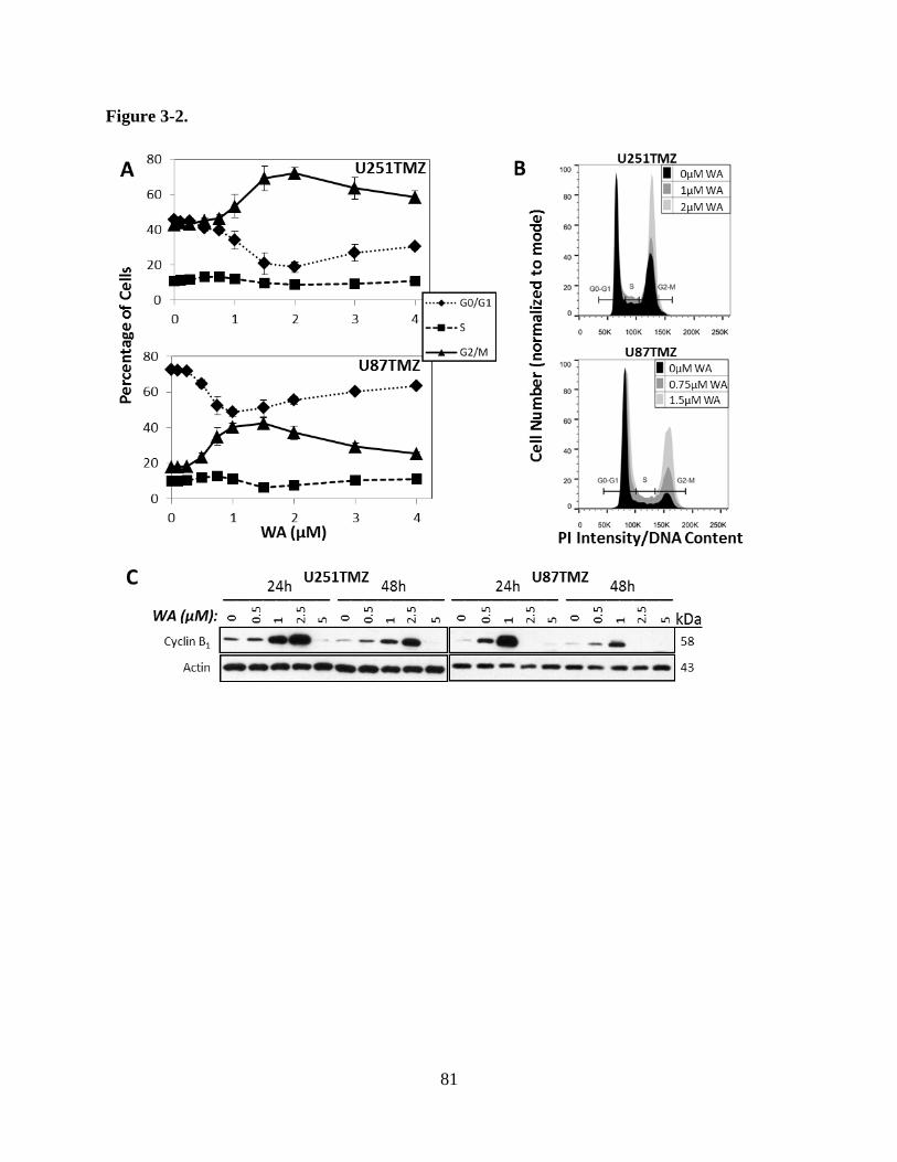
81
Figure 3-2.

82
Figure 3-2. A) Ability to induce cell cycle modulation was assessed by flow cytometry at 24 h
revealing that WA treatment results in dose-dependent G2/M cell cycle arrest. Maximum arrest
was observed at 2 µM and 1.5 µM for U251TMZ and U87TMZ cells, respectively. B)
Histograms demonstrate cell cycle distribution for U251TMZ and U87TMZ cells at
concentrations up to those yielding maximal arrest. C) Arrest in G2/M phase was molecularly
confirmed by immunoblotting for cyclin B1 at 24-48 h. Highest levels of cyclin B1 were observed
at 2.5 µM and 1 µM WA for U251TMZ and U87TMZ cells, respectively, confirming flow
cytometry findings.

83
3.4.4 Withaferin A induces cell death through both the intrinsic and extrinsic apoptotic pathways
PI and Annexin V-FITC dual staining analysis with flow cytometry was used to further
characterize the anti-proliferative effects of WA by assessing levels of apoptosis and necrosis
following drug exposure. After 24 h of 1-3 µM WA, U87TMZ and U251TMZ cells
demonstrated increases in Annexin V-only staining representative of early apoptotic processes
with increases in dual staining representative of late apoptosis present at higher concentrations
(2-6 µM) (Figures 3-3A and 3-3B). U87TMZ cells alone appear to also show mild increases in
levels of necrosis with WA exposure as demonstrated by staining with PI only. Total positive
staining of control U87TMZ and U251TMZ cells was 8.2% and 4.0% but increased to 45.5% (p
= 0.01: 14.7% early apoptosis; 23.0% late apoptosis; 7.7% necrosis) and 17.1% (p = 0.0004:
9.1% early apoptosis; 6.9% late apoptosis; 1.0% necrosis) with 2 µM WA, respectively. 6 µM
WA elevated total cell death to 80.0% for U87TMZ (p < 0.0001: 4.9% early apoptosis; 62.6%
late apoptosis; 12.5% necrosis) and 51.8% for U251TMZ cells (p < 0.0001: 4.0% early
apoptosis; 44.6% late apoptosis; 3.1% necrosis).
Molecular confirmation of an apoptotic response was conducted by Western blotting for
cleaved and/or total levels of the following proteins: extrinsic procaspase 8,
mitochondrial/intrinsic procaspase 9, effector procaspases 3 and 7, and downstream poly(ADP-
ribose) polymerase (PARP) cleavage (Figure 3-3C). At 24 h, increasing concentrations of WA
resulted in progressively decreased levels of all procaspases and PARP, suggesting both intrinsic
and extrinsic routes of apoptotic cell death. Maximum levels of cleaved caspase 8 were observed
at 1-2.5 µM WA for U251TMZ and 0.5-1 µM WA for U87TMZ while highest levels for cleaved

84
Figure 3-3.

85
Figure 3-3. A) WA treatment induced apoptotic cell death in U251TMZ and U87TMZ cells with
increasing concentrations. 1-3 µM WA induced early apoptotic processes with increasing levels
of late apoptosis observed at all concentrations examined. Only U87TMZ demonstrated elevated
necrosis with treatment. B) Dot plots demonstrating propidium iodide and Annexin V-FITC
staining show representative examples from both cell lines utilized. C) An apoptotic mechanism
was confirmed by Western blotting for total and cleaved initiator caspases 8 and 9, effector
caspases 3 and 7, and downstream PARP. Uncleaved proteins were decreased with increasing
WA concentration. U251TMZ and U87TMZ cells demonstrated optimal concentrations for
caspase cleavage with highest levels of PARP cleavage observed in both lines at 5 µM WA. D)
U251TMZ cells were pretreated with 50 µM of either caspase 8 inhibitor Z-IETD-FMK or
caspase 9 inhibitor Z-LEHD-FMK for 1 h followed by 2.5 µM WA for 24 h to determine if WA-
driven apoptosis was intrinsically or extrinsically mediated. Inhibition of both caspase 8 and 9
reduced WA-induced cleavage of caspases 3 and 7 as well as PARP.

86
caspases 3 and 7 were observed at 5 µM WA for U251TMZ and 1-2.5 µM WA for U87TMZ.
PARP cleavage increased up to 5 µM in both cell lines, the maximum concentration utilized, and
suggests that the absence of cleaved caspases despite depletion of the procaspase form at high
WA concentrations can be explained by a time-dependent nature of caspase signaling in which
the upstream cleaved caspases are ultimately degraded following their activity.
To further evaluate the nature of the intrinsic and extrinsic apoptotic processing in WA-
induced cytotoxicity, U251TMZ cells were pretreated with 50 µM of either caspase 8 inhibitor
Z-IETD-FMK or caspase 9 inhibitor Z-LEHD-FMK for 1 h followed by 2.5 µM WA for 24 h.
Pretreatment with both inhibitors resulted in a return to baseline for both cleaved caspase 3 and
PARP levels and decreased amounts of cleaved caspase 7 compared to WA alone (Figure 3-3D).
Inhibitors alone resulted in negligible changes. These data support that cytotoxicity at WA
concentrations above IC50 involves induction of intrinsically- and extrinsically-mediated
apoptosis in the GBM cell lines tested.
3.4.5 Withaferin A modulates the Akt/mTOR and MAPK pathways
Proteins in and related to the Akt/mTOR and mitogen-activated protein kinase (MAPK)
pathways are key to proliferation and survival of GBM cells and other cancers (Wen and
Kesari, 2008). These proteins were evaluated by Western blotting at 24-48 h post-treatment in
U251TMZ and U87TMZ cells in order to evaluate WA activity against total and phosphorylated
protein levels as a potential mechanism for the anti-proliferative effects observed (Figure 3-4A).
At 24 h, total levels of Akt and mTOR were decreased between ~1-5 µM WA with minimal
changes occurring in total p70 S6 kinase (S6K) in both cells lines. However, activation of each

87
Figure 3-4.

88
Figure 3-4. A) Key proteins of the survival and proliferation PI3K/Akt/mTOR and MAPK
pathways as well as common surface tyrosine kinase receptors were evaluated by
immunoblotting for total levels and activation by phosphorylation following treatment with WA
at 24-48 h in U251TMZ and U87TMZ cells. Total levels of specific proteins in both pathways
were decreased, but reduction of protein activation was only observed in the PI3K/Akt/MAPK
pathway whereas phosphorylation of Raf-1 and ERK1/2 was increased. B) To determine the
nature of MAPK pathway activation, U251TMZ and U87TMZ cells were pretreated with 50 µM
of the MEK inhibitor PD98059 for 1 h followed by 2.5 µM WA for 6 h or 24 h. PD98059
increased ERK1/2 phosphorylation but reduced WA-induced phosphorylation of ERK1/2 in a
time-dependent manner. WA alone induced caspase 3 and PARP cleavage in both cell lines in a
time-dependent manner that was increased when combined with MEK inhibitor. This
demonstrates that MAPK pathway activation with WA was acting in a pro-survival signal rather
than contributing to apoptosis.

89
protein via phosphorylation (p-Akt Ser473, p-mTOR Ser2448, and p-p70 S6K Thr389) was
similarly reduced over this range. Phosphorylation at Thr412 of the nuclear isoform of p70 S6K,
p85 S6K, was also reduced with increasing WA concentration (data not shown). These effects
were observed through the 48 h timepoint and demonstrate an inhibitory effect of WA on the
Akt/mTOR growth pathway. Additionally, phosphorylation (Thr172) of AMPKα, the catalytic
subunit of a negative regulator of the Akt/mTOR pathway that responds to cellular stressors, was
increased with WA treatment, peaking at 1 µM in U251TMZ cells (Supplemental Figure 3-1).
Despite depletion of total levels of the downstream tumor suppressor TSC2, phosphorylation at
Thr1462 was enhanced or maintained at all WA concentrations tested in U251TMZ cells. In
contrast to previous observations in parental U87 cells (Grogan et al., 2013), total and phospho-
levels of AMPKα were decreased in U87TMZ cells despite maintenance of p-TSC2 levels
(Supplemental Figure 3-1).
The MAPKKK Raf-1 and the MAPK ERK2 were assessed to evaluate the effects of WA
on the primary proliferative MAPK pathway (Figure 3-4A). Total Raf-1 levels decreased at ~2.5-
5 µM in both U251TMZ and U87TMZ cells, however, activating phosphorylation of the protein
at Ser338 increased over the same concentration range. Downstream, minimal changes were
observed in total ERK2 levels, but phosphorylation of ERK1/2 (Thr202/Tyr204) was notably
increased in a dose-dependent manner in both cell lines with a maximum level achieved at 2.5
µM WA in each at both 24 h and 48 h. Given that phosphorylation of MAPK proteins is typical
of a pro-survival response yet WA instead induces apoptosis in GBM cells, the MAPK pathway
was further explored to determine if its activation was necessary for the induction of apoptosis or
simply a compensatory pro-survival response. U251TMZ and U87TMZ cells were pretreated
with 50 µM of the MAPKK MEK inhibitor PD98059 for 1 h followed by 2.5 µM WA for 6h or

90
24 h. Pretreatment with the inhibitor alone resulted in increased ERK1/2 phosphorylation but
limited WA-induced phosphorylation in a time-dependent manner (Figure 3-4B). Alterations in
apoptosis were assessed by cleaved caspase 3 and cleaved PARP levels. WA alone induced
observable caspase 3 and PARP cleavage at 24 h in both cell lines and PARP cleavage alone at
6h in U87TMZ. However, cleaved caspase 3 and PARP levels were increased when combined
with the MEK inhibitor at 24 h in U251TMZ and 6 h in U87TMZ, demonstrating that MAPK
pathway activation with WA treatment appears to represent a compensatory response to the
apoptotic effects induced by the compound.
Finally, the tyrosine kinase receptors EGFR, Her2/ErbB2, and c-Met, known to signal to
both the Akt/mTOR and MAPK pathways, were evaluated due to their status as common
amplified, mutated, and/or drug-targeted surface proteins in GBM (Wullich et al., 1993; Potti et
al., 2004; Puputti et al., 2006; Guessous et al., 2010; Berezowska and Schlegel, 2011). Total
levels of EGFR and Her2/ErbB2 in both U251TMZ and U87TMZ decreased with 2.5-5 µM WA
treatment at 24 h and 48 h while total levels of c-Met decreased at 5 µM (Figure 3-4A).
Interestingly, increased phosphorylation of Tyr1068 on EGFR was observed at 2.5-5 µM
depending on the cell line as well as increased phosphorylation of Tyr1234/1235 on c-Met in
U87TMZ cells up to 2.5 µM WA at 48 h. Phosphorylation of c-Met was otherwise
downregulated with WA treatment in both lines.
3.4.6 Withaferin A elevates oxidative status and induces a heat shock stress response in TMZ-
resistant cells
91
We have previously demonstrated the ability of WA to elevate oxidative potential and
ROS-mediated cell death in TMZ-sensitive GBMs (Grogan et al., 2013). TMZ has also been
demonstrated to produce ROS that result in inhibitory activation of AMPK (Zhang et al., 2010).
We therefore evaluated the nature and ability of WA to continue to induce ROS in TMZ-resistant
U87TMZ and U251TMZ cells. Peroxide-type radical production was monitored with CM-
H2DCFDA conversion at 3 h post-WA exposure. Both cell lines showed statistically significant
increased detectible CM-H2DCFDA fluorescence with increasing WA exposure. ROS levels rose
20.9% (p = 0.001), 36.6% (p = 0.002), and 38.8% (p = 0.01) in U251TMZ and 21.3% (p =
0.003), 40.6% (p = 0.004), 47.3% (p = 0.001) in U87TMZ cells at 1, 3, and 5 µM WA compared
to control, respectively (Figure 3-5A). The thiol antioxidant and ROS scavenger NAC was added
simultaneously with WA and was shown to nearly complete abrogate WA-generated ROS,
further demonstrating the nature of the effect.
Various models of oxidative stress are known to be associated with a heat shock response
via induction of several HSPs (Ansari et al., 2011; Qiao et al., 2012), and alteration of HSPs
were previously reported following WA treatment (Khan et al., 2012; Grogan et al., 2013).
Treatment with WA yielded a unique pattern of HSP expression modulation in U251TMZ and
U87TMZ cells (Figure 3-5B). Total levels of HSP32/heme oxygenase 1 and HSP70, but not
HSP90, were observed to increase with increasing concentrations of WA at 24 h. HSP27 was
depleted at 24 h with 5 µM WA in both cell lines but remained largely unchanged in U251TMZ
and mildly elevated in U87TMZ with other concentrations at 24 h. The transcription factor heat
shock factor 1 (HSF1) showed dose-dependent reduction in expression between 1-5 µM WA.
Pretreatment with 5 mM NAC virtually eliminated all modulation of proteins in the heat shock
system (Figure 3-5B).

92
Figure 3-5.

93
Figure 3-5. A) The peroxide ROS indicator CM-H2DCFDA was preloaded into U251TMZ and
U87TMZ cells prior to WA exposure to determine alterations in oxidative potential. Elevations
in signal that were observed with increasing concentrations of WA at 3 h were reduced or
completely absent with co-treatment of 5mM NAC. B) Treatment with WA resulted in the
induction of an NAC-repressible cellular stress heat shock response. Proteins associated with
cellular stress and heat shock were evaluated by immunoblotting at 24 h and revealed elevated
levels of HSP32 and HSP70, known to be upregulated in response to oxidative stress, but
decreased transcription factor HSF1 with increasing WA exposure that could be completely
eliminated with 5 mM NAC pretreatment. NAC also prevented induction of cyclin B1 and
cleavage of PARP. Functionally, NAC pretreatment reduced the anti-proliferative (C) and pro-
cytotoxic (D) effects of WA as assessed by the CellTiter-Glo assay at 72 h and flow cytometry at
24 h, respectively. *p < 0.05, **p < 0.01, ***p < 0.001

94
Functionally, WA-induced depletion of ATP levels, a marker of reduced cellular viability
in the CellTiter-Glo assay, could be reduced or completely eliminated with 1-5 mM NAC
pretreatment (p < 0.0001) (Figure 3-5C). Such effects were seen with up to 6 µM WA, the
highest concentration examined. Additionally, re-evaluation of cell death by flow cytometry as
previously described following pretreatment with 5 mM NAC showed the elimination of death-
associated PI and Annexin V staining normally induced by 5 µM WA (p < 0.01) (Figure 3-5D).
This was supported by blockage of PARP cleavage via NAC pretreatment (Fig. 3-5B).
Additionally, WA-mediated cyclin B1 induction was blocked by NAC, demonstrating the
prevention of G2/M cell cycle arrest seen with WA treatment.
3.4.7 Withaferin A resensitizes TMZ-resistant GBM cells to TMZ through MGMT depletion
Expression of MGMT is associated with TMZ resistance in GBMs and therefore is an
attractive target for TMZ resistance resensitization and prevention (Kitange et al., 2009). WA
demonstrated the ability to induce depletion of MGMT at 48 h in cell lines (U251TMZ, T98G,
and U138) utilizing MGMT as the primary means of TMZ resistance (Figure 3-6A). U251TMZ
and U138 cells showed progressive depletion of MGMT from 0.5-10 µM WA with
approximately 5-fold and 20-fold less compared to control by 5 µM, respectively. At 5 µM WA
treatment, there was a 43% reduction in MGMT in T98G cells with complete elimination by 10
µM. U87TMZ cells, however, failed to express MGMT altogether.
To determine if these findings extended to functional resensitization of resistant cells to
WA, two cells lines resistant through MGMT (U251TMZ and T98G) and one cell line displaying
MGMT-independent resistance (U87TMZ) were treated with WA for 24 h followed by TMZ and

95
Figure 3-6.
96
Figure 3-6. A) WA treatment reduced protein levels of MGMT in TMZ-resistant U251TMZ,
T98G, and U138 cell lines at 48 h. MGMT was not observed in U87TMZ. B) 24 h pretreatment
with WA resensitized MGMT-expressing U251TMZ and T98G cells to TMZ in a dose-
dependent manner as assessed by a normalized MTS assay, but no resensitization was observed
in MGMT-deficient U87TMZ cells. C) TMZ-resistant cells were treated with WA for 24 h
followed by TMZ exposure for an additional 24 h. Pretreatment with WA potentiated and/or
synergized with TMZ to induce further depletion of MGMT in MGMT-expressing lines.
Increasing WA concentrations yielded higher levels of p-H2A.X and cleaved PARP in all cell
lines, but only T98G demonstrated WA-mediated increased levels with TMZ exposure. *p <
0.05, **p < 0.01, ***p < 0.001

97
evaluated with the MTS assay (Figure 3-6B). Both U251TMZ and T98G showed statistically
significant decreases in cell viability when 1, 1.5, or 2 µM WA was combined with 300 or 500
µM TMZ. Pretreatment with 2 µM WA followed by 300 µM TMZ yielded a 32% (p < 0.0001)
and 34% (p = 0.004) relative decrease in U251TMZ and T98G cell viability compared to TMZ
or WA alone, respectively. In contrast, U87TMZ cells only demonstrated a statistically
significant enhancement of combination therapy over TMZ alone – only 6% additional reduction
in relative viability – with a 2 µM WA and 300 µM TMZ combination (p = 0.02). 48 h
pretreatment with WA also demonstrated enhanced effect with significant elimination of the
benefit by 72 h pretreatment (data not shown). WA pretreatment did not prevent TMZ efficacy in
TMZ sensitive U251 and U87 parental cells (Supplemental Figure 3-2A).
Because TMZ also depletes MGMT (D'Atri et al., 2000), the effect of WA and TMZ
combination on MGMT levels was evaluated. Combination therapy demonstrated dose-
dependent synergy or potentiation to reduce total levels of MGMT in U251TMZ, T98G, and
U138 MGMT-expressing cells (Figure 3-6C). At 1 µM WA in U251TMZ and U138 cells and 2.5
µM WA in T98G cells, minimal decreases and/or increases of MGMT levels were observed. The
addition of 300 µM TMZ for 24 h resulted in 43%, 52%, and 34% reduction in MGMT protein
levels compared to TMZ alone in U251TMZ, T98G, and U138 cells, respectively.
Phosphorylation of H2A.X as a marker of double strand DNA damage and cleavage of
PARP during apoptosis were evaluated to look for molecular evidence of combinational efficacy.
Both markers were elevated in the setting of increasing concentrations of WA in U251TMZ,
T98G, U138, and U87TMZ cells. However, PARP cleavage was then diminished at the high
levels of U87TMZ and U138 cells suggesting non-apoptotic cell death or very late apoptosis at
this timepoint. Addition of TMZ was observed to increase p-H2A.X and PARP cleavage in only

98
T98G, particularly at 1 µM WA. Interestingly, despite similar or increased levels of cleaved
PARP within WA treatment groups, levels of uncleaved PARP increased with TMZ treatment in
each of the TMZ groups but not control in U251TMZ and T98G cells. Given the reported
oxidative effect induced by TMZ (Zhang et al., 2010), markers of oxidative stress including
HSP32, HSP70, and p-AMPKα as well as the AMPK-influenced downstream phosphorylation of
mTOR were evaluated to determine if combination of WA and TMZ demonstrated enhanced
oxidative effect in GBM. While WA increased HSP32 and HSP70 levels in all lines and TMZ
enhanced phosphorylation of AMPKα in U138, combination therapy did not result in additional
effect and failed to further diminish mTOR activation (Supplemental Figure 3-2B).
3.5 Discussion
While WA has demonstrated effectiveness as a promising new therapeutic agent in a
variety of cancer types including TMZ-sensitive GBM, this work is the first to explore its
efficacy in a known model of drug-resistance. Here, we show for the first time that WA induces
a potent cytotoxic effect against temozolomide resistant GBMs resulting in G2/M arrest and
apoptosis from both the intrinsic and extrinsic systems. These findings correspond with the
elevation of cellular oxidative potential and inhibitory modulation of the Akt/mTOR pathway but
not the MAPK pathway.
Since its approval by the FDA, TMZ remains the standard-of-care chemotherapeutic
agent in the treatment of primary GBM. Exposure to this agent results in DNA adducts such as
O6-methylation of guanine which, if not removed by MGMT, can mispair with thymine. Cells
with intact MMR undergo futile cycles of attempted repair with resultant replication-associated
99
DNA double strand breaks, G2/M arrest, and ultimately apoptosis (Hirose et al., 2003). Targeting
mechanisms of DNA damage repair with various inhibitors, such as those of PARP and ataxia
telangiectasia mutated (ATM), has provided potentially beneficial strategies in enhancing TMZ
effect, but comparatively limited effort has gone into addressing both inherent and acquired
resistance to TMZ by MGMT expression or, less frequently, MMR mutation in GBM. Despite
about half of patients displaying tumor phenotypes not conducive to TMZ use, it remains a first-
line agent, demonstrating the need for better tumor screening techniques. Furthermore, initially
responsive tumors are often quick to develop resistance to this line of therapy (Chamberlain,
2010; Quick et al., 2010).
Several experimental agents used to resensitize resistant cancer cells to TMZ or other
alkylating agents – such as valproic acid (Sztajnkrycer, 2002; Weller et al., 2011; Ryu et al.,
2012), O6-benzylguanine (Baer et al., 1993; Kaina et al., 2010), IL-24 (Zheng et al., 2008), and
silencing oligonucleotides (Citti et al., 1996; Xie et al., 2011; Kreth et al., 2013) – possess little
practical intrinsic anti-cancer cytotoxic properties or clinical applicability, however, some do
have the potential for adverse effects in the patient. Alternatively, dose-dense TMZ scheduling,
used to deplete MGMT levels quickly to enhance TMZ effects, has yielded disappointing
outcomes in several clinical trials (Strik et al., 2012). In this study, WA acted as both an
inherently cytotoxic agent and an enhancer of the efficacy of TMZ in MGMT-driven resistant
cell lines while maintaining TMZ sensitivity in parental lines. Such data conceptually support the
utility of combination therapy WA with TMZ in the first-line treatment of GBM. Such an
approach should maximize anti-cancer activity across broad tumor heterogeneity. Importantly,
while tumors with resistance due to MMR mutation may not be resensitized to TMZ by WA, the
presence of a second cytotoxic chemotherapeutic agent may provide significant benefit in the
100
context of ineffective TMZ treatment. It remains to be fully explored whether increased levels of
p-H2A.X observed with WA treatment are a result of direct DNA damage, perhaps through an
oxidative mechanism, or rather a known byproduct observed during cellular apoptosis for DNA
fragmentation (Rogakou et al., 2000), however, preliminary results in breast cancer support a
caspase-mediated mechanism (data not published). Elevated total PARP with increasing TMZ in
the presence of WA in MGMT-expressing lines may indicate the necessity for increased base
excision repair (BER) during combination therapy, alluding to induction of DNA damage as a
reason for combinational efficacy which will need to be validated by further scientific
investigation.
Several studies have implicated the role of WA in the disruption of HSP90 chaperone
functions through binding to the C-terminus of HSP90 (Yu et al., 2010; Grover et al., 2011).
While direct evidence of such an inhibitory effect has not yet been established, downstream
depletion of various HSP90-related client proteins suggests that WA may indeed act at least in
part as a novel HSP90 inhibitor. This could explain and support the wide-ranging effects
observed in this study, including the depletion in total levels of known HSP90 client proteins
such as Akt, mTOR, Raf-1, and EGFR. By depleting these key regulatory client proteins of
HSP90, the normal signaling axes of multiple proliferative pathways are simultaneously
modulated. MGMT, which is depleted in the presence of WA, has not, however, been reported as
a client protein of HSP90. Indeed, current studies demonstrate that total levels of MGMT are not
impacted by the widely-utilized HSP90-inhibitor 17-AAG until high concentrations (1-2 µM) are
achieved, suggesting that its stability and function are not directly maintained by HSP90 (data
not shown). Santagata, et al. demonstrated the thiol reactivity of WA (Santagata et al., 2012), and
our results implicate an oxidative mechanism, representing an alternative means by which

101
MGMT and other non-HSP90 client proteins may be targeted for degradation by WA.
Interestingly, valproic acid, which diminishes MGMT levels, has also been shown to cause
oxidative stress in glioma cell lines (Fu et al., 2010).
Induction of oxidation has become a promising mechanism by which to target cancer.
Alterations in the redox potential of cancer cells due to various molecular turnover and quick
replication results in increased cellular stress that can be exploited to shift cells into a state of
cytotoxicity (Laurent et al., 2005; Cabello et al., 2007). This approach circumvents traditional
therapeutic options such as antigen-targeting and DNA-damaging agents that have received the
most attention but are often rendered useless due to rapid development of resistance. In GBM,
such traditional agents have been explored in patients but have yielded unimpressive results in
clinical trials largely due to the development of tumor mutations that limit the effectiveness
and/or relevance of a particular targeted therapeutic (Wen and Kesari, 2008; Quick et al., 2010).
In the present study, thiol-reactivity and oxidation by WA resulted in depletion of
proteins in the Akt/mTOR and MAPK pathways including many cell surface receptors either
through direct interaction or as a downstream effect of the inhibition of other proteins. As noted
previously, other reports have shown additional pathways effected. Such an ability to target
multiple pathways simultaneously with a single compound has great potential to generate an
enhanced anti-cancer effect while reducing the development of resistance to a particular target.
The ability to modulate multiple components of the Akt/mTOR pathway simultaneously
provides a novel and important mechanism by which WA can manifest its beneficial effects
against GBMs. Because of mutations in proteins like the tumor suppressor phosphatase and
tensin homolog (PTEN), this pathway is frequently overactivated in GBM to drive proliferation,
as shown in both U251 and U87 cells (Koul, 2008; Lee et al., 2011; Lino and Merlo, 2011).

102
Interestingly, while we previously showed activation of AMPKα and downstream tumor
suppressor TSC2 which act to inhibit mTOR in both U251 and U87 parental cells (Grogan et al.,
2013), U87TMZ appears to have lost the ability to act through this pathway which suggests
direct inhibition of the Akt/mTOR axis.
WA treatment yields subsequent induction of the MAPK pathway as demonstrated by
activating phosphorylation of EGFR, Raf-1, and ERK1/2. Previous studies have shown that
reactive oxygen species generated by epoxide-containing compounds in GBM cells are
responsible for the phosphorylation of ERK1/2 which then drives apoptosis (Wu et al., 2011). In
contrast, inhibition of this pathway prior to WA exposure yields enhanced apoptosis, suggesting
that the activation of the MAPK pathway is a compensatory pro-survival response to apoptotic
shifts in the cell and may be a therapeutic target that would synergistically enhance the efficacy
of WA in future combinational therapy studies.
The studies presented here provide an important framework for the utilization of WA in
TMZ-resistant GBM as both a monotherapy and as a resensitizer in combination with the
standard chemotherapeutic agent TMZ. WA demonstrates an oxidative mechanism that leads to
anti-proliferative and pro-apoptotic effects largely through Akt/mTOR pathway modulation.
Simultaneously, depletion of MGMT allows for enhanced activity of TMZ. While future pre-
clinical animal studies will be necessary to determine its translational potential in GBMs,
evidence is supportive of a future clinical potential for the utilization of WA across the
heterogeneic spectrum of glioma patients.

103
Supplemental Figure 3-1.

104
Supplemental Figure 3-1. WA treatment resulted in depletion of total protein levels in both
AMPKα and TSC2 at 24-48 h in both U251TMZ and U87TMZ cells. U87TMZ cells also
demonstrated reduced phosphorylation of AMPKα in contrast to previous findings in U87
parental cells. U251TMZ cells display dose-dependent activating phosphorylation of AMPKα
and downstream TSC2.

105
Supplemental Figure 3-2.

106
Supplemental Figure 3-2. A) 24 h WA pretreatment did not prevent TMZ efficacy in TMZ-
sensitive U251 and U87 parental cells as assessed by a normalized MTS assay, suggesting a lack
of inhibition of functional MMR. In U251 cells, WA mildly sensitized cells to TMZ, but no
sensitization was observed in U87 cells. B) Given a described oxidative component of TMZ
therapy through activation of AMPKα, markers of oxidation were assessed during combination
therapy of WA and TMZ in TMZ-resistant cells. HSP32 and HSP70, known to be upregulated in
response to oxidative stress, increased with WA treatment to a maximal level but were not
further modulated by the presence of TMZ. Treatment with TMZ only resulted in elevated p-
AMPKα in only U138 cells, but WA failed to further increase this phosphorylation in
combination with TMZ. This was confirmed downstream by failure to further inhibit mTOR
activation with combination therapy, suggesting efficacy of such treatment was not due to
potentiated or synergized elevation in oxidation status. **p < 0.01

107
Chapter 4
Withaferin A is a novel inhibitor of the Wnt/β-catenin
signaling pathway in medulloblasoma through proteasome-
mediated degradation of TCF/LEF
108
4.1 Abstract
Pediatric medulloblastoma (MB) is a challenging tumor to approach therapeutically given
significant tumor heterogeneity across patients. While current treatment options have led to an
approximately 70% 5-year survival rate in children and adolescents, the off-target toxicity of
these agents frequently manifests with chronic quality-of life impairments in these vulnerable
patients, identifying a need for novel therapeutic options. Withaferin A (WA) has demonstrated
notable anti-cancer efficacy against multiple cancer types through several oncogenic pathways,
but its modulation of Wnt/β-catenin signaling, overactive in 10-15% of MB, has not been
explored. Here, WA inhibited MB cell proliferation through dose-dependent G2/M cell cycle
arrest and induction of apoptosis. Treatment with WA was demonstrated to function as a novel
direct inhibitor of Wnt signaling as assessed by TOP FLASH reporter activity. Reduced
signaling was mediated through proteasomal degradation the TCF/LEF transcription factors
TCF1, TCF3, TCF4, and LEF1 and reduced interaction with β-catenin without significant
alterations of β-catenin level or localization. This was associated with a reduction in total and
phosphorylated levels of Akt and mTOR but elevations in inhibitory p-GSK-3β which was
demonstrated pharmacologically to be independent of remaining Akt activity. Consistent with
previous findings in other models, WA treatment resulted in N-acetyl-L-cysteine-repressible
cellular oxidation and dissociation of HSP90/Cdc37. While TCF/LEF members were not
observed to directly associate with HSP90 or Cdc37, each was decreased following inhibition of
HSP90 with N-terminal inhibitor 17-AAG, suggesting an indirect role for HSP90 in their
stability. Taken together, these findings show both intriguing translational potential for WA as a

109
therapeutic agent against MB and other Wnt-driven cancers and a novel role for HSP90 in Wnt
signaling, each warranting further exploration.
4.2 Introduction
Central nervous system (CNS) tumors, including the grade IV medulloblastoma (MB) of
the posterior fossa, are the most common solid tumors of childhood, accounting for
approximately 20% of all pediatric malignancies (Linabery and Ross, 2008). Medulloblastomas
represent 25-30% of these primary lesions in children but can arising in individuals of all ages
(Khatua et al., 2012). Although the 5-year survival rate for medulloblastomas in both children
and adolescents is around 70%, therapeutic challenges remain to simultaneously address both
treating the tumor and ensuring a high long-term quality of life in the pediatric patient population
(Smoll, 2012). Current standards of care include surgical resection followed by craniospinal
radiation and chemotherapeutic agents such as vincristine, lomustine, cyclophosphamide,
carboplatin, and cisplatin (Khatua et al., 2012). However, because of significant morbidities
associated with these agents, it has been reported that long-term survivors of childhood
medulloblastoma suffer from endocrine, cognitive, and neurological complications with
significant deficits in daily psychosocial functioning including driving, employment,
independent living, and personal relationships (Boman et al., 2009; Frange et al., 2009).
Medulloblastomas are categorized in several unique molecular subgroups based on tumor
genetics, cell histology, growth, and clinical outcome including sonic hedgehog (SHH),
Wingless (Wnt), group 3, and group 4 (Northcott et al., 2012). SHH and Wnt are named due to
upregulation and overactivation of these key signaling pathways. Specifically, the Wnt subgroup

110
of MB patients is characterized by aberrant overactivation of the canonical Wnt/β-catenin
signaling pathway, often through inactivating mutations of the adenomatous polyposis coli
(APC) and Axin1/2 proteins or overexpression of β-catenin (Kim et al., 2011). Although the Wnt
subgroup represents 10-15% of total MB patients and is characterized by long-term survival rates
of 90%, the challenges of conventional therapy persist with current patient morbidity and
mortality largely due to the complications of therapy and the development of secondary
malignancies rather than recurrence of the MB (Ellison et al., 2011; Kim et al., 2011; Taylor et
al., 2012; Remke et al., 2013).
While a number of inhibitors have been identified that alter Wnt signaling at various
points in the cascade (Voronkov and Krauss, 2013), only four agents are currently in human
clinic trial against any malignacy: olaparib (poly(ADP-ribose) polymerase (PARP)/tankyrase
inhibitor), veliparib (PARP/tankyrase inhibitor), LGK974 (porcupine inhibitor), and OTSA101-
DTPA-90Y (radiolabeled antibody against frizzled-10) (MacDonald et al., 2014). Only PARP
inhibition, described to destabilize β-catenin through Axin (Waaler et al., 2012), is under trial for
MB with the latter two therapies failing to target the appropriate location in the signaling cascade
given the mutations of MB. Given the heterogeneic nature of the disease supported by multiple
subgroup classifications, there is a critical need for novel therapeutics that function to both target
this critical aberrant signaling to maximize Wnt subgroup effect while maintaining generalized
anti-tumor cytotoxicity in other subgroups lacking these specific signaling alterations.
Withaferin A (WA) is a cytotoxic steroidal lactone identified in several species of the
Solenceae plant family that has demonstrated the ability to modulate multiple signaling pathways
key to cancer proliferation and survival (Vanden Berghe et al., 2012; Vyas and Singh, 2014).
Notably, in the brain tumor glioblastoma multiforme (GBM), WA functioned to reduce total and
111
phosphorylated levels of key oncogenic proteins in the Akt/mammalian target of rapamycin
(mTOR) signaling pathway (Grogan et al., 2013; Grogan et al., 2014). The Akt/mTOR pathway
establishes cross-talk with the Wnt/β-catenin pathway through glycogen synthase kinase-3β
(GSK-3β) such that activation of Akt produces inhibitory phosphorylation of GSK-3β, a
reduction in the phosphorylation of β-catenin, and enhanced stability of β-catenin. As such,
inhibition of PI3 kinase (PI3K)/Akt signaling has been shown to inhibit the Wnt/β-catenin
pathway in MB (Baryawno et al., 2010). We hypothesized that WA could act as a novel inhibitor
of the Wnt pathway in MB by a similar mechanism. To date, WA has not been explored as a
Wnt/β-catenin signaling inhibitor nor examine against MB.
Studies by our group and others have suggested that modulation of the Akt/mTOR
pathway and the other diverse findings associated with WA treatment may be due to two
underlying mechanisms which are explored here in MB: enhanced cellular oxidation (Malik et
al., 2007; Widodo et al., 2010; Hahm et al., 2011; Mayola et al., 2011; Santagata et al., 2012;
Grogan et al., 2013; Grogan et al., 2014) and inhibition of the heat shock protein (HSP) 90
protein chaperone axis including its disruption of its interactions with co-chaperone cell division
cycle protein 37 (Cdc37) (Yu et al., 2010; Gu et al., 2014). Previous studies have identified that
HSP90 associates with Wnt/β-catenin pathways proteins Axin1, GSK-3β, and β-catenin but that
its inhibition only resulted in a cell line-dependent depletion of GSK-3β (Cooper et al., 2011; Liu
et al., 2012). p53-regulated HSP90 function was shown to enhance Wnt signaling, but this was
likely an indirect effect through phosphorylation of Akt and GSK-3β (Okayama et al., 2014).
Direct inhibition of the Wnt/β-catenin pathway through HSP90 inhibition has not been
demonstrated previously.

112
In this study, we explored the general cytotoxicity of WA, including evaluation of its
described oxidative and HSP90 inhibitory effects, as a potential chemotherapeutic for MB and
demonstrate for the first time that it functions in-part as a potent inhibitor of the canonical Wnt
signaling pathway. Interestingly, our studies provide evidence that WA induces degradation of
the transcription factor (TCF)/lymphoid enhancer-binding factor (LEF) family of transcription
factors critical in Wnt signaling to inhibit pathway activity directly rather than through
Akt/mTOR pathway cross-talk via GSK-3β. Maintenance of TCF/LEF proteins was shown to be
indirectly dependent on HSP90 function with WA inducing disruption of the HSP90/Cdc37
interaction.
4.3 Materials and methods
4.3.1 Cell culture and general reagents
Four human pediatric medulloblastoma cell lines were acquired. DAOY adherent cells
were obtained from American Type Culture Collection (ATCC; Manassas, VA). ONS76
adherent cells were generously donated by Dr. Michael D. Taylor (The Hospital for Sick
Children, Toronto, Ontario, Canada), and D425(MED) and D283(MED) semi-
adherent/suspension cells were kindly provided by Dr. Darell D. Begner (Duke University,
Durham, NC). All cells were maintained in Dulbecco’s modified Eagle’s medium (DMEM
#11995-065; Gibco, Grand Island, NY) supplemented with 10% fetal bovine serum (FBS;
Sigma-Aldrich, St. Louis, MO), 1% penicillin/streptomycin (Gibco), 2% L-glutamine (200 mM;
Gibco), 1% MEM-vitamin (100x; Hyclone, Logan, UT), and 1% MEM nonessential amino acids

113
(Sigma Aldrich) at a 37 °C humidified atmosphere of 5% CO2 in air. Cells were utilized in
experiments upon achieving exponential growth phase. Withaferin A was isolated as previously
described (Samadi et al., 2010b). Propidium iodide (PI), RNase, N-acetyl-L-cysteine (NAC), and
puromycin dihydrochloride were acquired from Sigma-Aldrich, Annexin V-FITC was obtained
from BD Biosciences (San Diego, CA), and MG132 was purchased from Selleckchem (Houston,
TX). 17-N-allylamino-17-demethoxygeldanamycin (17-AAG) was obtained from LC
Laboratories (Woburn, MA).
4.3.2 MTS assay
Cells were plated at 2,000-4,000 cells/well in 50 µL in a 96-well plate and allowed to
adhere and/or settle. After 6 h, cells were treated with various concentrations of WA to bring the
total well volume to 100 µL and allowed to incubate for 72 h. Cell viability and proliferation was
evaluated with the addition of the colorometric CellTiter96 Aqueous MTS assay reagent as per
the manufacturer’s instructions (Promega, Fitchburg, WI). Reagent was allowed to incubate with
the cells for 1-4 h in a cell line-dependent manner, and absorbance was quantified at 490 nm on a
BioTek Synergy 2 plate reader (BioTek, Winooski, VT).
4.3.3 CellTiter Glo assay
The CellTiter-Glo luminescent assay (Promega, Fitchburg, WI) was used to evaluate cell
viability and proliferation through assessment of cellular ATP levels in experiments with the
NAC pretreatment due to its reducing properties. Cells were plated as outlined in the MTS assay

114
in a white-walled 96-well plate. Where indicated, cells were pretreated with NAC for 1 h
followed by WA. After 72 h, assay reagent was added to the wells as per the manufacturer’s
instructions and gently shaken for 10 minutes in the dark at room temperature. Well
luminescence was measured on the BioTek Synergy 2 plate reader.
4.3.4 Cell cycle analysis
Cells were plated to ~50% confluency in the adherent lines or 125,000 cells/mL in the
suspension lines and treated with various concentrations of WA for 24 h. At that time, total cells
were collected, pelleted, fixed/permeablized, and stained with PI as previously described
(Grogan et al., 2013). Cells were analyzed by flow cytometry on a BD LSRII (Becton Dickinson,
San Diego, CA). Only living cells without DNA fragmentation were gated.
4.3.5 Apoptosis evaluation
Cells were plated and treated as described for cell cycle analysis. After 24 h, all cells
were collected and processed as previously described (Grogan et al., 2013). Briefly, cells were
washed once in Annexin binding buffer and subsequently stained with PI and Annexin V-FITV
as per the manufacturer’s instructions (BD Biosciences, San Diego, CA) for 20 minutes at 4 °C.
Stained cells were washed twice with the Annexin binding buffer, resuspended in 400 µL of the
buffer, and immediately analyzed by flow cytometry on the BD LSRII.
4.3.6 Immunoblotting
115
For general Western blotting, cells were plated as previously described for cell cycle
analysis before treatment with WA or 17-AAG. Where indicated, cells were pre-treated with 5
mM NAC for 1 h. After 24 h, cells were harvested, and proteins were isolated in lysis buffer (40
mM HEPES, 2 mM EDTA, 10 mM sodium pyrophosphate decahydrate, 10 mM β-
glycerophosphate disodium salt pentahydrate, 1% triton X-100 supplemented with 100 µM
phenylmethylsulfonyl fluoride (PMSF), 1 mM Na3VO4, and 2 µL/mL protease inhibitor
cocktail), quantified, separated by sodium dodecyl sulfate–polyacrylamide gel electrophoresis
(SDS–PAGE), and transferred onto a Hybond nitrocellulose membrane as previously described
(Samadi et al., 2011). 10-50 µg of protein sample was loaded per lane. Equal loading and
transfer of sample was confirmed by blotting for actin levels. Studies were repeated for accuracy.
For nuclear and cytoplasmic fractionation of proteins, cells were grown and harvested as
described. Washed cells were lysed in harvest buffer (10 mM HEPES pH 7.9, 50 mM NaCl, 0.5
M sucrose, 0.1 mM EDTA, and 0.5% triton X-100), incubated on ice for 5 minutes, and pelleted
at 1,000 rpm. The supernatant was collected as the cytoplasmic fraction. The pellet was
resuspended for one wash with buffer A (10 mM HEPES pH 7.9, 10 mM KCl, 0.1 mM EDTA,
0.1 mM EGTA) and subsequently repelleted. The supernatant was discarded. The pellet was then
resuspended in buffer C (10 mM HEPES pH 7.9, 500 mM NaCl, 0.1 mM EDTA, 0.1 mM
EGTA, 0.1% NP-40), vortexed periodically while incubating on ice for 15 minutes, and
centrifuged at 14,000 rpm for 10 minutes at 4 °C. The supernatant was collected as the nuclear
fraction. Proteins in the two fractions were quantified, prepared, separated, and immunoblotted
identically to what was previously described. Histone H4 was used as a nuclear-only control. All
buffers were supplemented with 100 µM PMSF, 1 mM Na3VO4, and 2 µL/mL protease inhibitor.

116
Triton X-100 soluble and insoluble fractionation was completed with triton soluble buffer
(50 mM Tris-HCl pH 6.8, 150 mM NaCl, 1mM EDTA, 1 mM EGTA, 1% triton X-100) and
insoluble buffer (50 mM Tris-HCl pH 6.8, 2% SDS) as previously described (Chen et al., 2008)
supplemented with 100 µM PMSF, 1 mM Na3VO4, and 2 µL/mL protease inhibitor cocktail. In
brief, cells were pre-treated with proteasome inhibitor MG132 for 1 h followed by WA for 24 h.
Cells were collected as previously described, lysed with triton soluble buffer, and centrifuged at
14,000 rpm for 20 minutes at 4 °C. The supernatant was collected as the soluble fraction. The
pellet was resuspended, washed once with soluble buffer, and repelleted by centrifuged at 14,000
rpm for 10 minutes. The pellet was resuspended in triton insoluble buffer and subsequently
ground with a pestle, vortexed, and heated at 100 °C repeatedly for ~15 minutes to promote
dissolution of the pellet. Any remaining debris was removed by centrifugation, and the
supernatant was collected as the insoluble fraction. Proteins in the two fractions were prepared
and evaluated as described above.
Primary rabbit antibodies against β-catenin (#9562; 1:1000), p-β-catenin
(Ser33/37/Thr41; #9561; 1:1000), TCF1 (#2203; 1:1000), TCF3 (#2883; 1:1000), TCF4 (#2569;
1:1000), LEF1 (#2230; 1:1000), GSK-3β (#9315; 1:1000), p-GSK-3β (Ser9; #9323; 1:1000), c-
myc (#9402; 1:1000), c-Met (#4560; 1:500), and Cdc37 (#4793; 1:1000) and primary mouse
antibodies against Cyclin D1 (#2926; 1:500) and histone H4 (#2935; 1:1000) were acquired from
Cell Signaling Technology (Danvers, MA). Primary rabbit antibody for Axin2 (#MA5-15015;
1:1000) was acquired from Pierce Biotechnology (Rockford, IL). Primary mouse antibodies for
survivin (sc-17779; 1:100) and FLAG (F1804; 1:1000) were purchased from Santa Cruz
Biotechnology (Santa Cruz, CA) and Sigma-Aldrich, respectively. Secondary mouse anti-rabbit
conformation specific HRP antibody (#5127; 1:1000) was obtained from Cell Signaling

117
Biotechnology and used for blotting in appropriate immunoprecipitation blotting. All other
primary and secondary antibodies were used as previously described (Grogan et al., 2013).
4.3.7 Reactive oxygen species measurement
WA-mediated production of intracellular reactive oxygen species (ROS), particularly
peroxides like H2O2, was measured through fluorescent conversion of the general oxidative
stress indicator CM-H2DCFDA (Molecular Probes, Grand Island, NY). All MB cells lines were
preloaded with 20 µM CM-H2DCFDA in 1x PBS at 37 °C for 1 h, washed once with phenol red-
free DMEM supplemented with all aforementioned additives, and resuspended in the same
medium. Cells were plated at 40,000 cells/well in a 96-well plate and treated with WA ± NAC
after 30 minutes. Evaluation of fluorescence was completed on a BioTek Synergy 2 plate reader
after 3-4 h with excitation and emission filters of 485 nm and 528 nm, respectively.
4.3.8 TOP/FOP FLASH reporter assay
A luciferase reporter vector for Wnt/β-catenin signaling activity through TCF/LEF DNA
binding sites, TOP FLASH, and its negative control, FOP FLASH, along with vectors for wild-
type (WT) β-catenin and overactive mutant S33Y-β-catenin (Kolligs et al., 1999) were
generously provided by Dr. Eric R. Fearon (University of Michigan, Ann Arbor, MI). DAOY
cells were transfected in bulk with reporter construct TOP FLASH or FOP FLASH and a renilla
luciferase construct at a ratio of 100:1 along with either β-catenin vector or a pcDNA3 control
vector using Lipofectamine 2000 (Life Technologies, Carlsbad, CA) as per the manufacturer’s

118
instructions and plated at 7,500 cells/well in a white-walled 96-well plate (50 ng reporter, 0.5 ng
renilla, and 400 ng respective β-catenin construct per well in 150 µL). The transfection and gene
expression were allowed to occur over 24 h before the cells were treated with WA. Reporter and
renilla luciferase activity were quantified after 24 h with the Dual-Luciferase Reporter Assay
System (Promega) on a BioTek Syngery NEO plate reader (Winooski, VT). Each reaction was
carried out in triplicate, and sample well values were normalized to the renilla signal.
4.3.9 Transfections
DAOY cells were transfected with S33Y-β-catenin-FLAG or a pcDNA3 vector and a
puromycin cassette at a 12:1 ratio (6 µg:500 ng in a 6 cm dish) using Lipofectamine 2000 as per
the manufacturer’s instructions (Life Technologies). The cells were selected with 1.5 ug/mL
puromycin, and surviving colonies were pooled. To verify expression of the S33Y-β-catenin,
Western blotting for the attached FLAG-tag was carried out. The transfected cells were used as a
model of constitutively active Wnt signaling for the outlined assays.
4.3.10 Real-time polymerase chain reaction (RT-PCR)
DAOY Vector or S33Y cells were plated, treated with WA, and harvested as described
for cell cycle analysis. RNA was isolated using Qiagen RNeasy Mini Kit as per the
manufacturer’s instructions (Venlo, Limburg, Netherlands). Quality of the RNA was confirmed
using a Nanodrop 2000 spectrophotometer (Thermo Scientific, Waltham, MA), and 400 ng of
each RNA was transformed to cDNA with the High Capacity RNA-to-cDNA Kit as per the

119
manufacturer’s instructions (Applied Biosystems, Foster City, CA) using a MyCycler Thermal
Cycler (Bio-Rad Laboratories, Hercules, CA). Power SYBR Master Mix (Applied Biosystems)
was combined with 10 ng of cDNA according to the manufacturer’s directions, and the following
primer pairs were used for amplification of mRNA/cDNA for survivin: sense 5’-
GGCATGGGTGCCCCGACGTTG-3’ and antisense 5’-CAGAGGCCTCAATCCATGGCA-3’
(Ikeguchi et al., 2002), cyclin D1: sense 5’-GCCGCAATGACCCCGCACGATTTC-3’ and
antisense 5’-TGCCTGGGCCCCTCAGATGTCCACACGT-3’ (Seki et al., 2006), and GAPDH:
sense 5’-TGATGACATCAAGAAGGTGGTGAAG-3’ and antisense 5’-
TCCTTGGAGGCCATGTGGGCCAT-3’ (Evron et al., 2012) (Invitrogen, Carlsbad, CA). RT-
PCR was carried out in a ViiA7 Real-Time PCR System (Applied Biosystems) with the standard
SYBR Green amplification settings.
4.3.11 Co-immunoprecipitation (co-IP)
DAOY cells were plated, allowed to adhere overnight, and grown to ~50% confluency
before treatment with WA. For co-IP of TCF/LEF proteins from IPed β-catenin, cells were
treated with 5 µM WA for 4-8 h before cells were trypsinized, washed twice with 1x PBS, and
pelleted. Pelleted cells were lysed in RIPA buffer (50 mM Tris-HCl pH 7.4, 150 mM NaCl, 1%
NP-40, 0.5% sodium deoxycholate, and 0.1% SDS) supplemented with 100 µM PMSF, 1 mM
Na3VO4, and 2 µL/mL protease inhibitor cocktail. Proteins were isolated and quantified as
described for immunoblotting. 750 µg of each protein sample was brought to 300 µL in RIPA
buffer, and 3 µL of anti-β-catenin antibody (1:100, Pierce Biotechnology #MA1-10056) was
added to each tube except for a no-antibody control tube with untreated cell lysate. Tubes were

120
rocked at 4°C overnight before the addition of 20 µL of protein A/G PLUS-Agarose beads (Santa
Cruz Biotechnology) for 2 h. Beads were washed 4x with RIPA buffer, and proteins were eluted
with the addition of SDS buffer as used in immunoblotting for 10 minutes at 100 °C. Beads were
cleared by centrifugation, and the supernatant was evaluated by immunoblotting.
Co-IP of Cdc37 from HSP90 was similar to this procedure but utilized cells treated for 4
h with varying concentrations of WA collected in a buffer consisting of 20 mM Tris HCl (pH
7.4), 100 mM NaCl, 2 mM DTT, 20 mM sodium molybdate dihydrate, and 0.5% NP-40
supplemented with 100 µM PMSF, 1 mM Na3VO4, and 2 µL/mL protease inhibitor cocktail. 2
mg of protein was IPed with 25 uL of anti-HSP90β antibody (1:12; Invitrogen #379400), and
IPed proteins were isolated using 25 µL of protein G magnetic bead system as described by the
manufacturer (Pierce Biotechnology). Proteins were eluted with the addition of SDS buffer as
used in immunoblotting for 15 minutes rocking at room temperature.
4.3.12 Immunocytochemistry (ICC)
DAOY and ONS76 cells were plated to ~50% confluency in 8-well chamber slides
(Nalge Nunc International, Penfield, NY) and treated with 1-2.5 µM WA for 24 h. Cells were
fixed in 4% PBS-buffered paraformaldehyde at room temperature for 30m and washed three
times with 1x PBS. Cell membranes were de-permeablized for 15 minutes in 1x PBS containing
0.3% triton X-100 and subsequently blocked in 5% goat serum (Cell Signaling Technology) in
1x PBS containing 0.15% triton X-100 shaking for 1 h. Cells were exposed to primary antibody
in blocking solution overnight at 4 °C. Primary antibodies included TCF1 (1:50; Cell Signaling
#2203), LEF1 (1:100; Cell Signaling #2230), and β-catenin (1:200; Pierce Biotechnology #MA1-
121
10056). After three washes with 1x PBS, secondary antibody (1:1000; anti-rabbit IgG Fab2
Alexa Fluor 555, Cell Signaling #4413; Anti-mouse IgG Fab2 Alexa Fluor 488, Cell Signaling
#4408) was added to each well for 1 h at room temperature followed by an additional three
washes. Coverslips were mounted on each slide with ProLong Gold Antifade with DAPI (Cell
Signaling Technology), allowed to dry, and visualized on a Leica DM IRB microscope (Wetzlar,
Germany) with Metamorph Basic software (version 7.7.3.0.; Molecular Devices; Sunnyvale,
CA).
4.3.13 Data and statistical analysis
GraphPad Prism 6 (version 6.02; GraphPad Inc., San Diego, CA) was used to generate
best-fit non-linear sigmoidal dose response curves for IC50 determination. Comparisons of
differences between two or more means/values were determined by Student’s unpaired t-test via
the statistical functions of GraphPad Prism or Microsoft Excel 2010 software (version
14.0.6129.5000; Microsoft Corporation Redmond, WA). Densitometry, where indicated, was
completed using ImageJ software (version 1.46r; Bethesda, MD). RT-PCR data was compared
via the ∆∆Ct method. Data are presented as mean values with error bars denoting standard
deviation or standard error of the mean where appropriate. The levels of significance were set at
*p < 0.05, **p < 0.01, and ***p < 0.001.
4.4 Results
4.4.1 WA reduces viability and proliferation of MB

122
General cytotoxicity and anti-proliferative effects induced by WA were evaluated by the
MTS assay after 72 h exposure. IC50 values were determined to be 168.5 µM ± 7.3, 518.4 µM ±
28.8, 661.7 µM ± 73.9, and 614.4 µM ± 71.1 in DAOY, ONS76, D425, D283 MB cell lines,
respectively, representing reduced cell number and viability with sub-micromolar concentrations
(Figure 4-1A).
4.4.2 WA prevents cell proliferation through G2/M cell cycle arrest
To examine the nature of the observed anti-proliferative effect of WA in MB, shifts in
cell cycle distribution were analyzed by flow cytometry after 24 h (Figure 4-1B and 4-1C).
Supporting the general cytotoxicity of the MTS assay, WA demonstrated the ability to induce
large elevations in the fraction of MB cells in G2/M, suggesting arrest of the cell cycle. DAOY
cells demonstrated a peak G2/M fraction of 77.6% at 0.5 µM compared to a baseline control of
22.2% (p = 0.004) and a statistically significant (p < 0.05) elevation from 0.5-1 µM. Similarly,
ONS76 cells demonstrated a peak G2/M fraction of 59.4% at 1 µM compared to a baseline
control of 25.3% (p = 0.002) and a statistically significant (p < 0.05) elevation from 0.75-2 µM.
The fraction of cells in S-phase was slight diminished, but G2/M elevation was shown to be
largely at the expense of G0/G1. WA levels above those inducing peak G2/M arrest showed
diminishing responses with increasing concentration. These findings were also observed in D425
and D283 cells (Supplemental Figure 4-1A).
Induction of G2/M cell cycle arrest was molecularly confirmed through evaluation of
G2/M-specific protein cyclin B1 (Figure 1D). Both DAOY and ONS76 cells showed WA dose-

123
Figure 4-1.

124
Figure 4-1. A) IC50 values were determined by MTS assay to be 168.5 µM ± 7.3, 518.4 µM ±
28.8, 661.7 µM ± 73.9, and 614.4 µM ± 71.1 in DAOY, ONS76, D425, D283 MB cell lines,
respectively. B) Cells stained with propidium iodide and analyzed by flow cytometry revealed
that WA induces a dose-dependent G2/M cell cycle at 24 h. Maximal induction of arrest above
baseline was observed at 0.5 µM in DAOY and 1 µM in ONS76. C) Cell cycle arrest
corresponded with induction of G2/M-specific protein cyclin B1 with highest levels at 0.5 µM
and 2.5 µM in DAOY and ONS76, respectively. D) Histograms depict representative examples
for the cell cycle distribution of control (white/transparent) and WA-treated (grey) DAOY and
ONS76 cells at concentrations inducing a peak effect. E) DAOY and ONS76 cells stained with
propidium iodide and annexin V-FITC and analyzed by flow cytometry revealed that WA
induced a dose-dependent increase of cell death with a progressive shift from early apoptosis to
late apoptosis at 24 h. F) The presence of WA-induced apoptotic processes in DAOY and
ONS76 was confirmed by Western blotting that demonstrated dose-dependent reduction in
initiator procaspases 8 and 9 and effector procaspases 3 and 7. Downstream cleavage of PARP
was observed by 0.25 µM and 2.5 µM in DAOY and ONS76 cells, respectively. G) Dot plots
display representative examples of each cell line demonstrating a progressive shift from early to
late apoptosis with increasing WA concentration. *p < 0.05, **p < 0.01, ***p < 0.001
125
dependent induction of the protein with peak levels observed at 0.5 and 2.5 µM, respectively,
supporting the flow cytometry data indicating that G2/M elevation is achieved at lower doses in
DAOY. These findings were supported with D425 cells (Supplemental Figure 4-1B).
4.4.3 WA induces dose-dependent cell death through apoptosis
Induction of cellular death through apoptotic and necrotic mechanisms was assessed by
flow cytometry with annexin V-FITC/PI staining to further characterize the general cytotoxic
effects observed from WA. WA induced dose-dependent cellular death with increasing
concentrations in both DAOY and ONS76 cells (Figures 4-1E and 4-1F). Elevation in early
apoptosis through annexin V-only staining was notable at low concentrations for DAOY (1-3
µM) at 24 h with increases in dual staining indicative of late apoptosis observed at the higher
concentrations evaluated in both DAOY (1-6 µM) and ONS76 (4-6 µM). Each cell line also
demonstrated small increases in the PI-only necrotic population with increasing concentrations
that did not reach above 11.5% and 7.3% in DAOY and ONS76, respectively. Total cell death
staining in control DAOY and ONS76 was 8.0% and 8.7% but elevated to 43.9% (p < 0.001;
9.4% early apoptosis, 6.9% necrosis, 27.6% late apoptosis) and 15.9% (p = 0.100; 3.7% early
apoptosis, 2.9% necrosis, 9.3% late apoptosis) with 3 µM WA, respectively. Cell death was
further increased with 6 µM WA to 63.2% (p < 0.001; 5.6% early apoptosis, 11.2% necrosis,
46.3% late apoptosis) and 85.5% (p < 0.001 ; 0.8% early apoptosis, 7.3% necrosis, 77.4% late
apoptosis), respectively. Consistent with the MTS findings, ONS76 cells required higher WA
concentrations than DAOY to achieve cytotoxicity. D425 and D283 cells also exhibited dose-
dependent elevations in cell death (Supplemental Figure 4-1C).
126
The role of apoptotic processes was confirmed molecularly through protein evaluation of
procaspase and poly(ADP-ribose) poylmerase biomarkers (Figure 4-1G). Consistent with our
previous report that WA-mediated apoptosis in GBM is driven through both the
mitochondrial/intrinsic and extrinsic apoptotic pathways, WA treatment of DAOY and ONS76
MB cells resulted in depletion of extrinsic procaspase 8, intrinsic procaspse 9, and effector
caspases 3 and 7 upon exposure to approximately 0.5-2.5 µM WA for 24 h with higher
concentrations consistently required for ONS76 to achieve the same effect. Downstream
cleavage of PARP was observed by 0.25 µM and 2.5 µM in DAOY and ONS76 cells,
respectively. Procaspase depletion and PARP cleavage were also observed in D425 and D283,
however, PARP cleavage was notably less in D283, consistent with flow cytometry findings
suggesting a greater necrotic role (Supplemental Figure 4-1D). Together, these data support the
role of apoptosis in the induction of WA-mediated cell death.
4.4.4 WA promotes an NAC-repressible elevation in cellular oxidation
Elevation of the oxidative potential has been a previously described effect in several
cancer cell lines following WA treatment and has been suggested to play a partial role in its
overall cytotoxicity profile. Therefore, the presence and role of WA-generated oxidative stress
was evaluated to determine if it could be functionally extended into a MB model. Peroxide-type
reactive oxygen species (ROS) were measured following 3-4 h WA treatment through activating
conversion of preloaded CM-H2DCFDA to a fluorescent byproduct (Figure 4-2A). ROS were
elevated at 1, 3, and 5 µM WA by 5.3% (p = 0.031), 16.3% (p < 0.001), and 33.8% (p = 0.009)
in DAOY and 14.6% (p = 0.037), 37.8% (p = 0.07), and 94.2% (p = 0.048), respectively.

127
Figure 4-2.

128
Figure 4-2. A) Increases in the concentration of WA generated peroxide-type radicals like H2O2
as quantified by CM-H2DCFDA fluorescent conversion in DAOY and ONS76 cells at 3-4 h
post-treatment. 5 mM NAC thiol antioxidant completely abrogated the elevation in ROS. B)
Consistent with a cellular oxidative stress response, HSP32 and HSP70 were induced with
increasing WA treatment with corresponding depletion of transcription factor HSF1 in both
DAOY and ONS76. Total levels of HSP90 remained largely unchanged in both lines. C)
Pretreatment of DAOY and ONS76 cells with 5 mM NAC eliminated WA-induced upregulation
of HSP32 and HSP70 and the decrease of HSF1. Similarly, NAC abrogated the cleavage of
PARP and induction of cyclin B1, demonstration reduction in the molecular apoptotic and
checkpoint processes. D) DAOY and ONS76 cell viability assessed by ATP levels in the
CellTiter Glo assay was increased with NAC pretreatment in the presence of WA. E) WA-
mediated induction of overall cell death assessed by flow cytometry of total propidium iodide
and annexin V-FITC staining was eliminated with NAC pretreatment. NAC alone had no effect
on cell viability. *p < 0.05, **p < 0.01, ***p < 0.001

129
Pretreatment with the thiol antioxidant NAC (5 mM) completely abrogated the elevation of ROS.
The pattern of NAC-repressible dose-dependent increases in cellular oxidative potential was also
observed in D425 and D283 cells (Supplemental Figure 4-2A).
Elevation in oxidative potential following WA was supported molecularly by the
induction of HSPs as has been previously described. Consistent with an oxidative stress-induced
heat shock response, DAOY and ONS76 cells displayed dose-dependent increases in HSP70 and
HSP32/heme oxygenase 1 with minimal changes in HSP90 levels by 0.1-0.25 µM and 0.5-1 µM
WA after 24 h, respectively (Figure 4-2B). Transcription factor heat shock factor 1 (HSF1), a
regulator of HSP70 expression, was reduced with an inverse correlation to HSP70 upregulation.
Pretreatment with 5 mM NAC completely eliminated induction of the WA-mediated heat shock
response (Figure 4-2C). This pattern of altered protein levels was also confirmed to occur in
D425 and D283 (Supplemental Figures 4-2B and 4-2C).
WA cytotoxicity in MB was functionally reduced or eliminated following pretreatment
with NAC. General cellular cytotoxicity was evaluated at 72 h by the ATP-measuring CellTiter-
Glo luminescent assay. Pretreatment with 1 mM NAC showed retention of viability in both
DAOY (p < 0.05) and ONS76 (p < 0.01) up to the highest WA concentration evaluated (6 µM),
but reduction in cell viability was completely abrogated with 5mM NAC (DAOY: p < 0.01;
ONS76: p < 0.001) (Figure 4-2D). Similarly, cell death evaluated by annexin V-FITC/PI staining
at 24 h revealed that 5 mM NAC pretreatment completely eliminated the effects of 5 µM WA in
both DAOY (p < 0.001) and ONS76 (p = 0.070) (Figure 4-2E). Cells treated with NAC ± WA
were not statistically different than control. NAC-mediated reduction of cell death was
confirmed in D425 (p < 0.001) and D283 (p < 0.001) cells (Supplemental Figure 4-2D and 4-
2E). Molecularly, diminished apoptosis was supported by the elimination of PARP cleavage with

130
NAC pretreatment. Similarly, NAC abrogated the cyclin B1 induction of 24 h WA treatment,
suggesting the prevention of G2/M cell cycle arrest (Figure 4-2C; Supplemental Figure 4-2C).
Together, these results suggest that WA may manifest its effects in MB through oxidation and
thiol reactivity, either directly or indirectly.
4.4.5 WA induces inhibitory phosphorylation of GSK-3β independent of alterations in Akt/mTOR
pathway signaling
Previous studies have demonstrated that WA can functionally alter signaling and total
proteins levels within the PI3K/Akt/mTOR proliferation pathway (Oh et al., 2008; Oh and
Kwon, 2009; Samadi et al., 2010a; Samadi et al., 2010b; Grogan et al., 2013; Grogan et al.,
2014). This pathway has been well-described in altering the Wnt/β-catenin signaling pathway, an
important and over-active cascade in a subset of MB patients, through inhibition of GSK-3β to
prevent proteasomal degradation of β-catenin (Baryawno et al., 2010). As such, the effects of
WA on total and phosphorylated protein levels in the Akt/mTOR cascade were evaluated along
with the resulting manifestations on GSK-3β and β-catenin.
Akt/mTOR signaling in MB cells lines DAOY, ONS76, D425, and D283 demonstrated
variable downstream responses to WA exposure after 24 h (Figure 4-3A). With the exception of
unchanged Akt levels in ONS76, total levels of Akt and mTOR were reduced in all cell lines in a
dose-dependent manner with complete depletion of Akt occurring at 2.5-5 µM WA. Complete
elimination of mTOR was observed at 2.5 µM in DAOY and D425 but was not achieved by 5
µM in ONS76 and D283 despite reduction. Activating phosphorylation of Akt (Ser473) and
mTOR (Ser2448) similarly patterned the loss of total protein except for a failure to decrease p-

131
Figure 4-3.

132
Figure 4-3. A) Akt/mTOR signaling in MB cells lines DAOY, ONS76, D425, and D283
demonstrated variable downstream responses to WA exposure after 24 h. All lines demonstrated
reduction or maintenance of total and phosphorylated/active levels of Akt (Ser473) and mTOR
(Ser2448) with WA treatment. While total p70 S6K was reduced or sustained in all cell lines,
activation of p70 S6K through phosphorylation (Thr389) demonstrated significant variability
with DAOY and D425, the cell lines with the highest WA-mediated Akt and mTOR depletion,
demonstrating reductionand ONS76 and D283 potently inducing this phosphorylation. Despite
diminished p-Akt, phosphorylation of GSK-3β (Ser9) was significantly induced with minimal
changes in total β-catenin levels. B) Pharmacological inhibition of PI3K, upstream of Akt, with
LY294002 or wortmannin was conducted 1 h prior to WA treatment to evaluate the
responsibility of residual p-Akt for phosphorylating GSK-3β. Rapamycin, an inhibitor of mTOR
downstream of Akt, was used as a pathway control. All inhibitors failed to prevent WA-induced
phosphorylation of GSK-3β, demonstrating that that the observation was independent of Akt
signaling.
133
mTOR in D283 by 5 µM WA. Downstream of mTOR, total p70 S6K was reduced or sustained in
all cell lines. However, activation of p70 S6K through phosphorylation (Thr389) demonstrated
significant variability. DAOY and D425, the cell lines with the highest WA-mediated Akt and
mTOR depletion, displayed consistent reduction of p-p70 S6K initiating at 0.5 µM and 2.5 µM,
respectively. In contrast, p-p70 S6K was potently induced through 5 µM WA in ONS76 despite
decreases in total protein and through 2.5 µM WA in D283 before reduction at 5 µM in the latter.
This suggests either maintained or enhanced activity of Akt/mTOR signaling despite diminished
phosphorylation or the presence of alternative and compensatory cell-specific signaling.
Despite decreased or sustained levels of p-Akt in all cell lines with WA treatment,
inhibitory phosphorylation of GSK-3β (Ser9), a target of Akt, was induced in all lines by 0.5-1
µM (Figure 4-3A). Total levels of GSK-3β were largely maintained except at high doses in D425
which also resulted in a loss of p-GSK-3β. Consistent with active GSK-3β normally functioning
to promote the proteasomal degradation of β-catenin, total levels of β-catenin after 24 h WA
treatment remained largely consistent with minimal increases and/or decreases in a cell-
dependent manner.
In order to determine if phosphorylation of GSK-3β was a result of residual Akt activity
following WA exposure, pharmacological inhibition of PI3K, upstream of Akt, with LY294002
or wortmannin was conducted prior to WA treatment. Rapamycin, an inhibitor of mTOR
downstream of Akt, was used as a pathway control. All inhibitors failed to prevent WA-induced
phosphorylation of GSK-3β, demonstrating that that the phenomenon was likely independent of
Akt signaling (Figure 4-3B). Treatment with the inhibitors alone actually induced mild to
moderate phosphorylation of GSK-3β, suggesting compensatory activation of the Wnt pathway
through an alternate mechanism. As expected, WA-induced p-GSK-3β correlated with
134
diminished phosphorylation of β-catenin (Ser33/37/Thr41), a marker for degradation, and
maintenance of total protein levels. Interestingly, LY294002 and rapamycin pretreatment before
2.5 µM WA in DAOY cells promoted diminished β-catenin despite not altering its
phosphorylation.
4.4.6 WA inhibits the Wnt/β-catenin signaling pathway
To evaluate if inhibition of GSK-3β and maintenance of β-catenin levels following WA
treatment led to altered signaling in the Wnt/β-catenin pathway, the TOP/FOP FLASH reporter
assay was employed to determine pathway activation and function (Figure 4-4A). DAOY cells
were co-transfected with either the TOP FLASH or FOP FLASH reporter and a renilla luciferase
control as well as WT-β-catenin, the constitutively active S33Y-β-catenin mutant, or a vector
control. After 24 h, cells were treated with 1 µM WA for an additional 24 h and quantified.
Negatively control reporter FOP FLASH failed to demonstrate significant signal induction under
all conditions. While vector DAOY cells displayed minimal endogenous TOP FLASH reporter
activation, the addition of extra WT and S33Y β-catenin increased reporter activation 17.1- and
118.8-fold, respectively. Treatment with WA reduced WT-β-catenin signal by 46.4% (p = 0.073)
and S33Y-β-catenin signal by 64.1% (p < 0.001), showing that WA, in part, acts as an inhibitor
of the Wnt/β-catenin signaling pathway. This is in contrast to the findings that demonstrate the
stabilization of β-catenin following WA treatment and suggests that this retention of β-catenin
may be a cellular compensatory attempt to maintain pathway activity despite downstream
inhibition.

135
Figure 4-4.

136
Figure 4-4. A) WA reduced activity of the TOP FLASH luciferase reporter assay for Wnt
signaling activity after 24 h when activated with excess WT-β-catenin and constitutively the
active S33Y-β-catenin mutant in DAOY cells. Negative control reporter FOP FLASH yielded
minimal luminescent signal. All signals were normalize to the renilla control. B) Protein levels of
known Wnt signaling target genes (axin2, survivin, cyclin D1, c-myc, and c-Met) were reduced
in DAOY Vector, DAOY S33Y, and ONS76 cells with increasing exposure to WA for 24h. C)
mRNA levels of Wnt signaling target genes survivin and cyclin D1 were evaluated through RT-
PCR following 24 h WA treatment in DAOY Vector and DAOY S33Y cells. Consistent with the
protein levels, both genes demonstrated a dose-dependent decrease in transcription of mRNA for
each gene. *p < 0.05, **p < 0.01, ***p < 0.001

137
Protein levels of known Wnt signaling target genes (Axin2, survivin, cyclin D1, c-myc,
and c-Met) were subsequently assessed to further evaluate and confirm functional manifestations
of the inhibition. DAOY cells were stably transfected with S33Y-β-catenin (“DAOY S33Y”) or
vector (“DAOY Vector”) to establish a model of constitutively active Wnt signaling. DAOY
Vector, DAOY S33Y, and ONS76 cells were treated with WA for 24 h and evaluated by
Western blotting. Each target protein was reduced in a dose-dependent manner with initial
efficacy seen between 1-2.5 µM in both DAOY lines and 1-5 µM in ONS76, supporting
inhibition of Wnt signaling (Figure 4-4B). Decreases in these proteins were also confirmed in
D425 and D283 (Supplemental Figure 4-3A).
To ensure protein decreases were not simply due to WA-induced degradation,
transcriptional level evaluation was performed through RT-PCR to quantify mRNA for target
genes survivin and cyclin D1 following WA treatment in DAOY Vector and DAOY S33Y cells
(Figure 4-4C). Consistent with the protein levels, 1 µM and 2.5 µM WA decreased mRNA levels
of survivin in DAOY Vector by 47.4% (p = 0.012) and 82.5% (p = 0.004), respectively, and in
DAOY S33Y by 34.0% (p = 0.162) and 73.0% (p = 0.028), respectively. Also supported by
protein reduction, mRNA for cyclin D1 was decreased only at 2.5 µM WA by 75.5% in DAOY
Vector (p = 0.019) and 40.4% in DAOY S33Y (p = 0.105).
4.4.7 WA disrupts Wnt signaling through depletion of TCF/LEF transcription factors and
without blocking nuclear translocation of β-catenin
Having established functional inhibition of the Wnt signaling pathway with WA
treatment despite the maintenance of total levels of β-catenin, the inhibitory mechanism of WA

138
on downstream Wnt signaling was evaluated. In addition to decreases of β-catenin levels, Wnt-
specific signaling reduction can manifest through failure of β-catenin to translocate to the
nucleus or subsequently associated with the necessary transcription factor of the TCF/LEF
family.
Changes in nuclear translocation of β-catenin in the presence of WA were assessed by
nuclear and cytoplasmic protein fractionation of DAOY Vector and S33Y lines (Figure 4-5A).
Consistent with the initial evaluation of β-catenin following WA treatment in DAOY cells, total
levels of the endogenous protein were slightly diminished but largely unchanged after 24 h. In
both lines, nuclear levels of total β-catenin were decreased at 5µM WA with slight increases in
cytoplasmic levels compared to 2.5 µM WA, suggesting possible disruption of translocation at
high doses. However, this was associated with slight reduced nuclear histone H4, and it is
unclear if the small shift is mechanistically relevant at a high concentration of WA. Similarly,
total levels of nuclear S33Y-β-catenin, observed only in the DAOY S33Y line, were reduced
only at 5 µM WA, but this was also observed in the cytoplasmic fraction, suggesting a global
decrease rather than alterations in translocation. Further, reduced levels of cytoplasmic WT- and
S33Y-β-catenin at 1-2.5 µM WA were not matched in the nuclear fraction, demonstrating
maintained translocation and localization of remaining β-catenin with WA treatment. ICC of β-
catenin with DAPI in DAOY and ONS76 cells confirmed minimal changes in nuclear β-catenin
levels up to 2.5µM WA (Figure 4-5B; Supplemental Figure 4-3B).
Upon entering the nucleus, β-catenin is known to function as a co-activator that
associates with members of the TCF/LEF transcription factor family to promote transcription of
Wnt pathway target genes (Voronkov and Krauss, 2013). Because failure of β-catenin to
associate with these factors prevents proper activation of transcription at these target genes, total

139
Figure 4-5.

140
Figure 4-5. A) Changes in nuclear translocation of β-catenin in the presence of WA were
assessed by nuclear and cytoplasmic protein fractionation of DAOY Vector and DAOY S33Y
lines. Total levels of the endogenous protein were slightly diminished after 24 h. In both lines,
nuclear levels of total β-catenin were decreased at 5 µM WA with slight increases in cytoplasmic
levels compared to 2.5 µM WA, suggesting possible disruption of translocation at high doses.
Similarly, total levels of nuclear S33Y-β-catenin, observed only in the DAOY S33Y line, were
reduced only at 5 µM WA in both fractions. B) ICC of β-catenin (green) with DAPI (blue) in
DAOY cells confirmed minimal changes in nuclear β-catenin levels. C) Total levels of TCF/LEF
transcription factors TCF1, TCF3, TCF4, and LEF1 were assessed in DAOY Vector, DAOY
S33Y, and ONS76. In each cell line, all proteins, including both detected isoforms of TCF4,
were notable depleted by WA in a dose-dependent manner after 24 h. LEF1 was not detected in
ONS76. D and E) ICC of TCF1 and LEF1 (red) in DAOY cells revealed diminished staining
intensity and/or a shift from the nucleus to the cytoplasm after 1-2.5 µM WA for 24 h. F) Co-IP
experiments revealed decreased β-catenin association with TCF/LEF members after treatment
with 5 µM WA for up to 8 h, demonstrating that reductions in total proteins also corresponded
with diminished functional interactions.

141
levels of TCF/LEF family members TCF1, TCF3, TCF4, and LEF1 were assessed in DAOY
Vector, DAOY S33Y, and ONS76. In each cell line, all proteins, including both detected
isoforms of TCF4, were notable depleted by 1µM WA after 24 h with near-complete elimination
by 5 µM except for both TCF3 and the 79kDa TCF4 isoform in ONS76. LEF1 was not detected
in ONS76 (Figure 4-5C). Depletion of each protein was confirmed in the additional MB cell
lines D425 and D283 (Supplemental Figure 4-3C).
ICC of TCF1 and LEF1 in DAOY cells revealed diminished staining intensity and/or
altered cellular localization of the proteins after 1-2.5 µM WA (Figures 4-5D and 4-5E). Control
cells showed near-complete overlap of nuclear DAPI staining and both TCF1 and LEF1.
Treatment with WA induced diffusion of both proteins into the cytoplasmic region with
diminished nuclear staining intensity. While negative for LEF1, ONS76 cells demonstrated the
same pattern of TCF1 alteration (Supplemental Figure 4-3D).
Depletion and delocalization of TCF/LEF proteins would be expected to diminish
transcription of Wnt signaling target genes given a reduced transcription factors available for β-
catenin association upon entering the nucleus. Co-IP was conducted to evaluate β-catenin
association with TCF/LEF members after treatment with WA (Figure 4-5F). DAOY cells were
treated with 5 µM WA for up to 8 h. At 8 h, total levels of TCF1, TCF3, and LEF1 input were
decreased 82.8%, 75.1%, and 71.9% normalized to actin, respectively. Conversely, total β-
catenin was elevated by 50.9%. IPed β-catenin showed levels of TCF1, TCF3, and LEF1 that
were decreased 70.1%, 58.1%, and 56.1% normalized to β-catenin, respectively. This confirms
that total depletion of TCF/LEF proteins also resulted in functional reduction of the association
between these transcription factors and β-catenin.

142
4.4.8 WA-mediated degradation of TCF/LEF proteins is mediated by the proteasome
Triton X-100 fractionation and pretreatment with proteasome inhibitor MG132 were
completed to further define WA-mediated alterations in TCF/LEF localization and degradation
(Figure 4-6A). Triton soluble proteins are largely regarded as functional, stable molecules,
whereas the insoluble fraction contains labile and aggregated proteins. Treatment of DAOY and
ONS76 cells with 2.5 µM WA for 24 h resulted in a depletion of triton soluble proteins with
variable accumulation in the insoluble fraction. Exploration of these findings suggested that early
timepoints (defined relatively in part by dose and cell line sensitivity) following treatment with
WA shifted proteins from the soluble to insoluble fractions with later timepoints resulting in
absolute depletion in both fractions (data not shown). Consistent with previous reports, MG132
blocked normal protein turnover, inducing cellular stress and a shift of proteins to the insoluble
fraction (Kawahara et al., 2008). Cells pretreated with MG132 before WA demonstrated
enhanced retention of all TCF/LEF proteins in the insoluble fraction at 24 h, including proteins
that were depleted by WA in both the soluble and insoluble fractions and demonstrating the
proteasome-dependent manner of WA-mediated TCF/LEF degradation.
4.4.9 TCF/LEF proteins require functional HSP90 for stability and WA disrupts the
HSP90/Cdc37 interaction in MB
Previous reports have suggested that WA may disrupt the association between HSP90
and co-chaperone Cdc37, thereby altering normal HSP90 function and resulting in widespread
protein misfolding and degradation. We evaluated this finding through co-IP in DAOY cells, and

143
Figure 4-6.

144
Figure 4-6. A) DAOY and ONS76 cells were pretreated with the proteasome inhibitor MG132
followed by WA for 24 h, and cellular proteins were fractionated by triton X-100 solubility.
Treatment with 2.5 µM WA or 20 µM MG132 for 24 h resulted in a depletion of triton soluble
TCF/LEF proteins with variable accumulation in the insoluble fraction. Cells pretreated with
MG132 before WA demonstrated enhanced retention of all TCF/LEF proteins in the insoluble
fraction at 24 h, including proteins that were depleted by WA in both the soluble and insoluble
fractions and demonstrating the proteasome-dependent manner of WA-mediated TCF/LEF
degradation. B) Co-IP experiments in DAOY cells demonstrated that WA reduced the
association between HSP90 and Cdc37 after a 4 h treatment. Total input Cdc37 was not
decreased with treatment. C) Treatment of DAOY cells with the N-terminal HSP90 inhibitor 17-
AAG demonstrated dose-dependent decreases in total levels of TCF1, TCF3, TCF4, and LEF1
but not β-catenin. TCF1 and TCF3 showed notably enhanced reduction by approximately 250
nM.

145
indeed, WA reduced the association between HSP90 and Cdc37. After 4 h, 0.5 µM and 2.5 µM
WA reduced IPed Cdc37 by 63.7% and 76.2%, respectively, compared to IPed HSP90 (Figure 4-
6B). Total input Cdc37 was not decreased with treatment.
To date, members of the TCF/LEF family have not been described as HSP90 client
proteins or known to associate with Cdc37. Co-IP for TCF1, TCF3, TCF4, and LEF1 with
HSP90 and Cdc37 was unable to identify any direct association between these proteins (data not
shown). However, treatment of DAOY cells with the N-terminal HSP90 inhibitor 17-AAG
demonstrated dose-dependent decreases in total levels of TCF1, TCF3, TCF4, and LEF1 up to
modest doses (1 µM) but not β-catenin (Figure 4-6C). TCF1 and TCF3 showed notable reduction
by approximately 250 nM. These findings suggest that while HSP90 and Cdc37 may not directly
associate with TCF/LEF members, intact HSP90 function is indirectly necessary for their
stability. Combined with the identification that WA works in part through disruption of the
HSP90 axis, these findings implicate a potential mechanism involving HSP90 for WA-driven
alterations in Wnt/β-catenin signaling.
4.5 Discussion
WA has been demonstrated to be a potent cytotoxic agent in a multitude of cancer
models. In this study, we are the first to demonstrate that this efficacy extends to the treatment of
MB and is manifested at least in-part through direct inhibition of the oncogenic Wnt/β-catenin
signaling pathway resulting in dose-dependent G2/M cell cycle arrest and ultimately apoptosis.
Inhibition of this signaling pathway was determined to be a result of proteasome-mediated

146
degradation of transcription factors from the TCF/LEF family. Consistent with previous findings
in other models, WA treatment resulted in cellular oxidation and dissociation of HSP90/Cdc37.
WA was demonstrated to function as a novel inhibitor of the Wnt/β-catenin signaling
pathway through downstream depletion and altered localization of TCF/LEF resulting in
diminished association with transcription activator β-catenin. Significant inhibition of reporter
TOP FLASH signaling in a model of constitutively active β-catenin as well as decreased Wnt
target mRNA and protein product were observed upon 24 h exposure to 1 µM WA, consistent
with levels necessary to decrease TCF/LEF proteins. Concentrations necessary to demonstrate
these findings were within the cytotoxically relevant dosing range, supporting that this inhibition
was not a high dose off-target drug effect. This study thoroughly evaluated the effects of WA at
different levels of the Wnt signaling cascade and demonstrated that this inhibition is not driven
by reduced total β-catenin levels or failure of β-catenin to translocate to the nucleus.
While WA also plays a role in altering normal Akt/mTOR signaling in MB, this pathway
is not responsible for inhibiting signaling of the Wnt/β-catenin pathway. Pharmacological
inhibition of PI3K failed to reduce phosphorylation of GSK-3β or alter levels of β-catenin,
suggesting that any remaining Akt activity that was not eliminated with WA treatment was not
responsible for this inhibition. It is likely that inhibition of GSK-3β, perhaps through the
upstream Wnt signaling protein dishevelled, is a compensatory response to the downstream
inhibition of the pathway and an attempt by the cells to overcome this bottleneck. Indeed, GSK-
3β phosphorylation was induced at low levels (0.5-1 µM WA) consistent with doses at which
TCF/LEF are depleted and suggesting a compensatory effect. Further exploration of the
inhibition of GSK-3β is necessary to explain and potentially exploit this finding. On the whole,
signaling through the Akt and mTOR proteins was shifted to an inhibitory state with reduced

147
and/or unchanged total and phosphorylated levels. However, in contrast to what has been
observed in GBM Akt/mTOR signaling (Grogan et al., 2013; Grogan et al., 2014), downstream
signaling to p70 S6K resulted in inconsistent activation/inhibition of the protein compared to
GSK-3β inhibition and TCF/LEF depletion, suggesting a greater role of Wnt signaling inhibition
in MB.
While a number of inhibitors have been identified that alter Wnt signaling at various
points in the cascade (Voronkov and Krauss, 2013), only four agents are currently in human
clinic trials not exclusively for MB: olaparib (PARP/tankyrase inhibitor), veliparib
(PARP/tankyrase inhibitor), LGK974 (porcupine inhibitor), and OTSA101-DTPA-90Y
(radiolabeled antibody against frizzled) (MacDonald et al., 2014). Only PARP inhibition,
described to destabilize β-catenin through Axin (Waaler et al., 2012), is under trial for MB with
the latter two therapies failing to target the appropriate location in the signaling cascade given
the mutations in MB. Such an approach, however, loses utility in presence of significantly
overexpressed β-catenin or otherwise mutated β-catenin and/or APC (Voronkov and Krauss,
2013). Of note is the fact that WA alters Wnt signaling at the transcriptional level by targeting
the ultimate transcription factors of the pathway. As such, it avoids the potential development of
resistance through upstream rerouting or mutations of the Wnt and Akt/mTOR signaling
pathways. In this way, WA functions differently than currently available therapeutics and avoids
these potential therapeutic pitfalls. Several small molecules have been identified that limit the
association of the TCF/LEF members with β-catenin, but to-date, these have not been explored
clinically (Voronkov and Krauss, 2013). Similarly, other compounds have been identified that
directly disrupt or induce degradation of β-catenin.

148
Exploration of HSP90 inhibition as a means to disrupt the Wnt pathway therapeutically
has not yet been explored. HSP90 was show to interact with Axin1, GSK-3β, and β-catenin but
its inhibition resulted in a cell line-dependent depletion of GSK-3β only, indicating that it likely
fails to function at that level of the cascade (Cooper et al., 2011; Liu et al., 2012). Further,
depletion of GSK-3β would be expected to promote stabilization of β-catenin and subsequent
activation of the pathway. The present study supports previous findings that WA disrupts the
interaction between HSP90 and Cdc37, a co-chaperone protein necessary for localizing target
kinases of the kinome with HSP90 for folding (Karnitz and Felts, 2007), and demonstrates this
finding in MB for the first time (Yu et al., 2010). Further alterations of the HSP90 axis beyond
this observation have remained unexplored. While direct interaction of HSP90 and Cdc37 with
members of TCF/LEF was unable to be demonstrated by co-IP, the importance of functional
HSP90 to promote the stability of the TCF/LEF members, likely indirectly, was identified
through treatment with the established N-terminal HSP90 inhibitor 17-AAG and subsequent
depletion of these transcription factors. TCF/LEF members in the absence of β-catenin have been
shown to interact with co-repressors like Groucho, C-terminal binding protein (CtBP), and
histone deacetylase (HDAC) with the latter being a known HSP90 modulator (Kekatpure et al.,
2009; Voronkov and Krauss, 2013). The role of HSP90 and/or Cdc37, if any, in the stability and
function of these proteins remains largely unexplored, especially Cdc37 which is thought to
primarily associate with kinome proteins instead. However, if inhibition of HSP90 function
results in a failure of the repressor complex or any of the numerous associated proteins to
adequately sequester TCF/LEF and result in its delocalization or degradation. Such a hypothesis
may explain our findings that TCF/LEF bound to β-catenin is somewhat less readily degraded

149
compared to total levels. Further exploration of the role of HSP90/Cdc37-mediated stabilization
of TCF/LEF directly or indirectly is necessary.
TCF3 is considered to be a repressor of the Wnt signaling transcription and is noted to be
less responsive to WA than activator LEF1 as well as context-dependent TCF1 and TCF4 (Arce
et al., 2006; Voronkov and Krauss, 2013). TCF3 is more potently decreased by 17-AAG
compared to the others. It is hypothesized that differences in currently undefined Cdc37-
dependence of the system or non-HSP90 axis effects of WA are likely responsible for this
difference and will need to be defined in future studies. However, given the eventual decrease of
all TCF/LEF members following WA treatment, it is unclear if this pattern is functionally
relevant on the context of general disarray.
Interestingly, ethacrynic acid was previously identified as an inhibitor of Wnt signaling
through direct targeting and binding of LEF1 that could be blocked by NAC (Lu et al., 2009).
Our findings demonstrate the thiol-reactive and oxidative properties of WA, the latter perhaps a
secondary result of the thiol reactivity, which may also play a more direct role in the depletion of
the TCF/LEF members. WA was previously shown to induce depletion of O6-methylguanine-
DNA methyltransferase (MGMT), a non-HSP90 client protein with known susceptibility to
oxidation (Fu et al., 2010; Ryu et al., 2012; Grogan et al., 2014). Further study is necessary to
determine if WA can directly interact with TCF/LEF proteins like ethacrynic acid, but while WA
exposure yields numerous alterations and depletions of cellular proteins, previous reports
utilizing biotinylated WA for protein pull-down have identified that WA protein-binding is
specific to a limited subgroup including HSP90 and vimentin rather than broad-spectrum non-
specific associations (Bargagna-Mohan et al., 2007; Yu et al., 2010). Further exploration of the
implications of the demonstrated general oxidative effects of WA on TCF/LEF is also necessary

150
as published reports on the effects of oxidation on TCF/LEF members is lacking. One study
utilized hydrogen peroxide treatment to assess oxidative stress on Wnt signaling and
demonstrated that it decreased the amount of nuclear β-catenin and resulted in diminished
TCF/LEF-mediated transcription (Shin et al., 2004). It did not, however, directly identify any
effects on TCF/LEF members. Failure of WA to significantly alter nuclear translocation of β-
catenin suggests that oxidation may be a byproduct of a greater mechanism, supporting a direct
or HSP90-mediated role for depletion of TCF/LEF by WA.
While the clinical importance of these findings has yet to be evaluated, they predict that
WA may be an effective treatment option for MB patients by targeting multiple critical signaling
pathways including Wnt/β-catenin which is known to be overactive in a significant subgroup of
these patients. Wnt signaling overactivation is most frequently regarded to occur though an
increase in total β-catenin, largely through overexpression of β-catenin itself or a failure to
properly degrade normal production of the protein through mutations in regulators like APC,
including in colorectal cancer as well as MB (Polakis, 2012). However, overexpression of
members of the TCF/LEF family like LEF1, which is potently targeted by WA, has also been
demonstrated as poor prognostic and disease progression marker in B-cell chronic lymphocytic
leukemia (CLL) and a potential option for a targeted therapeutic (Lu et al., 2009; Erdfelder et al.,
2010a; Erdfelder et al., 2010b; Wu et al., 2012). Taken together, this suggests potential
therapeutic value of WA in Wnt signaling-altered cancers beyond MB. Indeed, previous reports
have shown efficacy of WA in colon cancer models despite not evaluating Wnt signaling
modulation (Jayaprakasam et al., 2003). Importantly, WA exhibits intrinsic cytotoxicity
independent of Wnt signaling. Therefore, while utilization of WA against cancers such as MB or
colorectal cancer with frequent overactivation of the Wnt pathway may prove to be beneficial, it

151
retains potential utility for the population of patients with tumors lacking such characteristics for
which it may be challenging to screen prior to the initiation of therapy, including the other MB
subgroups representing ~85% of patients.
In this study, we identify the cytotoxic nature of WA against pediatric MB cells for the
first time. WA was identified to act as a novel and unique inhibitor of the Wnt/β-catenin
signaling pathway, overactivated in one of the MB subgroups, with associated induction of
oxidation and modulation of the HSP90/Cdc37 interaction. These findings suggest intriguing
clinical potential of the compound in MB and other cancers in which Wnt signaling is known to
be overactivated and warrant further translational exploration.

152
Supplemental Figure 4-1.

153
Supplemental Figure 4-1. A) Cells stained with propidium iodide and analyzed by flow
cytometry revealed that WA induces a dose-dependent G2/M cell cycle at 24 h. Maximal
induction of arrest above baseline was observed at 1 µM in D425 and 0.5 µM in D283. B) Cell
cycle arrest corresponded with induction of G2/M-specific protein cyclin B1 with highest levels
at 1 µM in D425 cells. C) D425 and D283 cells stained with propidium iodide and annexin V-
FITC and analyzed by flow cytometry revealed that WA induced a dose-dependent increase of
cell death with a progressive increase in late apoptotic/necrotic cells 24 h. D) The presence of
WA-induced apoptotic processes in D425 and D283 was confirmed by Western blotting that
demonstrated dose-dependent reduction in initiator procaspase 9 and effector procaspases 3 and
7. Downstream cleavage of PARP was observed by 1 µM and 0.25 µM in D425 and D283 cells,
respectively. Overall PARP cleavage was weaker in D283, consistent with flow cytometry
demonstrating a large necrotic response to treatment. *p < 0.05, **p < 0.01, ***p < 0.001

154
Supplemental Figure 4-2.

155
Supplemental Figure 4-2. A) Increases in the concentration of WA generated peroxide-type
radicals like H2O2 as quantified by CM-H2DCFDA fluorescent conversion in D425 and D283
cells at 3-4 h post-treatment. Pretreatment 5 mM NAC thiol antioxidant completely abrogated the
elevation in ROS. B) Consistent with a cellular oxidative stress response, HSP32 and HSP70
were induced with increasing WA treatment with corresponding depletion of transcription factor
HSF1 in both D425 and D283. Total levels of HSP90 remained largely unchanged in both lines.
C) Pretreatment of DAOY and ONS76 cells with 5 mM NAC eliminated WA-induced
upregulation of HSP32 and HSP70 and the decrease of HSF1 in D425 cells. Similarly, NAC
abrogated the cleavage of PARP and induction of cyclin B1, demonstration reduction in the
molecular apoptotic and checkpoint processes. D) D425 and D283 cell viability assessed by ATP
levels in the CellTiter Glo assay was increased with NAC pretreatment in the presence of WA.
E) WA-mediated induction of overall cell death assessed by flow cytometry of total propidium
iodide and annexin V-FITC staining was eliminated with NAC pretreatment. NAC alone had no
effect on cell viability. *p < 0.05, **p < 0.01, ***p < 0.001

156
Supplemental Figure 4-3.
157
Supplemental Figure 4-3. A) Protein levels of known Wnt signaling target genes (axin2,
survivin, c-myc, and c-Met) were reduced in D425 and D283 cells with increasing exposure to
WA for 24 h. B) ICC of β-catenin (green) with DAPI (blue) in ONS76 cells demonstrated
minimal changes in nuclear β-catenin levels and alterations in protein localization after 24 h of
2.5 µM WA. C) Total levels of TCF/LEF transcription factors TCF1, TCF3, TCF4, and LEF1
were assessed in D425 and D283. In each cell line, all proteins, including both detected isoforms
of TCF4, were notable depleted by WA in a dose-dependent manner after 24 h. LEF1 was
scarcely detected in D283. D) ICC of TCF1 (red) in ONS76 cells revealed diminished staining
intensity anr a shift from the nucleus to the cytoplasm after 2.5 µM WA for 24 h.

158
Chapter 5
Withaferin A promotes global proteasome-mediated protein
degradation through oxidation and inhibition of the HSP90
chaperone axis and is cytotoxically potentiated by inhibition
of the proteasome

159
5.1 Abstract
Withaferin A (WA), a 28-carbon steroidal lactone isolated from members of the
Solenceae plant family, has previously demonstrated significant anti-cancer efficacy in multiple
models through modulation and disruption of numerous oncogenic signaling pathways and
cellular events. The underlying mechanism behind these diverse effects, however, has been
incompletely explained. In this study, we follow up on previous findings suggesting that WA
functions through oxidation and modulation of the HSP90 protein chaperone. WA was
demonstrated to shift the HSP90/HSP70 balance with HSP70 upregulation and associated
increase in global protein ubiquitination in U87 and HeLa cancer cells. Similarly, WA treatment
yielded depletion of known HSP90 client proteins but not non-clients. Interestingly, this was
associated with no inhibition of intrinsic HSP90 function as evaluated by HSP90-mediated
glucocorticoid receptor (GR) steroid binding, but WA did induce dissociation of co-chaperone
Cdc37 from HSP90. This was confirmed by dissociation of Cdc37-dependent Cdk6 but not
Cdc37-independent GR from HSP90. Overall, WA promoted elevated cellular oxidation and
glutathione (GSH) depletion. Pretreatment with the thiol antioxidant completely eliminated WA-
induced oxidation and cytotoxicity, but non-thiol antioxidant ascorbic acid failed to prevent
cytotoxicity despite a reduction in general oxidation. Depletion of cellular GSH with γ-
glutamylcysteine inhibitor L-buthionine-sulfoximine enhanced the effect of WA. WA shifted
depleted proteins to a triton X-100 insoluble fraction with subsequent degradation through the
proteasome and, to a lesser extent, the lysosome. Functionally, pretreatment of U87 and HeLa
cells with proteasome inhibitor MG132 potentiated the cytotoxicity of WA with a resulting
decrease in proliferation, increased apoptosis, and an accumulation of ubiquitinated proteins.

160
This study further defines the mechanism of WA as a proteotoxic agent through increased
oxidation and modulation of the HSP90 axis to produce significant proteasome-mediated protein
degradation. Further exploration is warranted to fully explain the inhibitory effect on the HSP90
axis and the translational use of combination WA and MG132.
5.2 Introduction
The effective and sustained treatment of cancer remains an elusive and challenging goal
in the scientific community. While great strides have been made in recent decades to understand
the biological nature of initiation and progression of the disease at the molecular level, these
finds have arguably resulted in comparatively fewer breakthroughs in the management and
treatment of such malignancies.
Many current standard therapeutic agents target mechanisms necessary for cellular
replication or susceptibilities of cell cycling, often through induction of DNA damage or
inhibition of DNA replication (Kamal and Burrows, 2004). While these agents have shown
benefits in preventing disease progression and extending survival, they are often restricted by the
development of resistance or dose-limiting toxicity, including those affecting neuropsychological
and cognitive abilities (Ahles and Saykin, 2001). For example, the platinating agent cisplatin,
currently one of the most effective broad-spectrum agents, is frequently used in the management
and treatment of many solid tumors including lung, breast, ovarian, cervical, and testicular
among many others. However, its administration is associated with significant use-limiting
toxicity, most notably nephrotoxicity, and the ultimate development of resistance through a
spectrum of complex mechanisms (Stordal and Davey, 2007; Shen et al., 2012; Markman, 2013;
161
Wensing and Ciarimboli, 2013; Petrelli et al., 2014). Similarly, while the alkylating agent
temozolomide (TMZ) in combination with radiation currently represents the most efficacious
therapy for glioblastoma multiforme (GBM) patients and demonstrates limited toxicity, its
efficacy is often quickly limited by the expression of O6-methylguanine-DNA methyltransferase
(MGMT) (Paz et al., 2004; Hegi et al., 2005).
Advances in targeted therapy through both biological agents and small molecular
inhibitors allow for the specific and directed inhibition or modulation of many protein targets and
pathways including epidermal growth factor receptor (EGFR), vascular endothelial growth factor
(VEGF), BCR-ABL, and Her2/neu, among others (Stebbing et al., 2000; Herbst, 2004; Kamal
and Burrows, 2004; Baka et al., 2006; Jakopovic et al., 2014). While this results in
comparatively less toxicity due to the specificity of the protein target, it is frequently limited by
the development of resistance through tumor mutations or downregulation of the target protein.
Additional challenges arise with heterogeneity of target protein expression in the patient
population and the individual patient throughout the treatment period (Ellis and Hicklin, 2008;
Wheeler et al., 2010; Wong and Lee, 2012; Huang et al., 2014). These considerable challenges
demonstrate the need for therapeutic agents that can target multiple oncogenic and resistance
pathways simultaneously to circumvent the shortcomings of established therapeutic options.
Heat shock protein (HSP) 90, an evolutionarily well-conserved chaperone protein directly
responsible for the stability and/or function of over 400 key cellular proteins, represents an ideal
target for this purpose (Barrott and Haystead, 2013). Inhibition of HSP90 results in the depletion
of client proteins critical to the proliferation and ultimate survival of cancer cells such as pro-
growth signaling proteins, tyrosine kinases, cell cycle regulators, and steroid receptors (Zhang
and Burrows, 2004; Powers and Workman, 2006; Sidera and Patsavoudi, 2014). Cancer cells

162
demonstrate enhanced responsiveness to HSP90 inhibition compared to normal tissue due to
enhanced reliance on HSP90 for stabilization of oncoproteins, localization in stressful
microenvironments, and selective accumulation of inhibitor in cancer cells (Sidera and
Patsavoudi, 2014). To date, 17 HSP90 inhibitors, all N-terminal inhibitors of ATPase activity
have been brought to clinical trial, and while some have shown therapeutic promise despite
formulation challenges, none have been approved for standard use against any cancer. Further
development of HSP90 axis modulators, both directly and indirectly through co-chaperones, will
be necessary to better target this potential vulnerability of cancer.
Withaferin A (WA), a 28-carbon steroidal lactone isolated from members of the
Solenceae plant family, has shown promise as an experimental anti-cancer agent by inducing
protein degradation and subsequently disrupting numerous signaling pathways including those
served by Akt/mammalian target of rapamycin (mTOR), nuclear factor kappa (NFκB), and
mitogen-activated protein kinase (MAPK) (Vyas and Singh, 2014). These alterations ultimately
result in pro-apoptotic, anti-proliferative, and anti-angiogenic manifestations both in vitro and in
vivo (Vanden Berghe et al., 2012; Vyas and Singh, 2014). While this cytotoxic capacity of WA
has been well-defined in a broad spectrum of cancer types, the underlying mechanism
responsible for these observations remains incompletely defined. It has been noted that WA may
function in part through disruption of the HSP90 chaperone axis either directly or through
disruption of the HSP90/Cdc37 interaction (Yu et al., 2010; Grover et al., 2011; Gu et al., 2014).
Cdc37 (cell division cycle protein 37) functions as a co-chaperone to HSP90 and plays a vital
role in localizing target kinases of the kinome with HSP90 for folding (Karnitz and Felts, 2007).
Additionally, WA has demonstrated intrinsic thiol-reactivity and oxidizing properties which have
been thought to, in part, play a role in the spectrum of effects observed (Malik et al., 2007;

163
Widodo et al., 2010; Hahm et al., 2011; Mayola et al., 2011; Santagata et al., 2012; Grogan et al.,
2013).
In this study, we further define the mechanism of WA responsible for protein depletion
through oxidation and modulation of the HSP90 axis and demonstrate its therapeutic utility in
combination with an inhibitor of the proteasome.
5.3 Materials and methods
5.3.1 Cell culture and general reagents
The human GBM cell line U87 was generously provided by Dr. Jann N. Sarkaria (Mayo
Clinic, Rochester, MN), and the human embryonic kidney cell line 293T was kindly provided by
Dr. Roland Kwok (University of Michigan, Ann Arbor, MI). The human cervical cancer line
HeLa was obtained through the American Type Culture Collection (ATCC; Manassas, VA). All
cell lines were grown in Dulbecco’s modified Eagle’s medium (DMEM #11995-065; Gibco,
Grand Island, NY) supplemented with 10% fetal bovine serum (FBS; Sigma-Aldrich, St. Louis,
MO) and 1% penicillin/streptomycin (Gibco), at a 37 °C humidified atmosphere of 5% CO2 in
air. Medium for HeLa cells was further supplemented with 2% L-glutamine (200 mM; Gibco),
1% MEM-vitamin (100x; Hyclone, Logan, UT), and 1% MEM nonessential amino acids (Sigma
Aldrich). WA was isolated as previously described (Samadi et al., 2010b). Propidium iodide (PI),
DL-buthionine-(S,R)-sulfoximine (BSO), ascorbic acid (AA), N-acetyl-L-cysteine (NAC),
monochlorobimane, chloroquine diphosphate salt, and puromycin dihydrochloride were acquired
from Sigma-Aldrich, Annexin V-FITC was obtained from BD Biosciences (San Diego, CA), and

164
MG132 was purchased from Selleckchem (Houston, TX). 17-N-allylamino-17-
demethoxygeldanamycin (17-AAG) was obtained from LC Laboratories (Woburn, MA).
17-AAG resistant HeLa cells were generated through serial escalations in drug exposure
to the parental HeLa line. Cells were initially exposed to 250 nM 17-AAG and allowed to
recover. All surviving cells remained pooled. Escalation ultimately reached 5 µM and was
maintained at this level until the cells were observed to demonstrate minimal change in response
to the drug. Resistant cells were not treated with 17-AAG for at least one month before any
functional studies.
5.3.2 MTS assay
HeLa and U87 cells were plated at 1,500 and 3,000 cells/well, respectively, in 100 µL in
a 96-well plate. After 5-6 h, cells were treated with various concentrations of MG132 for 1 h
followed by the addition of WA to bring the total well volume to 200 µL. Similarly, parental and
17-AAG resistant HeLa cells were plated at 1,500 cells/well in 50 µL, allowed to settle for 5-6 h,
and treated with varying concentrations of WA or 17-AAG. After a 72 h incubation, viable cell
levels were quantified with the addition of the colorometric CellTiter96 Aqueous MTS assay
reagent as per the manufacturer’s instructions (Promega, Fitchburg, WI). Reagent was allowed to
incubate with the cells for 1-2 h, and absorbance was quantified at 490 nm on a BioTek Synergy
2 plate reader (BioTek, Winooski, VT).
5.3.3 CellTiter Glo assay

165
The CellTiter-Glo luminescent assay (Promega, Fitchburg, WI) was used to evaluate cell
viability and proliferation through assessment of cellular ATP levels in assays with the use of
reducing agents that would otherwise interfere with the MTS assay mechanism. Cells were
plated as outlined in the MTS assay in a white-walled 96-well plate. Where indicated, cells were
pretreated with AA or NAC for 1h followed by WA. After 72 h, assay reagent was added to the
wells as per the manufacturer’s instructions and gently shaken for 10 minutes in the dark at room
temperature. Well luminescence was measured on the BioTek Synergy 2 plate reader.
5.3.4 Evaluation of cell death
HeLa and U87 cells were plated and allowed to adhere and grow to ~50% confluency
overnight. Cells were then pretreated with MG132 for 1 h followed by WA. DMSO was used as
a vehicle control for both agents. After 24 h, all cells were collected and processed as previously
described (Grogan et al., 2013). Briefly, cells were washed once in Annexin binding buffer and
subsequently stained with PI and Annexin V-FITC as per the manufacturer’s instructions (BD
Biosciences, San Diego, CA) for 30 minutes at 4 °C. Stained cells were washed twice with the
Annexin binding buffer, resuspended in 400 µL of the buffer, and immediately analyzed by flow
cytometry on a Beckman Coulter CyAn ADP analyzer (Brea, CA).
5.3.5 Immunoblotting
For general Western blotting, cells were plated as previously described for the evaluation
of cell death. Cells were treated with WA or 17-AAG for 24 h and pretreated with 20 µM

166
MG132, 50 µM chloroquine, or 5 mM NAC for 1 h or 500 µM BSO for 24 h where indicated.
After 24 h, cells were harvested, and proteins were isolated in lysis buffer (40 mM HEPES, 2
mM EDTA, 10 mM sodium pyrophosphate decahydrate, 10 mM β-glycerophosphate disodium
salt pentahydrate, 1% triton X-100 supplemented with 100 µM phenylmethylsulfonyl fluoride
(PMSF), 1 mM Na3VO4, and 2 µL/mL protease inhibitor cocktail), quantified, separated by
sodium dodecyl sulfate–polyacrylamide gel electrophoresis (SDS–PAGE), and transferred onto a
Hybond nitrocellulose membrane as previously described (Samadi et al., 2011). 20-50 µg of
protein sample was loaded per lane. Equal loading and transfer of sample was confirmed by
blotting for total actin levels. Studies were repeated for accuracy.
Triton X-100 soluble and insoluble fractionation was completed with triton soluble buffer
(50 mM Tris-HCl pH 6.8, 150 mM NaCl, 1 mM EDTA, 1 mM EGTA, 1% triton X-100) and
insoluble buffer (50 mM Tris-HCl pH 6.8, 2% SDS) as previously described (Chen et al., 2008)
supplemented with 100 µM PMSF, 1 mM Na3VO4, and 2 µL/mL protease inhibitor cocktail. In
brief, cells were pre-treated with proteasome inhibitor MG132 for 1 h where indicated followed
by WA for 24 h. Cells were collected as previously described, lysed with triton soluble buffer,
and centrifuged at 14,000 rpm for 20 minutes at 4°C. The supernatant was collected as the
soluble fraction. The pellet was resuspended and washed once with soluble buffer and repelleted
by centrifuged at 14,000 rpm for 10 minutes. The pellet was resuspended in triton insoluble
buffer and subsequently ground with a pestle, vortexed, and heated at 100 °C repeatedly for ~15
minutes to promote dissolution of the pellet. Any remaining debris was removed by
centrifugation, and the supernatant was collected as the insoluble fraction. Proteins in the two
fractions were prepared and evaluated as described above. For electrophoresis under non-
reducing conditions, β-mercaptoethanol was not added to the SDS loading buffer.

167
Primary antibodies against ubiquitin (#3936; 1:2000), glucocorticoid receptor (GR;
#12041; 1:500), Akt (#4691; 1:2000), mTOR (#2972; 1:1000), EGFR (#4267; 1:1000), c-myc
(#9402; 1:1000), Cdk4 (#2906; 1:2000), Cdk6 (3136; 1:1000), BRCA1 (#9010; 1:500), Chk1
(#2360; 1:1000), GAPDH (#2118; 1:1000), Cdc37 (#4793; 1:1000), PARP (#9542; 1:1000), and
caspase 3 (#9665; 1:1000) were acquired from Cell Signaling Technology (Danvers, MA).
HSP40 (#ADI-SPA-400; 1:1000), HSP90 (SPA-836; 1:1000), and HSP70 (ADI-SPA-810;
1:1000) antibodies were collectively obtained from Enzo Life Sciences (Farmingdale, NY). Total
actin antibody (#MAB1501; 1:50000) was obtained from EMD Millipore (Billerica, MA), and
FLAG antibody (F1804; 1:500) was acquired from Sigma-Aldrich. The primary antibodies for
ERK2 (sc-154; 1:5000)and Raf-1 (sc-7267; 1:250) and the secondary antibodies donkey anti-
rabbit IgG HRP (sc-2313; 1:2500-1:10000) and goat anti-mouse IgG HRP (sc-2005; 1:5000-
1:20000) were purchased from Santa Cruz Biotechnology (Santa Cruz, CA).
5.3.6 Immunocytochemistry (ICC)
U87 and HeLa cells were plated to ~50% confluency in 8-well chamber slides (Nalge
Nunc International, Penfield, NY) and treated with 1-2 µM WA for 24h. Cells were fixed in 4%
PBS-buffered paraformaldehyde at room temperature for 30 minutes and washed 3x with 1x
PBS. Cell membranes were de-permeablized for 15 minutes in 1x PBS containing 0.3% triton X-
100 and subsequently blocked in 5% goat serum (Cell Signaling Technology) in 1x PBS
containing 0.15% triton X-100 shaking for 1 h. Cells were exposed to primary antibody in
blocking solution overnight at 4 °C. Primary antibodies included HSP90 (1:100; Cell Signaling
#4877), HSP70 (1:250; Enzo Life Sciences #SPA-810), and ubiquitin (1:200; Biolegend

168
#646301; San Diego, CA). After three washes with 1x PBS, secondary antibody (1:1000; anti-
rabbit IgG Fab2 Alexa Fluor 555, Cell Signaling #4413; Anti-mouse IgG Fab2 Alexa Fluor 488,
Cell Signaling #4408) was added to each well for 1 h at room temperature followed by an
additional 3 washes. Coverslips were mounted on each slide with ProLong Gold Antifade with
DAPI (Cell Signaling Technology), allowed to dry, and visualized on a Leica DM IRB
microscope (Wetzlar, Germany) with Metamorph Basic software (version 7.7.3.0.; Molecular
Devices; Sunnyvale, CA).
5.3.7 Steroid-binding assay
The GR steroid binding assay was performed as previously described (Murphy et al.,
2011). U87 and HeLa cells were plated to ~50% confluency, treated with WA or 17-AAG,
collected after 24 h, washed and pelleted, and snap frozen in liquid nitrogen. Homogenization
HEM buffer (10 mM HEPES pH 7.35, 1 mM EDTA, 20 mM sodium molybdate, 1 mM PMSF,
1x protease inhibitor cocktail) was added at 1.5 volumes of the pellet, and the protein was
isolated by centrifugation. FiGR antibody producing cells were purchased at American Type
Culture Collection (ATCC; Manassas, VA; FiGR ATCC CRL-2173), and ascites was produced
at the University of Michigan Hybridoma Core as described (Morishima et al., 2003). 50 µL
murine GR from the lysate of overexpressing SF9 cells as previously produced (Morishima et al.,
2003) was combined with 178 µL TEG buffer (8 mM TES pH 7.6, 4 mM EDTA, 50 mM NaCl,
10% (v/v) glycerol), 70 µL protein A sepharose (Sigma-Aldrich), and 2 µL FiGR and rotated at 4
°C for 2 h to promote immunoadsoption of the GR. Immunoadsorbed GR was salt-stripped of
associated HSP90 complex proteins with 0.625 M NaCl in TEG buffer at 4 °C. After 2 h, the

169
beads were washed with TEG buffer followed by 10 mM HEPES (pH 7.4) and reconstituted in
50 µL of U87 (185 µg) or HeLa (250 µg) lysate volume-equilibrated amongst treatment samples
with 5µL of an ATP-regenerating system (10 mM HEPES pH 7.4, 50 mM ATP, 250 mM
creatine phosphate, 20 mM magnesium acetate, and 100 U/mL creatine phosphokinase). Each
sample was incubated at 30 °C for 20 minutes with agitation every 2 minutes. After 20 minutes,
samples were washed twice with TEGM buffer (TEG buffer with 20 mM sodium molybdate).
To evaluate the steroid binding ability of the immunopellets, each pellet was resuspended
in 100 µL of HEM buffer with 100 nM [H3]dexamethasone (PerkinElmer, Waltham, MA) and
incubated overnight at 4 °C. Samples were then washed three times with TEGM buffer and
measured by liquid scintillation spectrometry on a Beckman LS 5000CE counter. Steroid binding
was identified as counts/minute. Negative and positive controls were control lysate without FiGR
and 50 µL reticulocyte lysate, respectively.
5.3.8 Co-immunoprecipitation (co-IP)
HeLa cells were plated, allowed to adhere overnight, and grown to ~50% confluency
before treatment with WA. For co-IP of Cdc37, Cdk6, and GR proteins from IPed HSP90, cells
were treated with 5µM WA for 1-16 h or 0.5-5 µM WA for 4 h before cells were trypsinized,
washed twice with 1x PBS, and pelleted. Pelleted cells were lysed in a buffer consisting of 20
mM Tris HCl (pH 7.4), 100 mM NaCl, 2 mM DTT, 20 mM sodium molybdate dihydrate, and
0.5% NP-40 supplemented with 100 µM PMSF, 1 mM Na3VO4, and 2 µL/mL protease inhibitor
cocktail. Proteins were isolated and quantified as described for immunoblotting. 2 mg of each
protein sample was brought to 300 µL in buffer, and 25 uL of anti-HSP90β antibody (1:12;

170
Invitrogen #379400) was added to each tube except for a no-antibody control tube with untreated
cell lysate. Tubes were rocked at 4 °C overnight before the addition of 25 µL of protein G
magnetic bead system as described by the manufacturer (Pierce Biotechnology, Rockford, IL) for
2 h. Beads were washed 4x with buffer, and proteins were eluted with the addition of SDS buffer
as used in immunoblotting for 15 minutes rocking at room temperature. Beads were cleared, and
the supernatant was evaluated by immunoblotting. For immunoblotting, secondary mouse anti-
rabbit conformation specific HRP antibody (#5127; 1:1000) was obtained from Cell Signaling
Biotechnology and used for blotting in appropriate to limit heavy-chain interference.
5.3.9 Reactive oxygen species (ROS) measurement
WA-mediated production of intracellular ROS, particularly peroxides like H2O2, was
measured through fluorescent conversion of the general oxidative stress indicator CM-
H2DCFDA (Molecular Probes, Grand Island, NY). U87 cells were preloaded with 20 µM CM-
H2DCFDA in 1x PBS at 37 °C for 1 h, washed once with phenol red-free DMEM supplemented
with 10% FBS and 1% penicillin/streptomycin, and resuspended in the same medium. Cells were
plated at 20,000 cells/well in a 96-well plate and treated with WA ± 5 mM NAC or 100 µM AA
after 30 minutes. Evaluation of fluorescence was completed on a BioTek Synergy 2 plate reader
after 4 h with excitation and emission filters of 485 nm and 528 nm, respectively.
5.3.10 Determination of glutathione (GSH) levels

171
U87 and HeLa cells were plated at 10,000 cells/well using phenol red-free medium in 50
µL in a 96-well plate and allowed to adhere overnight before treatment with WA in a 50 µL
volume. Monoclorobimane, a non-fluorescent compound until reacting with thiols like GSH, was
made up in DMSO at a stock concentration of 25 mM and diluted to 200 µM in 1xPBS before
immediately before use. After 6 h of WA exposure, 100 µL of the diluted monoclorobimane was
added to each well, and plates were incubated at 37 °C for 30 minutes. Evaluation of
fluorescence, corresponding with free cellular GSH levels, was then completed on a BioTek
Synergy NEO plate reader excitation and emission filters of 390 nm and 478 nm, respectively.
5.3.11 Transfections
c-myc has previously been shown to require amino acid sites T58 and S62 for
phosphorylation-directed ubiquitination and subsequent degradation via the proteasome (Gregory
and Hann, 2000; Sears et al., 2000; Kamemura et al., 2002). 293T cells were transfected with
pcDNA3-c-mycT58A/S62A
-FLAG, a mutant for both sites, or the pcDNA3 control vector and a
puromycin cassette at a 20:1 ratio (4 µg:200 ng in a 6 cm dish) using Lipofectamine 2000 as per
the manufacturer’s instructions (Life Technologies, Carlsbad, CA). The constructs were
previously produced as described (Sundberg et al., 2006). The cells were selected with 1.5
ug/mL puromycin, and surviving colonies were pooled. To verify expression of c-mycT58A/S62A
,
Western blotting for the attached FLAG tag was carried out. The transfected cells were used as a
model of altered proteasomal degradation in the present of WA.
5.3.12 Data and statistical analysis

172
GraphPad Prism 6 (version 6.02; GraphPad Inc., San Diego, CA) was used to generate
best-fit non-linear sigmoidal dose response curves for IC50 determination. Comparisons of
differences between two or more means/values were determined by Student’s unpaired t-test via
the statistical functions of GraphPad Prism or Microsoft Excel 2010 software (version
14.0.6129.5000; Microsoft Corporation Redmond, WA). Densitometry, where indicated, was
completed using ImageJ software (version 1.46r; Bethesda, MD). Data are presented as mean
values with error bars denoting standard deviation or standard error of the mean where
appropriate. Unless otherwise noted, all experiments were performed minimally in triplicate. The
levels of significance were set at *p < 0.05, **p < 0.01, and ***p < 0.001.
5.4 Results
5.4.1 Withaferin A alters the HSP90/HSP70 balance to favor HSP70 upregulation and molecular
ubiquitination
While HSP90 and HSP70 associate in a heteroprotein chaperone complex, it has been
described that each, when favored, provides a contrasting dominant function in response to the
presence of misfolded proteins. HSP90 promotes selective protein refolding and function,
whereas HSP70 mediates aberrant protein degradation. WA has previously demonstrated the
ability to reduce total levels of key proteins in multiple signaling pathways, but the underlying
mechanisms remain incompletely defined. In order to determine if changes in the HSP90/HSP70
axis are observed with WA treatment, total levels of HSP90 and HSP70 were evaluated in HeLa

173
Figure 5-1.

174
Figure 5-1. A) ICCrevealed dose-dependent increases of HSP70 in both cell lines with visible
induction throughout the nucleus and cytoplasm occurring at 2 µM WA in HeLa and elevation at
1 µM in U87 after 24 h. Baseline HSP70 staining was notably higher in U87 compared to HeLa.
Total HSP90 levels were largely unchanged in HeLa but slightly elevated in U87 with significant
staining observed at baseline in both lines. Merged images revealed the shift in the
HSP90/HSP70 balance toward HSP70 with the predominant red HSP90 staining in control cells
replaced by the green staining of HSP70 and yellow staining of combined HSP90/HSP70. B)
Immunocytochemistry results were confirmed by Western blotting demonstrating minimal
changes in total HSP90 levels but dose-dependent increases in HSP70 and HSP40. C) Dose-
dependent increases of ubiquitin staining with WA treatment were observed in both HeLa and
U87 after 24 h. Notable aggregation of ubiquitinated proteins was visible at 2 µM WA in both
lines.

175
and U87 cells by ICC and Western blotting (Figure 5-1A). ICC revealed dose-dependent
increases of HSP70 in both cell lines with visible induction throughout the nucleus and
cytoplasm occurring at 2 µM WA in HeLa and elevation at 1 µM in U87. Baseline HSP70
staining was notably higher in U87 compared to HeLa. Total HSP90 levels were largely
unchanged in HeLa but slightly elevated in U87 with significant staining observed at baseline in
both lines. Merged images revealed the shift in the HSP90/HSP70 balance. Predominant red
HSP90 staining in control cells was replaced by the notable green staining of HSP70 and yellow
staining of combined HSP90/HSP70.
Western blotting confirmed the ICC observations (Figure 5-1B). Total levels of HSP90
were predominantly unchanged with WA concentrations up to 5 µM with mild increases noted in
U87. Consistent with ICC findings, baseline levels of HSP70 were not noted in HeLa cells but
were observed in U87. HSP70 was significantly induced at 2.5 µM WA in both HeLa and U87.
Levels in U87 cells were moderately increased between 0.25-1 µM WA. Additionally, HSP40, a
co-chaperone known to aid in the function of HSP70, was notably elevated from 1-5 µM WA in
U87 but only 2.5 µM WA in HeLa, consistent with highest levels of HSP70 upregulation.
Given the WA-mediated shift toward the HSP70 axis which favors proteasome-mediated
protein degradation, total cellular ubiquitination, a signaling marker for this degradation, was
evaluated by ICC following WA treatment. Staining for ubiquitin was increased in a dose-
dependent manner in both HeLa and U87 (Figure 5-1C). By 2 µM, notable aggregates of
ubiquitination were seen in both cell lines, suggesting accumulation of normal and/or aberrant
proteins targeted for degradation.
5.4.2 Withaferin A inhibits the HSP90 axis through disruption of the HSP90/Cdc37 interaction
176
Several previous investigations have identified a pattern of protein degradation
characteristic of HSP90 inhibition following exposure to WA (Samadi et al., 2009; Yu et al.,
2010; Gu et al., 2014). Yu, et al. suggested that WA binds HSP90 and reduces its association
with co-chaperone Cdc37 but questioned whether alterations in the stability of HSP90 client
proteins were a result of this disruption or rather inhibition of HSP90 (Yu et al., 2010). To
evaluate this, the effects of WA were examined in Cdc37-dependent and independent models.
WA was first demonstrated to induce protein depletion of known HSP90 clients Akt,
mTOR, Raf-1, EGFR, c-myc, Cdk6, BRCA1, and Chk1 by 2.5 µM in both HeLa and U87 cells
(Figure 5-2A). In contrast, WA treatment failed to decrease levels of non-HSP90 client proteins
ERK2, GAPDH, and actin. The role of HSP90 in the stability of each protein was confirmed by
treating HeLa cells with known HSP90 inhibitor 17-AAG.
The glucocorticoid receptor (GR) has been well-described as requiring an association
with HSP90 for its steroid binding activity. In contrast to the androgen receptor, this activity
does not require the presence of Cdc37 (Rao et al., 2001). HSP90 function was measured through
the ability of purified GR to bind [3H]dexamethasone following re-exposure to cellular lysate
containing HSP90 and other co-chaperones as previously described (Murphy et al., 2011) (Figure
5-2B). Lysate was obtained from HeLa and U87 cells treated with vehicle, WA, and 17-AAG for
24 h. Reticulocyte lysate was used as a positive control and confirmed model efficacy (data not
shown). WA failed to demonstrate inhibition of HSP90 activity at cytotoxically relevant
concentrations 2-5 µM and rather displayed elevated promotion of steroid binding by 4-10% and
49-57% with HeLa and U87 lysates, respectively (p > 0.05 expected 2 µM WA in U87 cells: p =
0.047). At 10 µM WA, steroid binding in U87 was seen to return to baseline but reduced to 45%
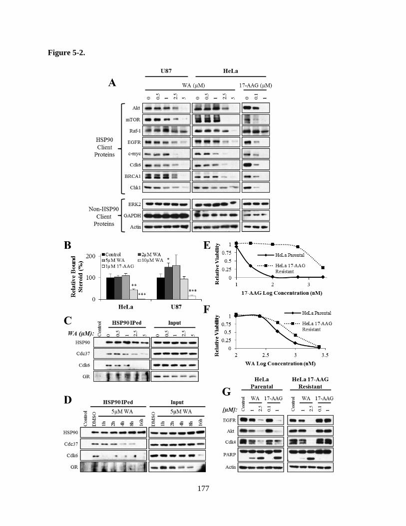
177
Figure 5-2.

178
Figure 5-2. A) WA treatment for 24 h in U87 and HeLa caused protein depletion of known
HSP90 clients Akt, mTOR, Raf-1, EGFR, c-myc, Cdk6, BRCA1, and Chk1 but failed to
decrease levels of non-HSP90 client proteins ERK2, GAPDH, and actin. HSP90-mediated
stability of each protein was confirmed by treating HeLa cells with known N-terminal HSP90
inhibitor 17-AAG. B) HSP90 function was measured through the ability of purified GR to bind
[3H]dexamethasone following re-exposure to cellular lysate containing HSP90 and other co-
chaperones treated with WA or 17-AAG for 24 h. WA failed to demonstrate inhibition of HSP90
activity at cytotoxically relevant concentrations 2-5 µM and rather displayed elevated promotion
of steroid binding with HeLa and U87 lysates. At 10 µM WA, steroid binding in U87 was seen
to return to baseline but reduced to 45% of baseline in HeLa. In contrast, 1 µM 17-AAG, a
positive control for HSP90 inhibition, significantly reduced HSP90-mediated GR steroid binding
with HeLa and U87 lysates. C and D) Co-IP experiments in HeLa cells using an HSP90 antibody
pull-down demonstrated that WA reduced the association between HSP90 and Cdc37 in a dose-
dependent manner after 4 h and in a time-dependent manner when exposed to 5 µM WA. Total
input HSP90 and Cdc37 was not decreased with treatment. Similar to Cdc37, the Cdc37-
dependent client protein Cdk6 was also shown to dissociate with WA treatment, but Cdc37-
independent protein GR did not. Total levels of both Cdk6 and GR were reduced over time. E) A
17-AAG resistant HeLa cell line was developed and shown to have a 72 h IC50 to 17-AAG of
1380 nM compared to 21 nM for the parental line, a 66-fold difference. F) In contrast to findings
with 17-AAG, WA IC50s in the parental and resistant lines were 503 nM and 841 nM,
respectively, a different of only 1.7-fold. G) Both WA and 17-AAG decreased total levels of
HSP90 client proteins EGFR, Akt, and Cdk4 and increased the apoptotic cleavage of PARP in

179
parental HeLa cells, but only WA demonstrated these findings in the resistant line, showing its
maintained utility. *p < 0.05, **p < 0.01, ***p < 0.001

180
of baseline in HeLa (p = 0.002). In contrast, 1 µM 17-AAG, a positive control for HSP90
inhibition, reduced HSP90-mediated GR steroid binding in HeLa and U87 by 99% and 83%,
respectively (p < 0.001). These findings suggest that WA does not directly inhibit HSP90
function.
The ability of WA to alter the association between HSP90 and Cdc37 was confirmed in
HeLa cells by co-immunoprecipitation for HSP90 (Figures 5-2C and 5-2D). After 4 h WA
treatment, co-IPed Cdc37 was reduced 36.9% with 1 µM up to 67.0% with 5 µM relative to IPed
HSP90, demonstrating a dose-dependent effect. Similarly, a time-dependent effect was observed
by maintaining WA at 5 µM with diminished association from 1-16 h post-treatment. IPed
Cdc37 was reduced 23.8%, 72.7%, 77.2%, and 99.4% after 2 h, 4 h, 8 h, and 16 h relative to IPed
HSP90, respectively. Total input HSP90 and Cdc37 remained consistent with WA treatment.
To further define the importance of this co-chaperone disruption, IPed HSP90 was also
evaluated for a Cdc37-dependent protein, Cdk6, and a Cdc37-independent protein, GR (Figures
5-2C and 5-2D) (Lamphere et al., 1997; Rao et al., 2001). Treatment with WA in both dose- and
time-dependent manners demonstrated a progressive failure of HSP90 to associate with client
protein Cdk6. This association was completely eliminated after 4 h with 2.5 µM WA. Similarly,
association was nearly completely eliminated with 5µM WA after 1 h and remained consistently
reduced at latter timepoints. Total input levels of Cdk6 were constant among samples except for
5 µM WA at 16 h which demonstrated near-complete elimination of the protein. In contrast, co-
IPed GR in both the dose- and time-dependence studies was maintained under all conditions.
Interestingly, total input GR was decreased with time and concentration in both experiments with
near-complete depletion with 5 µM at 16 h, but this did not result in a corresponding decrease in
HSP90-GR association, suggesting the retention of HSP90 function independent of Cdc37.

181
In contrast to 17-AAG, WA has been shown to associate with the C-terminus of HSP90
and did not affect molecular ATPase function (Yu et al., 2010). To demonstrate if WA’s unique
disruption of the HSP90 axis could be utilized in overcoming inhibition to 17-AAG while still
generating a similar effect, a HeLa cell line resistant to 17-AAG was developed through serial
escalations in drug exposure. Evaluation of 17-AAG cytotoxicity revealed 72 h IC50 values of the
parental and resistant lines to be 21 nM and 1380 nM, respectively, for a 66-fold change in
response (Figure 5-2E). WA sensitivity, however, was only elevated 1.7-fold from 503 nM in the
parental line to 841 nM in the 17-AAG resistant derivative (Figure 5-2F).
Maintained cytotoxicity of WA in the 17-AAG resistant line was confirmed molecularly
through evaluation of HSP90 client proteins EGFR, Akt, and Cdk4 as well as cleavage of the
apoptotic biomarker PARP (Figure 5-2G). Depletion of the client proteins and increased PARP
cleavage observed with 1 µM 17-AAG after 24 h in the parental HeLa line was completely
absent in the resistant line, but WA-mediated client degradation remained consistent with near-
complete elimination by 2.5 µM. WA also maintained the ability to induce PARP cleavage in the
resistant sub-line.
5.4.3 Cytotoxicity of withaferin A is driven by thiol-reactivity and cellular oxidation
Previous studies have identified an oxidative component resulting from treatment with
WA (Malik et al., 2007; Widodo et al., 2010; Hahm et al., 2011; Mayola et al., 2011; Santagata
et al., 2012; Grogan et al., 2013; Grogan et al., 2014). Given that certain proteins are susceptible
to oxidative stress and may become misfolded, the nature of this oxidative component was
explored. Treatment of U87 cells with WA resulted in a dose-dependent increase in oxidation as
182
measured by CM-H2DCFDA conversion to a fluorescent product by 4 h (Figure 5-3A). 5 µM
WA increased fluorescence 1.9-fold (p = 0.038). This enhanced oxidative signal was reduced by
pretreatment with 100 µM non-thiol antioxidant AA by 17.2% (p = 0.233) and completely
returned to baseline with 5 mM thiol antioxidant NAC (p = 0.037), suggesting a greater role of
thiol reactivity in the manifestation of WA’s effects.
Further evaluation of thiol reactivity and changes in the redox states following WA
exposure was accomplished through measurements of GSH levels with monochlorobimane
which fluoresces when conjugated to GSH (Figure 5-3B). GSH is a protective component of
cells that sequesters thiol reactive agents to prevent cellular damage. After 6 h, 2.5 µM WA
reduced total cellular glutathione in both HeLa and U87 cells by 41.1% (p < 0.001) and 31.9% (p
= 0.012), respectively, further indicating WA-mediated thiol reactivity and disruption of the
redox balance.
Evaluation of anti-oxidant pretreatment was completed with the CellTier-Glo assay to
define the functional cytotoxic effects of WA treatment (Figure 5-3C). In the assay, cellular
viability was assessed by total ATP levels 72 h after treatment. WA alone exhibited significant (p
< 0.001) reduction in the viability of HeLa and U87 cells at the concentrations tested (2.5-5 µM).
While 100 µM AA previously reduced WA-mediated oxidation, it was unable to limit
cytotoxicity, mirroring the cell viability reduction of WA alone. However, 5 mM NAC
completely abrogated WA cytotoxicity, returning cell viability to baseline and demonstrating the
predominant thiol reactive mechanism of WA.
Triton X-100 fractionation under non-reducing conditions was completed to define WA-
mediated redox alterations in HSP90 localization. While triton soluble proteins are largely
regarded as functional, stable molecules, the insoluble fraction is thought to contain labile and

183
Figure 5-3.

184
Figure 5-3. A) WA generated peroxide-type radicals like H2O2 as quantified by CM-H2DCFDA
fluorescent conversion with increasing concentrations of WA in U87 cells 4 h post-treatment.
The thiol antioxidant NAC (5 mM) completely abrogated the elevation in ROS, while 100 µM of
the non-thiol antioxidant AA produced a small reduction in overall oxidation. B) GSH levels
were measured by monochlorobimane conversion to a fluorescent product in the presence of a
thiol. WA treatment was shown to significantly reduce GSH levels in both HeLa and U87 after
treatment for 6 h. HeLa (C) and U87 (D) cell viability was assessed by total ATP levels with the
CellTiter Glo assay. WA-induced reduction of viability was completely eliminated with NAC
but not AA pretreatment. E) Triton X-100 fractionation under non-reducing conditions was
completed to define WA-mediated redox alterations in HSP90 localization. Slower migrating
proteins aggregates of heterogeneic molecular weight positive for HSP90 were dose-dependently
increased by WA in the triton insoluble fraction and was reduced with NAC pretreatment. F) In
contrast, triton insoluble HSP90 aggregation was enhanced by reduction in GSH through
inhibition of γ-glutamylcysteine synthetase by BSO. *p < 0.05, **p < 0.01, ***p < 0.001

185
aggregated proteins. Additionally, previous reports have demonstrated increased disulfide-
bonded HSP90 in the insoluble fraction in response to oxidative stress (Cumming et al., 2004;
Chen et al., 2008). Enhanced exposure of Western blots for HSP90 in HeLa and U87 revealed
minimal changes to total levels of HSP90 in both the soluble and insoluble fractions 24 h after
2.5-5 µM WA. Slower migrating disulfide-bonded HSP90 was not elevated following WA
exposure. However, slower migrating proteins of heterogeneic molecular weight positive for
HSP90 were dose-dependently increased by WA in the triton insoluble fraction indicating
damaged, aggregated, and delocalized HSP90 protein complexes. Pretreatment with NAC
completely eliminated this protein aggregation (Figure 5-3D). In contrast, reduction in GSH
through inhibition of γ-glutamylcysteine synthetase by BSO potentiated the total level of WA-
induced HSP90 aggregate in the insoluble fraction, demonstrating the thiol-dependent nature of
HSP90 disruption (Figure 5-3E). While BSO enhanced WA-induced HSP70 upregulation and
NAC prevented HSP70 induction, changes in disulfide-bonded or otherwise aggregated HSP70
was not detected (data not shown).
5.4.4 Withaferin A-mediated protein degradation requires the proteasome
Cellular protein elimination is largely driven through two primary pathways: proteasome-
and lysosome-mediated degradation. Given the observation that WA drives upregulation of
HSP70 and ubiquitination of proteins globally and that the latter targets proteins to the
proteasome, the mechanism of WA-driven protein depletion was evaluated.
Triton X-100 fractionation under reducing conditions was utilized to evaluate WA-altered
protein localization and depletion in the presence of proteasome inhibitor MG132 and lysosome

186
inhibitor chloroquine (Figure 5-4A). As previously described, 20 µM MG132 alone, mimicking
cellular stress and altering normal protein turnover, reduced soluble levels of the proteins
evaluated (Cdk6, Cdk4, mTOR, c-myc, Akt, and EGFR) and promoted elevated levels in the
insoluble fraction (Kawahara et al., 2008). Also, consistent with previous reports (Carter and
Sorkin, 1998), 50 µM chloroquine, an inhibitor of lysosomal protein degradation, blocked
endogenous EGFR turnover driven in part through endocytosis and lysosomal degradation and
enhanced the triton insoluble fraction. WA treatment promoted depletion of soluble proteins with
variable shift to the insoluble fraction. Complete elimination after 24 h WA exposure was
favored in the more sensitive HeLa cells compared to U87. However, extended treatment
duration in U87 cells demonstrated diminished levels of insoluble proteins (data not shown).
Compared to WA alone, the combination of WA and MG132 demonstrated increased retention
of all evaluated proteins in the insoluble fraction, suggesting that WA-mediate protein depletion
occurred at least in part through proteasomal degradation. Protein retention with chloroquine
pretreatment was variable and demonstrated larger effect in the HeLa cells, suggesting a greater
cellular role of WA-induced lysosomal degradation in addition to proteasomal degradation.
Combination of MG132 and chloroquine to rescue WA-induced protein depletion was not shown
to be greater than either compound alone.
HSP90 client protein c-myc has previously been shown to require amino acid sites T58
and S62 for phosphorylation-directed ubiquitination and subsequent degradation via the
proteasome (Gregory and Hann, 2000; Sears et al., 2000; Kamemura et al., 2002). 293T cells
were stably transfected with T58A/S62A FLAG-tagged c-myc (c-mycT58A/S62A
-FLAG), a variant
with mutations to disrupt the normal c-myc proteasomal degradation, and treated with WA. Both
vector control and mutant transfected cells demonstrated dose-dependent depletion of

187
Figure 5-4.

188
Figure 5-4. A) HeLa and U87 cells were pretreated with the proteasome inhibitor MG132 or the
lysosome inhibitor chloroquine for 1 h followed by WA for 24 h, and cellular proteins were
fractionated by triton X-100 solubility. Treatment with WA induced a loss of HSP90 client
proteins (Cdk6, Cdk4, mTOR, c-myc, Akt, EGFR) in the soluble fraction with a shift to either
the insoluble fraction or a depletion in both fractions. MG132 also acted to shift proteins to the
insoluble fraction. Combination of WA and MG132 demonstrated increased retention of all
evaluated proteins in the insoluble fraction. Protein retention with chloroquine pretreatment
followed by WA was variable and demonstrated greater effect in the HeLa cells. Combination of
MG132 and chloroquine to rescue WA-induced protein depletion was not shown to be greater
than either compound alone. B) 293T cells were transfected with c-mycT58A/S62A
-FLAG, a variant
with mutations to disrupt the normal c-myc proteasomal degradation, and treated with WA.
Endogenous wild-type c-myc was depleted by WA in a dose-dependent manner, but the mutant
failed to decrease after 24 h.
189
endogenous wild-type c-myc by 2.5 µM WA. The c-mycT58A/S62A
mutant, however, showed no
decrease in response to WA treatment, demonstrating the functional requirement for
proteasomal-targeting in WA-mediated protein degradation (Figure 5-4B).
5.4.5 Proteasomal inhibition potentiates the cytotoxicity of withaferin A
Given the apparent importance of the HSP70 chaperone axis and the proteasome in
regulating protein degradation following WA treatment, simultaneous inhibition of the
proteasome with MG132 to prevent degradation of aberrant protein accumulation was evaluated
to determine the utility of combination therapy. Cell viability was assessed by MTS assay after
72 h (Figure 5-5A). HeLa and U87 cells displayed no significant decrease in viability with 25-75
nM and 75-125 nM MG132 alone, respectively. However, 1h sub-cytotoxic MG132 pretreatment
potentiated the anti-proliferative response to treatment with escalating concentrations of WA.
62.5 nM WA reduced HeLa cell viability by 15.5% after 72 h but was enhanced to 38.2% with
25 nM MG132 (p = 0.010) and 42.2% with 75 nM MG132 (p = 0.043). Comparable
enhancement was observed with 125-250 nM WA. In U87, 750 nM WA decreased cell viability
by 25.0% but was enhanced to 56.3% with 75 nM MG132 (p = 0.072) and 83.1% with 125 nM
MG132 (p = 0.016).
Similarly, evaluation of drug-induced cell death by flow cytometry revealed that
pretreatment with MG132 potentiated the cytotoxicity of WA (Figures 5-5B and 5-5C). In HeLa,
combination therapy induced 45.8% cell death (25.3% early apoptosis, 7.7% necrosis, 12.9% late
apoptosis/necrosis) compared to only 2.4% in control (1.2% early apoptosis, 0.5% necrosis, 0.7%
late apoptosis/necrosis; p = 0.015), 2.6% with 125 nM MG132 (0.6% early apoptosis, 1.1%

190
Figure 5-5.

191
Figure 5-5. A) HeLa and U87 cell viability following combination therapy with proteasome
inhibitor MG132 and WA was evaluated by MTS assay after 72 h. HeLa and U87 cells displayed
no significant decrease in viability with 25-75 nM and 75-125 nM MG132 alone, respectively.
However, MG132 pretreatment at these concentrations potentiated the anti-proliferative response
to treatment with escalating concentrations of WA. B) Efficacy of combination MG132 and WA
therapy on HeLa and U87 cells after 24 h was further evaluated by flow cytometry with
propidium iodide and annexin V-FITC doe cell death. Staining revealed that pretreatment with
125 nM MG132 potentiated the cytotoxicity of 1 µM WA in both lines. MG132 alone had no
statistically significant effect compared to control, and only WA demonstrated elevated death in
U87 cells, but combined, the cytotoxic effects were significantly elevated. C) A representative
example of the flow cytometry results in HeLa cells. D) Flow cytometry data was supported by
molecular evaluation of apoptotic markers procaspase 3 and PARP cleavage in both HeLa and
U87 cells. After 24 h, treatment with 125-250 nM MG132 in both lines as well as 0.5-1 µM WA
in HeLa and 1-2 µM WA in U87 failed to induce significant cleavage of either protein but
yielded large increases in the cleaved product when combined. E) Combined treatment with
MG132 and WA for 24 h resulted in the accumulation of ubiquitinated proteins at all
concentrations evaluated in both HeLa and U87 cells, demonstrating retention of proteins
targeted for degradation. *p < 0.05, **p < 0.01, ***p < 0.001

192
necrosis, 0.9% late apoptosis/necrosis; p = 0.016), and 3.2% with 1 µM WA (1.2% early
apoptosis, 1.3% necrosis, 0.7% late apoptosis/necrosis; p = 0.016). WA and WA+RT
demonstrated no statistically significant difference over control. Similarly, in U87, combination
therapy induced 50.2% cell death (37.3% early apoptosis, 2.2% necrosis, 10.6% late
apoptosis/necrosis) compared to only 4.7% in control (2.8% early apoptosis, 0.8% necrosis, 1.1%
late apoptosis/necrosis; p = 0.013), 8.3% with 125nM MG132 (5.4% early apoptosis, 1.2%
necrosis, 1.8% late apoptosis/necrosis; p = 0.024), and 20.9% with 1 µM WA (14.6% early
apoptosis, 3.0% necrosis, 3.3% late apoptosis/necrosis; p = 0.015). WA alone also demonstrated
a statistically significant cytotoxic advantage over control (p = 0.011). Together, these data
demonstrate that sub-cytotoxic pre-exposure to MG132 potentiates minimally or non-lethal WA
concentrations.
Flow cytometry data was supported by molecular evaluation of apoptotic markers
procaspase 3 and PARP cleavage in both HeLa and U87 cells (Figure 5-5D). After 24 h,
treatment with 125-250 nM MG132 in both lines failed to induce significant cleavage of either
protein. Similar findings were observed with 0.5-1 µM WA in HeLa and 1-2 µM WA in U87.
However, combination of these agents at the noted concentrations revealed large increases in the
cleaved product of both procaspase 3 and PARP, demonstrating the advantage of combination
therapy.
Consistent with the expectation that combination therapy would result in enhanced
cytotoxicity through an accumulation of aberrant proteins generated by WA treatment and unable
to be properly degraded via the proteasome due to MG132, dual treatment over 24 h resulted in
the accumulation of ubiquitinated proteins at all concentrations evaluated (Figure 5-5E).
Potentiation of the effect was particularly notable at modest concentration of each compound that

193
alone produced minimal yet early indications of ubiquitination. By denisometry, 125 nM MG132
increased ubiquitination 2.2-fold in HeLa and 1.8-fold in U87, while 1 µM and 2 µM WA
resulted in 3.0- and 3.3-fold elevations in HeLa and U87, respectively. Combination of MG132
and WA showed further enhanced levels of global protein ubiquination by 11.4-fold in HeLa and
10.7-fold in U87, demonstrating the value of this approach.
5.5 Discussion
WA is a promising naturally-derived anti-cancer compound that has demonstrated
efficacy across a spectrum of cancer types both in vitro and in vivo. Underlying WA’s
antiproliferative, pro-apoptotic, and anti-angiogenic effects is disruption of various signaling
pathways including those utilizing Akt/mTOR, MAPK, Notch, NFκB, JAK/STAT, and more
(Vanden Berghe et al., 2012; Vyas and Singh, 2014). However, details of the mechanism driving
these diverse effects have remained poorly defined to-date. Previous studies have identified a
pro-oxidant effect (Malik et al., 2007; Widodo et al., 2010; Hahm et al., 2011; Mayola et al.,
2011; Santagata et al., 2012; Grogan et al., 2013) and suggested that alterations in HSP90
function occurs with WA treatment (Yu et al., 2010; Gu et al., 2014). Here, we further
characterized these findings and propose each as the two of the dominant mechanisms by which
WA mediates in anti-cancer effects: Induction of cellular oxidation and disruption of the
interaction between HSP90 and its co-chaperone Cdc37 independent of direct HSP90 inhibition.
These proposed mechanisms explain the numerous descriptions of alterations in oncogenic
proteins and signaling pathways that have come to define the cellular response to WA.

194
HSP90 functions as a chaperone to promote the folding, refolding, and function of a
specific set of client proteins (Pearl et al., 2008). This ability is dependent on the association of
HSP90 with several co-chaperone proteins that promote this function (Taipale et al., 2010).
Although not yet fully understood mechanistically, Cdc37 is thought to act largely as a kinase-
specific co-chaperone to promote the folding and maturation of these proteins through
localization with HSP90 with enhanced client loading though modulation of HSP90 ATPase
activity (Siligardi et al., 2002; Roe et al., 2004). Cdc37 has also demonstrated chaperoning
functions independent of HSP90 (MacLean and Picard, 2003). Here, we demonstrate that
pharmacological disruption of this interaction between HSP90 and Cdc37 with WA is associated
with a failure of HSP90 to associate with Cdc37-dependent protein Cdk6 but not Cdc37-
independent protein GR as well as degradation of multiple HSP90 client proteins. Additionally,
depletion of HSP90 client proteins known to interact with Cdc37 (Akt, Raf-1, EGFR, Cdk4,
Cdk6, and Chk1) was observed. Intrinsic HSP90 chaperone function, assessed by supporting GR
steroid binding, was not impaired at relevant concentrations of WA, although several HSP90
client proteins not previously described as Cdc37-dependent (BRCA1, c-myc, GR) were also
depleted suggesting an alternate mechanism of protein modulation and/or a previously
undescribed requirement for Cdc37. These findings, if not mediated through an alternative
mechanism, suggest that WA may still maintain a direct role in the inhibition of HSP90’s folding
mechanism independent of its GR support. Cdc37 has been primarily reported to facilitate
folding of newly translated proteins (MacLean and Picard, 2003; Karnitz and Felts, 2007), but its
importance has been demonstrated in the stability of existing proteins (Jinwal et al., 2011).
Further studies are necessary to determine if either situation represents the predominant
inhibitory effect of WA.

195
Interestingly, a recent study demonstrated that mutant Cdc37 lacking the C-
terminal/middle HSP90-association domain maintained cellular levels of client proteins and
augmented the interaction between HSP90 and client Cdk4 despite diminished client protein
interaction itself (Smith et al., 2013). However, this directly contrasts previous reports about the
importance of the C-terminal/middle domain for association with HSP90 in maintaining the
stability of client proteins like Raf-1 (Grammatikakis et al., 1999; Rao et al., 2001). It is possible
that the client proteins evaluated have different co-chaperone requirements for interacting with
HSP90 or can be maintained through Cdc37’s inherent chaperone activity given that only
approximately half of mammalian Cdc37/Cdk4 complexes contain HSP90 (Dai et al., 1996).
However, the latter is likely not the case here given the reduced client affinity of the mutant
Cdc37 despite maintained client levels (Smith et al., 2013). Cdc37 has also been shown to
interact with other co-chaperone proteins, suggesting another way by which it can still join the
HSP90 chaperone complex, although its ability to alter HSP90 ATPase activity and otherwise
function properly through this indirect association is unknown (Abbas-Terki et al., 2002).
Clearly, further study of this system is required to better understand the variations observed.
Regardless of the nature of the HSP90/Cdc37 interaction, its disruption and the resulting
cytotoxicity by exposure to WA and celastrol, as previously studied, demonstrate
pharmacological proof-of-concept of this target (Zhang et al., 2008; Zhang et al., 2009; He et al.,
2013).
While computational analysis revealed a potential binding site for WA on Cdc37 to
disrupt the HSP90/Cdc37 interaction (Grover et al., 2011), IP studies with biotinylated WA
showed that it actually bound to the C-terminus of HSP90 (Yu et al., 2010). The C-terminus of
HSP90 is responsible for the association with various co-chaperones (Young et al., 1998; Sidera

196
and Patsavoudi, 2014), however, WA does not decrease HSP90 association with co-chaperones
Hop or p23 but rather Cdc37 which interacts primarily on the N-terminus of HSP90 (Yu et al.,
2010; Eckl et al., 2013). This unexpected finding requires further study to explain but may be a
result of WA-mediated direct steric hindrance, conformational alterations in HSP90 structure that
prevent effective binding of Cdc37 but maintain HSP90 function, or changes in the ability of
Cdc37 to associate with C-terminal co-chaperones. Interestingly, celastrol also binds to the C-
terminus of HSP90 to disrupt HSP90/Cdc37 interaction but fails to disrupt association with
HSP70, Hop, or p23 (Zhang et al., 2009). Novobiocin, also known to associate with the C-
terminus, demonstrates completely opposite effects, suggesting specific sites of co-chaperone
regulation on HSP90 (Yun et al., 2004).
HSP90 represents an intriguing anti-cancer target given that it is endogenously expressed
in all cells, typically accounting for 1-3% of the total cellular protein (Welch and Feramisco,
1982; Workman et al., 2007; Moulick et al., 2011; Barrott and Haystead, 2013). However, most
cancer cells demonstrate significantly greater sensitivity to HSP90 inhibitors than non-cancerous
cells with sub-toxic doses resulting in anti-tumor efficacy. This is thought to likely be the result
of several factors that frequently differ in cancer cells including upregulation of HSP90 protein
and mRNA, enhanced basal activation of HSP90 through post-translational modifications and/or
enhanced association with various co-chaperones and client proteins, and altered localization to
the ectopic portion of the cell (Barrott and Haystead, 2013). These considerations for enhancing
inhibitory selectivity of HSP90 alone would appear to translate well to the disruption of co-
chaperones such as Cdc37, particularly HSP90 dependency and localization. Further studies are
necessary to identify whether WA affinity favors disruption of HSP90 and Cdc37 that are
already associated or prevention of the initiation of the association. Treatment with WA in

197
several murine models demonstrates an appropriate therapeutic window against several cancer
types with minimal off-target toxicity and shows the translatable promise of this approach (Vyas
and Singh, 2014).
WA also demonstrated the ability to induce cellular oxidation. AA was used as a non-
thiol antioxidant to evaluate the oxidative effects of WA independent of concerns that NAC may
simply sequester WA, preventing the availability of free drug rather than actually directly
reducing generated oxidation (Santagata et al., 2012; Vyas and Singh, 2014). AA at a previously-
identified effective concentration (100 µM) was capable of reducing total oxidative potential
within U87 GBM cells but was unable to completely eliminate the effect like NAC. Similarly,
the cytotoxicity of WA was completely blocked by NAC but not AA. These findings suggest that
while oxidation does indeed play a part in the mechanism of WA, the generation of free reactive
oxygen species likely does not have nearly as significant of an impact in the manifestations of
WA as its thiol reactivity and thiol-mediated redox alterations. The observation that WA depletes
GSH suggests that the oxidation may be a general effect of an altered redox state. Interestingly,
overexpression of Cu,Zn-superoxide dismutase demonstrated a partial protective effect against
WA treatment in breast cancer cells (Hahm et al., 2011). The same study suggested that WA-
mediated ROS generation and subsequent toxicity was a result of mitochondrial respiration
inhibition, although the impact of the mitochondrial alterations on normal intrinsic/mitochondrial
apoptotic pathway function, shown to be partially necessary for apoptosis following WA
exposure (Grogan et al., 2014), was not evaluated. Taken together, these findings support an
important role for oxidation following WA treatment but suggest it may be more critical for
cytotoxicity in certain cancer types.

198
We have also previously demonstrated WA-mediated depletion of MGMT, a non-HSP90
client protein with known susceptibility to oxidation (Fu et al., 2010; Ryu et al., 2012; Grogan et
al., 2014). Similarly, GR has been shown to be modulated by redox alteration, especially with
thiol-modifying agents (Carter and Ragsdale, 2014). Despite not requiring Cdc37 for its function
and stability and maintaining association with HSP90, we demonstrated in the present study that
GR is eventually depleted with WA treatment, suggesting either an extended role of WA as an
HSP90 inhibitor beyond Cdc37 effects or oxidation-meditated proteotoxicity. Utilization of
oxidative therapies to induce proteotoxicity has gained interest as an anti-cancer approach given
the frequently altered redox states and exposure to increased cellular stress which can be
exploited in cancer cells (Laurent et al., 2005; Cabello et al., 2007).
Treatment with WA resulted in the accumulation of aggregated HSP90 of heterogeneic
molecular weight in the triton insoluble fraction of the cell, often regarded as the biologically
inactive compartment. Previous studies have suggested that chaperone proteins form disulfide
bonds in response to oxidative insults (Cumming et al., 2004; Chen et al., 2008), and while the
described homodimeric complex of HSP90 was not readily observed in this study, significant
aggregation was noted. Further study will be necessary to determine whether this pattern of
aggregation was related to dissociation from Cdc37 (showing direct interplay between the two
proposed mechanisms of WA), a byproduct of oxidative stress, or misfolding and destabilization
guided by direct WA binding. While Cdc37 plays a role in the appropriate client targeting for
HSP90 and has been shown to localize in the cell for recruitment of HSP90-mediated refolding
(Jinwal et al., 2011), it has not been shown necessary to promote HSP90 complex stability or
intrinsic function. While pretreatment with NAC reduced or eliminated the general cytotoxicity
of WA, it also prevented this alteration in HSP90 localization. Conversely, depletion of cellular

199
GSH through inhibition of γ-glutamylcysteine synthetase by BSO greatly enhanced this HSP90
alteration. Together, these findings support an oxidative or thiol-reactive role of WA in
promoting insoluble complexation of HSP90. Of note, BSO also enhanced general WA
cytotoxicity in several additional cancer models (data not published). Combined with the
observation that WA depleted GSH, this suggests that GSH plays a protective role against WA.
Future studies are necessary to determine how basal cellular oxidation, GSH levels, and
susceptibility to oxidative stress enhance or resist the cytotoxic sequelae of WA exposure.
Several early studies attempting to identify the mechanism of WA suggested that it
functions through inhibition of the proteasome (Yang et al., 2007; Khan et al., 2012; Yang et al.,
2012). Our data suggests that the proteasome is still functionally intact and necessary for protein
degradation at cytotoxically-relevant concentrations of WA. While the findings in these reports
are likely accurate, they have consistently utilized concentrations of WA well-above those
considered therapeutically relevant – including in cell-free models – to achieve modest
proteasomal inhibition, suggesting that these findings may simply be off-target effects that
manifest at high concentrations. Interestingly, our studies demonstrate direct inhibition of the
HSP90 chaperone heterocomplex independent of Cdc37 in a cell-free system with an IC50 of
<10µM (data not shown), well above physiologically relevant dosing but still lower than
observed in the GR-reactivation cell lysate system. Combined, these findings indicate non-
specific targeting by WA at high concentrations and suggest caution is necessary during the
interpretation of data at these levels.
While the suggestion that HSP90 promotes protein refolding while HSP70 favors
degradation oversimplifies the complex mechanism of the protein chaperone machinery, these
generalizations accurately describe the accepted primary roles that these chaperones play in

200
protein homeostasis (Pratt et al., 2010). In the presence of oxidative stress, proteins are prone to
misfold with greater frequency. Simultaneous inhibition of the HSP90 axis, as observed with
WA, would suggest the accumulation of many aberrant proteins requiring proper degradation to
limit cellular cytotoxicity. Previous studies have identified upregulation of HSP70, as observed
in the present study, to be a biomarker of HSP90 inhibition and/or oxidative stress (Zhang et al.,
2006; Workman et al., 2007; Ansari et al., 2011; Qiao et al., 2012), acting as a compensatory
mechanism for protein dysregulation and promoting this necessary degradation. Indeed,
inhibition of HSP70 or the proteasome in the presence of traditional HSP90 inhibitors promotes a
synergistic effect (Minnnaugh et al., 2004; Ma et al., 2014). Given the promotion of proteasome-
mediated protein degradation induced by WA, the addition of a proteasome inhibitor was a
logical progression in the development of a clinical utility for WA. Here, we further demonstrate
the efficacy of this approach with WA, an oxidizing inhibitor of the HSP90/Cdc37 association.
This suggests a potential clinical utility for combining a co-chaperone disruptor with already
established chemotherapeutic agents like the proteasome inhibitor bortezomib. While the value
of this combination therapy was demonstrated in highly aggressive solid tumor cervical and
brain cancer cell lines, perhaps the greatest early clinically applicability lies in the treatment of
multiple myeloma (MM). Proteasome inhibitors have been approved for use as standard agents
in the treatment of MM (Chauhan et al., 2005), and an early human trial with combination
bortezomib and the HSP90 inhibitor tanespimycin, a promising formulation of 17-AAG with a
Cremophor-containing vehicle (Modi et al., 2007; Modi et al., 2011), has shown promise
(Richardson et al., 2011). Given the enhanced efficacy of many agents in combination with
traditional HSP90 inhibitors, further exploration of combination therapy with WA in alternative
models is also warranted (Neckers, 2002).

201
The studies here provide an important framework for further defining the mechanisms by
which WA induces significant protein depletion. We demonstrate that WA acts to produce thiol-
mediated oxidation and disrupt the interaction between HSP90 and Cdc37 but fails to inhibit
intrinsic HSP90 function assessed by HSP90-mediated GR steroid binding. Ultimately, this shifts
the HSP90/HSP70 balance toward HSP70-domination with associated protein degradation that
can be cytotoxically potentiated therapeutically with simultaneous inhibition of the proteasome.
While further pre-clinical studies will be necessary to fully evaluate the therapeutic potential of
WA alone and in combination with proteasome inhibitors, current findings support the
translational utility of this approach.

202
Chapter 6
Withaferin A disrupts the cellular DNA-damage response to
promote radiosensitization
203
6.1 Abstract
Radiation therapy (RT) plays in important role in the treatment of cancer for an estimated
50% of patients, including as a standard therapy for the brain tumor glioblastoma multiforme
(GBM). Withaferin A (WA) is a naturally-derived steroidal lactone that has demonstrated
significant intrinsic anti-cancer activity. Several preliminary reports have identified WA as a
potential radiosensitizer, but the underlying mechanism has remained largely unexplored. In this
study, WA potentiated or otherwise enhanced the anti-proliferative activity of RT in both U87
GBM and HeLa cervical cancer cells. These findings were associated with reduction of cells in
the DNA-synthesis phase of the cell cycle and increased levels of cell cycle checkpoint inhibitors
p21 and p27 with combination therapy through 72 h. Combination therapy failed to enhance
PARP cleavage and LC3B conversion, demonstrating that the effect was not mediated through
apoptosis or autophagy. Treatment with WA reduced total levels of DNA-damage response
(DDR) proteins involved in damage recognition (Mre11, p95/NBS1, Rad50), signaling (ATM,
Chk1), and repair through homologous recombination (BRCA1, BRCA2, Rad51), non-
homologous end-joining (DNA-PKcs, XLF, Ku70, and Ku80), and mismatch repair (MLH1,
MSH2, MSH6) in both U87 and HeLa. This protein depletion was an expansion of that observed
with traditional HSP90 inhibitor 17-AAG. WA differentially modulated normal the normal
recognition response to RT-mediated DNA damage with U87 and HeLa cells showing a
reduction and an increase of p-H2A.X compared to RT alone, respectively, with WA
pretreatment. Both lines showed alterations of normal signal transduction through ATM and
Chk1. WA pretreatment also enhanced the level of RT-induced double strand DNA damage after
4 h measured by a neutral comet assay and was associated with functional inhibition of

204
homologous recombination through a DR-GFP reporter. These findings demonstrate that WA-
driven radiosensitization is a direct result of an impaired DDR. Given the widespread success of
WA as a monotherapy in numerous cancer models and the large therapeutic spectrum for RT,
further translational exploration of WA as an adjuvant agent is warranted.
6.2 Introduction
In recent years, significant effort has been invested in the development of more effective
therapeutic and diagnostic techniques to better the outcomes of cancer patients. While numerous
advances have adjusted the treatment paradigms of various malignancies, the management and
treatment of many cancers depends on the use of radiation therapy (RT), including an indication
in an estimated 50% of patients (Delaney et al., 2005). Given that survival rates following RT,
which in many cases represents a component of the base available approved therapeutic regimen,
remain low for certain cancer types like glioblasoma multiforme (GBM), any means by which to
elevate RT efficacy would be critical in helping a wide population of patients (Begg et al., 2011).
With the widespread importance and critical role of RT, efforts have been made to
maximize its therapeutic potential through adjustments of dose intensity and fractionation as well
as addition of chemotherapeutics. Such therapies focus on modulating the DNA damage
response (DDR), signal transduction pathways critical for radioresistance, and oxygen levels
necessary for generating an effect (Begg et al., 2011). Additionally, several commonly utilized
DNA-modifying anticancer agents, including 5-fluorouracil (5-FU) and temozolomide (TMZ),
have shown radiosensitization effects with unclear mechanisms. Similar agents like gemcitabine
demonstrate off-target inhibition of HR as a mechanism of radiosensitization (Byfield, 1989;

205
Wachters et al., 2003; Chakravarti et al., 2006; Bobola et al., 2010). Other agents that directly
target elements of the cellular DDR, including the recognition and repair of the damage, have
generated significant interest as well (Begg et al., 2011).
RT utilizes the energy of ionizing radiation (IR) to induce DNA lesions in targeted cells.
Genomic instability through double-strand DNA (dsDNA) breaks (DSBs) is largely recognized
as the primary cytotoxic event of IR, but other lesions such as single-strand DNA (ssDNA)
breaks (SSBs) and damage to individual bases has been reported. DSBs are repaired by two
primary pathways: homologous recombination (HR) and non-homologous end joining (NHEJ).
While HR maintains the fidelity of the DNA sequence at the break site, NHEJ simply anneals
two ends of DNA without respect to sequence homology and is therefore intrinsically mutagenic.
The cellular response to DNA damage, including that induced by IR, involves an
immensely complex network of signaling pathways that recognize and repair the damage with
associated arrest of the cell cycle or induction of death (Lamarche et al., 2010; Baskar et al.,
2012). Very generally, following DSBs, damage is recognized by the MRN complex (Mre11,
Rad50, p95/NBS1) which functions to coordinate signal transduction through ATM to arrest cell
growth at specific cell cycle checkpoints. This complex also functions directly and indirectly to
mediate dsDNA damage repair through HR and NHEJ (Weterings and Chen, 2008; Krejci et al.,
2012). Appropriate coordination of these efforts in normal tissue usually results in either
effective DNA damage repair and cell survival or failure to appropriately correct the insult
followed by the controlled induction of cellular senescence or death.
Several studies have identified the potential utility of withaferin A (WA), a 28-carbon
steroidal lactone identified from genera of the Solenceae plant family with intrinsic cytotoxicty,
as a potential radiosensitizer (Vanden Berghe et al., 2012; Vyas and Singh, 2014). One study that

206
utilized a knockout model suggested WA may alter NHEJ (Devi et al., 2008), while another
group identified enhanced oxidation and modulation of the mitogen-activiated protein kinase
(MAPK) and Akt pathways as markers of combination therapy efficacy (Yang et al., 2011b;
Yang et al., 2011a). However, to date, the underlying mechanism behind this effect has not been
explored. Studies from our group and others have identified two key components of WA
monotherapy that are hypothesized to lead to the inhibition and modulation of many oncogenic
proteins that have been observed: oxidation and inhibition of the HSP90 axis through disruption
of the HSP90/Cdc37 association (Malik et al., 2007; Widodo et al., 2010; Yu et al., 2010; Hahm
et al., 2011; Mayola et al., 2011; Santagata et al., 2012; Grogan et al., 2013). Indeed, direct
inhibition of HSP90 has been previously noted to function in a radiosensitizing role (Dote et al.,
2006; Kabakov et al., 2010; Ko et al., 2012; Stecklein et al., 2012), suggesting a potential
mechanism by which WA may function and a novel role for disruption of Cdc37.
In this study, we identify WA as a potent radiosensitizer of two highly aggressive cancer
cell lines, the GBM U87 and the cervical cancer HeLa with the latter being utilized as a known
model of highly competent HR, through depletion of key DDR proteins that prevent the proper
recognition and repair of DSBs.
6.3 Materials and methods
6.3.1 Cell culture and reagents
The human GBM cell line U87 was generously provided by Dr. Jann N. Sarkaria (Mayo
Clinic, Rochester, MN), and the human cervical cancer line HeLa was obtained through the

207
American Type Culture Collection (ATCC; Manassas, VA). These cell lines were chosen given
their DNA damage repair preferences: HeLa is more competent at HR than GBM cells while the
latter favor NHEJ after RT (Kachhap et al., 2001; Golding et al., 2004; Ransburgh et al., 2010;
Quiros et al., 2011; Lim et al., 2012; Stecklein et al., 2012). Both cell lines were grown in
Dulbecco’s modified Eagle’s medium (DMEM #11995-065; Gibco, Grand Island, NY)
supplemented with 10% fetal bovine serum (FBS; Sigma-Aldrich, St. Louis, MO) and 1%
penicillin/streptomycin (Gibco), at a 37 °C humidified atmosphere of 5% CO2 in air. Medium for
HeLa cells was further supplemented with 2% L-glutamine (200 mM; Gibco), 1% MEM-vitamin
(100x; Hyclone, Logan, UT), and 1% MEM nonessential amino acids (Sigma Aldrich). Where
indicated, cells were irradiated with a Kimtron IC-320 orthovoltage x-ray device (Oxford, CT) at
a calibrated dose-rate of 452.23 cGy/minute. WA was isolated as previously described (Samadi
et al., 2010b). Propidium iodide (PI), RNase, and puromycin dihydrochloride were acquired from
Sigma-Aldrich, and 17-N-allylamino-17-demethoxygeldanamycin (17-AAG) was obtained from
LC Laboratories (Woburn, MA).
6.3.2 Clonogenic assay
HeLa and U87 cells in exponential growth phase were plated in 6cm dishes at densities
expected to yield approximately 20-50 colonies per plate following exposure to WA, RT, or
combination therapy based on preliminary studies. After 6 h, plates were changed to WA- or
DMSO-containing media and allowed to incubate for 2 h before exposure to 2-4 Gy of radiation.
24 h post-RT, media was changed on each plate to remove WA. Cells were incubated for 10-14 d
at which time they were gently washed with 1x phosphate-buffered saline (PBS), stained and
208
fixed with Coomassie brilliant blue, and quantified. Colonies were considered viable if they
consisted of approximately > 50 cells.
6.3.3 Cell cycle analysis
U87 and HeLa cells were plated to ~20-50% confluency depending on the collection
timepoint and treated with WA for 2 h followed by 2.5-5 Gy RT. After 24-72 h, cells were
collected, pelleted, fixed/permeablized in 70% ice-cold ethanol and stored at -20 °C. Cells were
subsequently stained with PI as previously described (Grogan et al., 2013) and analyzed by flow
cytometry on a Beckman Coulter CyAn ADP analyzer (Brea, CA). Only living cells without
DNA fragmentation were gated for analysis.
6.3.4 Immunoblotting
Cells were plated to appropriate confluencies as outlined for cell cycle analysis and
treated with various concentrations of WA or 17-AAG. Where indicated, cells were irradiated
with 5-10 Gy 2 h post-WA. After 1-72 h, cells were trypsinized and washed twice with 1x PBS.
Proteins were isolated in a HEPES-based lysis buffer, quantified, separated by sodium dodecyl
sulfate–polyacrylamide gel electrophoresis (SDS–PAGE), and transferred onto a Hybond
nitrocellulose membrane as previously described (Samadi et al., 2011; Grogan et al., 2014). 25-
50 µg of protein sample was loaded per lane with equal loading and transfer of sample confirmed
by total actin levels. Studies were repeated for accuracy.

209
Primary antibodies kits were acquired from Cell Signaling Technology (Danvers, MA)
and used as per the manufacturer’s recommendations: DNA damage (#9947), double stand
breaks (DSB) repair (#9653), MRN complex (#8344), mismatch repair (#9786), and Rb (#9969).
Additional primary antibodies obtained from Cell Signaling Technology included ATM (#2873;
1:1000), Chk1 (#2360; 1:1000), Chk2 (#2662; 1:1000), Ku70 (#4588; 1:1000), BRCA1 (#9010;
1:500), BRCA2 (#9012; 1:1000), p27 (#2552; 1:1000), cyclin B1 (#4138; 1:1000), cyclin D1
(#2926; 1:500), cyclin E1 (#4129; 1:1000), poly(ADP-ribose) polymerase (PARP; #9542;
1:1000), and LC3B (#3868; 1:1000). Rad51antibody (#ab213; 1:1000) was purchased from
Abcam (Cambridge, England). p-H2A.X antibody (Ser139; #613401; 1:1000) was acquired from
Biolegend (San Diego, CA). Total actin antibody (#MAB1501; 1:50000) was obtained from
EMD Millipore (Billerica, MA). The primary antibody for p21 (#sc-6246; 1:200) and the donkey
anti-rabbit IgG HRP (sc-2313; 1:2500-1:10000) and goat anti-mouse IgG HRP (sc-2005; 1:5000-
1:20000) secondary antibodies were purchased from Santa Cruz Biotechnology (Santa Cruz,
CA).
6.3.5 Immunocytochemistry (ICC)
U87 and HeLa cells were plated to ~50% confluency in 8-well chamber slides (Nalge
Nunc International, Penfield, NY) and treated with 2.5 µM WA for 2 h. Slides then received 10
Gy RT. After 1 h, cells were fixed in 4% PBS-buffered paraformaldehyde at room temperature
for 30 minutes and washed 3x with 1x PBS. Cell membranes were de-permeablized for 15
minutes in 1x PBS containing 0.3% triton X-100 and subsequently blocked in 5% goat serum
(Cell Signaling Technology) in 1x PBS containing 0.15% triton X-100 shaking for 1 h. Cells

210
were exposed to p-H2A.X (Ser139) primary antibody (1:500; Biolegend #613401) in blocking
solution overnight at 4 °C. After three washes with 1x PBS, secondary antibody (1:1000; anti-
mouse IgG Fab2 Alexa Fluor 488, Cell Signaling #4408) was added to each well for 1h at room
temperature followed by an additional 3 washes. Coverslips were mounted on each slide with
ProLong Gold Antifade with DAPI (Cell Signaling Technology), allowed to dry, and visualized
on a Leica DM IRB microscope (Wetzlar, Germany) with Metamorph Basic software (version
7.7.3.0.; Molecular Devices; Sunnyvale, CA)
6.3.6 Comet assay
U87 cells were assessed for dsDNA damage by a neutral comet assay. Cells were plated
to 50% confluency and treated with WA. After 12 h, cells were irradiated 5 Gy via the
orthovoltage device and allowed to incubate for 4 h. Evaluation of DNA damage through
migration was conducted according to the manufacturer’s instructions (Trevigen, Gaithersburg,
MD). Briefly, cells were collected, combined with agar, and plated on slides (~500 cells/slide).
Agar was solidified at 4 °C for 10 minutes, and slides were immersed in a lysis buffer solution
for 1 h at room temperature. Slides were then washed with neutral electrophoresis buffer and
underwent electrophoresis for 45 minutes at 4 °C with the recommended voltage. At room
temperature, slides were then immersed in DNA precipitation buffer for 30 minutes followed by
70% ethanol for 30 minutes. Samples were dried at 37 °C for 15 minutes, stained with SYBR
Gold (Molecular Probes, Eugene, OR) as outlined in the instructions, and visualized on the Leica
DM IRB microscope with Metamorph Basic software. Comets from eight independent visual
211
fields per sample were quantified with ImageJ software (version 1.46r; Bethesda, MD) using the
automated scoring of the OpenComet software macro (version 1.3) (Gyori et al., 2014).
6.3.7 DR-GFP reporter assay
The DR-GFP reporter construct, pCAGGS empty vector, and pCBASce I-SceI vector
were generously provided by Dr. Maria Jasin (Memorial Sloan-Kettering Cancer Center, New
York, NY). Adenovirus was kindly provided by Dr. Jeffrey Parvin (The Ohio State University,
Columbus, OH). The production and titering of HA-tagged I-SceI or empty vector adenovirus
was described previously (Stecklein et al., 2012). U87 and HeLa cells were transfected with the
DR-GFP and selected with 2-5 µg/mL puromycin. Surviving colonies were collected and
expanded under puromycin selection. The empty control vector or HA-I-SceI construct (~1
µg/1x105 cells) were introduced into the U87-DR-GFP stable transfectants via electroporation
using the Amaxa Cell Line Nucleaofector Kit V according to the manufacturer’s instructions
(Lonza, Bazel, Switzerland). The vectors were introduced into the HeLa-DR-GFP cells via the
adenovirus. Both cells were immediately plated and treated with DMSO or various
concentrations of WA. After 72 h, cells were collected, and HR was evaluated visually by
microscopy and by determining the number of GFP+ cells on the Beckman Coulter CyAn ADP
analyzer. Western blotting for HA was used to confirm that WA had no effect on the expression
level of the HA-tagged I-SceI.
6.3.8 Data and statistical analysis

212
Comparisons of differences between two or more means/values were determined by
Student’s unpaired t-test via the statistical functions of GraphPad Prism 6 (version 6.02;
GraphPad Inc., San Diego, CA) or Microsoft Excel 2010 software (version 14.0.6129.5000;
Microsoft Corporation Redmond, WA). Densitometry, where indicated, was completed using
ImageJ software (version 1.46r; Bethesda, MD). Data are presented as mean values with error
bars denoting standard deviation or standard error of the mean where appropriate. The levels of
significance were set at *p < 0.05, **p < 0.01, and ***p < 0.001.
6.4 Results
6.4.1 WA potentiates the cytotoxicity of radiation therapy
Combinational efficacy of WA and RT was evaluated by the clonogenicity survival assay
for single cell colony formation (Figure 6-1). Plated cells of appropriate number were pretreated
with WA for 2 h followed by RT at various doses, washed after 24 h, and quantified after a
growth period of 10-14 days. WA demonstrated the ability to potentiate or otherwise enhance the
anti-proliferative effects of RT in both U87 and HeLa cells at concentrations as low as 125 nM.
U87 cells showed no significant changes in cell survival between control and 2 Gy alone groups
with a colony growth efficiency reduction of only 1.4% (p = 0.947). The addition of 125 nM and
250 nM WA reduced colony formation efficiency by 10.8% (p = 0.538) and 74.5% (p = 0.003) in
U87 cells not treated with RT and 57.3% (p = 0.010 compared to RT only; p < 0.001 compared
to WA only) and 84.8% (p = 0.002 compared to RT only; p = 0.026 compared to WA only) in 2
Gy treated cells, respectively. 4 Gy RT reduced colony formation efficiency to 29.1%, however,

213
Figure 6-1.

214
Figure 6-1. U87 and HeLa cells were treated with WA and irradiated 2 h later. After 10-14 d,
surviving cells demonstrating growth clonogenicity through the formation of a colony were
scored. WA potentiated or otherwise enhanced the anti-proliferative effects of RT in both U87
and HeLa cells at concentrations as low as 125 nM. Notably, no significant decrease in cell
survival was observed with 2 Gy RT in U87 and 125 nM WA in HeLa. Addition of the other
agent in each line significantly potentiated the anti-proliferative effect. *p < 0.05, **p < 0.01,
***p < 0.001 (Statistics indicate significant difference from the WA alone treatment.)

215
the relative enhancement of treatment efficacy mediated by WA was comparable to that of the 2
Gy treated cells. HeLa cells showed a similar pattern of WA-mediated radiosensitization. 2 Gy
and 4 Gy RT reduced colony formation by 13.2% (p = 0.002) and 26.7% (p < 0.001) compared
to control, respectively, while 125 nM WA reduced this efficacy by 7.7% (p = 0.126). The
combination of 125nM WA with 2 Gy RT reduced colony formation by 35.0% (p < 0.001
compared to control and WA only) and 25.2% relative to 2 Gy alone (p < 0.001). Similarly, The
combination of 125 nM WA with 4 Gy RT showed additional efficacy, reducing colony
formation efficiency by 67.7% (p < 0.001 compared to control and WA only) and 56.0% relative
to 4 Gy alone (p < 0.001). Further extension of these WA sensitizing effects were observed at
higher WA concentrations with enhanced combinational efficacy compared both to control and
when normalized to individual RT groups.
6.4.2 Combination WA and RT favors reduced cell cycling but not enhanced apoptosis or
autophagy
To determine the nature of the reduced clonogenicity following combination WA and RT
treatment for 24-72 h, cell cycle evaluation was conducted by PI staining and subsequent flow
cytometry to quantify levels of DNA. As demonstrated previously, WA treatment resulted in an
increased percentage of cells in the G2/M-phase of the cell cycle with minimal changes in S-
phase percentage (Supplemental Figure 6-1) (Grogan et al., 2013). RT-treated U87 and HeLa
cells both demonstrated small reductions in S-phase percentage, maximally at 24 h or 48 h, but
showed variable shifts with cell predominantly moving to G0/G1 (2N DNA) in U87 and G2/M
(4N DNA) in HeLa. Combination of WA and RT produced enhanced depletion of S-phase cells

216
at 24 h and maintained the depletion at 72 h despite normalizing levels in the RT-only groups in
both U87 and HeLa cells (Figure 6-2A). Depletion of S-phase cells was primarily associated
with maintained or minimally decreased levels of G0/G1-phase cells compared to WA-only with
displacement to G2/M after 24 h. U87 cells demonstrated maintained elevation of G0/G1-phase
cells at 24 h only with combination therapy, consistent with a predominant response to RT.
At 24 h, control HeLa cells displayed 22.3% of cells in S-phase. Treatment with 250 nM
WA maintained the S-phase fraction at 21.8% (p = 0.205), while 5Gy RT reduced S-phase to
19.5% (p = 0.010). Combined, the treatment yielded 16.1% of cells in S-phase (p = 0.006 vs.
control; p = 0.002 vs. WA; p = 0.005 vs. RT). This efficacy was even more pronounced after 72
h in HeLa cells with respective depletion of the S-phase fraction of WA, RT, and WA+RT to be
1.7% (p = 0.075), 4.5% (p = 0.036), and 10.6% (p = 0.019 vs. control; p = 0.022 vs. WA; p =
0.002 vs. RT) (Figure 6-2A). Comparable findings were observed in U87 cells, notably at 24 h
when the percentage of S-phase cells increased by 0.5% following 500 nM WA (p = 0.648 vs.
control) but was decreased by 3.5% with 2.5 Gy RT (p = 0.064 vs. control) and 6.8% with
combination therapy (p = 0.045 vs. control; p = 0.019 vs. WA; p = 0.025 vs. RT) (Figure 6-2A).
Reduction in S-phase percentage was maintained at 72 h post-treatment. Complete changes in
G0/G1 and G2/M levels are presented in Supplemental Figure 6-1.
Shifts in the cell cycle at different levels were then evaluated molecularly through
Western blotting for cyclins B1 (G2/M transition), D1 (G1/S transition), E1 (G1/S transition) and
cyclin-dependent kinase inhibitors p21/cip1 (G1/S transition) and p27/kip1 (G1/S transition)
(Sgambato et al., 2000; Cazzalini et al., 2010) (Figure 6-2B). Consistent with the induction of
G2/M cell cycle arrest, WA treatment in U87 increased levels of cyclin B1, but this was not
observed in HeLa at the moderate concentrations tested. Cyclin B1 was also mildly induced with
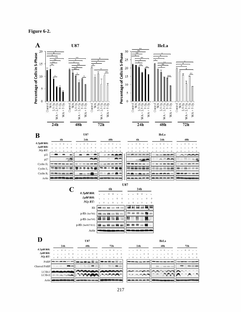
217
Figure 6-2.

218
Figure 6-2. A) Cell cycle analysis was conducted by propidium iodide staining and subsequent
flow cytometry following combination WA and RT treatment for 24-72 h. RT-treated U87 and
HeLa cells both demonstrated small reductions in S-phase percentage, maximally at 24 h or 48 h,
but combination of WA and RT produced enhanced depletion of S-phase cells at 24 h and
maintained the depletion after 72 h despite normalizing levels in the RT-only groups in both
cells. B) Shifts in the cell cycle at different levels were then evaluated molecularly through
Western blotting for cyclins B1 (G2/M transition), D1 (G1/S transition), E1 (G1/S transition) and
cyclin-dependent kinase inhibitors p21/cip1 (G1/S transition) and p27/kip1 (G1/S transition).
Consistent with G2/M arrest, WA alone induced elevated cyclin B1 in U87 cells but was
abrogated with RT. While Cyclin E1 was largely unchanged, cyclin D1 was elevated in a WA
dose-dependent manner at 24-48 h in both cell lines. At 24 h in both lines, small increases in
cyclin D1 were noted with the addition of RT after WA pretreatment. Negative regulators of the
G1/S transition, p21 and p27, demonstrated notable induction with combination WA and RT
compared to either agent alone in a time-dependent manner. C) Treatment with RT diminished
total levels of total Rb protein and phosphorylation at Ser780, Ser795, and Ser807/811 after 6-24
h. Pretreatment with WA further enhanced this decreased Rb phorphorylation with observable
depletion of Ser780 and Ser795 at 6h and Ser807/811 at 24h. D) Combination of WA with RT
failed to enhance apoptotic PARP cleavage or the autophagy-mediated conversion of LC3B
compared to either agent alone, suggesting the cytotoxic effect of combination therapy was not
through these mechanisms. *p < 0.05, **p < 0.01, ***p < 0.001

219
5 Gy RT in U87 through 24h and in HeLa through 6 h. WA-mediated induction of cyclin B1 in
U87 cells was repressed with RT co-treatment. Cyclin E1 was largely unchanged with small RT-
induced elevation at 6 h and 1 µM WA-induced cell-dependent increases at 24-48 h. Cyclin D1
was elevated in a WA dose-dependent manner at 24-48 h in both cell lines. At 24 h in both lines,
small increases in cyclin D1 were noted with the addition of RT to WA, suggesting a potential
block in the G1/S transition and an enhanced attempt to enter S-phase, consistent with the
reduced S-phase fraction noted by flow cytometry findings.
Levels of p21 and p27, negative regulators of the G1/S transition which function to block
cyclin-Cdk1/2/4/6 activity (Sgambato et al., 2000; Cazzalini et al., 2010), supported this claim,
demonstrating notable induction with combination WA and RT compared to either agent alone
(Figure 6-2B). p21 was significantly induced by RT in U87 after only 6 h as previously reported
(Lee et al., 2011) and mildly increased in HeLa. Induction of p21 by WA alone was not observed
until 24 h in HeLa and 48h in U87, and the enhanced combinational effect, particularly
distinguishable at 0.5 µM WA, was consistent with these timepoints. p27 induction was driven
by WA in a dose-dependent manner in both U87 and HeLa cells. Combination with radiation
enhanced this induction at 6 h but failed to show notable benefit at later timepoints except with 1
µM WA in HeLa at 48 h as total induced levels were nearing depletion, suggesting that
predominant early combinational effects are mediated by p27 while long-term responses are p21-
driven.
The retinoblastoma protein (Rb) prevents progression from G1- to S-phase through
inactivation of the transcription factor E2F (Cazzalini et al., 2010). Because cyclin-Cdk1/2/4/6
promotes inactivation of Rb through hyperphosphorylaton and is inhibited by p21 and p27, both
of which were elevated with WA+RT therapy, the downstream status of Rb was evaluated in
220
U87 (Figure 6-2C). Consistent with a previous report demonstrating the activated status of Rb
(Orr et al., 1997), treatment with RT paradoxically diminished total levels of the protein. RT also
decreased inhibitory phosphorylation of Rb at Ser780, Ser795, and Ser807/811 after 6-24 h.
Pretreatment with WA further decreased Rb phorphorylation with observable depletion of
Ser780 and Ser795 at 6 h and Ser807/811 at 24 h. Phosphorylation of Ser795 was nearly
completely eliminated in all RT treatment groups at 24 h. Notably, 1 µM WA alone increased
phosphorylation at all measured sites after 24 h, suggesting a single agent-mediated inverse
effect when not used in combination with RT.
To evaluate whether enhanced apoptosis and/or autophagy also played a role in the
cytotoxicity of combination therapy, cleavage of PARP (apoptosis marker) and conversion of
LC3B-I to LC3B-II (autophagy marker) were examined by Western blotting from 24-72 h
(Figure 6-2D). PARP cleavage was noted with increasing concentrations of WA in both cell lines
but only in HeLa with RT as previously reported (Eriksson et al., 2009; Lee et al., 2011; Grogan
et al., 2013). WA+RT failed to increase PARP cleavage above RT alone in HeLa cells and
decreased WA-mediated PARP cleavage in U87 cells. Despite dose-dependent increases with
WA alone, conversion of LC3B-I to LC3B-II did not demonstrate a consistent pattern of
enhancement with combination therapy. In U87 cells at 48-72 h, 1 µM WA with RT
demonstrated reduced levels of LC3B-II compared to WA alone, and by 72 h in HeLa cells,
LC3B-II levels were consistently depleted compared to control. These findings suggest that
combination of WA and RT favors a mechanism of diminished cellular replication over
apoptosis or autophagy in U87 and HeLa cells.
6.4.3 WA induces depletion of key proteins in HR, NHEJ, and MMR
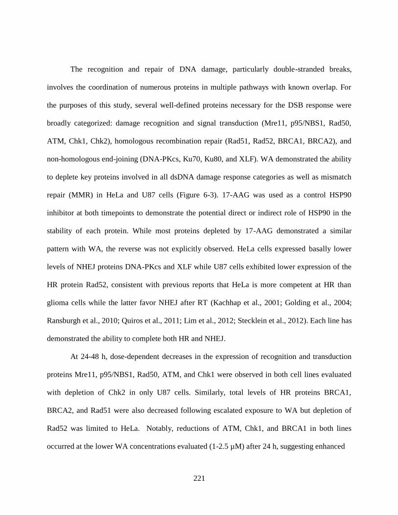
221
The recognition and repair of DNA damage, particularly double-stranded breaks,
involves the coordination of numerous proteins in multiple pathways with known overlap. For
the purposes of this study, several well-defined proteins necessary for the DSB response were
broadly categorized: damage recognition and signal transduction (Mre11, p95/NBS1, Rad50,
ATM, Chk1, Chk2), homologous recombination repair (Rad51, Rad52, BRCA1, BRCA2), and
non-homologous end-joining (DNA-PKcs, Ku70, Ku80, and XLF). WA demonstrated the ability
to deplete key proteins involved in all dsDNA damage response categories as well as mismatch
repair (MMR) in HeLa and U87 cells (Figure 6-3). 17-AAG was used as a control HSP90
inhibitor at both timepoints to demonstrate the potential direct or indirect role of HSP90 in the
stability of each protein. While most proteins depleted by 17-AAG demonstrated a similar
pattern with WA, the reverse was not explicitly observed. HeLa cells expressed basally lower
levels of NHEJ proteins DNA-PKcs and XLF while U87 cells exhibited lower expression of the
HR protein Rad52, consistent with previous reports that HeLa is more competent at HR than
glioma cells while the latter favor NHEJ after RT (Kachhap et al., 2001; Golding et al., 2004;
Ransburgh et al., 2010; Quiros et al., 2011; Lim et al., 2012; Stecklein et al., 2012). Each line has
demonstrated the ability to complete both HR and NHEJ.
At 24-48 h, dose-dependent decreases in the expression of recognition and transduction
proteins Mre11, p95/NBS1, Rad50, ATM, and Chk1 were observed in both cell lines evaluated
with depletion of Chk2 in only U87 cells. Similarly, total levels of HR proteins BRCA1,
BRCA2, and Rad51 were also decreased following escalated exposure to WA but depletion of
Rad52 was limited to HeLa. Notably, reductions of ATM, Chk1, and BRCA1 in both lines
occurred at the lower WA concentrations evaluated (1-2.5 µM) after 24 h, suggesting enhanced

222
Figure 6-3.

223
Figure 6-3. Treatment with increasing concentrations of WA for 24-48 h reduced total levels of
DNA-damage response (DDR) proteins involved in damage recognition (Mre11, p95/NBS1,
Rad50), signaling (ATM, Chk1), and repair through homologous recombination (BRCA1,
BRCA2, Rad51), non-homologous end-joining (DNA-PK, XLF, Ku70, and Ku80), and
mismatch repair (MLH1, MSH2, MSH6) in both U87 and HeLa. This protein depletion was an
expansion of that observed with 1 µM of the traditional HSP90 inhibitor 17-AAG.

224
and consistent selectivity for these targets. Reduced expression of these three proteins was also
observed with 17-AAG. NHEJ proteins XLF, Ku70, Ku80 were depleted with WA dose
escalation in both cells lines after 24-48 h, however, the effect in Ku70/80 was observed at 2.5
µM WA in HeLa (lower baseline expression) compared 10µM WA in U87 (higher expression).
Higher DNA-PKcs expression was detected in U87 cells and was decreased at 1 µM WA. WA-
mediated decreases in NHEJ protein levels corresponded with the effects of 17-AAG treatment.
MMR proteins MLH1, MSH2, and MSH6 showed reduction in expression at 2.5 µM WA at 24 h
with complete protein ablation by 5-10 µM in both HeLa and U87 cells. Treatment with 17-AAG
induced minimal MMR protein reduction after 24 h, however, this effect was more pronounced
in HeLa cells after 48 h. Overall, most proteins evaluated demonstrated reduced or absent
expression at 10 µM WA, suggesting limited selectivity of effect at high concentrations. HeLa
cells also demonstrated enhanced response to 1µM 17-AAG compared to U87 cells which was
confirmed by a cell viability assay (data not shown).
6.4.4 WA alters radiation-induced DNA damage recognition
Following the induction of dsDNA damage and recognition by the MRN complex, ATM
is recruited to the site of the break where it is phosphorylated and can then phosphorylate
Chk1/Chk2 for cell cycle checkpoint activation and H2A.X at the site of damage for recruitment
of repair proteins (Lamarche et al., 2010). Normalized phosphorylation of ATM at Ser1981 was
measured to evaluate the response to RT-mediated DNA damage following WA pretreatment.
Compared to control 1 h post-RT (5 Gy in U87; 10 Gy in HeLa), p-ATM was induced 15.4-fold
in U87 and 8.8-fold in HeLa. The presence of 1 µM WA reduced this activation 56.3% and

225
14.9% in the two lines, respectively (Figure 6-4A). By 6 h, reduction of ATM activation by WA
was no longer observed in U87 cells, showing a delayed enhancement of this phosphorylation
with combination therapy. In HeLa cells, despite diminished total activation of ATM after 6h
(2.2-fold above control) and 24 h (1.89-fold above control), WA 1 µM WA reduced this
activation 66.2% and 74.6%, respectively, showing a delayed response to 2 h WA pretreatment.
U87 cells demonstrated increased p-ATM with WA exposure after 24 h, but this still
demonstrated a 33.6% and 16.9% reduction in the residual p-ATM induced by RT (5.09-fold
above control) with 0.5 µM and 1 µM WA, respectively, by despite the increase induced by
combination therapy at 6 h.
To further evaluate the downstream effects of altered ATM signaling, normalized
phosphorylation of Chk1 at Ser296, an autophosphorylation site requiring previous ATM-
mediated signaling, was measured (Okita et al., 2012) (Figure 6-4A). Minimal changes were
observed in U87 cells 1 h post-RT with slight elevation of p-Chk1. After 6 h, however, total RT-
mediated induction was 2.82-fold compared to control and was reduced by 58.5% with 1µM WA
pretreatment, demonstrating a delayed response compared to p-ATM. Despite relative WA-
mediated reductions in p-ATM largely manifesting at 6 h in HeLa cells, induction of p-Chk1 by
RT at 1 h (2.46-fold above control) was reduced by 40.2% with 1 µM WA pretreatment,
suggesting WA-mediated inhibition of signal transduction. Overall, high induction of p-Chk1 at
1 h corresponded with the largest levels of p-ATM 1h post-RT. Induction by RT at 6 h was
reduced to 1.74-fold above control and only slightly diminished with WA pretreatment.
Phosphorylation of H2A.X (Ser139) is regarded as a marker of DNA damage for
recruitment of repair machinery and is mediated, at least in part, by the activity of ATM
(Lamarche et al., 2010). Recognition of DNA damage was evaluated by immunocytochemistry

226
Figure 6-4.
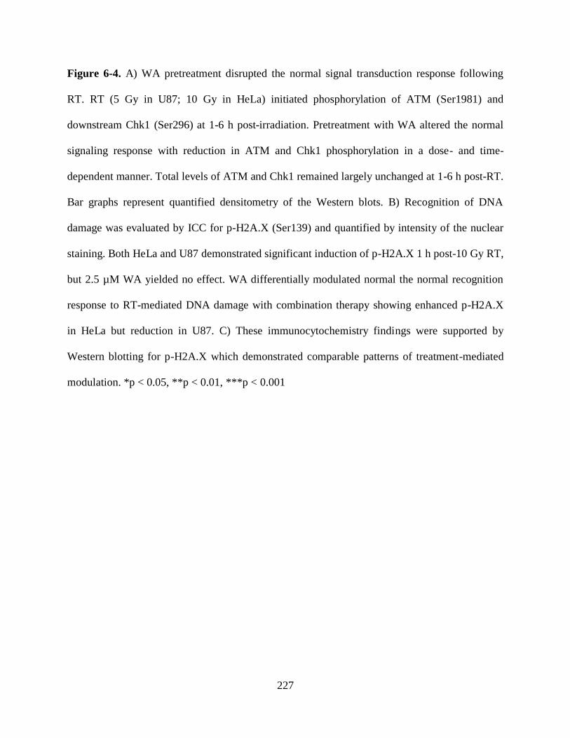
227
Figure 6-4. A) WA pretreatment disrupted the normal signal transduction response following
RT. RT (5 Gy in U87; 10 Gy in HeLa) initiated phosphorylation of ATM (Ser1981) and
downstream Chk1 (Ser296) at 1-6 h post-irradiation. Pretreatment with WA altered the normal
signaling response with reduction in ATM and Chk1 phosphorylation in a dose- and time-
dependent manner. Total levels of ATM and Chk1 remained largely unchanged at 1-6 h post-RT.
Bar graphs represent quantified densitometry of the Western blots. B) Recognition of DNA
damage was evaluated by ICC for p-H2A.X (Ser139) and quantified by intensity of the nuclear
staining. Both HeLa and U87 demonstrated significant induction of p-H2A.X 1 h post-10 Gy RT,
but 2.5 µM WA yielded no effect. WA differentially modulated normal the normal recognition
response to RT-mediated DNA damage with combination therapy showing enhanced p-H2A.X
in HeLa but reduction in U87. C) These immunocytochemistry findings were supported by
Western blotting for p-H2A.X which demonstrated comparable patterns of treatment-mediated
modulation. *p < 0.05, **p < 0.01, ***p < 0.001

228
for p-H2A.X and quantified by intensity of the nuclear staining (Figure 6-4B). HeLa and U87
cells displayed 8.1-fold (p = 0.017) and 11.1-fold (p = 0.021) increases in p-H2A.X 1 h post-10
Gy RT compared to controls, respectively. Pretreatment with 2.5 µM WA for 2 h, which alone
produced no deviation from control in either line, resulted in diverging effects when followed by
RT. In HeLa cells, combination therapy further induced expression of p-H2A.X 3.0-fold greater
than RT alone (p = 0.006) and 24.7-fold above control (p = 0.007). In contrast, the addition of
WA to RT in U87 cells resulted in a 62.5% decrease in the phosphorylation of H2A.X compare
to RT alone (p = 0.049) but was still significantly greater than control (p = 0.048). These results
were supported by Western blotting for p-H2A.X which demonstrated comparable patterns of
treatment-mediated modulation (Figure 6-4C). Modulation of H2A.X phosphorylation was
consistent with the large early impairment of ATM activation in U87 but not HeLa, suggesting
that the effects of WA are manifest at different signaling levels in various cells, such that
diminished ability to recognize DNA damage is observed in U87 whereas a failure to response to
and repair the damage is observed and in HeLa despite enhanced phosphorylation of H2A.X.
6.4.5 WA blocks dsDNA damage repair
Direct measurement of dsDNA damage was evaluated through the neural comet assay,
measuring the movement of the DNA content of single cells through electrophoresis (Figure 6-
5A and 5B). U87 cells were pretreated with WA for 12 h, irradiated, and collected after a 4 h
incubation. Enhanced DNA damage was associated with a higher percentage of total DNA
shifted and a greater distance traveled, quantified as the tail moment. Control cells displayed a
baseline tail moment of 1.1 which was increased 2.3-fold to 2.6 with 0.5 µM WA (p = 0.028),

229
Figure 6-5.

230
Figure 6-5. A) Direct measurement of dsDNA damage was evaluated through the neural comet
assay, measuring the movement of the DNA content of single cells through electrophoresis
followed by nucleic acid staining with SYBR Gold. U87 cells were pretreated with WA for 12 h,
irradiated with 5 Gy, and collected after a 4 h incubation. Mildly increased DNA damage was
observed with treatment of either WA or RT, but combination of the agents significantly
enhanced the average comet tail moment, demonstrating enhanced DNA damage. B)
Representative examples of comets from each treatment group. SYBR Gold = green. C) HeLa
and U87 cells stably expressing the DR-GFP reporter were transfected with an empty vector
control or an I-SceI construct, plated, and treated immediately with multiple concentrations of
WA for 72 h before analysis of GFP expression, a marker of successful HR, by flow cytometry.
WA demonstrated significant dose-dependent reduction in HR function through decreases in
GFP+ cells in both lines. Cells transfected with the control empty vector demonstrated no or
minimal GFP at all concentrations. D) Representative examples of the flow cytometry results for
HeLa and U87. *p < 0.05, **p < 0.01, ***p < 0.001

231
likely a result of minor DNA fragmentation characteristic of intrinsically cytotoxic compounds,
and 2.6-fold to 2.9 with 5Gy RT (p = 0.023). Combination of these two agents resulted in a tail
moment of 6.9, a 6.1-fold increase over baseline (p < 0.001) and statistically greater than either
agent alone (p < 0.001). These findings demonstrate that reduced phosphorylation of H2A.X
resulting from WA+RT compared to RT alone in U87 cells is the result of reduced DDR
signaling rather than enhanced repair.
These findings were supported functionally by the WA-mediated inhibition of HR as
evaluated through the DR-GFP reporter assay (Figure 6-5C and 6-5D; Supplemental Figure 6-2).
U87 and HeLa cells stably expressing the reporter following clonal selection were transfected
with a vector for the restriction enzyme I-SceI or a control vector. Expression of I-SceI in the cell
functioned to cut the reporter assay at a specific site, mimicking a dsDNA break with functional
HR repair that could be monitored through ultimate expression of GFP by flow cytometry and
microscopy. HR-mediated repair was shown to occur in 1.10% of U87 and 1.12% of HeLa
control cells after 72 h. This was significantly reduced by treatment with WA. At 0.5 µM, U87
and HeLa cell repair of the break through HR was reduced to 0.07% (p = 0.017) and 0.59% (p =
0.003), respectively which was further reduced by 1 µM WA to 0.02% (p = 0.018) and 0.20% (p
< 0.001), respectively. Cells transfected with the control empty vector demonstrated no GFP+
cells at all concentrations in U87 and 0.03% at all concentrations in HeLa. Preliminary data in
the murine GBM cell line GL26 demonstrated the same pattern of HR reduction with WA
treatment. These findings support the results demonstrating enhanced dsDNA damage with
combination of WA and RT as well as reductions in DNA damage repair proteins following WA
treatment. This suggests that WA-mediated radiosensitization functions in-part through
inhibition of these repair pathways.

232
6.5 Discussion
WA is a promising novel anti-cancer therapeutic agent that has shown both in vitro and in
vivo promise in a variety of models by targeting a multitude of proliferation, survival, and
growth progression pathways (Vanden Berghe et al., 2012; Vyas and Singh, 2014). While
monotherapy with WA in animal models has yielded impressive results, single agent therapy in
the treatment of human cancer often demonstrates disappointing outcomes and the rapid
development of resistance. Treatment of many cancers now relies on the utilization of several
adjuvant therapies to target multiple cellular susceptibilities simultaneously, often through
production of DNA damage or alterations of DNA/cellular replication (Kamal and Burrows,
2004). Here, we demonstrated that WA inhibits normal pathways of DSB repair through
depletion of total proteins involved in HR and NHEJ as well as alterations and delays in DDR
signaling in two highly aggressive cancer cell lines, HeLa and U87. Combining WA and RT
potentiated the efficacy of both agents leading to enhanced DNA damage and diminished
viability through failure of cells to synthesize DNA and progress through the cell cycle.
Several early studies on WA identified it as a possible radiosensitizer, including its
potential in vivo utility as such (Devi et al., 1995; Devi et al., 1996; Devi et al., 2000; Devi and
Kamath, 2003; Devi et al., 2008). This research group then demonstrated that knockout of
Rad54, but not Ku70 or both Ku70 and Rad54, could sensitize cells to radiation and concluded
that WA may alter normal homologous recombination repair (Devi et al., 2008). Since then,
several additional studies further evaluated the merit of combination therapy and suggested that
it increases efficacy through enhanced oxidation and modulation of the MAPK and Akt

233
pathways (Yang et al., 2011b; Yang et al., 2011a). A lack of adequate controls and data
quantification limits the overall interpretation of the data in these studies, but each demonstrated
the promise of co-treatment with both agents.
Ionizing RT is a frequently utilized treatment modality against many primary cancer
types and some metastatic disease. While certain cancers are quite responsive to the DNA
damaging effects of RT, others show only modest sensitivity or even radio-resistance (Begg et
al., 2011). Given the widespread importance of RT for cancer patients, efforts have been made to
identify means by which to enhance its effect, frequently through the additional of specific
adjuvant chemotherapies. For example, the addition of TMZ to RT in the treatment of GBM
patients extended average post-diagnosis survival by approximately two months, although the
specific mechanism for this radiosensitization is unclear (Chakravarti et al., 2006; Stupp et al.,
2009; Bobola et al., 2010). Other agents have been developed to specifically target mechanisms
of the DDR such as direct inhibitors of ATM and Chk1 (Dent et al., 2011; Nadkarni et al., 2012).
In the present study, WA demonstrated the ability to reduce expression of multiple
proteins in the HR, NHEJ, and MMR DDR pathways. Such findings suggest that WA may
sensitize cancer cells to radiotherapy and/or other DNA-altering chemotherapeutic agents
through induction of instability and promotion of degradation of proteins critical in the
recognition and response to such an insult. WA has previously shown the ability to disrupt the
interaction between HSP90 and Cdc37 with direct binding to the C-terminus of HSP90, a
chaperone protein that functions to fold, refold, and stabilize a specific set of client proteins (Yu
et al., 2010). This disruption corresponds with subsequent degradation of various client proteins
despite no direct inhibition of HSP90 function through its mediation of GR steroid binding
(unpublished data). However, total GR and levels of proteins not previously described to be

234
associated with Cdc37 were depleted, suggesting that WA may also directly inhibit HSP90
activity through a separate mechanism or induce protein depletion through an alternative means.
While Cdc37 is thought to have minor intrinsic protein chaperone properties, it has traditionally
been considered a co-chaperone of HSP90 that promotes localization of the HSP90 chaperone
complex to various protein targets, largely kinases. Such a binding effect of WA to HSP90
appears to be specific to a small subset of proteins such as HSP90 and vimentin (Bargagna-
Mohan et al., 2007), demonstrating the specificity of WA targeting. We hypothesize that the
depletion of a large number proteins globally, including those involved in the DDR, is not due to
direct interaction with the compound but rather the secondary effect of HSP90 axis modulation.
Indeed, WA-mediated depletion of DDR proteins patterned HSP90 inhibition with
enhanced decreases in known HSP90 clients BRCA1 (Stecklein et al., 2012), Chk1 (Arlander et
al., 2003), and DNA-PK (Solier et al., 2012) compared to other proteins at 1-2.5 µM after 24 h.
Other HSP90 client or interacting proteins such as BRCA2 (Noguchi et al., 2006), members of
the MRN complex (Dote et al., 2006), and Rad51 (Ko et al., 2012) demonstrated enhanced
depletion upon WA exposure or responded in a cell line-dependent manner. This suggests either
diminished cellular importance on the HSP90 axis for stability as frequently confirmed with 17-
AAG treatment such as for BRCA2, Cdc37-independent protein targeting, or alternate WA-
induced effects. The importance of the roles of HSP90 and Cdc37 for the stability and function
of proteins involved in the DNA damage recognition and repair pathway is incompletely defined,
and these findings suggest that it may be more significant than currently understood. Consistent
with these findings, direct inhibition of HSP90 has been shown to result in radiosensitization
with inhibition or depletion of proteins in the DDR such as Akt, BRCA1, and Rad51 (Dote et al.,
2006; Kabakov et al., 2010; Ko et al., 2012; Stecklein et al., 2012). To date, no studies have been

235
completed to identify a role for Cdc37 in the interaction with the altered DDR proteins either
directly or indirectly. Further study is also necessary to identify why ATM is significantly
depleted with both WA and 17-AAG treatment but has previously been shown to not interact
with HSP90 (Dote et al., 2006). Importantly, WA modulates proteins from all pathways of the
DDR, including both HR and NHEJ, demonstrating its potential broad-spectrum effectiveness as
a radiosensitizer against various malignancies regardless of the favored repair pathway(s).
Additionally, our group has demonstrated the ability of WA to elevate the oxidative
potential in cancer cells which suggests an alternative mechanism by which DDR proteins can be
degraded (Grogan et al., 2013; Grogan et al., 2014). O6-methylguanine-DNA methyltransferase
(MGMT), a protein involved in the removal of TMZ-mediated DNA alkylation, was depleted by
WA in a dose-dependent manner. While MGMT has not been described as a client of HSP90 or
Cdc37 and was not significantly decreased with 17-AAG exposure, it had previously shown
sensitivity to compounds inducing oxidative stress such as valproic acid (Fu et al., 2010; Ryu et
al., 2012). Further studies are necessary to identify whether the pro-oxidant capacity of WA
results in protein misfolding and/or depletion and if it is a direct consequence of reactive oxygen
species (ROS) production through its epoxide group or an indirect shift in background potential
resulting from thiol sequestering of the compound. Regardless, such a dual mechanism of
oxidation and HSP90 axis inhibition provides great potential for enhanced efficacy while
minimizing the development of resistance. In addition, oxygen is a known radiosensitizer
(Teicher, 1995; Okunieff et al., 1996), and the known oxidizing effects of WA may play an
indirect role in further enhancing the efficacy of RT through increased formation of DNA-
damaging free radicals, although it is unclear if introduction of oxygen in this form will indeed
be beneficial. While WA elevates cellular oxidative potential and increases p-H2A.X at high

236
concentrations after 24 h (Grogan et al., 2014), this p-H2A.X induction could be completely
eliminated with pretreatment of a global caspase inhibitor in a breast cancer model, suggesting
the effect was an apoptosis-related phenomenon rather than direct damage to the DNA (data not
published).
Interestingly, the results of this study suggest that WA not only functions to deplete total
levels of proteins critical to the DDR but can also alter normal signaling at concentrations below
those that alter protein levels. In the evaluation of ATM and Chk1 activation and signaling, total
levels of each protein at low-dose WA remain largely unchanged while phosphorylation is
altered. If these findings are related to inhibition of the HSP90 chaperone axis induced by WA,
this may suggest general mild destabilization of some fraction of ATM and its upstream kinases
that alters function but remains detectable in total level analysis. Consistent with this
observation, disruption of Cdc37 has previously been demonstrated in prostate cancer to inhibit
cellular kinase activity without significantly depleting certain HSP90 client proteins (Gray et al.,
2007).
Despite the WA-mediated modulation of ATM and Chk1 signaling following RT, each
cell line showed an enhanced the ability to undergo cell cycle arrest at 24-72 h with a notably
reduced fraction of DNA synthesis phase cells. Diminished phosphorylation of Chk1 after
combination therapy was observed between 1-6 h post-RT but not after 24 h, suggesting delayed
but not eliminated cell cycle checkpoint signaling which may perpetuate error generation or lack
of timely repair early post-RT as the cell continues to cycle. Further, additional signaling
proteins such as ataxia telangiectasia and Rad3-related protein (ATR) and Chk2 may compensate
for this effect, and it is unclear if the mild reduction in p-Chk1 was functionally significant. It is
hypothesized that the maintained low dose WA-mediated reduction of p-ATM after RT favors

237
reduction in DNA damage recognition and ultimately repair, both effects demonstrated in this
study, compared to inhibited checkpoint modulation and may eventually overcome any
diminished regulation of the checkpoints. Higher dose WA, which leads to significant direct
depletion of Chk1 and/or Chk2 but manifests notable cytotoxicity independent of RT, may
eventually limit the functional execution of these checkpoints.
Interestingly, the data suggest that combination of WA and RT results in a reduction of
cells entering the DNA synthesis phase of the cell cycle with cells existing in the S-phase
shifting to a non-mitotic population containing 4N DNA. The notable accumulation of cyclin B1
in U87 cells treated with WA for 24-48 h is nearly completely reduced to baseline when
simultaneously treated with RT. This suggests that the 4N DNA cells are not appropriately
progressing in the G2/M phase as has previously been demonstrated with the flavoprotein-
specific inhibitor diphenyl eneiodonium (Scaife, 2004). While p21, which is increased in this
study with WA treatment and further with combination therapy, has traditionally been regarded
as a regulator of the G1/S transition, it has also been demonstrated to play a role during the
G2/M phase (Sherr and Roberts, 1995; Dulic et al., 1998; Cazzalini et al., 2010). Indeed,
knockout of p21 prevented cells from appropriately arresting at the G2/M checkpoint following
the induction of DNA damage. This was similarly identified in p27 which has also been
traditionally regarded as an inhibitor of the G1/S transition and is increased in the present study
(Sgambato et al., 2000; Payne et al., 2008). Further, this induction of p21 and repression of
cyclin B1 to maintain G2 arrest has been demonstrated to be a p53-mediated effect (Levesque et
al., 2008), a protein that of wild-type status in both U87 and HeLa albeit of low expression in the
latter. In addition to functioning as a transient inhibitor of the cell cycle, p21 elevation is also a
marker of cellular senescence. While further studies are necessary to address this, the enhanced

238
retention of cell cycle arrest and elevated p21 with combination WA and RT supports this idea.
A previous study has demonstrated that WA alone is capable of inducing senescence with
induction of marker β-galactosidase (Vaishnavi et al., 2012).
Previous reports from Yang, et al. showed that combination of WA and RT in U937 and
Caki cells resulted in enhanced markers of apoptosis without cell cycle radiosensitization
contributions from p21 (Yang et al., 2011b). In contrast, we demonstrated in HeLa and U87 cells
that cleavage of PARP as an apoptotic marker was either not enhanced or otherwise reduced with
combination therapy compared to either agent alone in favor of increased expression of p21 and
p27 and diminished progression of the cell cycle. These differences may represent intrinsic
difference among cells or variability in dosing timepoints and concentration as analysis was only
completed up to 24 h in the previous evaluation, however, both studies demonstrate the
therapeutic value of combining WA and RT. Of note, U937 cell are a known p53-mutant cell line
(Sugimoto et al., 1992), suggesting one potential intrinsic explanation for the difference. Caki
cells contain wild-type p53 comparable to the cells used in the present study which was indeed
shown to be activated through phosphorylation in the Yang, et al. study, indicating the potential
role of an alternative mechanism not yet identified (Warburton et al., 2005). The Caki study did
not evaluate changes in cell cycle distribution resulting from combination therapy, so it is
possible that p53/p21 may still demonstrate comparable effects at later timepoints.
Future studies will be necessary to further define the dosing schedule that will yield
optimal potentiation or synergy in the combination of WA and RT, particularly in vivo. Our in
vitro studies determined that 2 h pretreatment was sufficient to all for this enhanced efficacy.
Depending on the tissue levels achieved and the time necessary to manifest effects in the DDR
pathways, a build-up period may be necessary in translational studies. WA dosing prior to rounds

239
of RT appear to be necessary to fully take advantage of the blockage of DNA damage repair.
Studies are also necessary to identify the ability of WA to penetrate the blood-brain barrier
(BBB) to evaluate its potential utility against brain tumors such as GBM which is indicated
based on these studies, although tumors such as these are frequently described to disrupt the
normal architecture of the BBB (Zhang et al., 1992). One preliminary study of WA in a murine
orthotopic GBM model demonstrated a survival advantage with treatment, indicating that WA
was reaching its target (Santagata et al., 2012). Further, evaluation of WA against the Lipinski
Rule of Five, typically utilized for predicting favorability of drug absorption oral but often
applied to the BBB, demonstrates that it does not violate any rule (Banks, 2009; Vaishnavi et al.,
2012).
In this study, WA was determined for the first time to be a highly effective radiosensitizer
through modulation of the normal DDR. Following WA treatment, proteins involved in DNA
damage recognition and repair through HR and NHEJ were depleted, resulting in altered
recognition and functionally inhibited repair of RT-induced DNA damage. Our previous findings
show that WA acts as a potent cytotoxic monotherapy against GBM and re-sensitizes resistant
cells to TMZ, the standard chemotherapeutic along with RT against GBM (Grogan et al., 2013;
Grogan et al., 2014). Other groups have demonstrated WA efficacy other non-central nervous
system (CNS) cancers and identified that WA enhances the efficacy of the broad-spectrum agent
cisplatin, an agent often used against cervical cancer (Kakar et al., 2012; Vanden Berghe et al.,
2012; Vyas and Singh, 2014). Taken together with WA-mediated enhancement of RT, these
findings suggest the great clinical potential of WA as an adjuvant agent for brain tumors and
non-CNS solid tumors like cervical cancer that warrant further translational exploration.

240
Supplemental Figure 6-1.

241
Supplemental Figure 6-1. Complete cell cycle analysis of the percentages of cells in the G0/G1
(2N DNA), S, and G2/M (4N DNA) phases with WA and RT treatment. WA treatment results in
an increased percentage of cells in the G2/M-phase of the cell cycle with minimal changes in S-
phase percentage. RT-treated U87 and HeLa cells both demonstrated small reductions in S-phase
percentage, maximally at 24 h or 48 h, but showed variable shifts with cell predominantly
moving to G0/G1 in U87 and G2/M in HeLa. Combination of WA and RT produced enhanced
depletion of S-phase cells at 24 h and maintained the depletion at 72 h despite normalizing levels
in the RT-only groups in both U87 and HeLa cells. Depletion of S-phase cells was primarily
associated with maintained or minimally decreased levels of G0/G1-phase cells compared to
WA-only with displacement to G2/M after 24 h. U87 cells demonstrated maintained elevation of
G0/G1-phase cells at 24 h only with combination therapy.

242
Supplemental Figure 6-2.

243
Supplemental Figure 6-2. HeLa cells stably expressing the DR-GFP reporter were transfected
with an empty vector control or an I-SceI construct, plated, and treated immediately with 1 µM
WA for 72 h before analysis of GFP expression, a marker of successful HR. WA demonstrated
significant reduction in HR function with undetectable GFP+ cells as assessed by microscopy.
Cells transfected with the control empty vector demonstrated no detected GFP by microscopy.
GFP = green.

244
Chapter 7
Summary, Significance, and Future Directions
245
7.1 Summary and significance
7.1.1 Introduction
The treatment of brain tumors, particularly high grade malignancies like glioblastoma
multiforme (GBM) and medulloblastoma (MB), continues to remain a tremendous clinical
challenge. While surgical resection followed by radiation therapy (RT) and temozolomide
(TMZ) in GBM patients limits disease progression, the average post-diagnosis survival time is
still only approximately 14 months with rapid and frequent development of TMZ resistance
(Stupp et al., 2002; DeAngelis, 2005; Stupp et al., 2005; Taphoorn et al., 2005; Chamberlain et
al., 2007). TMZ was approved for use in GBM patients in 2004, but little therapeutic progress
has been achieved since that time. Avastin® (bevacizumab), an angiogenesis inhibitor, was
approved after TMZ but is largely utilized to reduce disease progression during the management
phase rather than to provide a long-term disease response (Friedman et al., 2009; Zhang et al.,
2012a). MB, largely but not exclusively a pediatric disease, demonstrates a greater response to
currently available chemotherapies and RT, but patients are shown to frequently develop short-
term and chronic quality of life issues that manifest from the off-target effects of the treatment in
a young patient population (Khatua et al., 2012; Smoll, 2012). The research of the present study
demonstrates the potent in vitro utility of WA as a therapeutic agent against the high-grade
malignant brain tumors GBM and MB. The following briefly summarizes relevant finds in each
of the chapters and identifies key significance and utility of the data. Further detailed
interpretation and discussion can be found in each chapter.

246
7.1.2 Chapter 2: Cytotoxicity of withaferin A in glioblastomas involves induction of an oxidative
stress-mediated heat shock response while altering Akt/mTOR and MAPK signaling pathways
Given the promising efficacy of WA in several non-brain tumor models and the identified
need for novel therapeutic approaches in these cancers, the effects of WA were evaluated in a
GBM model. The aim of this chapter was to determine if these previously observed cytotoxic
effects could be translated to GBM with subsequent analysis of the nature of these effects. In this
study, WA was indeed described to have an anti-proliferative effect on GBM cells with induction
of G2/M cell cycle arrest at correspondingly low-to-moderate concentrations with a transition to
cell death through apoptotic mechanisms at moderate-to-high doses. These findings were
associated with changes in expression of Akt/mTOR and MAPK pathway proteins and upstream
tyrosine kinase receptors. Notably, WA reduced activation of oncoproteins in the Akt/mTOR
pathway but enhanced phosphorylation in MAPK proteins despite a reduction of total Raf-1.
Simultaneously, WA produced significant cellular oxidation with induction of biomarkers
HSP27, HSP32, and HSP70. All effects could be completely abrogated with N-acetyl-L-cysteine
(NAC) pretreatment.
These findings demonstrated the in vitro utility of WA against GBM and provided a
broad and general evaluation of its utility. Notably it also expanded on the limited data available
addressing the effects of WA on the Akt/mTOR pathway. Here, WA not only decreased total and
phosphorylated Akt as previously described but also diminished activation of downstream
mTOR and p70 S6K. The α-subunit of the stress-response protein AMPK and its downstream
tumor suppressor TSC2 that ultimately signal to mTOR were also activated via phosphorylation.
This demonstrates that WA functions, in part, to inhibit the the Akt/mTOR pathway in the

247
manifestation of its effects. This study also showed the potential utility of thiol-reactive oxidative
agents against GBM, further enhancing the altered redox state that has been described in cancer
cells.
7.1.3 Chapter 3: Oxidative cytotoxic agent withaferin A resensitizes temozolomide-resistant
glioblastomas via MGMT depletion and induces apoptosis through Akt/mTOR pathway
inhibitory modulation
Intrinsic or acquired therapeutic resistance to TMZ has been shown to represent a major
problem in the treatment of GBM patients (Hansen et al., 2007; Kitange et al., 2009). With a
poor system of pre-therapy tumor screening methods currently in place, patients frequently
receive TMZ when it would be contraindicated either early in the treatment process or after
multiple cycles of chemotherapy (Chamberlain, 2010; Quick et al., 2010; Zhang et al., 2012c).
Given the in vitro success of WA in known TMZ-sensitive GBM cell lines, the aim of this
chapter was to evaluate the potential use of WA in developed TMZ-resistant GBM as a
monotherapy and as a potential re-sensitizer to TMZ. As a monotherapy, WA demonstrated the
same pattern of efficacy in resistant cells as was seen in the sensitive lines but with enhanced
potency. This was expanded to demonstrate that phosphorylation of components of the MAPK
cascade was a compensatory activation of the pathway in response to the cytotoxicity of WA,
identifying a potentially effective target for the use of WA in synergistic combination therapy.
Additionally, inhibition of both the intrinsic and extrinsic apoptotic pathways resulted in reduced
downstream cleavage of PARP and effector caspases, demonstrating the role of both pathways in
WA-mediated apoptosis.

248
Most importantly, WA demonstrated the ability to deplete cells of O6-methylguanine-
DNA-methyltransferase (MGMT) which confers resistance to TMZ. This effect was associated
with re-sensitization to TMZ and was potentiated with TMZ combination therapy. Re-
sensitization was not observed in a TMZ-resistant line with resistance through mismatch repair
(MMR) alterations, and TMZ-sensitive lines were inconsistently further sensitized to TMZ by
WA but never desensitized. These results demonstrate that WA, in addition to functioning as an
effective monotherapy in a model of TMZ resistance, can be utilized to enhance the utility of
WA in a setting where it would otherwise be contraindicated. Given that around 50% of patients
are thought to display intrinsic characteristics for poor response to TMZ but receive TMZ
regardless (Chamberlain, 2010; Quick et al., 2010; Zhang et al., 2012c), addition of WA to GBM
treatment has the potential to function as 1) a cytotoxic therapeutic addition in a system that
would otherwise be using an ineffective chemotherapeutic agent, and 2) as an agent to establish
value with for the inclusion of TMZ. Of note, while MMR-driven resistance may not be re-
sensitized to TMZ with WA, the presence of WA potentially provides a valuable cytotoxic
component in a system with otherwise limited efficacy beyond RT. In cases where tumors
display TMZ sensitivity, despite the inconsistent synergistic benefit of combination therapy, the
additive presence of two agents may still be beneficial. Overall, the addition of WA, if tolerated,
would appear to be universally beneficial.
7.1.4 Chapter 4: Withaferin A is a novel inhibitor of the Wnt/β-catenin signaling pathway in
medulloblasoma through proteasome-mediated degradation of TCF/LEF

249
With the effective in vitro utilization of WA against GBM and its observed inhibition of
Akt/mTOR pathway signaling, the use of WA was expanded to a model of MB. The aim of this
chapter was to confirm the known cytotoxicity of WA in MB and evaluate the effects of WA in
the Wnt/β-catenin signaling pathway. Indeed, WA demonstrated the anti-proliferative, pro-
apoptotic, G2/M cell cycle arresting, and oxidative effects identified previously in GBM. Given
the well-described cross-talk between the Akt/mTOR and Wnt/β-catenin pathways through GSK-
3β and the overactivation of Wnt signaling in a subgroup of MBs (Baryawno et al., 2010;
Northcott et al., 2012), inhibition of Akt as previously described in GBM was expected to result
in inhibition of Wnt signaling through diminished phosphorylation of GSK-3β and subsequent
degradation of β-catenin. WA did in fact reduce Wnt signaling through a reporter assay.
However, in contrast to this original expectation, WA induced significant phosphorylation of
GSK-3β and failed to significantly alter β-catenin levels or nuclear translocation in the cells.
Rather, WA promoted proteasomal degradation of the TCF/LEF transcription factors that
associate with β-catenin for the transcription of Wnt target genes.
These findings not only demonstrate the general efficacy of WA against MB but also
identify it as a novel inhibitor of the Wnt/β-catenin signaling pathway through a unique
mechanism. Further, WA was shown to disrupt the interaction between HSP90 and co-chaperone
Cdc37 in MB, and inhibition of HSP90 with the established small molecule 17-AAG resulted in
depletion of TCF/LEF proteins. Previous studies have identified the interaction of HSP90 with
upstream components of the pathway like β-catenin and GSK-3β but have failed to conclusively
demonstrate the role of HSP90 in their stability (inhibition of HSP90 did not consistently reduce
levels of these proteins) (Cooper et al., 2011; Liu et al., 2012). Despite our inability to co-IP
TCF/LEF members with HSP90 or Cdc37, these findings demonstrate a previous undescribed

250
role for HSP90 in the function of the Wnt signaling pathway which will require further
exploration but suggests a novel role for HSP90 inhibitors against Wnt-driven malignancies like
MB and colorectal cancer (Polakis, 2012). While WA targets this important signaling pathway
overactivated in a subset of MB, it is also intrinsically cytotoxic through alternative pathways
suggesting that it can be utilized against all MB subgroups without pathological pre-
identification of subgroup. Its known selectivity for cancer cells with minimal off-target toxicity
in murine models, as currently described, could reduce quality of life issues plaguing pediatric
MB patients with current treatments while maintaining efficacy, although more studies are
required in regard to translational toxicity and efficacy.
7.1.5 Chapter 5: Withaferin A promotes global proteasome-mediated protein degradation
through oxidation and inhibition of the HSP90 chaperone axis and is cytotoxically potentiated by
inhibition of the proteasome
WA has demonstrated the ability to yield diverse effects throughout the cell, frequently
with protein depletion and modulation of multiple oncogenic signaling pathways (Vanden
Berghe et al., 2012; Vyas and Singh, 2014). Several studies have suggested that WA alters the
HSP90 chaperone axis and promotes cellular oxidation to promote the broad effects that are
observed (Samadi et al., 2009; Yu et al., 2010; Wang et al., 2012). The aim of this chapter was to
identify and explore the role of WA on cellular proteotoxicity through these mechanisms in
GBM and the well-established HeLa cervical cancer line. WA favored a shift to HSP70 in the
HSP90/HSP70 balance, promoting enhanced global protein ubiquitination. This was associated
with degradation of HSP90 client proteins via the proteasome with inhibition of the HSP90 axis

251
through disruption of the HSP90/Cdc37 interaction but not direct reduction in intrinsic HSP90
function. Cytotoxicity and oxidation events following WA exposure could be completely
abrogated with thiol antioxidant NAC pretreatment and enhanced with reduction in cellular
glutathione. Non-thiol antioxidant ascorbic acid reduced oxidation but failed to alter cytotoxicity.
These findings further define the novel mechanism by which WA manifests its cytotoxic
effects and provide details regarding both the oxidative and HSP90 inhibitory components which
are likely not mutually exclusive. Such an effect on the HSP90 axis may predict WA utility in
altering additional proteins and pathways critical to cancer progression subsequently revealing
appropriate clinical uses. Future studies identifying other HSP90 inhibitory aspects given the
depletion of non-Cdc37 proteins will be necessary. Additional experiments were completed to
evaluate WA in combination with a proteasome inhibitor. Given that WA promotes widespread
protein degradation via the proteasome, simultaneous inhibition of the proteasome promoted
accumulation of ubiquitinated aberrant proteins, induced cytotoxicity with reduced cell viability,
and ultimately potentiated the induction of apoptosis compared to either agent as a monotherapy.
Proteasome inhibitors are currently approved for clinical use in some cancer types including
multiple myeloma (Chauhan et al., 2005), identifying an opportunity for rapid WA translation to
human use and establishment of proof-of-concept. This enhanced efficacy of combination with a
proteasome inhibitor has been supported with the use of traditional HSP90 inhibitors
(Minnnaugh et al., 2004). These data support translational utility derived from the identified
mechanisms of WA.
7.1.6 Chapter 6: Withaferin A disrupts the cellular DNA-damage response to promote
radiosensitization

252
RT represents a key therapeutic component in the treatment of a significant percentage of
primary tumors including GBM and MB (Delaney et al., 2005). Therefore, efforts to enhance
RT-induced DNA damage and limit the repair of this damage by cancer cells has been seen as
means by which to effectively enhance cytotoxicity and efficacy of clinical RT in patients (Begg
et al., 2011). Given that inhibition of HSP90, among other targets, can radiosensitize (Dote et al.,
2006; Kabakov et al., 2010; Ko et al., 2012; Stecklein et al., 2012), the aim of this chapter was to
evaluate the radiosensitization properties of WA against GBM cells and the homologous
recombination (HR)-competent HeLa cervical cancer line. Indeed, low-dose WA potentiated the
efficacy of RT in both lines. This was associated with a reduction in cells synthesizing new DNA
and elevation of the checkpoint inhibitors p21 and p27. Functionally, WA reduced total levels of
key proteins involved in the recognition and repair of DNA damage through HR and non-
homologous end-joining (NHEJ) and modulated normal signaling in response to RT. This lead to
altered recognition of the damage assessed by p-H2A.X, enhanced and sustained DNA damage
with combination therapy, and a functional block of HR.
These findings further demonstrate the potential clinical application of WA. Given that
RT is used in the treatment of an estimated 50% of cancer patients (Delaney et al., 2005),
enhancing the efficacy of this therapeutic modality is an extremely important translational
prospect and has been a consideration in the selection of adjuvant chemotherapy previously,
including the addition of TMZ to RT (Chakravarti et al., 2006). The data suggests that WA is
likely to enhance DNA damage induced by other agents and repaired through a range of
modalities including HR, NHEJ, and MMR which identifies a potential for broad spectrum
combination use clinically alone with the intrinsic cytotoxicity of WA as a monotherapy.

253
Because augmented DNA damage response mechanisms are known to play a key role in chemo-
and radio-resistance, we believe that modulation and depletion of these pathways with WA may
provide enhanced efficacy of therapeutics in refractory or resistant GBM or other cancer types.
Indeed, we demonstrated WA’s ability to re-sensitize resistant GBM cells to TMZ through
depletion of MGMT, showing proof-of-concept (Grogan et al., 2014).
7.2 Future directions
The conducted research identified, defined, and utilized the cytotoxic anti-cancer
properties of WA in a model of high-grade brain tumors. Among the results, WA induced
inhibitory modulation of the PI3K/Akt/mTOR and Wnt/β-catenin oncogenic signaling pathways.
These are the first studies to thoroughly evaluate the influence of WA on both pathways in any
cancer model and potentially define several therapeutic roles for the compound. Given the broad
effects of WA on multiple oncogenic or otherwise cancer-promoting pathways in our studies and
those published by others (Vanden Berghe et al., 2012; Vyas and Singh, 2014), particularly with
noted activity against the HSP90 chaperone axis with over 400 known client or interacting
proteins, future experiments are necessary to further expand on the effects of WA against other
proteins that have yet to be explored and the resulting changes in signal transduction efficiency.
Notably, there are currently no published reports on the effect of WA on the SHH pathway,
important in a subgroup of MB. Similarly, the inhibition of the pathways identified with this
research can be expanded to other relevant cancer models. For example, the Wnt/β-catenin
pathway is overactivated in a significant percentage of colorectal cancers (Polakis, 2012).
Targeting this pathway with WA along with the general cytotoxicity of the compound through
254
additional mechanisms would be thought to have great therapeutic benefits clinically by blocking
a key driving oncogenic pathway and inducing additional anti-cancer manifestations.
The efficacy of WA was also demonstrated to be enhanced in combination with both RT
and a proteasome inhibitor and functioned to re-sensitize resistant GBM cells to TMZ. Previous
studies have identified that WA synergizes with other chemotherapeutic agents such as sorafenib
and cisplatin (Cohen et al., 2012; Kakar et al., 2012). HSP90 inhibition, a described function of
WA, has also been shown to potentiate, enhance, or re-sensitize the activity of numerous agents
(Lu et al., 2012). These observations suggest that WA may function well in combination with
other therapeutic agents, particularly those inducing DNA damage, generating a large expansion
in the potential for studying and ultimately using this compound. Additionally, because WA was
demonstrated to promote activation of the MAPK pathway through phosphorylation of Raf-1 and
ERK1/2, relevant inhibitors of these proteins would be expected to yield favorable efficacy in
combination with WA. Given the apparent protective effect of GSH against WA, selective
depletion of GSH in redox susceptible cancer cells, perhaps through inhibition of γ-
glutamylcysteine synthetase, could serve to enhance the cellular cytotoxicity to WA, although
the potential for off-target toxicity would need to be evaluated if similar reduction in GSH was
observed in normal tissue. These combinations also establish greater therapeutic relevance for
WA given the problems of ineffectiveness and resistance frequently associated with
monotherapy. Interestingly, however, our in vitro attempts to develop cell line resistance to WA
have been largely unsuccessful and warrant further exploration.
These findings open some additional questions regarding the role of WA in the inhibition
of the HSP90 axis. While these results clearly indicate that WA eliminates the interaction
between HSP90 and the co-chaperone Cdc37 at high enough concentrations and does not appear
255
to alter the intrinsic activity of HSP90 at least in regard to steroid receptor association, protein
depletion mirrors total inhibition of HSP90 (and perhaps more) rather than a prevention of Cdc37
function. Cdc37 primarily functions as a kinase-specific co-chaperone to promote the folding and
maturation of these proteins through localization with HSP90 with enhanced client loading
though modulation of HSP90 ATPase activity (Siligardi et al., 2002; Roe et al., 2004). However,
proteins not previously known to associate with Cdc37 and non-kinase proteins such as c-myc
and the glucocorticoid receptor were dose-dependently degraded with WA exposure. Such
findings suggest that while the disruption of the HSP90/Cdc37 interaction was real, the effects of
WA extend beyond that observation or the role of Cdc37 is greater than currently thought. Cdc37
has also been shown to interact with other co-chaperone proteins, suggesting another way by
which it can still join the HSP90 chaperone complex (Abbas-Terki et al., 2002). Given that WA
interacts with the C-terminus of HSP90 in the location that other co-chaperones are known to
associate, this represents a potential explanation of these expanded findings (Yu et al., 2010).
Interestingly p23 and Hop were not dissociated from HSP90 in the presence of WA (Yu et al.,
2010), but their maintained functionality directly with HSP90 was not addressed. Clearly, further
study of this system is required to better understand the variations observed whether due to
additional modulation of the HSP90 axis or indirect effect from associated observations like
cellular oxidation.
Additionally, while alterations in the HSP90 chaperone axis have been clearly
demonstrated to play a role in the cytotoxicity of WA, there have been arguments regarding the
specificity of this effect. In order to determine whether the cytotoxic manifestations are primarily
driven by its association with HSP90, it is proposed that future studies be conducted with cells in
which endogenous HSP90 has been replaced or supplemented with HSP90 mutants retaining

256
function but lacking the appropriate WA binding site, likely through mutation of an existing C-
terminal cysteine. As such, the HSP90-independent effects of WA could be studied. Further,
enrichment or depletion of GSH in the context of non-thiol antioxidants could further help define
the nature of the effect as it relates to general oxidation and redox alterations. To date, few
proteins have been shown to directly associate with WA, including via a broad spectrum analysis
of biotinylated-WA binding, suggesting some specificity of is binding (Bargagna-Mohan et al.,
2007; Yu et al., 2010). Such studies could further demonstrate the importance of the HSP90
target with WA treatment.
Studies from our group and others have evaluated novel natural and semi-synthetic
withanolides beyond WA (Zhang et al., 2011a; Santagata et al., 2012; Zhang et al., 2012b;
Wijeratne et al., 2014). Notably, significant cytotoxicity is lost with the elimination of the A-ring
enone and/or the epoxide group. Our preliminary studies in triple negative breast cancer and
several other models demonstrate that acetylation of one or more of the hydroxide groups
enhance cytotoxic activity and anti-tumor efficacy in vivo. A recent study also identified that
synthetic acetylation or other modifications of functional groups on WA or the withanolide
structure can differentially alter biological cytotoxicity and/or heat shock-inducing activity
(Wijeratne et al., 2014). It is unclear if this heat shock response is a function of HSP90
inhibition, general oxidation, or perhaps more likely, both. Such structure-activity relationship
(SAR) studies will be necessary going forward to identify improved therapeutic withanolides
with better anti-cancer efficacy, more favorable pharmacokinetics for a particular malignancy
[especially for crossing the blood-brain-barrier (BBB)], and diminished off-target toxicity. To
date, SAR studies with withanolides have primarily been assessed in vitro. While these studies
have yield valuable information, they have failed to address the pharmacokinetic aspect for

257
translational use. Withanolides, primarily WA, have demonstrated in vivo efficacy against a
number of murine cancer models (Vyas and Singh, 2014) but drug absorption, distribution, and
elimination have remained largely unexplored.
Pharmacokinetic studies, particularly those involving the metabolism of WA and these
other novel withanolides, will shed light on the body’s ability to activate and/or detoxify these
compounds, thereby suggesting potential benefits or limitations to various dosing schemes. To
date, very few studies have evaluated the metabolism of WA with the last studies emerging from
the 1980s. Those studies, conducted in Cunninghamella elegans and Arthrobacter simplex,
revealed that WA can be metabolized via hydroxylation, hydrogenation, and hydrolysis (Rosazza
et al., 1978; Fuska et al., 1982; Proksa et al., 1986; Fuska et al., 1987). Because WA shares many
structural similarities with endogenous steroids given its steroidal lactone classification, the
study of its metabolism remains a critical element necessary for its potential use in the clinic as
steroids are often metabolized in oxidative reactions by cytochrome P450 (CYP450) enzymes
(Gonzalez, 1990). On top of that, an estimated minimum of 75-90% of the reactions in the
metabolism of pharmaceutical drugs are by CYP450s, demonstrating an integral role for their
study in the development of any compound (Guengerich, 2008; Sun and Scott, 2010). An overlap
in the metabolic processes serving WA with other chemotherapeutic agents whose metabolism
has been previously well-characterized could suggest successful pairing schemes on the basis of
toxicokinetics or generating more favorable pharmacokinetic parameters (Cheng et al., 2010).
Additionally, studies on the distribution of WA and its metabolites, particularly to the
brain and more specifically during the diseased state of a brain tumor, have remained largely
unpublished, although one preliminary study utilizing a murine orthotopic glioma model
demonstrated a survival benefit with WA (Santagata et al., 2012). Given this survival benefit, it
258
is evident that the compound or an active metabolite likely reaches the site of the tumor lesion
regardless of whether the BBB lacks integrity. What is less clear is whether WA is capable of
crossing an intact BBB peritumorally where tumor invasion has occurred or in healthy brain
tissue with relatively normal and maintained architecture. A recent bioinformatics study
suggested that WA’s structure fails to violate Lipinski’s Rule of Five and has a favorable
blood/brain partition coefficient (Vaishnavi et al., 2012), but translation to a biological system
still needs to be examined. Other active withanolides in which polar functional groups are
masked or simply absence may yield more favorable results if not ultimately achieved with WA,
and indeed, preliminary studies in several non-brain tumor murine models show this.
Encouragingly, multiple additional non-brain tumor murine models have demonstrated
promising responses to WA treatment with multiple dosing schedules and levels (Vyas and
Singh, 2014). One report identified a maximum plasma concentration of WA following a single
intraperitoneal injection of the moderate dose of 4 mg/kg to be 1.8 µM, well-above typical in
vitro IC50 values for cancer cells, with a plasma half-life of 1.3 h. Tissue distribution has not yet
been reported. Overall, further translational studies with WA and other withanolides, particularly
in an orthotopic brain tumor model, are necessary to evaluate both the pharmacokinetics and
pharmacodynamics of the compound and establish maximally effective and minimally toxic
dosing levels and schedules. Such studies can be followed up with efficacy and toxicity studies
for combination therapy as previously discussed and utilization of NAC as a rescue agent in
cases of toxicity or attempted high-dose regimens.

259
References
Abbas-Terki T, Briand PA, Donze O and Picard D (2002) The Hsp90 co-chaperones Cdc37 and Sti1 interact
physically and genetically. Biol Chem 383:1335-1342. ACS (2013) Cancer Facts and Figures 2013, in Atlanta, GA: American Cancer Society, American Cancer
Society, Atlanta, GA. Agarwal S, Sane R, Oberoi R, Ohlfest JR and Elmquist WF (2011) Delivery of molecularly targeted therapy
to malignant glioma, a disease of the whole brain. Expert Rev Mol Med 13:e17. Ahles TA and Saykin A (2001) Cognitive effects of standard-dose chemotherapy in patients with cancer.
Cancer Invest 19:812-820. Alam MI, Beg S, Samad A, Baboota S, Kohli K, Ali J, Ahuja A and Akbar M (2010) Strategy for effective
brain drug delivery. Eur J Pharm Sci 40:385-403. Ansari N, Khodagholi F and Amini M (2011) 2-Ethoxy-4,5-diphenyl-1,3-oxazine-6-one activates the
Nrf2/HO-1 axis and protects against oxidative stress-induced neuronal death. Eur J Pharmacol 658:84-90.
Antosiewicz J, Ziolkowski W, Kar S, Powolny AA and Singh SV (2008) Role of reactive oxygen intermediates in cellular responses to dietary cancer chemopreventive agents. Planta Med 74:1570-1579.
Arce L, Yokoyama NN and Waterman ML (2006) Diversity of LEF/TCF action in development and disease. Oncogene 25:7492-7504.
Arlander SJH, Eapen AK, Vroman BT, McDonald RJ, Toft DO and Karnitz LM (2003) Hsp90 inhibition depletes Chk1 and sensitizes tumor cells to replication stress. J Biol Chem 278:52572-52577.
Au Q, Zhang Y, Barber JR, Ng SC and Zhang B (2009) Identification of inhibitors of HSF1 functional activity by high-content target-based screening. J Biomol Screen 14:1165-1175.
Avruch J, Khokhlatchev A, Kyriakis JM, Luo Z, Tzivion G, Vavvas D and Zhang XF (2001) Ras activation of the Raf kinase: tyrosine kinase recruitment of the MAP kinase cascade. Recent Prog Horm Res 56:127-155.
Baer JC, Freeman AA, Newlands ES, Watson AJ, Rafferty JA and Margison GP (1993) Depletion of O6-alkylguanine-DNA alkyltransferase correlates with potentiation of temozolomide and CCNU toxicity in human tumour cells. Br J Cancer 67:1299-1302.
Bailey CK, Andriola IF, Kampinga HH and Merry DE (2002) Molecular chaperones enhance the degradation of expanded polyglutamine repeat androgen receptor in a cellular model of spinal and bulbar muscular atrophy. Hum Mol Genet 11:515-523.
Baka S, Clamp AR and Jayson GC (2006) A review of the latest clinical compounds to inhibit VEGF in pathological angiogenesis. Expert Opin Ther Targets 10:867-876.
Banks WA (2009) Characteristics of compounds that cross the blood-brain barrier. BMC Neurol 9 Suppl 1.
Bargagna-Mohan P, Hamza A, Kim YE, Khuan Abby Ho Y, Mor-Vaknin N, Wendschlag N, Liu J, Evans RM, Markovitz DM, Zhan CG, Kim KB and Mohan R (2007) The tumor inhibitor and antiangiogenic agent withaferin A targets the intermediate filament protein vimentin. Chem Biol 14:623-634.
Bargagna-Mohan P, Ravindranath PP and Mohan R (2006) Small molecule anti-angiogenic probes of the ubiquitin proteasome pathway: potential application to choroidal neovascularization. Invest Ophthalmol Vis Sci 47:4138-4145.
Barrott JJ and Haystead TA (2013) Hsp90, an unlikely ally in the war on cancer. FEBS J 280:1381-1396.
260
Baryawno N, Sveinbjornsson B, Eksborg S, Chen CS, Kogner P and Johnsen JI (2010) Small-Molecule Inhibitors of Phosphatidylinositol 3-Kinase/Akt Signaling Inhibit Wnt/beta-Catenin Pathway Cross-Talk and Suppress Medulloblastoma Growth. Cancer Res 70:266-276.
Baskar R, Lee KA, Yeo R and Yeoh KW (2012) Cancer and radiation therapy: current advances and future directions. Int J Med Sci 9:193-199.
Begg AC, Stewart FA and Vens C (2011) Strategies to improve radiotherapy with targeted drugs. Nat Rev Cancer 11:239-253.
Berezowska S and Schlegel J (2011) Targeting ErbB receptors in high-grade glioma. Curr Pharm Des 17:2468-2487.
Bobola MS, Kolstoe DD, Blank A and Silber JR (2010) Minimally cytotoxic doses of temozolomide produce radiosensitization in human glioblastoma cells regardless of MGMT expression. Mol Cancer Ther 9:1208-1218.
Boman KK, Hoven E, Anclair M, Lannering B and Gustafsson G (2009) Health and persistent functional late effects in adult survivors of childhood CNS tumours: a population-based cohort study. Eur J Cancer 45:2552-2561.
Buonerba C, Di Lorenzo G, Marinelli A, Federico P, Palmieri G, Imbimbo M, Conti P, Peluso G, De Placido S and Sampson JH (2011) A comprehensive outlook on intracerebral therapy of malignant gliomas. Crit Rev Oncol Hematol 80:54-68.
Butler JM, Rapp SR and Shaw EG (2006) Managing the cognitive effects of brain tumor radiation therapy. Curr Treat Options Oncol 7:517-523.
Byfield JE (1989) 5-Fluorouracil radiation sensitization--a brief review. Invest New Drugs 7:111-116. Cabello CM, Bair WB, 3rd and Wondrak GT (2007) Experimental therapeutics: targeting the redox
Achilles heel of cancer. Curr Opin Investig Drugs 8:1022-1037. Carter EL and Ragsdale SW (2014) Modulation of nuclear receptor function by cellular redox poise. J
Inorg Biochem 133:92-103. Carter RE and Sorkin A (1998) Endocytosis of functional epidermal growth factor receptor-green
fluorescent protein chimera. J Biol Chem 273:35000-35007. Cazzalini O, Scovassi AI, Savio M, Stivala LA and Prosperi E (2010) Multiple roles of the cell cycle inhibitor
p21(CDKN1A) in the DNA damage response. Mutat Res 704:12-20. Chakravarti A, Erkkinen MG, Nestler U, Stupp R, Mehta M, Aldape K, Gilbert MR, Black PM and Loeffler
JS (2006) Temozolomide-mediated radiation enhancement in glioblastoma: a report on underlying mechanisms. Clin Cancer Res 12:4738-4746.
Chamberlain MC (2010) Temozolomide: therapeutic limitations in the treatment of adult high-grade gliomas. Expert Rev Neurother 10:1537-1544.
Chamberlain MC, Glantz MJ, Chalmers L, Van Horn A and Sloan AE (2007) Early necrosis following concurrent Temodar and radiotherapy in patients with glioblastoma. J Neurooncol 82:81-83.
Chang L and Karin M (2001) Mammalian MAP kinase signalling cascades. Nature 410:37-40. Chauhan D, Hideshima T, Mitsiades C, Richardson P and Anderson KC (2005) Proteasome inhibitor
therapy in multiple myeloma. Mol Cancer Ther 4:686-692. Chen WY, Chang FR, Huang ZY, Chen JH, Wu YC and Wu CC (2008) Tubocapsenolide A, a novel
withanolide, inhibits proliferation and induces apoptosis in MDA-MB-231 cells by thiol oxidation of heat shock proteins. J Biol Chem 283:17184-17193.
Cheng JW, Frishman WH and Aronow WS (2010) Updates on cytochrome p450-mediated cardiovascular drug interactions. Dis Mon 56:163-179.
Citti L, Boldrini L, Preuss I, Kaina B, Mariani L and Rainaldi G (1996) Targeting of O6-methylguanine-DNA methyltransferase (MGMT) activity by antimessenger oligonucleotide sensitizes CHO/Mex+ transfected cells to mitozolomide. Carcinogenesis 17:25-29.

261
Cohen SM, Mukerji R, Timmermann BN, Samadi AK and Cohen MS (2012) A novel combination of withaferin A and sorafenib shows synergistic efficacy against both papillary and anaplastic thyroid cancers. Am J Surg 204:895-900.
Cooper LC, Prinsloo E, Edkins AL and Blatch GL (2011) Hsp90alpha/beta associates with the GSK3beta/axin1/phospho-beta-catenin complex in the human MCF-7 epithelial breast cancer model. Biochem Biophys Res Commun 413:550-554.
Cumming RC, Andon NL, Haynes PA, Park M, Fischer WH and Schubert D (2004) Protein disulfide bond formation in the cytoplasm during oxidative stress. J Biol Chem 279:21749-21758.
D'Atri S, Graziani G, Lacal PM, Nistico V, Gilberti S, Faraoni I, Watson AJ, Bonmassar E and Margison GP (2000) Attenuation of O(6)-methylguanine-DNA methyltransferase activity and mRNA levels by cisplatin and temozolomide in jurkat cells. J Pharmacol Exp Ther 294:664-671.
Dai K, Kobayashi R and Beach D (1996) Physical interaction of mammalian CDC37 with CDK4. J Biol Chem 271:22030-22034.
DeAngelis LM (2005) Chemotherapy for brain tumors--a new beginning. N Engl J Med 352:1036-1038. Delaney G, Jacob S, Featherstone C and Barton M (2005) The role of radiotherapy in cancer treatment:
estimating optimal utilization from a review of evidence-based clinical guidelines. Cancer 104:1129-1137.
Deneke SM (2000) Thiol-based antioxidants. Curr Top Cell Regul 36:151-180. Dent P, Tang Y, Yacoub A, Dai Y, Fisher PB and Grant S (2011) CHK1 inhibitors in combination
chemotherapy: thinking beyond the cell cycle. Mol Interv 11:133-140. Desjardins A, Reardon DA, Herndon JE, 2nd, Marcello J, Quinn JA, Rich JN, Sathornsumetee S,
Gururangan S, Sampson J, Bailey L, Bigner DD, Friedman AH, Friedman HS and Vredenburgh JJ (2008) Bevacizumab plus irinotecan in recurrent WHO grade 3 malignant gliomas. Clin Cancer Res 14:7068-7073.
Devi PU, Akagi K, Ostapenko V, Tanaka Y and Sugahara T (1996) Withaferin A: a new radiosensitizer from the Indian medicinal plant Withania somnifera. Int J Radiat Biol 69:193-197.
Devi PU and Kamath R (2003) Radiosensitizing effect of withaferin A combined with hyperthermia on mouse fibrosarcoma and melanoma. J Radiat Res 44:1-6.
Devi PU, Kamath R and Rao BS (2000) Radiosensitization of a mouse melanoma by withaferin A: in vivo studies. Indian J Exp Biol 38:432-437.
Devi PU, Sharada AC and Solomon FE (1995) In vivo growth inhibitory and radiosensitizing effects of withaferin A on mouse Ehrlich ascites carcinoma. Cancer Lett 95:189-193.
Devi PU, Utsumi H, Takata M and Takeda S (2008) Enhancement of radiation induced cell death in chicken B lymphocytes by withaferin A. Indian J Exp Biol 46:437-442.
Dhillon AS, Hagan S, Rath O and Kolch W (2007) MAP kinase signalling pathways in cancer. Oncogene 26:3279-3290.
Dote H, Burgan WE, Camphausen K and Tofilon PJ (2006) Inhibition of hsp90 compromises the DNA damage response to radiation. Cancer Res 66:9211-9220.
Dulic V, Stein GH, Far DF and Reed SI (1998) Nuclear accumulation of p21(Cip1) the onset of mitosis: a role at the G(2)/M-phase transition. Mol Cell Biol 18:546-557.
Eckl JM, Rutz DA, Haslbeck V, Zierer BK, Reinstein J and Richter K (2013) Cdc37 (cell division cycle 37) restricts Hsp90 (heat shock protein 90) motility by interaction with N-terminal and middle domain binding sites. J Biol Chem 288:16032-16042.
Ellis LM and Hicklin DJ (2008) Pathways mediating resistance to vascular endothelial growth factor-targeted therapy. Clin Cancer Res 14:6371-6375.
Ellison DW, Kocak M, Dalton J, Megahed H, Lusher ME, Ryan SL, Zhao W, Nicholson SL, Taylor RE, Bailey S and Clifford SC (2011) Definition of disease-risk stratification groups in childhood

262
medulloblastoma using combined clinical, pathologic, and molecular variables. J Clin Oncol 29:1400-1407.
Erdfelder F, Hertweck M, Filipovich A, Uhrmacher S and Kreuzer KA (2010a) High lymphoid enhancer-binding factor-1 expression is associated with disease progression and poor prognosis in chronic lymphocytic leukemia. Hematol Rep 2:e3.
Erdfelder F, Poll-Wolbeck SJ, Gehrke I, Hallek M and Kreuzer KA (2010b) High Lymphoid Enhancer-Binding Factor-1 (LEF1) Expression Is Associated with ZAP70 Positivity, Requirement of Treatment, and Fibromodulin (FMOD) Expression In Chronic Lymphocytic Leukemia (CLL). Blood 116:718-718.
Eriksson D, Lofroth PO, Johansson L, Riklund K and Stigbrand T (2009) Apoptotic signalling in HeLa Hep2 cells following 5 Gy of cobalt-60 gamma radiation. Anticancer Res 29:4361-4366.
Evans SM, Judy KD, Dunphy I, Jenkins WT, Hwang WT, Nelson PT, Lustig RA, Jenkins K, Magarelli DP, Hahn SM, Collins RA, Grady MS and Koch CJ (2004a) Hypoxia is important in the biology and aggression of human glial brain tumors. Clin Cancer Res 10:8177-8184.
Evans SM, Judy KD, Dunphy I, Jenkins WT, Nelson PT, Collins R, Wileyto EP, Jenkins K, Hahn SM, Stevens CW, Judkins AR, Phillips P, Geoerger B and Koch CJ (2004b) Comparative measurements of hypoxia in human brain tumors using needle electrodes and EF5 binding. Cancer Res 64:1886-1892.
Evron A, Goldman S and Shalev E (2012) Human amniotic epithelial cells differentiate into cells expressing germ cell specific markers when cultured in medium containing serum substitute supplement. Reprod Biol Endocrinol 10.
Frange P, Alapetite C, Gaboriaud G, Bours D, Zucker JM, Zerah M, Brisse H, Chevignard M, Mosseri V, Bouffet E and Doz F (2009) From childhood to adulthood: long-term outcome of medulloblastoma patients. The Institut Curie experience (1980-2000). J Neurooncol 95:271-279.
Friedman HS, Prados MD, Wen PY, Mikkelsen T, Schiff D, Abrey LE, Yung WK, Paleologos N, Nicholas MK, Jensen R, Vredenburgh J, Huang J, Zheng M and Cloughesy T (2009) Bevacizumab alone and in combination with irinotecan in recurrent glioblastoma. J Clin Oncol 27:4733-4740.
Fu J, Shao CJ, Chen FR, Ng HK and Chen ZP (2010) Autophagy induced by valproic acid is associated with oxidative stress in glioma cell lines. Neuro Oncol 12:328-340.
Furgason JM and Bahassi EM (2012) Targeting DNA repair mechanisms in cancer. Pharmacol Ther. Fuska J, Proska B, Williamson J and Rosazza JP (1987) Microbiological and chemical dehydrogenation of
withaferin A. Folia Microbiol (Praha) 32:112-115. Fuska J, Prousek J, Rosazza J and Budesinsky M (1982) Microbial transformations of natural antitumor
agents. 23. Conversion of withaferin-A to 12 beta- and 15 beta-hydroxy derivatives of withaferin-A. Steroids 40:157-169.
Giese A, Bjerkvig R, Berens ME and Westphal M (2003) Cost of migration: invasion of malignant gliomas and implications for treatment. J Clin Oncol 21:1624-1636.
Gilbertson RJ and Ellison DW (2008) The origins of medulloblastoma subtypes. Annu Rev Pathol 3:341-365.
Gilbertson RJ, Langdon JA, Hollander A, Hernan R, Hogg TL, Gajjar A, Fuller C and Clifford SC (2006) Mutational analysis of PDGFR-RAS/MAPK pathway activation in childhood medulloblastoma. Eur J Cancer 42:646-649.
Gillingham FJ and Yamashita J (1975) [The effect of radiotherapy for glioblastoma: a review of 516 cases (author's transl)]. No Shinkei Geka 3:329-336.
Golding SE, Rosenberg E, Khalil A, McEwen A, Holmes M, Neill S, Povirk LF and Valerie K (2004) Double strand break repair by homologous recombination is regulated by cell cycle-independent signaling via ATM in human glioma cells. J Biol Chem 279:15402-15410.
Gonzalez FJ (1990) Molecular genetics of the P-450 superfamily. Pharmacol Ther 45:1-38.

263
Grammatikakis N, Lin JH, Grammatikakis A, Tsichlis PN and Cochran BH (1999) p50(cdc37) acting in concert with Hsp90 is required for Raf-1 function. Mol Cell Biol 19:1661-1672.
Gray PJ, Jr., Stevenson MA and Calderwood SK (2007) Targeting Cdc37 inhibits multiple signaling pathways and induces growth arrest in prostate cancer cells. Cancer Res 67:11942-11950.
Gregory MA and Hann SR (2000) c-Myc proteolysis by the ubiquitin-proteasome pathway: stabilization of c-Myc in Burkitt's lymphoma cells. Mol Cell Biol 20:2423-2435.
Grogan PT, Samadi AK and Cohen MS (2010) A novel cytotoxic agent induced apoptosis in malignant gliomas in vitro, in Academic Surgical Congress pp 341-342, Journal of Surgical Research, San Antonio, TX.
Grogan PT, Sarkaria JN, Timmermann BN and Cohen MS (2014) Oxidative cytotoxic agent withaferin A resensitizes temozolomide-resistant glioblastomas via MGMT depletion and induces apoptosis through Akt/mTOR pathway inhibitory modulation. Invest New Drugs [Epub ahead of print].
Grogan PT, Sleder KD, Samadi AK, Zhang H, Timmermann BN and Cohen MS (2013) Cytotoxicity of withaferin A in glioblastomas involves induction of an oxidative stress-mediated heat shock response while altering Akt/mTOR and MAPK signaling pathways. Invest New Drugs 31:545-557.
Grogan PT, Sleder KD, Stecklein SR and Cohen MS (2011) Vassobia Breviflora Root-Extract Withaferin A As A Novel Cytotoxic And Synergistic Agent Against Malignant Gliomas, in Academic Surgical Congress p 311, Journal of Surgical Research, Huntington Beach, CA.
Grover A, Shandilya A, Agrawal V, Pratik P, Bhasme D, Bisaria VS and Sundar D (2011) Hsp90/Cdc37 chaperone/co-chaperone complex, a novel junction anticancer target elucidated by the mode of action of herbal drug Withaferin A. BMC Bioinformatics 12 Suppl 1.
Gu M, Yu Y, Gunaherath GM, Gunatilaka AA, Li D and Sun D (2014) Structure-activity relationship (SAR) of withanolides to inhibit Hsp90 for its activity in pancreatic cancer cells. Invest New Drugs 32:68-74.
Guengerich FP (2008) Cytochrome p450 and chemical toxicology. Chem Res Toxicol 21:70-83. Guessous F, Zhang Y, diPierro C, Marcinkiewicz L, Sarkaria J, Schiff D, Buchanan S and Abounader R
(2010) An orally bioavailable c-Met kinase inhibitor potently inhibits brain tumor malignancy and growth. Anticancer Agents Med Chem 10:28-35.
Gyori BM, Venkatachalam G, Thiagarajan PS, Hsu D and Clement MV (2014) OpenComet: An automated tool for comet assay image analysis. Redox Biol 2:457-465.
Hahm ER, Moura MB, Kelley EE, Van Houten B, Shiva S and Singh SV (2011) Withaferin A-Induced Apoptosis in Human Breast Cancer Cells Is Mediated by Reactive Oxygen Species. PLoS One 6.
Hamilton A and Sibson NR (2013) Role of the systemic immune system in brain metastasis. Mol Cell Neurosci 53:42-51.
Hansen RJ, Nagasubramanian R, Delaney SM, Samson LD and Dolan ME (2007) Role of O6-methylguanine-DNA methyltransferase in protecting from alkylating agent-induced toxicity and mutations in mice. Carcinogenesis 28:1111-1116.
Harper JW and Elledge SJ (2007) The DNA damage response: ten years after. Mol Cell 28:739-745. Hartmann W, Digon-Sontgerath B, Koch A, Waha A, Endl E, Dani I, Denkhaus D, Goodyer CG, Sorensen N,
Wiestler OD and Pietsch T (2006) Phosphatidylinositol 3'-kinase/AKT signaling is activated in medulloblastoma cell proliferation and is associated with reduced expression of PTEN. Clin Cancer Res 12:3019-3027.
He J, Niu X, Hu C, Zhang H, Guo Y, Ge Y, Wang G and Jiang Y (2013) Expression and purification of recombinant NRL-Hsp90alpha and Cdc37-CRL proteins for in vitro Hsp90/Cdc37 inhibitors screening. Protein Expr Purif 92:119-127.
Heath EI, Gaskins M, Pitot HC, Pili R, Tan W, Marschke R, Liu G, Hillman D, Sarkar F, Sheng S, Erlichman C and Ivy P (2005) A phase II trial of 17-allylamino-17- demethoxygeldanamycin in patients with hormone-refractory metastatic prostate cancer. Clin Prostate Cancer 4:138-141.

264
Heath EI, Hillman DW, Vaishampayan U, Sheng S, Sarkar F, Harper F, Gaskins M, Pitot HC, Tan W, Ivy SP, Pili R, Carducci MA, Erlichman C and Liu G (2008) A phase II trial of 17-allylamino-17-demethoxygeldanamycin in patients with hormone-refractory metastatic prostate cancer. Clin Cancer Res 14:7940-7946.
Hegi ME, Diserens AC, Gorlia T, Hamou MF, de Tribolet N, Weller M, Kros JM, Hainfellner JA, Mason W, Mariani L, Bromberg JE, Hau P, Mirimanoff RO, Cairncross JG, Janzer RC and Stupp R (2005) MGMT gene silencing and benefit from temozolomide in glioblastoma. N Engl J Med 352:997-1003.
Herbst RS (2004) Review of epidermal growth factor receptor biology. Int J Radiat Oncol Biol Phys 59:21-26.
Hirose Y, Kreklau EL, Erickson LC, Berger MS and Pieper RO (2003) Delayed repletion of O6-methylguanine-DNA methyltransferase resulting in failure to protect the human glioblastoma cell line SF767 from temozolomide-induced cytotoxicity. J Neurosurg 98:591-598.
Hu Y and Mivechi NF (2011) Promotion of heat shock factor Hsf1 degradation via adaptor protein filamin A-interacting protein 1-like (FILIP-1L). J Biol Chem 286:31397-31408.
Huang M, Shen AJ, Ding J and Geng MY (2014) Molecularly targeted cancer therapy: some lessons from the past decade. Trends Pharmacol Sci 35:41-50.
Ikeguchi M, Ueta T, Yamane Y, Hirooka Y and Kaibara N (2002) Inducible nitric oxide synthase and survivin messenger RNA expression in hepatocellular carcinoma. Clin Cancer Res 8:3131-3136.
Jakopovic M, Thomas A and Lopez-Chavez A (2014) From platinum compounds to targeted therapies in advanced thoracic malignancies. Anticancer Res 34:477-482.
Jana NR, Tanaka M, Wang G and Nukina N (2000) Polyglutamine length-dependent interaction of Hsp40 and Hsp70 family chaperones with truncated N-terminal huntingtin: their role in suppression of aggregation and cellular toxicity. Hum Mol Genet 9:2009-2018.
Jayaprakasam B, Zhang YJ, Seeram NP and Nair MG (2003) Growth inhibition of human tumor cell lines by withanolides from Withania somnifera leaves. Life Sci 74:125-132.
Jiang YW and Uhrbom L (2012) On the origin of glioma. Ups J Med Sci 117:113-121. Jinwal UK, Trotter JH, Abisambra JF, Koren J, Lawson LY, Vestal GD, O'Leary JC, Johnson AG, Jin Y, Jones
JR, Li QY, Weeber EJ and Dickey CA (2011) The Hsp90 Kinase Co-chaperone Cdc37 Regulates Tau Stability and Phosphorylation Dynamics. J Biol Chem 286:16976-16983.
Kabakov AE, Kudryavtsev VA and Gabai VL (2010) Hsp90 inhibitors as promising agents for radiotherapy. J Mol Med (Berl) 88:241-247.
Kachhap SK, Vetale SP, Dange P and Ghosh SN (2001) Reduced expression of the BRCA1 gene and increased chromosomal instability in MCF-7 cell line. Cell Biol Int 25:547-551.
Kaina B, Margison GP and Christmann M (2010) Targeting O(6)-methylguanine-DNA methyltransferase with specific inhibitors as a strategy in cancer therapy. Cell Mol Life Sci 67:3663-3681.
Kakar SS, Jala VR and Fong MY (2012) Synergistic cytotoxic action of cisplatin and withaferin A on ovarian cancer cell lines. Biochem Biophys Res Commun 423:819-825.
Kamal A and Burrows FJ (2004) Hsp90 inhibitors as selective anticancer drugs. Discov Med 4:277-280. Kamemura K, Hayes BK, Comer FI and Hart GW (2002) Dynamic interplay between O-glycosylation and
O-phosphorylation of nucleocytoplasmic proteins - Alternative glycosylation/phosphorylation of Thr-58, a known mutational hot spot of c-Myc in lymphomas, is regulated by mitogens. J Biol Chem 277:19229-19235.
Karnitz LM and Felts SJ (2007) Cdc37 regulation of the kinome: when to hold 'em and when to fold 'em. Sci STKE 2007:pe22.
Kawahara K, Hashimoto M, Bar-On P, Ho GJ, Crews L, Mizuno H, Rockenstein E, Imam SZ and Masliah E (2008) alpha-Synuclein aggregates interfere with Parkin solubility and distribution: role in the pathogenesis of Parkinson disease. J Biol Chem 283:6979-6987.

265
Kekatpure VD, Dannenberg AJ and Subbaramaiah K (2009) HDAC6 Modulates Hsp90 Chaperone Activity and Regulates Activation of Aryl Hydrocarbon Receptor Signaling. J Biol Chem 284:7436-7445.
Khan S, Rammeloo AW and Heikkila JJ (2012) Withaferin A induces proteasome inhibition, endoplasmic reticulum stress, the heat shock response and acquisition of thermotolerance. PLoS One 7:e50547.
Khatua S, Sadighi ZS, Pearlman ML, Bochare S and Vats TS (2012) Brain Tumors in Children- Current Therapies and Newer Directions. Indian J Pediatr 79:922-927.
Kim W, Choy W, Dye J, Nagasawa D, Safaee M, Fong B and Yang I (2011) The tumor biology and molecular characteristics of medulloblastoma identifying prognostic factors associated with survival outcomes and prognosis. J Clin Neurosci 18:886-890.
Kitange GJ, Carlson BL, Schroeder MA, Grogan PT, Lamont JD, Decker PA, Wu W, James CD and Sarkaria JN (2009) Induction of MGMT expression is associated with temozolomide resistance in glioblastoma xenografts. Neuro Oncol 11:281-291.
Klucken J, Shin Y, Masliah E, Hyman BT and McLean PJ (2004) Hsp70 Reduces alpha-Synuclein Aggregation and Toxicity. J Biol Chem 279:25497-25502.
Ko JC, Chen HJ, Huang YC, Tseng SC, Weng SH, Wo TY, Huang YJ, Chiu HC, Tsai MS, Chiou RYY and Lin YW (2012) HSP90 inhibition induces cytotoxicity via down-regulation of Rad51 expression and DNA repair capacity in non-small cell lung cancer cells. Regul Toxicol Pharm 64:415-424.
Koduru S, Kumar R, Srinivasan S, Evers MB and Damodaran C (2010) Notch-1 inhibition by Withaferin-A: a therapeutic target against colon carcinogenesis. Mol Cancer Ther 9:202-210.
Kolligs FT, Hu G, Dang CV and Fearon ER (1999) Neoplastic transformation of RK3E by mutant beta-catenin requires deregulation of Tcf/Lef transcription but not activation of c-myc expression. Mol Cell Biol 19:5696-5706.
Koul D (2008) PTEN signaling pathways in glioblastoma. Cancer Biol Ther 7:1321-1325. Krakstad C and Chekenya M (2010) Survival signalling and apoptosis resistance in glioblastomas:
opportunities for targeted therapeutics. Mol Cancer 9. Krejci L, Altmannova V, Spirek M and Zhao X (2012) Homologous recombination and its regulation.
Nucleic Acids Res 40:5795-5818. Kreth S, Limbeck E, Hinske LC, Schutz SV, Thon N, Hoefig K, Egensperger R and Kreth FW (2013) In human
glioblastomas transcript elongation by alternative polyadenylation and miRNA targeting is a potent mechanism of MGMT silencing. Acta Neuropathol 125:671-681.
Lamarche BJ, Orazio NI and Weitzman MD (2010) The MRN complex in double-strand break repair and telomere maintenance. FEBS Lett 584:3682-3695.
Lamphere L, Fiore F, Xu X, Brizuela L, Keezer S, Sardet C, Draetta GF and Gyuris J (1997) Interaction between Cdc37 and Cdk4 in human cells. Oncogene 14:1999-2004.
Laurent A, Nicco C, Chereau C, Goulvestre C, Alexandre J, Alves A, Levy E, Goldwasser F, Panis Y, Soubrane O, Weill B and Batteux F (2005) Controlling tumor growth by modulating endogenous production of reactive oxygen species. Cancer Res 65:948-956.
Lee JJ, Kim BC, Park MJ, Lee YS, Kim YN, Lee BL and Lee JS (2011) PTEN status switches cell fate between premature senescence and apoptosis in glioma exposed to ionizing radiation. Cell Death Differ 18:666-677.
Levesque AA, Fanous AA, Poh A and Eastman A (2008) Defective p53 signaling in p53 wild-type tumors attenuates p21(waf1) induction and cyclin B repression rendering them sensitive to Chk1 inhibitors that abrogate DNA damage-induced S and G(2) arrest. Mol Cancer Ther 7:252-262.
Lim YC, Roberts TL, Day BW, Harding A, Kozlov S, Kijas AW, Ensbey KS, Walker DG and Lavin MF (2012) A Role for Homologous Recombination and Abnormal Cell-Cycle Progression in Radioresistance of Glioma-Initiating Cells. Mol Cancer Ther 11:1863-1872.

266
Linabery AM and Ross JA (2008) Trends in childhood cancer incidence in the U.S. (1992-2004). Cancer 112:416-432.
Lino MM and Merlo A (2011) PI3Kinase signaling in glioblastoma. J Neurooncol 103:417-427. Liu KS, Ding WC, Wang SX, Liu Z, Xing GW, Wang Y and Wang YF (2012) The heat shock protein 90
inhibitor SNX-2112 inhibits B16 melanoma cell growth in vitro and in vivo. Oncol Rep 27:1904-1910.
Lu D, Liu JX, Endo T, Zhou H, Yao S, Willert K, Schmidt-Wolf IG, Kipps TJ and Carson DA (2009) Ethacrynic acid exhibits selective toxicity to chronic lymphocytic leukemia cells by inhibition of the Wnt/beta-catenin pathway. PLoS One 4:e8294.
Lu XY, Xiao L, Wang L and Ruden DM (2012) Hsp90 inhibitors and drug resistance in cancer: The potential benefits of combination therapies of Hsp90 inhibitors and other anti-cancer drugs. Biochem Pharmacol 83:995-1004.
Luo J, Solimini NL and Elledge SJ (2009) Principles of cancer therapy: oncogene and non-oncogene addiction. Cell 136:823-837.
Ma L, Sato F, Sato R, Matsubara T, Hirai K, Yamasaki M, Shin T, Shimada T, Nomura T, Mori K, Sumino Y and Mimata H (2014) Dual targeting of heat shock proteins 90 and 70 promotes cell death and enhances the anticancer effect of chemotherapeutic agents in bladder cancer. Oncol Rep.
MacDonald TJ, Aguilera D and Castellino RC (2014) The rationale for targeted therapies in medulloblastoma. Neuro Oncol 16:9-20.
MacLean M and Picard D (2003) Cdc37 goes beyond Hsp90 and kinases. Cell Stress Chaperones 8:114-119.
Malik F, Kumar A, Bhushan S, Khan S, Bhatia A, Suri KA, Qazi GN and Singh J (2007) Reactive oxygen species generation and mitochondrial dysfunction in the apoptotic cell death of human myeloid leukemia HL-60 cells by a dietary compound withaferin A with concomitant protection by N-acetyl cysteine. Apoptosis 12:2115-2133.
Mandal C, Dutta A, Mallick A, Chandra S, Misra L and Sangwan RS (2008) Withaferin A induces apoptosis by activating p38 mitogen-activated protein kinase signaling cascade in leukemic cells of lymphoid and myeloid origin through mitochondrial death cascade. Apoptosis 13:1450-1464.
Markman M (2013) Chemoradiation in the management of cervix cancer: current status and future directions. Oncology 84:246-250.
Mayola E, Gallerne C, Esposti DD, Martel C, Pervaiz S, Larue L, Debuire B, Lemoine A, Brenner C and Lemaire C (2011) Withaferin A induces apoptosis in human melanoma cells through generation of reactive oxygen species and down-regulation of Bcl-2. Apoptosis 16:1014-1027.
McCarthy N (2011) Medulloblastoma Origins. Nature Reviews Cancer 11:80-80. Mihaylova MM and Shaw RJ (2011) The AMPK signalling pathway coordinates cell growth, autophagy
and metabolism. Nat Cell Biol 13:1016-1023. Minnnaugh EG, Xu WP, Vos M, Yuan XT, Isaacs JS, Bisht KS, Gius D and Neckers L (2004) Simultaneous
inhibition of hsp 90 and the proteasome promotes protein ubiquitination, causes endoplasmic reticulum-derived cytosolic vacuolization, and enhances antitumor activity. Mol Cancer Ther 3:551-566.
Mladenov E, Magin S, Soni A and Iliakis G (2013) DNA double-strand break repair as determinant of cellular radiosensitivity to killing and target in radiation therapy. Front Oncol 3:113.
Modi S, Stopeck A, Linden H, Solit D, Chandarlapaty S, Rosen N, D'Andrea G, Dickler M, Moynahan ME, Sugarman S, Ma WN, Patil S, Norton L, Hannah AL and Hudis C (2011) HSP90 Inhibition Is Effective in Breast Cancer: A Phase II Trial of Tanespimycin (17-AAG) Plus Trastuzumab in Patients with HER2-Positive Metastatic Breast Cancer Progressing on Trastuzumab. Clin Cancer Res 17:5132-5139.
267
Modi S, Stopeck AT, Gordon MS, Mendelson D, Solit DB, Bagatell R, Ma W, Wheler J, Rosen N, Norton L, Cropp GF, Johnson RG, Hannah AL and Hudis CA (2007) Combination of trastuzumab and tanespimycin (17-AAG, KOS-953) is safe and active in trastuzumab-refractory HER-2-overexpressing breast cancer: A phase I Dose-Escalation study. J Clin Oncol 25:5410-5417.
Mohan AL, Friedman MD, Ormond DR, Tobias M, Murali R and Jhanwar-Uniyal M (2012) PI3K/mTOR signaling pathways in medulloblastoma. Anticancer Res 32:3141-3146.
Mohan R, Hammers HJ, Bargagna-Mohan P, Zhan XH, Herbstritt CJ, Ruiz A, Zhang L, Hanson AD, Conner BP, Rougas J and Pribluda VS (2004) Withaferin A is a potent inhibitor of angiogenesis. Angiogenesis 7:115-122.
Morishima Y, Kanelakis KC, Murphy PJM, Lowe ER, Jenkins GJ, Osawa Y, Sunahara RK and Pratt WB (2003) The Hsp90 cochaperone p23 is the limiting component of the multiprotein Hsp90/Hsp70-based chaperone system in vivo where it acts to stabilize the client protein center dot Hsp90 complex. J Biol Chem 278:48754-48763.
Moulick K, Ahn JH, Zong H, Rodina A, Cerchietti L, Gomes DaGama EM, Caldas-Lopes E, Beebe K, Perna F, Hatzi K, Vu LP, Zhao X, Zatorska D, Taldone T, Smith-Jones P, Alpaugh M, Gross SS, Pillarsetty N, Ku T, Lewis JS, Larson SM, Levine R, Erdjument-Bromage H, Guzman ML, Nimer SD, Melnick A, Neckers L and Chiosis G (2011) Affinity-based proteomics reveal cancer-specific networks coordinated by Hsp90. Nat Chem Biol 7:818-826.
Murphy PJ, Franklin HR and Furukawa NW (2011) Biochemical reconstitution of steroid receptor*Hsp90 protein complexes and reactivation of ligand binding. J Vis Exp.
Nadkarni A, Shrivastav M, Mladek AC, Schwingler PM, Grogan PT, Chen J and Sarkaria JN (2012) ATM inhibitor KU-55933 increases the TMZ responsiveness of only inherently TMZ sensitive GBM cells. J Neurooncol 110:349-357.
Neckers L (2002) Hsp90 inhibitors as novel cancer chemotherapeutic agents. Trends Mol Med 8:S55-S61. Noguchi M, Yu D, Hirayama R, Ninomiya Y, Sekine E, Kubota N, Ando K and Okayasu R (2006) Inhibition
of homologous recombination repair in irradiated tumor cells pretreated with Hsp90 inhibitor 17-allylamino-17-demethoxygeldanamycin. Biochem Biophys Res Commun 351:658-663.
Nogueira V, Park Y, Chen CC, Xu PZ, Chen ML, Tonic I, Unterman T and Hay N (2008) Akt determines replicative senescence and oxidative or oncogenic premature senescence and sensitizes cells to oxidative apoptosis. Cancer Cell 14:458-470.
Nokia MS, Anderson ML and Shors TJ (2012) Chemotherapy disrupts learning, neurogenesis and theta activity in the adult brain. Eur J Neurosci 36:3521-3530.
Northcott PA, Jones DT, Kool M, Robinson GW, Gilbertson RJ, Cho YJ, Pomeroy SL, Korshunov A, Lichter P, Taylor MD and Pfister SM (2012) Medulloblastomics: the end of the beginning. Nat Rev Cancer 12:818-834.
Oh JH and Kwon TK (2009) Withaferin A inhibits tumor necrosis factor-alpha-induced expression of cell adhesion molecules by inactivation of Akt and NF-kappaB in human pulmonary epithelial cells. Int Immunopharmacol 9:614-619.
Oh JH, Lee TJ, Kim SH, Choi YH, Lee SH, Lee JM, Kim YH, Park JW and Kwon TK (2008) Induction of apoptosis by withaferin A in human leukemia U937 cells through down-regulation of Akt phosphorylation. Apoptosis 13:1494-1504.
Okayama S, Kopelovich L, Balmus G, Weiss RS, Herbert BS, Dannenberg AJ and Subbaramaiah K (2014) p53 Regulates Hsp90 ATPase Activity and Thereby Wnt Signaling by Modulating Aha1 Expression. J Biol Chem 289:6513-6525.
Okita N, Minato S, Ohmi E, Tanuma S and Higami Y (2012) DNA damage-induced CHK1 autophosphorylation at Ser296 is regulated by an intramolecular mechanism. FEBS Lett 586:3974-3979.

268
Okunieff P, de Bie J, Dunphy EP, Terris DJ and Hockel M (1996) Oxygen distributions partly explain the radiation response of human squamous cell carcinomas. Br J Cancer Suppl 27:S185-190.
Onoyama Y, Abe M, Yabumoto E, Sakamoto T and Nishidai T (1976) Radiation therapy in the treatment of glioblastoma. AJR Am J Roentgenol 126:481-492.
Orr MS, Watson NC, Sundaram S, Randolph JK, Jain PT and Gewirtz DA (1997) Ionizing radiation and teniposide increase p21(waf1/cip1) and promote Rb dephosphorylation but fail to suppress E2F activity in MCF-7 breast tumor cells. Mol Pharmacol 52:373-379.
Pacey S, Gore M, Chao D, Banerji U, Larkin J, Sarker S, Owen K, Asad Y, Raynaud F, Walton M, Judson I, Workman P and Eisen T (2012) A Phase II trial of 17-allylamino, 17-demethoxygeldanamycin (17-AAG, tanespimycin) in patients with metastatic melanoma. Invest New Drugs 30:341-349.
Patel M, McCully C, Godwin K and Balis FM (2003) Plasma and cerebrospinal fluid pharmacokinetics of intravenous temozolomide in non-human primates. J Neurooncol 61:203-207.
Payne SR, Zhang S, Tsuchiya K, Moser R, Gurley KE, Longton G, deBoer J and Kemp CJ (2008) p27kip1 deficiency impairs G2/M arrest in response to DNA damage, leading to an increase in genetic instability. Mol Cell Biol 28:258-268.
Paz MF, Yaya-Tur R, Rojas-Marcos I, Reynes G, Pollan M, Aguirre-Cruz L, Garcia-Lopez JL, Piquer J, Safont MJ, Balana C, Sanchez-Cespedes M, Garcia-Villanueva M, Arribas L and Esteller M (2004) CpG island hypermethylation of the DNA repair enzyme methyltransferase predicts response to temozolomide in primary gliomas. Clin Cancer Res 10:4933-4938.
Pearl LH, Prodromou C and Workman P (2008) The Hsp90 molecular chaperone: an open and shut case for treatment. Biochem J 410:439-453.
Petrelli F, Coinu A, Borgonovo K, Cabiddu M, Ghilardi M, Lonati V and Barni S (2014) The value of platinum agents as neoadjuvant chemotherapy in triple-negative breast cancers: a systematic review and meta-analysis. Breast Cancer Res Treat 144:223-232.
Polakis P (2012) Wnt Signaling in Cancer. Cold Spring Harb Perspect Biol 4. Potti A, Forseen SE, Koka VK, Pervez H, Koch M, Fraiman G, Mehdi SA and Levitt R (2004) Determination
of HER-2/neu overexpression and clinical predictors of survival in a cohort of 347 patients with primary malignant brain tumors. Cancer Invest 22:537-544.
Pouratian N, Crowley RW, Sherman JH, Jagannathan J and Sheehan JP (2009) Gamma Knife radiosurgery after radiation therapy as an adjunctive treatment for glioblastoma. J Neurooncol 94:409-418.
Powers MV and Workman P (2006) Targeting of multiple signalling pathways by heat shock protein 90 molecular chaperone inhibitors. Endocr Relat Cancer 13:S125-S135.
Pratt WB, Morishima Y, Peng HM and Osawa Y (2010) Proposal for a role of the Hsp90/Hsp70-based chaperone machinery in making triage decisions when proteins undergo oxidative and toxic damage. Exp Biol Med 235:278-289.
Proksa B, Uhrin D and Fuska J (1986) Hydrolysis of withaferin A-4,27-diacetate. Pharmazie 41:282. Puputti M, Tynninen O, Sihto H, Blom T, Maenpaa H, Isola J, Paetau A, Joensuu H and Nupponen NN
(2006) Amplification of KIT, PDGFRA, VEGFR2, and EGFR in gliomas. Mol Cancer Res 4:927-934. Qiao SX, Lamore SD, Cabello CM, Lesson JL, Munoz-Rodriguez JL and Wondrak GT (2012) Thiostrepton is
an inducer of oxidative and proteotoxic stress that impairs viability of human melanoma cells but not primary melanocytes. Biochem Pharmacol 83:1229-1240.
Quick A, Patel D, Hadziahmetovic M, Chakravarti A and Mehta M (2010) Current therapeutic paradigms in glioblastoma. Rev Recent Clin Trials 5:14-27.
Quiros S, Roos WP and Kaina B (2011) Rad51 and BRCA2--New molecular targets for sensitizing glioma cells to alkylating anticancer drugs. PLoS One 6:e27183.
Ransburgh DJR, Chiba N, Ishioka C, Toland AE and Parvin JD (2010) Identification of Breast Tumor Mutations in BRCA1 That Abolish Its Function in Homologous DNA Recombination. Cancer Res 70:988-995.

269
Rao J, Lee P, Benzeno S, Cardozo C, Albertus J, Robins DM and Caplan AJ (2001) Functional interaction of human Cdc37 with the androgen receptor but not with the glucocorticoid receptor. J Biol Chem 276:5814-5820.
Remke M, Ramaswamy V and Taylor MD (2013) Medulloblastoma molecular dissection: the way toward targeted therapy. Curr Opin Oncol 25:674-681.
Richardson PG, Chanan-Khan AA, Lonial S, Krishnan AY, Carroll MP, Alsina M, Albitar M, Berman D, Messina M and Anderson KC (2011) Tanespimycin and bortezomib combination treatment in patients with relapsed or relapsed and refractory multiple myeloma: results of a phase 1/2 study. Br J Haematol 153:729-740.
Robinson KE, Fraley CE, Pearson MM, Kuttesch JF and Compas BE (2012) Neurocognitive Late Effects of Pediatric Brain Tumors of the Posterior Fossa: A Quantitative Review. J Int Neuropsychol Soc:1-10.
Roe SM, Ali MM, Meyer P, Vaughan CK, Panaretou B, Piper PW, Prodromou C and Pearl LH (2004) The Mechanism of Hsp90 regulation by the protein kinase-specific cochaperone p50(cdc37). Cell 116:87-98.
Rogakou EP, Nieves-Neira W, Boon C, Pommier Y and Bonner WM (2000) Initiation of DNA fragmentation during apoptosis induces phosphorylation of H2AX histone at serine 139. J Biol Chem 275:9390-9395.
Rosazza JP, Nicholas AW and Gustafson ME (1978) Microbial transformations of natural antitumor agents. 7. 14-alpha-Hydroxylation of withaferin-A by Cunninghamella elegans (NRRL 1393). Steroids 31:671-679.
Ryu CH, Yoon WS, Park KY, Kim SM, Lim JY, Woo JS, Jeong CH, Hou Y and Jeun SS (2012) Valproic acid downregulates the expression of MGMT and sensitizes temozolomide-resistant glioma cells. J Biomed Biotechnol 2012:987495.
Samadi A, Loo P, Mukerji R, O'Donnell G, Tong X, Timmermann BN and Cohen MS (2009) A novel HSP90 modulator with selective activity against thyroid cancers in vitro. Surgery 146:1196-1207.
Samadi AK, Mukerji R, Shah A, Timmermann BN and Cohen MS (2010a) A novel RET inhibitor with potent efficacy against medullary thyroid cancer in vivo. Surgery 148:1228-1236; discussion 1236.
Samadi AK, Tong X, Mukerji R, Zhang H, Timmermann BN and Cohen MS (2010b) Withaferin A, a cytotoxic steroid from Vassobia breviflora, induces apoptosis in human head and neck squamous cell carcinoma. J Nat Prod 73:1476-1481.
Samadi AK, Zhang X, Mukerji R, Donnelly AC, Blagg BS and Cohen MS (2011) A novel C-terminal HSP90 inhibitor KU135 induces apoptosis and cell cycle arrest in melanoma cells. Cancer Lett 312:158-167.
Santagata S, Xu YM, Wijeratne EM, Kontnik R, Rooney C, Perley CC, Kwon H, Clardy J, Kesari S, Whitesell L, Lindquist S and Gunatilaka AA (2012) Using the heat-shock response to discover anticancer compounds that target protein homeostasis. ACS Chem Biol 7:340-349.
Scaife RM (2004) G2 cell cycle arrest, down-regulation of cyclin B, and induction of mitotic catastrophe by the flavoprotein inhibitor diphenyleneiodonium. Mol Cancer Ther 3:1229-1237.
Sears R, Nuckolls F, Haura E, Taya Y, Tamai K and Nevins JR (2000) Multiple Ras-dependent phosphorylation pathways regulate Myc protein stability. Genes Dev 14:2501-2514.
Seki Y, Yamamoto H, Ngan CY, Yasui M, Tomita N, Kitani K, Takemasa I, Ikeda M, Sekimoto M, Matsuura N, Albanese C, Kaneda Y, Pestell RG and Monden M (2006) Construction of a novel DNA decoy that inhibits the oncogenic beta-catenin/T-cell factor pathway. Mol Cancer Ther 5:985-994.
Sepp-Lorenzino L, Ma Z, Lebwohl DE, Vinitsky A and Rosen N (1995) Herbimycin A induces the 20 S proteasome- and ubiquitin-dependent degradation of receptor tyrosine kinases. J Biol Chem 270:16580-16587.

270
Serwer LP and James CD (2012) Challenges in drug delivery to tumors of the central nervous system: an overview of pharmacological and surgical considerations. Adv Drug Deliv Rev 64:590-597.
Sgambato A, Cittadini A, Faraglia B and Weinstein IB (2000) Multiple functions of p27(Kip1) and its alterations in tumor cells: a review. J Cell Physiol 183:18-27.
Shah N, Kataria H, Kaul SC, Ishii T, Kaur G and Wadhwa R (2009) Effect of the alcoholic extract of Ashwagandha leaves and its components on proliferation, migration, and differentiation of glioblastoma cells: combinational approach for enhanced differentiation. Cancer Sci 100:1740-1747.
Sheline GE (1977) Radiation therapy of brain tumors. Cancer 39:873-881. Shen DW, Pouliot LM, Hall MD and Gottesman MM (2012) Cisplatin Resistance: A Cellular Self-Defense
Mechanism Resulting from Multiple Epigenetic and Genetic Changes. Pharmacol Rev 64:706-721.
Sherr CJ and Roberts JM (1995) Inhibitors of mammalian G1 cyclin-dependent kinases. Genes Dev 9:1149-1163.
Shin SY, Kim CG, Jho EH, Rho MS, Kim YS, Kim YH and Lee YH (2004) Hydrogen peroxide negatively modulates Wnt signaling through downregulation of beta-catenin. Cancer Lett 212:225-231.
Sidera K and Patsavoudi E (2014) HSP90 Inhibitors: Current Development and Potential in Cancer Therapy. Recent Pat Anticancer Drug Discov 9:1-20.
Siligardi G, Panaretou B, Meyer P, Singh S, Woolfson DN, Piper PW, Pearl LH and Prodromou C (2002) Regulation of Hsp90 ATPase activity by the co-chaperone Cdc37p/p50cdc37. J Biol Chem 277:20151-20159.
Singh D, Aggarwal A, Maurya R and Naik S (2007) Withania somnifera inhibits NF-kappaB and AP-1 transcription factors in human peripheral blood and synovial fluid mononuclear cells. Phytother Res 21:905-913.
Smith JR, de Billy E, Hobbs S, Powers M, Prodromou C, Pearl L, Clarke PA and Workman P (2013) Restricting direct interaction of CDC37 with HSP90 does not compromise chaperoning of client proteins. Oncogene {Epub ahead of print].
Smoll NR (2012) Relative survival of childhood and adult medulloblastomas and primitive neuroectodermal tumors (PNETs). Cancer 118:1313-1322.
Solier S, Kohn KW, Scroggins B, Xu W, Trepel J, Neckers L and Pommier Y (2012) Heat shock protein 90alpha (HSP90alpha), a substrate and chaperone of DNA-PK necessary for the apoptotic response. Proc Natl Acad Sci U S A 109:12866-12872.
Solit DB, Osman I, Polsky D, Panageas KS, Daud A, Goydos JS, Teitcher J, Wolchok JD, Germino FJ, Krown SE, Coit D, Rosen N and Chapman PB (2008) Phase II trial of 17-allylamino-17-demethoxygeldanamycin in patients with metastatic melanoma. Clin Cancer Res 14:8302-8307.
Stadtman ER (1986) Oxidation of Proteins by Mixed-Function Oxidation Systems - Implication in Protein-Turnover, Aging and Neutrophil Function. Trends Biochem Sci 11:11-12.
Stan SD, Zeng Y and Singh SV (2008) Ayurvedic medicine constituent withaferin a causes G2 and M phase cell cycle arrest in human breast cancer cells. Nutr Cancer 60 Suppl 1:51-60.
Stebbing J, Copson E and O'Reilly S (2000) Herceptin (trastuzamab) in advanced breast cancer. Cancer Treat Rev 26:287-290.
Stecklein SR, Kumaraswamy E, Behbod F, Wang W, Chaguturu V, Harlan-Williams LM and Jensen RA (2012) BRCA1 and HSP90 cooperate in homologous and non-homologous DNA double-strand-break repair and G2/M checkpoint activation. Proc Natl Acad Sci U S A 109:13650-13655.
Stordal B and Davey M (2007) Understanding cisplatin resistance using cellular models. IUBMB Life 59:696-699.
Strik HM, Marosi C, Kaina B and Neyns B (2012) Temozolomide dosing regimens for glioma patients. Curr Neurol Neurosci Rep 12:286-293.

271
Stupp R, Dietrich PY, Ostermann Kraljevic S, Pica A, Maillard I, Maeder P, Meuli R, Janzer R, Pizzolato G, Miralbell R, Porchet F, Regli L, de Tribolet N, Mirimanoff RO and Leyvraz S (2002) Promising survival for patients with newly diagnosed glioblastoma multiforme treated with concomitant radiation plus temozolomide followed by adjuvant temozolomide. J Clin Oncol 20:1375-1382.
Stupp R, Hegi ME, Mason WP, van den Bent MJ, Taphoorn MJ, Janzer RC, Ludwin SK, Allgeier A, Fisher B, Belanger K, Hau P, Brandes AA, Gijtenbeek J, Marosi C, Vecht CJ, Mokhtari K, Wesseling P, Villa S, Eisenhauer E, Gorlia T, Weller M, Lacombe D, Cairncross JG, Mirimanoff RO, European Organisation for R, Treatment of Cancer Brain T, Radiation Oncology G and National Cancer Institute of Canada Clinical Trials G (2009) Effects of radiotherapy with concomitant and adjuvant temozolomide versus radiotherapy alone on survival in glioblastoma in a randomised phase III study: 5-year analysis of the EORTC-NCIC trial. Lancet Oncol 10:459-466.
Stupp R, Mason WP, van den Bent MJ, Weller M, Fisher B, Taphoorn MJ, Belanger K, Brandes AA, Marosi C, Bogdahn U, Curschmann J, Janzer RC, Ludwin SK, Gorlia T, Allgeier A, Lacombe D, Cairncross JG, Eisenhauer E, Mirimanoff RO, European Organisation for R, Treatment of Cancer Brain T, Radiotherapy G and National Cancer Institute of Canada Clinical Trials G (2005) Radiotherapy plus concomitant and adjuvant temozolomide for glioblastoma. N Engl J Med 352:987-996.
Sugimoto K, Toyoshima H, Sakai R, Miyagawa K, Hagiwara K, Ishikawa F, Takaku F, Yazaki Y and Hirai H (1992) Frequent mutations in the p53 gene in human myeloid leukemia cell lines. Blood 79:2378-2383.
Sun H and Scott DO (2010) Structure-based drug metabolism predictions for drug design. Chem Biol Drug Des 75:3-17.
Sundberg TB, Ney GM, Subramanian C, Opipari AW and Glick GD (2006) The immunomodulatory benzodiazepine Bz-423 inhibits B-cell proliferation by targeting c-Myc protein for rapid and specific degradation. Cancer Res 66:1775-1782.
Sztajnkrycer MD (2002) Valproic acid toxicity: Overview and management. J Toxicol Clin Toxicol 40:789-801.
Taipale M, Jarosz DF and Lindquist S (2010) HSP90 at the hub of protein homeostasis: emerging mechanistic insights. Nat Rev Mol Cell Bio 11:515-528.
Taphoorn MJ, Stupp R, Coens C, Osoba D, Kortmann R, van den Bent MJ, Mason W, Mirimanoff RO, Baumert BG, Eisenhauer E, Forsyth P, Bottomley A, European Organisation for R, Treatment of Cancer Brain Tumour G, Group ER and National Cancer Institute of Canada Clinical Trials G (2005) Health-related quality of life in patients with glioblastoma: a randomised controlled trial. Lancet Oncol 6:937-944.
Taylor MD, Northcott PA, Korshunov A, Remke M, Cho YJ, Clifford SC, Eberhart CG, Parsons DW, Rutkowski S, Gajjar A, Ellison DW, Lichter P, Gilbertson RJ, Pomeroy SL, Kool M and Pfister SM (2012) Molecular subgroups of medulloblastoma: the current consensus. Acta Neuropathol 123:465-472.
Teicher BA (1995) Physiologic mechanisms of therapeutic resistance. Blood flow and hypoxia. Hematol Oncol Clin North Am 9:475-506.
Tew KD and Townsend DM (2011) Redox platforms in cancer drug discovery and development. Curr Opin Chem Biol 15:156-161.
Thaiparambil JT, Bender L, Ganesh T, Kline E, Patel P, Liu Y, Tighiouart M, Vertino PM, Harvey RD, Garcia A and Marcus AI (2011) Withaferin A inhibits breast cancer invasion and metastasis at sub-cytotoxic doses by inducing vimentin disassembly and serine 56 phosphorylation. Int J Cancer 129:2744-2755.
Vaishnavi K, Saxena N, Shah N, Singh R, Manjunath K, Uthayakumar M, Kanaujia SP, Kaul SC, Sekar K and Wadhwa R (2012) Differential activities of the two closely related withanolides, Withaferin A and Withanone: bioinformatics and experimental evidences. PLoS One 7:e44419.

272
Vanden Berghe W, Sabbe L, Kaileh M, Haegeman G and Heyninck K (2012) Molecular insight in the multifunctional activities of Withaferin A. Biochem Pharmacol 84:1282-1291.
Voronkov A and Krauss S (2013) Wnt/beta-Catenin Signaling and Small Molecule Inhibitors. Curr Pharm Des 19:634-664.
Vyas AR and Singh SV (2014) Molecular targets and mechanisms of cancer prevention and treatment by withaferin a, a naturally occurring steroidal lactone. AAPS J 16:1-10.
Waaler J, Machon O, Tumova L, Dinh H, Korinek V, Wilson SR, Paulsen JE, Pedersen NM, Eide TJ, Machonova O, Gradl D, Voronkov A, von Kries JP and Krauss S (2012) A Novel Tankyrase Inhibitor Decreases Canonical Wnt Signaling in Colon Carcinoma Cells and Reduces Tumor Growth in Conditional APC Mutant Mice. Cancer Res 72:2822-2832.
Wachters FM, van Putten JW, Maring JG, Zdzienicka MZ, Groen HJ and Kampinga HH (2003) Selective targeting of homologous DNA recombination repair by gemcitabine. Int J Radiat Oncol Biol Phys 57:553-562.
Wang HC, Tsai YL, Wu YC, Chang FR, Liu MH, Chen WY and Wu CC (2012) Withanolides-Induced Breast Cancer Cell Death Is Correlated with Their Ability to Inhibit Heat Protein 90. PLoS One 7.
Warburton HE, Brady M, Vlatkovic N, Linehan WM, Parsons K and Boyd MT (2005) p53 regulation and function in renal cell carcinoma. Cancer Res 65:6498-6503.
Welch WJ and Feramisco JR (1982) Purification of the major mammalian heat shock proteins. J Biol Chem 257:14949-14959.
Weller M, Gorlia T, Cairncross JG, van den Bent MJ, Mason W, Belanger K, Brandes AA, Bogdahn U, Macdonald DR, Forsyth P, Rossetti AO, Lacombe D, Mirimanoff RO, Vecht CJ and Stupp R (2011) Prolonged survival with valproic acid use in the EORTC/NCIC temozolomide trial for glioblastoma. Neurology 77:1156-1164.
Wen PY and Kesari S (2008) Malignant gliomas in adults. N Engl J Med 359:492-507. Wensing KU and Ciarimboli G (2013) Saving Ears and Kidneys from Cisplatin. Anticancer Res 33:4183-
4188. Westphal M, Hilt DC, Bortey E, Delavault P, Olivares R, Warnke PC, Whittle IR, Jaaskelainen J and Ram Z
(2003) A phase 3 trial of local chemotherapy with biodegradable carmustine (BCNU) wafers (Gliadel wafers) in patients with primary malignant glioma. Neuro Oncol 5:79-88.
Weterings E and Chen DJ (2008) The endless tale of non-homologous end-joining. Cell Res 18:114-124. Wheeler DL, Dunn EF and Harari PM (2010) Understanding resistance to EGFR inhibitors-impact on
future treatment strategies. Nat Rev Clin Oncol 7:493-507. Whitesell L, Mimnaugh EG, De Costa B, Myers CE and Neckers LM (1994) Inhibition of heat shock protein
HSP90-pp60v-src heteroprotein complex formation by benzoquinone ansamycins: essential role for stress proteins in oncogenic transformation. Proc Natl Acad Sci U S A 91:8324-8328.
Widodo N, Priyandoko D, Shah N, Wadhwa R and Kaul SC (2010) Selective Killing of Cancer Cells by Ashwagandha Leaf Extract and Its Component Withanone Involves ROS Signaling. PLoS One 5.
Wijeratne EM, Xu YM, Scherz-Shouval R, Marron MT, Rocha DD, Liu MX, Costa-Lotufo LV, Santagata S, Lindquist S, Whitesell L and Gunatilaka AA (2014) Structure-Activity Relationships for Withanolides as Inducers of the Cellular Heat-Shock Response. J Med Chem [Epub ahead of print].
Wondrak GT (2009) Redox-directed cancer therapeutics: molecular mechanisms and opportunities. Antioxid Redox Signal 11:3013-3069.
Wong AL and Lee SC (2012) Mechanisms of Resistance to Trastuzumab and Novel Therapeutic Strategies in HER2-Positive Breast Cancer. Int J Breast Cancer 2012:415170.
Workman P (2003) Auditing the pharmacological accounts for Hsp90 molecular chaperone inhibitors: unfolding the relationship between pharmacokinetics and pharmacodynamics. Mol Cancer Ther 2:131-138.

273
Workman P, Burrows F, Neckers L and Rosen N (2007) Drugging the cancer chaperone HSP90: combinatorial therapeutic exploitation of oncogene addiction and tumor stress. Ann N Y Acad Sci 1113:202-216.
Wu J, Chien CC, Yang LY, Huang GC, Cheng MC, Lin CT, Shen SC and Chen YC (2011) Vitamin K3-2,3-epoxide induction of apoptosis with activation of ROS-dependent ERK and JNK protein phosphorylation in human glioma cells. Chem Biol Interact 193:3-11.
Wu W, Zhu HY, Fu Y, Xu JD, Shen WY, Xu W, Liu P and Li JY (2012) High Expression of Lymphoid Enhancer-Binding Factor 1 (LEF1) Is Related to Ummutated Status of Igvh and Is an Unfavorable Prognostic Marker for Aggressive CLL Patients. Blood 120.
Wullich B, Muller HW, Fischer U, Zang KD and Meese E (1993) Amplified met gene linked to double minutes in human glioblastoma. Eur J Cancer 29A:1991-1995.
Xie SM, Fang M, Guo H and Zhong XY (2011) Silencing of MGMT with small interference RNA reversed resistance in human BCUN-resistant glioma cell lines. Chin Med J (Engl) 124:2605-2610.
Xu W, Trepel J and Neckers L (2011) Ras, ROS and proteotoxic stress: a delicate balance. Cancer Cell 20:281-282.
Yang ES, Choi MJ, Kim JH, Choi KS and Kwon TK (2011a) Combination of withaferin A and X-ray irradiation enhances apoptosis in U937 cells. Toxicol In Vitro 25:1803-1810.
Yang ES, Choi MJ, Kim JH, Choi KS and Kwon TK (2011b) Withaferin A enhances radiation-induced apoptosis in Caki cells through induction of reactive oxygen species, BcI-2 downregulation and Akt inhibition. Chem Biol Interact 190:9-15.
Yang H, Shi G and Dou QP (2007) The tumor proteasome is a primary target for the natural anticancer compound Withaferin A isolated from "Indian winter cherry". Mol Pharmacol 71:426-437.
Yang H, Wang Y, Cheryan VT, Wu W, Cui CQ, Polin LA, Pass HI, Dou QP, Rishi AK and Wali A (2012) Withaferin a inhibits the proteasome activity in mesothelioma in vitro and in vivo. PLoS One 7:e41214.
Young JC, Obermann WM and Hartl FU (1998) Specific binding of tetratricopeptide repeat proteins to the C-terminal 12-kDa domain of hsp90. J Biol Chem 273:18007-18010.
Yu Y, Hamza A, Zhang T, Gu M, Zou P, Newman B, Li Y, Gunatilaka AA, Zhan CG and Sun D (2010) Withaferin A targets heat shock protein 90 in pancreatic cancer cells. Biochem Pharmacol 79:542-551.
Yun BG, Huang W, Leach N, Hartson SD and Matts RL (2004) Novobiocin induces a distinct conformation of Hsp90 and alters Hsp90-cochaperone-client interactions. Biochemistry 43:8217-8229.
Zhang G, Huang S and Wang Z (2012a) A meta-analysis of bevacizumab alone and in combination with irinotecan in the treatment of patients with recurrent glioblastoma multiforme. J Clin Neurosci 19:1636-1640.
Zhang H, Bazzill J, Gallagher RJ, Subramanian C, Grogan PT, Day VW, Kindscher K, Cohen MS and Timmermann BN (2013) Antiproliferative withanolides from Datura wrightii. J Nat Prod 76:445-449.
Zhang H and Burrows F (2004) Targeting multiple signal transduction pathways through inhibition of Hsp90. J Mol Med (Berl) 82:488-499.
Zhang H, Chung D, Yang YC, Neely L, Tsurumoto S, Fan J, Zhang L, Biamonte M, Brekken J, Lundgren K and Burrows F (2006) Identification of new biomarkers for clinical trials of Hsp90 inhibitors. Mol Cancer Ther 5:1256-1264.
Zhang H, Samadi AK, Cohen MS and Timmermann BN (2012b) Anti-proliferative withanolides from the Solanaceae: a structure-activity study. Pure Appl Chem 84:1353-1367.
Zhang H, Samadi AK, Gallagher RJ, Araya JJ, Tong X, Day VW, Cohen MS, Kindscher K, Gollapudi R and Timmermann BN (2011a) Cytotoxic withanolide constituents of Physalis longifolia. J Nat Prod 74:2532-2544.
274
Zhang J, Stevens MF and Bradshaw TD (2012c) Temozolomide: mechanisms of action, repair and resistance. Curr Mol Pharmacol 5:102-114.
Zhang RD, Price JE, Fujimaki T, Bucana CD and Fidler IJ (1992) Differential permeability of the blood-brain barrier in experimental brain metastases produced by human neoplasms implanted into nude mice. Am J Pathol 141:1115-1124.
Zhang T, Hamza A, Cao X, Wang B, Yu S, Zhan CG and Sun D (2008) A novel Hsp90 inhibitor to disrupt Hsp90/Cdc37 complex against pancreatic cancer cells. Mol Cancer Ther 7:162-170.
Zhang T, Li Y, Yu Y, Zou P, Jiang Y and Sun D (2009) Characterization of celastrol to inhibit hsp90 and cdc37 interaction. J Biol Chem 284:35381-35389.
Zhang WB, Wang Z, Shu F, Jin YH, Liu HY, Wang QJ and Yang Y (2010) Activation of AMP-activated protein kinase by temozolomide contributes to apoptosis in glioblastoma cells via p53 activation and mTORC1 inhibition. J Biol Chem 285:40461-40471.
Zhang X, Mukerji R, Samadi AK and Cohen MS (2011b) Down-regulation of estrogen receptor-alpha and rearranged during transfection tyrosine kinase is associated with withaferin a-induced apoptosis in MCF-7 breast cancer cells. BMC Complement Altern Med 11:84.
Zheng M, Bocangel D, Ramesh R, Ekmekcioglu S, Poindexter N, Grimm EA and Chada S (2008) Interleukin-24 overcomes temozolomide resistance and enhances cell death by down-regulation of O6-methylguanine-DNA methyltransferase in human melanoma cells. Mol Cancer Ther 7:3842-3851.
275
Appendix I: List of Publications during Graduate Training
(In chronological order; June 2009-May 2014)
Carlson BL, Grogan PT, Mladek AC, Schroeder MA, Kitange GJ, Decker PA, Giannini C, Wu
W, Ballman KA, James CD and Sarkaria JN (2009) Radiosensitizing effects of temozolomide
observed in vivo only in a subset of O6-methylguanine-DNA methyltransferase methylated
glioblastoma multiforme xenografts. Int J Radiat Oncol Biol Phys 75:212-219.
Pluhar GE, Grogan PT, Seiler C, Goulart M, Santacruz KS, Carlson C, Chen W, Olin MR,
Lowenstein PR, Castro MG, Haines SJ and Ohlfest JR (2010) Anti-tumor immune response
correlates with neurological symptoms in a dog with spontaneous astrocytoma treated by gene
and vaccine therapy. Vaccine 28:3371-3378.
Staley J, Grogan P, Samadi AK, Cui H, Cohen MS and Yang X (2010) Growth of melanoma
brain tumors monitored by photoacoustic microscopy. J Biomed Opt 15:040510.
Olin MR, Andersen BM, Zellmer DM, Grogan PT, Popescu FE, Xiong Z, Forster CL, Seiler C,
SantaCruz KS, Chen W, Blazar BR and Ohlfest JR (2010) Superior efficacy of tumor cell
vaccines grown in physiologic oxygen. Clin Cancer Res 16:4800-4808.
Olin MR, Andersen BM, Litterman AJ, Grogan PT, Sarver AL, Robertson PT, Liang X, Chen
W, Parney IF, Hunt MA, Blazar BR and Ohlfest JR (2011) Oxygen is a master regulator of the
immunogenicity of primary human glioma cells. Cancer Res 71:6583-6589.

276
Nadkarni A, Shrivastav M, Mladek AC, Schwingler PM, Grogan PT, Chen J and Sarkaria JN
(2012) ATM inhibitor KU-55933 increases the TMZ responsiveness of only inherently TMZ
sensitive GBM cells. J Neurooncol 110:349-357.
Grogan PT, Sleder KD, Samadi AK, Zhang H, Timmermann BN and Cohen MS (2013)
Cytotoxicity of withaferin A in glioblastomas involves induction of an oxidative stress-mediated
heat shock response while altering Akt/mTOR and MAPK signaling pathways. Invest New
Drugs 31:545-557.
Zhang H, Bazzill J, Gallagher RJ, Subramanian C, Grogan PT, Day VW, Kindscher K, Cohen
MS and Timmermann BN (2013) Antiproliferative withanolides from Datura wrightii. J Nat
Prod 76:445-449.
Cen L, Carlson BL, Pokorny JL, Mladek AC, Grogan PT, Schroeder MA, Decker PA, Anderson
SK, Giannini C, Wu W, Ballman KV, Kitange GJ and Sarkaria JN (2013) Efficacy of protracted
temozolomide dosing is limited in MGMT unmethylated GBM xenograft models. Neuro Oncol
15:735-746.
Cao CM, Zhang H, Gallagher RJ, Day VW, Kindscher K, Grogan P, Cohen MS and
Timmermann BN (2014) Withanolides from Physalis hispida. J Nat Prod [Epub ahead of print].
Grogan PT, Sarkaria JN, Timmermann BN and Cohen MS (2014) Oxidative cytotoxic agent
withaferin A resensitizes temozolomide-resistant glioblastomas via MGMT depletion and

277
induces apoptosis through Akt/mTOR pathway inhibitory modulation. Invest New Drugs [Epub
ahead of print].

278
Appendix II: License agreements for published papers and
copyrighted materials

279

280

281

282

283

284

285

286

287

288

289

290

291

292

293

294

295

296
297

298

299

300