women from covid-19 placental pathology of the third
TRANSCRIPT

Page 1/19
Placental Pathology of the Third Trimester PregnantWomen from COVID-19Likun Gao
Renmin Hospital of Wuhan University: Wuhan University Renmin HospitalJiacai Ren
Renmin Hospital of Wuhan University: Wuhan University Renmin HospitalLi Xu
Renmin Hospital of Wuhan University: Wuhan University Renmin HospitalXiaokang Ke
Renmin Hospital of Wuhan University: Wuhan University Renmin HospitalLin Xiong
Renmin Hospital of Wuhan University: Wuhan University Renmin HospitalXiaoli Tian
Renmin Hospital of Wuhan University: Wuhan University Renmin HospitalCuifang Fan
Renmin Hospital of Wuhan University: Wuhan University Renmin HospitalHonglin Yan
Wuhan University Renmin Hospital https://orcid.org/0000-0001-7645-9692Jingping Yuan ( [email protected] )
Renmin Hospital of Wuhan University: Wuhan University Renmin Hospital
Case Report
Keywords: severe acute respiratory syndrome coronavirus 2 (SARS-CoV-2), placenta pathology, the thirdtrimester pregnancy, Hofbauer cells, syncytial knots
Posted Date: November 13th, 2020
DOI: https://doi.org/10.21203/rs.3.rs-104837/v1
License: This work is licensed under a Creative Commons Attribution 4.0 International License. Read Full License
Version of Record: A version of this preprint was published on January 14th, 2021. See the publishedversion at https://doi.org/10.1186/s13000-021-01067-6.

Page 2/19
AbstractAims: To explore the clinical characteristics and placental pathological changes of pregnant women with2019 novel coronavirus (CoV) disease (COVID-19) in the third trimester, and to assess the possibility ofvertical transmission.
Methods and results: The placenta tissues were evaluated by using immunohistochemistry forin�ammatory cells and Hofbauer cells, and using severe acute respiratory syndrome (SARS) CoV-2 RNAFluorescence In-Situ Hybridization (FISH) and SARS-CoV-2 spike protein immunofluorescence (IF) doublestaining. All Eight placentas from the third trimester pregnancy women were studied. all patients werecured, no clinical or serological evidence pointed to vertical transmission of SARS-CoV-2. Features ofmaternal vascular malperfusion (MVM) such as increased syncytial knots were present in all 8 cases(8/8), and increased focal perivillous �brin depositions were presented in 7 cases (7/8). No signi�catein�ammatory cell reaction was noted in the placenta. The number of macrophages and in�ammatorycells such as T cells, B cells and plasma cells in the placental villous was not signi�cantly increased in allcases. Moreover, all of eight cases demonstrated negative results by FISH using a SARS-CoV-2 virus RNAprobe and by IF using a monoclonal antibody against SARS-CoV-2 spike protein.
Conclusions: We found no evidence of vertical transmission and adverse maternal-fetal outcomes in theplacentas of third trimester COVID-19 pregnancy women, which provided further information for theclinical management of those women in the third trimester. However, further studies are still needed forpatients with infections in different stage of gestation, especially in �rst and second trimester.
IntroductionSince December 2019, the highly contagious 2019 novel coronavirus (CoV) disease (COVID-19) 1, 2 hasaffected more than 17 060 000 persons and the number of death cases has reached more than 663 000worldwide, as of July 30, 2020. Most of COVID-19 patients showed mild upper respiratory infectionsymptom, but occasional might progress to severe illness even respiratory failure in some individuals 3, 4.Contrasted to the overall population, pregnant women are a special group with a signi�cantly higher riskof viral pneumonia with the changes of their bodies 5 6, 7, and intrauterine infection is one of the mostserious complications of viral diseases during pregnancy. Since the evidence of coronaviruses infectionsuch as severe acute respiratory syndrome (SARS) and Middle East respiratory syndrome (MERS)showed severe adverse pregnancy outcomes8, 9, the effects of the severe acute respiratory syndromecoronavirus 2 (SARS-COV-2) infection on the pregnant women and their fetus have caught worldwideattention. In order to protect the fetus from various pathogens that may be infected during pregnancy, theplacenta plays an important role as a natural barrier10. Recently, the SARS-COV-2 invasion of the placentain a second trimester pregnant woman has been con�rmed clearly 11, but whether the SARS-COV-2invasion of the placenta can occur in late pregnancy women is not completely clear. Therefore, toinvestigate the above question, further studies is needed to examine the structure of the placenta and toexplore the role of the placenta in the vertical transmission mechanism of COVID-19 pregnancy women.

Page 3/19
Notably, placental pathology can provide vital information on the change of the human placenta structureand the mechanisms of maternal-fetal transmission for pathogens infection 12, as well as the effects ofthe organisms on the placenta to the virus infection such as the in�ammatory response change, necrosis,hemorrhage, or vascular disease13. In addition, recent studies have shown that placenta has a uniquecapacity to prevent expansion of the virus and transmission to the fetus10, 14, 15. So, focusing on placentafeature may contribute to understand the effect of virus infection on maternal and fetus safety. As thefew existing studies are not only limited to the number of patients, but also to simple microscopicobservations, it is very necessary to further study the placenta of the late pregnancy women with SARS-COV-2 infection from the three levels of histology, immunohistochemistry, and molecular genetics.
In this study, we collected placenta tissues from 8 cases of pregnant women with COVID-19 in their thirdtrimester. We aimed to analyze the clinical characteristics of SARS-COV-2 infected pregnant women andtheir neonates, and to detail the placental pathological changes by histology observations andimmunohistochemical detection of in�ammatory cells and fetal-derived placental macrophages(Hofbauer cells) in the placenta, and to determine the occurrence of SARS-CoV-2 infection of placentas byFluorescence In-Situ Hybridization (FISH). Our study tried to establish corresponding clinicopathologicallinks, evaluate the possibility of the intrauterine vertical transmission and provide a basis for the optimalmanagement of such pregnant women.
Materials And Methods
Clinical data collectionThis study was conducted following expedited instigutional review board approval. Pregnant women withCOVID-19 con�rmed by Renmin Hospital of Wuhan University between January 30 and April 23, 2020,were included. The main contents in clues corresponding clinical history of the mother and infant,laboratory test results and chest CT scan data were abstracted from the electronic medical recordsystem. In addition, this study has gathered the information on obstetric and neonatal outcomes. Majormedical complications also be identi�ed. Finally, SARS-CoV-2 RNA real-time reverse transcriptionpolymerase chain reaction (RT-PCR) was performed to con�rm whether there was evidence of perinataltransmission.
Histopathological examinationAccording to the recommendation of SARS-CoV-2 surgical specimen speci�cation �xation, all freshplacentas of COVID-19 pregnant women in third trimester were collected and delivered to the Departmentof Pathology for comprehensive pathological examination after standard clinical precautions. Thestandard examination protocol mainly consisted of �xation in 3.7% formaldehyde solution, sectioning,and careful examination the cut surface. Sections submitted included extraplacental membranes,umbilical cord, placenta and representative sampling of any lesions present. After para�n embedding,

Page 4/19
routine H&E staining protocol was performed. The section thickness was 4 µm. All cases were reviewedby 2 pathologists to con�rm the diagnosis.
Immunohistochemical studiesBrie�y, the placenta samples of all 8 patients were �xed with formalin, taken and prepared into para�nblocks according to the standard procedure. Then the blocks were cut into 4 µm thick sections for thenext immunohistochemistry (IHC) operation. IHC staining was performed in a DAKO Autostainer system(DAKO, Glostrup, Denmark) according to the manufacturer’s protocol instructions. The list of primaryantibodies was as follows: anti-CD3, anti-CD20, anti-CD163, anti-CD68, anti-CD138, which were allpurchased from DAKO (Glostrup, Denmark). In addition, 3 cases of para�n-embedded placenta tissues inthe third trimester without abnormal histology served as controls.
SARS-CoV-2 RNA Fluorescence In-Situ Hybridization andSARS-CoV-2 spike protein immuno�uorescence doublestaining for placenta tissueFirstly, immuno�uorescence (IF) with an antibody against SARS-CoV-2 spike protein was conducted inaccordance with the manufacturer’s protocol (Servicebio, Wuhan, China). Secondly, Fluorescence In-SituHybridization (FISH) was performed for detecting the genomic RNA of SARS-CoV-2 virus from placentaformalin-�xed, para�n-embedded (FFPE) tissues in accordance with the manufacturer’s protocol(Servicebio, Wuhan, China). The RNA probe oligonucleotides which carrys one CY3 �uorophore targetingspeci�c areas of SARS-CoV-2 virus and contains 22 nucleotides were designed and synthesized byServicebio. The probe sequence is 5’-CY3-CCGUC UGCGG UAUGU GGAAA GGUUA UGG-3’. Brie�y, tissuesections were handled by the FISH standard examination protocol which consisted of depara�nizing,washing by ethanol, and blocking endogenous peroxidase. To induce epitope retrieval, the sections wereheated in buffer. Then all sections were digested by proteinase and incubated with probe over night at50 °C. After washing, FISH signals in cells were analyzed by �uorescence microscopy. A para�n-embedded colon tissue with SARS-CoV-2 infection from another COVID-19 patient served as a positivecontrol for detection of SARS-CoV-2 mRNA expression 16. The results were viewed and visualized by anOlympus Eclipse 55i microscope (Olympus, Tokyo, Japan).
Result
Clinical features of all 8 pregnant women with SARS-CoV-2infectionDuring the study period, there was a total of 8 pregnant women who were hospitalized for COVID-19 inRenmin Hospital of Wuhan University in Wuhan, Table 1 summarized detailed clinical relevant data. Eightpatients ranged in age from 25 to 40 years. The stage of gestation at admission ranged between 33weeks to 40 weeks plus 1 day. Four patients had mild symptoms related to COVID-19 pneumonia. None

Page 5/19
of the 8 patients showed symptoms of high fever (body temperature > 39℃). Only one patient hadcontinuous fever for 3 days before delivery (case 8, temperature was 38.5℃), but no fever after delivery.One patient had cough (case 3). RT-PCR showed positivity for SARS-CoV-2 RNA in all patients in theirthird trimester. The major complications of all 8 patients were as follows: anemia (Case 1 and 3),hypertension and low amniotic �uid (Case 2), pericardial effusion(Case 3), thrombocytopenia(Case 3),glomerulonephritis and hypothyroidism(Case 4).

Page 6/19
Table 1Demographical and clinical characteristics of the recovered COVID-19 infected pregnant women
Characteristics patient
1 2 3 4 5 6 7 8
Date of admission(month-day) 4–18 4–6 3–24 3–24
3–18
4–23
1–30 1–31
Age at admission (year) 34 25 27 37 32 32 29 40
Gestational age (weeks) onadmission
39+ 2 39+ 5 39+ 1 33+
140+
139+
537+ 1 37+
6
Recovery Y Y Y Y Y Y Y Y
Complications AN HT
LAF
AN;PE
TB
GR
HT
N N N N
Clinical classi�cationAsymptomatic
A A M M A A M M
signs and symptoms
Fever on admission N N N 37.4 N N 38.5
(3days)
N
Post-partum fever N N N N N N N N
Myalgia N N N N N N N N
Malaise N N N N N N N N
Cough N N Y N N N N N
Chest pain N N N N N N N N
Sore throat /muscle pains N N N N N N N N
Diarrhoea N N N N N N N N
abdominal pains N N N N N N N N
chills N N N N N N N N
Laboratory characteristics
/: not applicable or data missing; Y: yes; N: no; P: positive; Clinical classi�cation: asymptomatic (A),mild syndrome (M); AN: anemia; HT: hypertension ; LAF: low amniotic �uid; PE: pericardial effusion;TB: thrombocytopenia; GR: glomerulonephritis; HT: hypothyroidism; IA, Induced abortion; CS:Caesarean section; SD: spontaneous delivery ; SR: self-request; CoV: COVID-19 pneumonia; NE:negative ; #: Case 2 had a 2 × 2 cm defect on the top of the head at birth

Page 7/19
Characteristics patient
1 2 3 4 5 6 7 8
White blood cell count(× 109) 3.45 10.89 9.84 7.41 NA 6.33 NA 19.8
lymphocyte count(× 109) 0.93 1.72 1.15 1.29 NA 0.96 NA 0.89
Elevated C-reactive proteinconcentration(mg/L)
<5.0 <5.0 27.5 <5.0 18.9 <5.0 NA NA
ALT(U/L) 9 11 NA 14 NA 7 NA NA
AST(U/L) 15 23 NA 17 NA 14 NA NA
SARS-CoV-2 quantitative RT-PCR P P P P P P P P
CT typical evidence of viralinfection pneumonia
Y N Y Y Y N N Y
Delivery
Method of delivery CS CS CS CS CS SD CS CS
Indication for delivery CoV CoV CoV CoV CoV / CoV CoV
Date of delivery(month-day) 4–18 4–6 3–25 3–21
3–18
4–23
1–30 1–31
Diagnosis to termination ofpregnancy(day)
0 0 1 5 0 0 0 0
Newborn infants
Apgar score(1 min) 8 9 9 9 9 9 9 9
Apgar score(5 min) 10 10 10 10 10 10 10 10
Neonatal weight (kg) 3.55 3.3 3.23 2.17 3.35 3.45 3.1 2.95
Neonatal asphyxia N N N N N N N N
Neonatal death N N N N N N N N
Neonatal birth defect Y# N N N N N N N
SARS-CoV-2 quantitative RT-PCRfor infant
NE NE NE NE NE NE NE NE
/: not applicable or data missing; Y: yes; N: no; P: positive; Clinical classi�cation: asymptomatic (A),mild syndrome (M); AN: anemia; HT: hypertension ; LAF: low amniotic �uid; PE: pericardial effusion;TB: thrombocytopenia; GR: glomerulonephritis; HT: hypothyroidism; IA, Induced abortion; CS:Caesarean section; SD: spontaneous delivery ; SR: self-request; CoV: COVID-19 pneumonia; NE:negative ; #: Case 2 had a 2 × 2 cm defect on the top of the head at birth

Page 8/19
Laboratory examination showed that some patients with COVID-19 had slightly elevated C-reactiveprotein (2/8,>5.0 mg/L). Additionally, no one presented leukopenia and lymphopenia, and all patients hadnormal concentrations of alanine aminotransaminase (ALT) and aspartate aminotransferase (AST). All 8patients underwent chest CT scan. Five patients (Case 1, 3, 4, 5 and 8) had multiple patchy ground-glassdensity shadows in both lungs, which is the typical manifestation of chest CT images of SARS-CoV-2lung infection.
Seven patients terminated the pregnancy by cesarean section, but one patient (Case 6) by naturaldelivery. All newborns met the Apgar score at birth, and underwent SARS-CoV-2 pharyngeal swab nucleicacid testing, all of which were negative for SARS-CoV-2 infection. Case 1 had a 2 × 2 cm defect on the topof the head at birth. Up to date, all patients have been cured, no clinical or serological evidence pointed tovertical transmission of SARS-CoV-2.
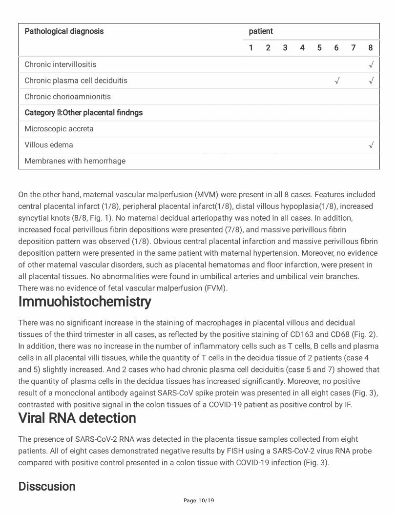
Pathological �ndings in the placentas of the pregnantwomen with SARS-CoV-2 infectionAll cases were intact placenta. In general, 8 cases of intact placenta tissues were all sponge-like and darkred, grossly normal from the appearance. Under the microscopic examination of the placental disc, asshown in Table 2, only 1 case (case 7) showed edema in the villous stroma and chronic intervillositis, 2cases showed chronic plasma cell deciduitis, which was the feature of chronic in�ammation. In addition,2 cases showed maternal in�ltrating in�ammatory cells in the subchorionic �brin, but there was noevidence of acute villitis. As for the ascending intrauterine infection, placenta membrane examinationshowed that only 2 cases (case 4 and 5) presented neutrophils in�ltration (more than 30 neutrophils perhigh-power �eld) in the �brin-deposited fetal membrane tissues. However, neutrophils in�ltration waslimited to the �brin under the chorionic lamina or the decidual layer of the fetal membrane, showing acutechorioamnionitis, maternal in�ammatory response, stage 1 (acute chorionitis). Above of all, no obviousin�ammatory response was noted in the placenta of the pregnant women with SARS-CoV-2 infection.

Page 9/19
Table 2Pathological examination of the placental samples of the pregnant women with COVID-19 pneumoniaPathological diagnosis patient
1 2 3 4 5 6 7 8
Category I: Maternal vascular malperfusion
Central placental infarct(s) √
Peripheral placental infarct √
Distal villous hypoplasia √
Accelerated villous maturation pattern
Increased syncytial knots √ √ √ √ √ √ √ √
Villous agglutination
Category :maternal decidual arteriopathy
Insu�cient vessel remodelling
Fibrinoid necrosis
Category :Fetal vascular malperfusion(FVM)
Avascular �brotic villi
Thrombosis
Intramural �brin deposition
Villous stromal-vascular karyorrhexis
Stem villous vascular obliteration
High-grade fetal vascular malperfusion
Category :Ascending intrauterine infection
Maternal in�ammatory response (mild) √ √
Fetal in�ammatory response
Category :Fibrinoid
Increased focal perivillous �brin depositions(perivillous �brin plaque)
√ √ √ √ √ √ √
Massive perivillous �brin deposition pattern √
Maternal �oor infarct pattern
Category :Chronic in�ammation
Page 10/19
Pathological diagnosis patient
1 2 3 4 5 6 7 8
Chronic intervillositis √
Chronic plasma cell deciduitis √ √
Chronic chorioamnionitis
Category :Other placental �ndngs
Microscopic accreta
Villous edema √
Membranes with hemorrhage
On the other hand, maternal vascular malperfusion (MVM) were present in all 8 cases. Features includedcentral placental infarct (1/8), peripheral placental infarct(1/8), distal villous hypoplasia(1/8), increasedsyncytial knots (8/8, Fig. 1). No maternal decidual arteriopathy was noted in all cases. In addition,increased focal perivillous �brin depositions were presented (7/8), and massive perivillous �brindeposition pattern was observed (1/8). Obvious central placental infarction and massive perivillous �brindeposition pattern were presented in the same patient with maternal hypertension. Moreover, no evidenceof other maternal vascular disorders, such as placental hematomas and �oor infarction, were present inall placental tissues. No abnormalities were found in umbilical arteries and umbilical vein branches.There was no evidence of fetal vascular malperfusion (FVM).
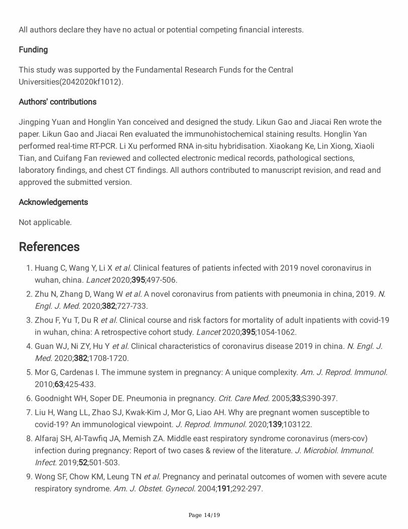
ImmuohistochemistryThere was no signi�cant increase in the staining of macrophages in placental villous and decidualtissues of the third trimester in all cases, as re�ected by the positive staining of CD163 and CD68 (Fig. 2).In addition, there was no increase in the number of in�ammatory cells such as T cells, B cells and plasmacells in all placental villi tissues, while the quantity of T cells in the decidua tissue of 2 patients (case 4and 5) slightly increased. And 2 cases who had chronic plasma cell deciduitis (case 5 and 7) showed thatthe quantity of plasma cells in the decidua tissues has increased signi�cantly. Moreover, no positiveresult of a monoclonal antibody against SARS-CoV spike protein was presented in all eight cases (Fig. 3),contrasted with positive signal in the colon tissues of a COVID-19 patient as positive control by IF.
Viral RNA detectionThe presence of SARS-CoV-2 RNA was detected in the placenta tissue samples collected from eightpatients. All of eight cases demonstrated negative results by FISH using a SARS-CoV-2 virus RNA probecompared with positive control presented in a colon tissue with COVID-19 infection (Fig. 3).
Disscusion

Page 11/19
This study retrospectively analyzed the clinical characteristics of 8 cases of COVID-19 pregnant womenin third trimester. The limited data showed that the clinical manifestations of SARS-CoV-2 infectedpregnant women were basically similar to those of the general infected population, and there were noserious adverse mother-infant outcomes. As far as we know, for the �rst time, we performed microscopicobservations and immunohistochemical tests at the same time to determine whether the number ofin�ammatory cells and fetal-derived placental macrophages (Hofbauer cells) in the placenta frompregnant women with COVID-19 has increased. No speci�c in�ammatory pathological changessuggesting SARS-CoV-2 invasion of the placenta were present through the microscopic observation andIHC detection. FISH detection of SARS-CoV-2 RNA in placental tissues and RT-PCR detection of neonatalpharyngeal swabs in all cases were negative. This study suggested no de�nite evidence pointing tomaternal-fetal vertical transmission in pregnant women with COVID-19 in late pregnancy, and providedimportant clues for further understanding of the clinical characteristics, pregnancy outcomes, andevaluation of intrauterine transmission of SARS-CoV-2 infection in late pregnancy.
The main histopathological features of placenta viral infection showed signi�cant fetal originin�ammatory abnormalities, such as chronic villitis, intervillositis, and funisitis, which occurred in someTORCH agents17, such as cytomegalovirus, Treponema pallidum, Toxoplasma, rubella virus infection andother hematogenously transmitted infections through the placenta18. Fortunately, in our study, no speci�cpathological changes of in�ammatory reactions and no evidence of worse maternal disease werepresent. Although individual cases showed corresponding in�ammation changes in the placental tissue,such as maternal in�ammatory response (mild), chronic intervillositis and chronic plasma cell deciduitis,they were not universal. This phenomenon was consistent with the results of the existing limited studies19, 20. In addition, we were the �rst to perform IHC analysis on in�ammatory cells, especially Hofbauercells, in late placenta tissues from COVID-19 pregnant women. As Hofbauer cells can harbor live virussuch as ZIKA virus13, HIV virus21 and Cytomegalo virus22, and serve as reservoirs within the placenta, it isone of the important ways to transmit pathogens to fetal-placenta tissues by infecting Hofbauercells23,24. However, H&E staining and IHC showed no signi�cant in�ltration of T cell or evidence of villousstomal macrophages hyperplasia. FISH analysis further enhanced the evidence that no virus directlyinfected the placenta, which was similar to the results of previous limited studies19, 25.
On the other hand, the stage of gestation at the time of infection may affect whether SARS-CoV-2 viruswas vertical transmission. Stage of gestation has been proved as an important factor affecting themechanisms of maternal-fetal vertical transmission 26. For example, in the early infection of rubella virus,more than 50% of fetuses were infected vertically through the uterus, but as the pregnancy timeincreases, the risk of vertical transmission was signi�cantly reduced27. The phenomenon was alsopresent in the ZIKA virus. Since higher ZIKA virus titers were detected in amniotic epithelial cells from mid-gestation, suggesting a greater susceptibility of virus infection in the placenta from the second trimesteror earlier compared to late-gestation placentas10. As with previous studies19, 28, 29, our study mainlyincluded pregnancy women with infection in the third trimester and found no no evidence of verticaltransmission, further suggesting that the placenta may play a greater and powerful barrier role to prevent

Page 12/19
SARS–CoV2 infection in the third trimester, and the speci�c resistance mechanism still needs to befurther studied.
Although the defense mechanism of placenta to restrict microorganisms from entering the fetus is largelyunclear, existing evidence suggested that syncytiotrophoblasts can effectively resist numerouspathogens, and CTB also has an innate defense mechanism against intracellular pathogens10.Impressively, the syncytiotrophoblast layer has strong resistance to various viruses such as HCMV, HSV1,and ZIKA30–32 in the late pregnancy. For example, trophoblasts are sensitive to ZIKA virus at the earlieststage of trophoblast development, but become more and more resistant when the syncytium forms in latepregnancy33. So whether trophoblasts play a part in the mechanism of placental resistance of the SARS–CoV2 virus in late pregnancy will be the direction of our further research.
Notably, another most striking observation in the placentas (all 8 cases) was the prominent and diffuseincrease of syncytial knots, which was one of the features of MVM. As �rst described by Tenney andParker 34, syncytial knots were the aggregations of syncytiotrophoblast nuclei, and their increase mayinvolve nearly all terminal villi in preeclampsia, whereas they were only appeared in 10%-15% normalterminal villi 35. Moreover, exposure of the placenta to conditions such as hypoxia, hyperoxemia, oroxidative stress may cause an increase in syncytial knots 36. And our results were consistent with theexisting evidence on the pathology of placentas with coronavirus infection, which exhibited a fewabnormalities about MVM19, 37, such as increased syncytial knots, different degrees of �brin deposition inintervillous and subchorion, which could also be observed in this study. Given that all cases collected inthis study were asymptomatic or with mild syndrome, so the results suggested that mild symptoms ofSARS-CoV-2 infection might induce the decline in oxygenation within the intervillous space and cause adegree of placental injury, although there was no clear evidence of SARS-CoV-2 infection of the placentain the third trimester. This is of great signi�cance to the safety of mothers and fetuses in late pregnancy.
Consistent with a recent case report11, FISH was performed to detect SARS-CoV-2 RNA in the placenta,and no evidence of SARS-CoV-2 invasion in the late gestation placenta was present. None havedemonstrated the presence of the SARS-CoV-2 virus by RT-PCR from existing limited studies in theplacenta tissue 19. Although the recent case report suggested the presence of SARS-CoV-2 in 3/11 swabsof the placenta or membrane by RT-PCR 38, swab samples rather than tissue samples of the placenta ormembranes might increase the possibility of virus droplet contamination in the hospital environment orvirus exposure during delivery, so they could not be used as direct evidence of vertical transmission.Compared with RT-PCR, FISH analysis directly used tissue samples for detection, which displayed theprecise cell location of fusion genes and relevant information on the anatomical distribution of theplacenta23, and helped to provide clues for exploring the mechanism of placental virus infection ordefense. Above all, it can be seen that FISH is practicable and can provide more information to diagnosisSARS-CoV-2 invasion of the placenta.

Page 13/19
This study still has some limitations. First of all, the cases collected in this study were all mild patients,and it was still unknown whether patients with severe infections in pregnancy will develop intrauterineinfection, which is the direction for further research in later research. Secondly, a recent report suggestedthat positive SARS-CoV-2 infection in the second trimester pregnancy women can lead to miscarriage,and the evidence of SARS-CoV-2 infection in the placenta had aslo been found 11. So further casesincluding different gestation stage women of COVID-19, especially in the �rst and second trimester, needto be collected to study the effect to maternal and fetus safety.
In summary, we found no evidence of vertical transmission in the third trimester placenta of COVID-19pregnancy women by observing histological changes and nucleic acid test, we also analyzed whether thenumber of the in�ammatory cells and macrophages cells increased by immunohistochemistry. Althoughthe sample size of this study was limited, considering the important adverse effects of this ongoingglobal public health emergency, our results were very useful for understanding the clinical characteristicsof COVID-19 infection in late-stage pregnant women and whether it has the potential for verticaltransmission. It was important and provided a certain basis for the best clinical management of latepregnant women.
AbbreviationsCoronavirus: CoV; 2019 novel coronavirus (CoV) disease: COVID-19; Severe acute respiratory syndrome:SARS; Fluorescence In-Situ Hybridization: FISH; Immuno�uorescence: IF; Maternal vascular malperfusion:MVM; Severe acute respiratory syndrome coronavirus 2: SARS-CoV-2; Middle East respiratory syndrome:MERS; Real-time reverse transcription polymerase chain reaction: RT-PCR; Immunohistochemistry: IHC;Formalin-�xed, para�n-embedded: FFPE; Alanine aminotransaminase: ALT; Aspartate aminotransferase:AST; Fetal vascular malperfusion: FVM
DeclarationsEthics approval and consent to participate
This study was approved by the Ethical Committee of Renmin Hospital of Wuhan University (WDRY2020-K201). The written informed consents were obtained from all the patients.
Consent for publication
The parents of patient agreed to publication of this case.
Availability of data and materials
All data generated or analysed during this study are included in this published article.
Competing interests
Page 14/19
All authors declare they have no actual or potential competing �nancial interests.
Funding
This study was supported by the Fundamental Research Funds for the CentralUniversities(2042020kf1012).
Authors' contributions
Jingping Yuan and Honglin Yan conceived and designed the study. Likun Gao and Jiacai Ren wrote thepaper. Likun Gao and Jiacai Ren evaluated the immunohistochemical staining results. Honglin Yanperformed real-time RT-PCR. Li Xu performed RNA in-situ hybridisation. Xiaokang Ke, Lin Xiong, XiaoliTian, and Cuifang Fan reviewed and collected electronic medical records, pathological sections,laboratory �ndings, and chest CT �ndings. All authors contributed to manuscript revision, and read andapproved the submitted version.
Acknowledgements
Not applicable.
References1. Huang C, Wang Y, Li X et al. Clinical features of patients infected with 2019 novel coronavirus in
wuhan, china. Lancet 2020;395;497-506.
2. Zhu N, Zhang D, Wang W et al. A novel coronavirus from patients with pneumonia in china, 2019. N.Engl. J. Med. 2020;382;727-733.
3. Zhou F, Yu T, Du R et al. Clinical course and risk factors for mortality of adult inpatients with covid-19in wuhan, china: A retrospective cohort study. Lancet 2020;395;1054-1062.
4. Guan WJ, Ni ZY, Hu Y et al. Clinical characteristics of coronavirus disease 2019 in china. N. Engl. J.Med. 2020;382;1708-1720.
5. Mor G, Cardenas I. The immune system in pregnancy: A unique complexity. Am. J. Reprod. Immunol.2010;63;425-433.
�. Goodnight WH, Soper DE. Pneumonia in pregnancy. Crit. Care Med. 2005;33;S390-397.
7. Liu H, Wang LL, Zhao SJ, Kwak-Kim J, Mor G, Liao AH. Why are pregnant women susceptible tocovid-19? An immunological viewpoint. J. Reprod. Immunol. 2020;139;103122.
�. Alfaraj SH, Al-Taw�q JA, Memish ZA. Middle east respiratory syndrome coronavirus (mers-cov)infection during pregnancy: Report of two cases & review of the literature. J. Microbiol. Immunol.Infect. 2019;52;501-503.
9. Wong SF, Chow KM, Leung TN et al. Pregnancy and perinatal outcomes of women with severe acuterespiratory syndrome. Am. J. Obstet. Gynecol. 2004;191;292-297.

Page 15/19
10. Arora N, Sadovsky Y, Dermody TS, Coyne CB. Microbial vertical transmission during humanpregnancy. Cell Host Microbe 2017;21;561-567.
11. Hosier H, Farhadian SF, Morotti RA et al. Sars-cov-2 infection of the placenta. J. Clin. Invest. 2020.
12. Heerema-McKenney A. Defense and infection of the human placenta. APMIS 2018;126;570-588.
13. Rosenberg AZ, Yu W, Hill DA, Reyes CA, Schwartz DA. Placental pathology of zika virus: Viralinfection of the placenta induces villous stromal macrophage (hofbauer cell) proliferation andhyperplasia. Arch. Pathol. Lab. Med. 2017;141;43-48.
14. Bayer A, Delorme-Axford E, Sleigher C et al. Human trophoblasts confer resistance to virusesimplicated in perinatal infection. Am. J. Obstet. Gynecol. 2015;212;71 e71-71 e78.
15. Cardenas I, Means RE, Aldo P et al. Viral infection of the placenta leads to fetal in�ammation andsensitization to bacterial products predisposing to preterm labor. J. Immunol. 2010;185;1248-1257.
1�. Liu YL, Ren J, Yuan JP et al. Postoperative onset and detection of SARS-CoV-2 in surgically resectedspecimens from gastrointestinal cancer patients with pre/asymptomatic COVID-19. Ann. Surg.[published online ahead of print, 2020 Oct 14]. doi:10.1097/SLA.0000000000004362.
17. Costa ML, de Moraes Nobrega G, Antolini-Tavares A. Key infections in the placenta. Obstet. Gynecol.Clin. North Am. 2020;47;133-146.
1�. Robbins JR, Bakardjiev AI. Pathogens and the placental fortress. Curr. Opin. Microbiol. 2012;15;36-43.
19. Chen S, Huang B, Luo DJ et al. [pregnant women with new coronavirus infection: A clinicalcharacteristics and placental pathological analysis of three cases]. Zhonghua Bing Li Xue Za Zhi2020;49;E005.
20. Shanes ED, Mithal LB, Otero S, Azad HA, Miller ES, Goldstein JA. Placental pathology in covid-19. Am.J. Clin. Pathol. 2020;154;23-32.
21. Villegas-Castrejon H, Paredes-Vivas Y, Flores-Rivera E, Gorbea-Robles MC, Arredondo-Garcia JL.[comparative study of the placenta from hiv+ mothers. Ultrastructural analysis]. Ginecol. Obstet.Mex. 1996;64;167-176.
22. Schwartz DA, Khan R, Stoll B. Characterization of the fetal in�ammatory response tocytomegalovirus placentitis. An immunohistochemical study. Arch. Pathol. Lab. Med. 1992;116;21-27.
23. Schwartz DA. Viral infection, proliferation, and hyperplasia of hofbauer cells and absence ofin�ammation characterize the placental pathology of fetuses with congenital zika virus infection.Arch. Gynecol. Obstet. 2017;295;1361-1368.
24. Reyes L, Golos TG. Hofbauer cells: Their role in healthy and complicated pregnancy. Front. Immunol.2018;9;2628.
25. Schwartz DA. An analysis of 38 pregnant women with covid-19, their newborn infants, and maternal-fetal transmission of sars-cov-2: Maternal coronavirus infections and pregnancy outcomes. Arch.Pathol. Lab. Med. 2020.

Page 16/19
2�. Langel SN, Paim FC, Alhamo MA et al. Stage of gestation at porcine epidemic diarrhea virus infectionof pregnant swine impacts maternal immunity and lactogenic immune protection of neonatalsuckling piglets. Front. Immunol. 2019;10;727.
27. Bouthry E, Picone O, Hamdi G, Grangeot-Keros L, Ayoubi JM, Vauloup-Fellous C. Rubella andpregnancy: Diagnosis, management and outcomes. Prenat. Diagn. 2014;34;1246-1253.
2�. Chen H, Guo J, Wang C et al. Clinical characteristics and intrauterine vertical transmission potentialof covid-19 infection in nine pregnant women: A retrospective review of medical records. Lancet2020;395;809-815.
29. Karimi-Zarchi M, Neamatzadeh H, Dastgheib SA et al. Vertical transmission of coronavirus disease19 (covid-19) from infected pregnant mothers to neonates: A review. Fetal Pediatr. Pathol. 2020;1-5.
30. Maidji E, Nigro G, Tabata T et al. Antibody treatment promotes compensation for humancytomegalovirus-induced pathogenesis and a hypoxia-like condition in placentas with congenitalinfection. Am. J. Pathol. 2010;177;1298-1310.
31. Delorme-Axford E, Donker RB, Mouillet JF et al. Human placental trophoblasts confer viral resistanceto recipient cells. Proc. Natl. Acad. Sci. U. S. A. 2013;110;12048-12053.
32. Bayer A, Lennemann NJ, Ouyang Y et al. Type iii interferons produced by human placentaltrophoblasts confer protection against zika virus infection. Cell Host Microbe 2016;19;705-712.
33. Sheridan MA, Yunusov D, Balaraman V et al. Vulnerability of primitive human placental trophoblastto zika virus. Proc. Natl. Acad. Sci. U. S. A. 2017;114;E1587-E1596.
34. Fogarty NM, Ferguson-Smith AC, Burton GJ. Syncytial knots (tenney-parker changes) in the humanplacenta: Evidence of loss of transcriptional activity and oxidative damage. Am. J. Pathol.2013;183;144-152.
35. Loukeris K, Sela R, Baergen RN. Syncytial knots as a re�ection of placental maturity: Referencevalues for 20 to 40 weeks' gestational age. Pediatr. Dev. Pathol. 2010;13;305-309.
3�. Heazell AE, Moll SJ, Jones CJ, Baker PN, Crocker IP. Formation of syncytial knots is increased byhyperoxia, hypoxia and reactive oxygen species. Placenta 2007;28 Suppl A;S33-40.
37. Ng WF, Wong SF, Lam A et al. The placentas of patients with severe acute respiratory syndrome: Apathophysiological evaluation. Pathology 2006;38;210-218.
3�. Pen�eld CA, Brubaker SG, Limaye MA et al. Detection of sars-cov-2 in placental and fetal membranesamples. Am J Obstet Gynecol MFM 2020;100133.
Figures
Page 17/19
Figure 1
Microscopy of the placentas. A, Low power view with the chorionic plate of case 3 (39+1 weeksgestation). Increased focal perivillous �brin depositions and increased syncytial knots were presented(H&E, original magni�cations ×100). B, Close up view of increased syncytial knots in the terminal villi withthe chorionic plate of case 3 (H&E, original magni�cations ×200). C, Low power view with the chorionicplate of case 2 (39+5 weeks gestation). Central placental infarct was presented (H&E, originalmagni�cations ×40). D, Low power view with the chorionic plate of case 8 (37+6 weeks gestation). Distalvillous hypoplasia was presented (H&E, original magni�cations ×100).
Page 18/19
Figure 2
Immunohistochemical staining of in�ammatory cells and Hofbauer cells. A and B, CD3 (A) and CD20 (B)staining revealed only occasional in�ltration of T lymphocytes and B lymphocytes in the middle ofterminal villi (Envision, Original magni�cations ×200). C and D, CD163 (C) and CD68 (D) staining revealednone prominently increased numbers of Hofbauer cells present in the stroma of all villi (Envision, originalmagni�cations ×200)

Page 19/19
Figure 3
IF and FISH double staining result of SARS-CoV spike protein(green) and SARS-CoV-2 RNA (red) in alleight cases and positive control. A, Original magni�cations ×200) and B, Original magni�cations ×400):no positive signal was presented of monoclonal antibody against SARS-CoV spike protein(green) andSARS-CoV-2 RNA (red) in all eight cases by IF and FISH double staining. C, Original magni�cations ×200)and D, Original magni�cations ×400): both of positive signal were presented in a colon tissue with COVID-19 infection as positive control.