women issues and thrombosis
TRANSCRIPT

Women Issues and Thrombosis
Sabine Eichinger
Dept. of Medicine IMedical University of Vienna/Austria
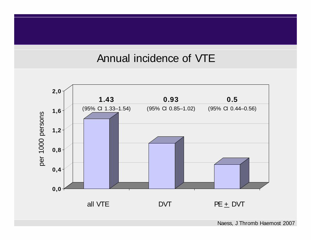
Annual incidence of VTE
0,0
0,4
0,8
1,2
1,6
2,0
DVT PE + DVTall VTE
Naess, J Thromb Haemost 2007
1.43(95% CI 1.33–1.54)
0.93(95% CI 0.85–1.02)
0.5(95% CI 0.44–0.56)
per
1000
per
sons
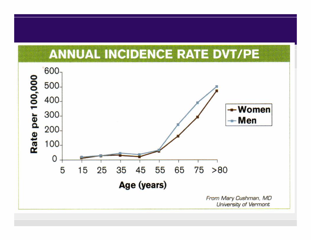

Risk conditions / risk factors of VTEAdvancing age
Obesity
Previous venous thromboembolism
Surgery
Trauma
Active cancer
Acute medical illnesses—eg, acute myocardial infarction,
Heart failure, respiratory failure, infection
Inflammatory bowel disease
Antiphospholipid syndrome
Dyslipoproteinaemia
Nephrotic syndrome
Paroxysmal nocturnal haemoglobinuria
Myeloproliferative diseases
Behçet’s syndrome
Varicose veins
Superficial vein thrombosis
Congenital venous malformation
Long-distance travel
Prolonged bed rest
Immobilisation
Limb paresis
Chronic care facility stay
Pregnancy/puerperium
Hormone contraceptives
Hormone replacement therapy
Heparin-induced thrombocytopenia
Other drugs
Chemotherapy
Tamoxifen
Thalidomide
Antipsychotics
Central venous catheter
Vena cava filter
Intravenous drug abuse
Factor V Leiden
Factor II G20210A
Natural inhibitor deficiency
High factor VIII, factor IX, or factor XI
Lupus anticoagulant
High thrombin activatable fibrinolysis inhibitor
Hyperhomocysteinaemia
Dysfibrinogenaemia or hyperfibrinogenaemia
Plasminogen deficiency
from Kyrle & Eichinger, Lancet 2005
• Sex related differences in hemostasis
Women issues and thrombosis

Coagulation factors in women compared to men
Sex-related differences
FibrinogenF VII, VIII, IX
Lowe, Br J Haematol 1997
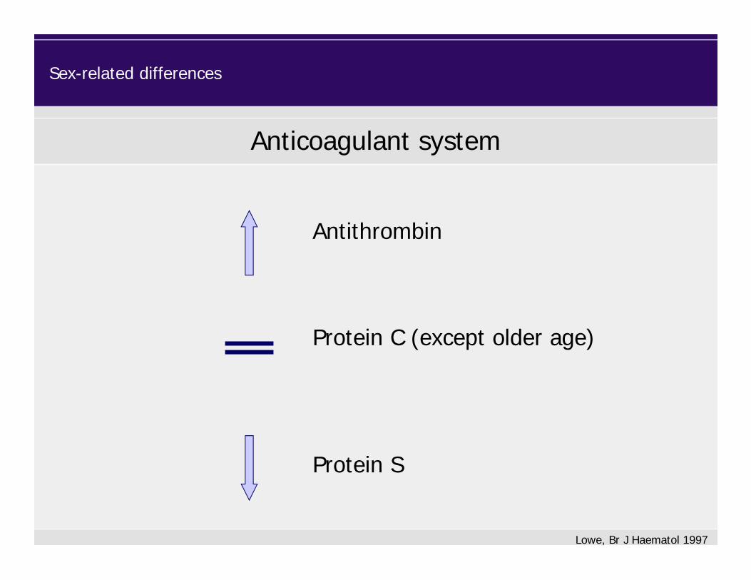
Anticoagulant system
Antithrombin
Protein S
Protein C (except older age)
Sex-related differences
Lowe, Br J Haematol 1997
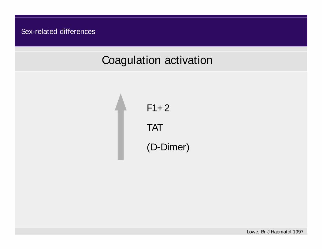
F1+2
TAT
(D-Dimer)
Coagulation activation
Sex-related differences
Lowe, Br J Haematol 1997

• Sex related differences in hemostasis
• Specific hormone-related issues– Pregnancy
Women issues and thrombosis
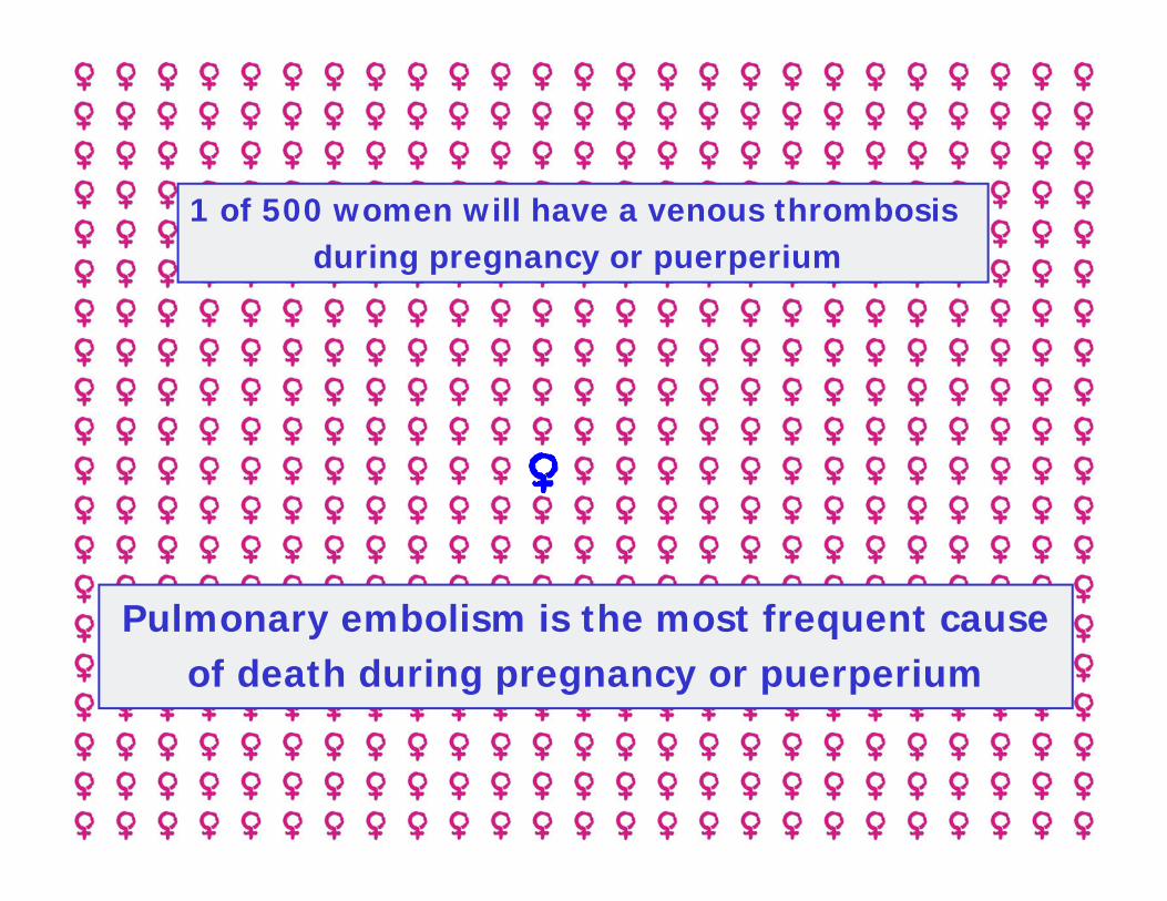
Pulmonary embolism is the most frequent cause of death during pregnancy or puerperium
1 of 500 women will have a venous thrombosis during pregnancy or puerperium

Altered Rheology
Pregnancy
Vascular Injury
Altered Hemostasis
Prothrombotic State
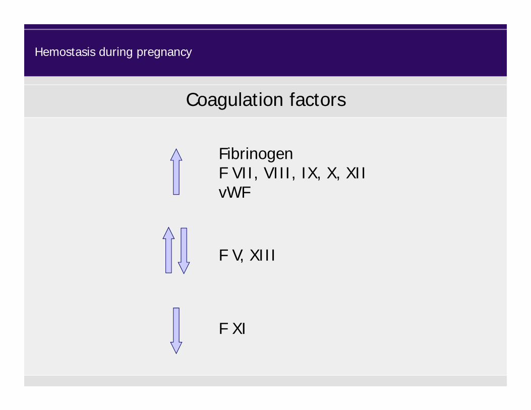
Coagulation factors
Hemostasis during pregnancy
FibrinogenF VII, VIII, IX, X, XIIvWF
F V, XIII
F XI

Anticoagulant system
Hemostasis during pregnancy
ThrombomodulinTFPI
Protein SAPC-ratio
Protein C Antithrombin

Fibrinolytic system
Hemostasis during pregnancy
PlasminogentPA
PAI-1PAI-2
TAFI

F1+2
TAT
FPA
Soluble fibrin
D-Dimer
Plasmin-antiplasmin complexes
Coagulation activation
Increased fibrin generation Increased fibrinolysis
Hemostasis during pregnancy
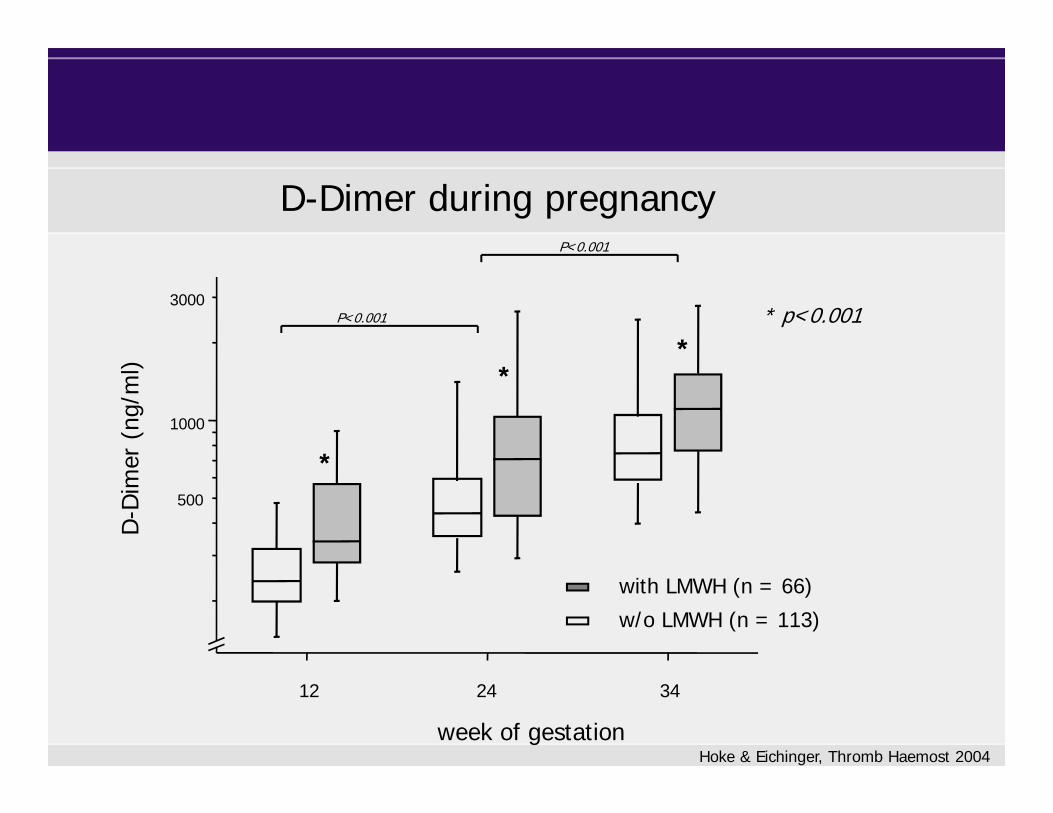
12 24 34
week of gestation
1000
3000
500
D-D
imer
(ng
/ml)
*
**
P<0.001
P<0.001
with LMWH (n = 66)
w/o LMWH (n = 113)
* p<0.001
Hoke & Eichinger, Thromb Haemost 2004
D-Dimer during pregnancy
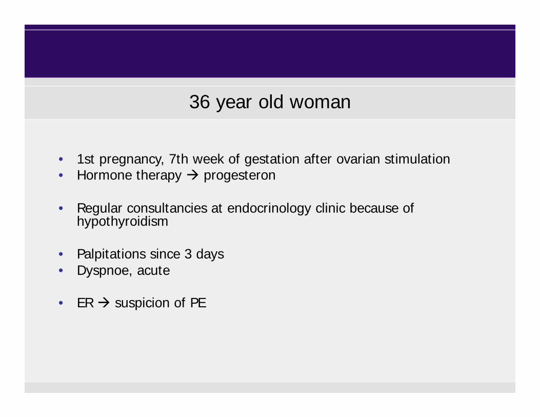
36 year old woman
• 1st pregnancy, 7th week of gestation after ovarian stimulation • Hormone therapy progesteron
• Regular consultancies at endocrinology clinic because of hypothyroidism
• Palpitations since 3 days• Dyspnoe, acute
• ER suspicion of PE

36 year old woman
• Otherwise healthy • No previous VTE• Father PE, FV Leiden heterozygous
• Patient‘s thrombophilia screen normal• Heart rate 101/min• ECG: normal

Approach to a patient with suspicion of VTE
Clinic ImagingLab
Diagnostic issues of VTE in pregnancy
Not validated for pregnant women
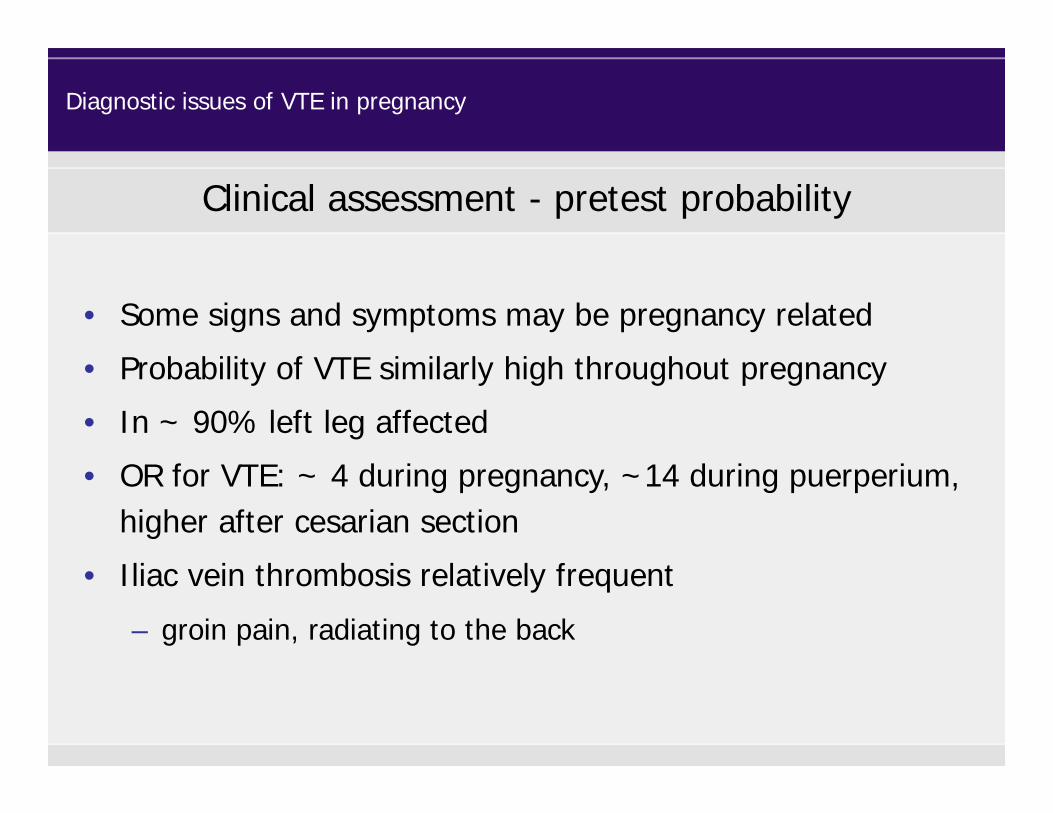
• Some signs and symptoms may be pregnancy related
• Probability of VTE similarly high throughout pregnancy
• In ~ 90% left leg affected
• OR for VTE: ~ 4 during pregnancy, ~14 during puerperium, higher after cesarian section
• Iliac vein thrombosis relatively frequent
– groin pain, radiating to the back
Clinical assessment - pretest probability
Diagnostic issues of VTE in pregnancy
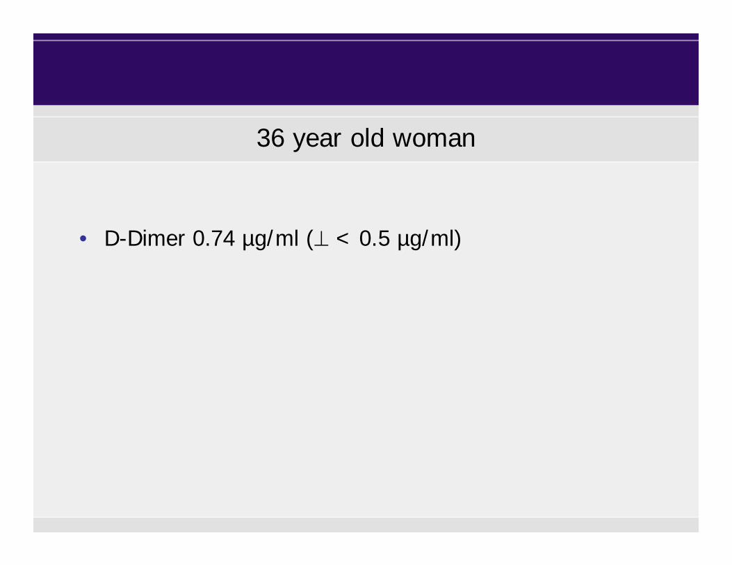
• D-Dimer 0.74 µg/ml ( < 0.5 µg/ml)
36 year old woman

Week of gestation Patients, n 95% CI
<12 0 (0%) 0 - 60
13-28 12 (24%) 14 - 37
>28 41 (51%) 40 - 61
Chan W, Ann Intern Med 2007
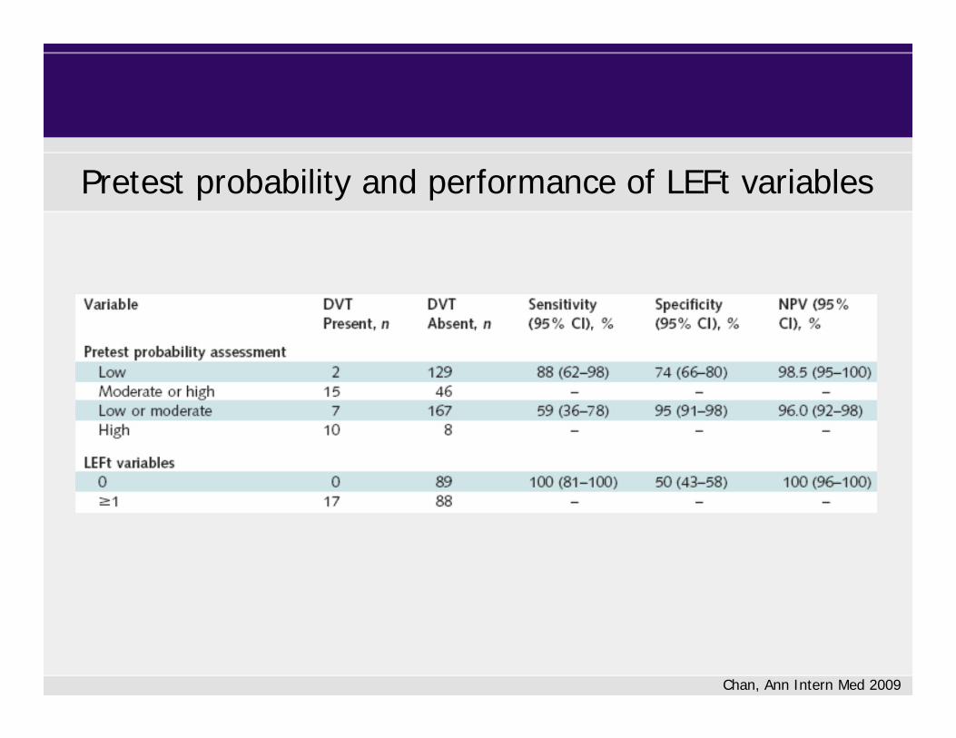
Positive D-Dimer result in pregnant women
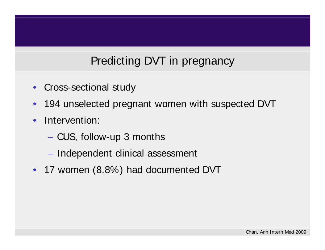
• Cross-sectional study
• 194 unselected pregnant women with suspected DVT
• Intervention:
– CUS, follow-up 3 months
– Independent clinical assessment
• 17 women (8.8%) had documented DVT
Chan, Ann Intern Med 2009
Predicting DVT in pregnancy

Chan, Ann Intern Med 2009
Potential predictive variables for DVT in pregnancy
Chan, Ann Intern Med 2009
Pretest probability and performance of LEFt variables

Approach to a woman with suspicion of VTE
Imaging
Diagnostic issues of VTE in pregnancy
DVTcompression ultrasoundphlebography
PEventilation/perfusion scancomputed tomography

Compression ultrasound to exclude DVT
Chan, CMAJ 2013
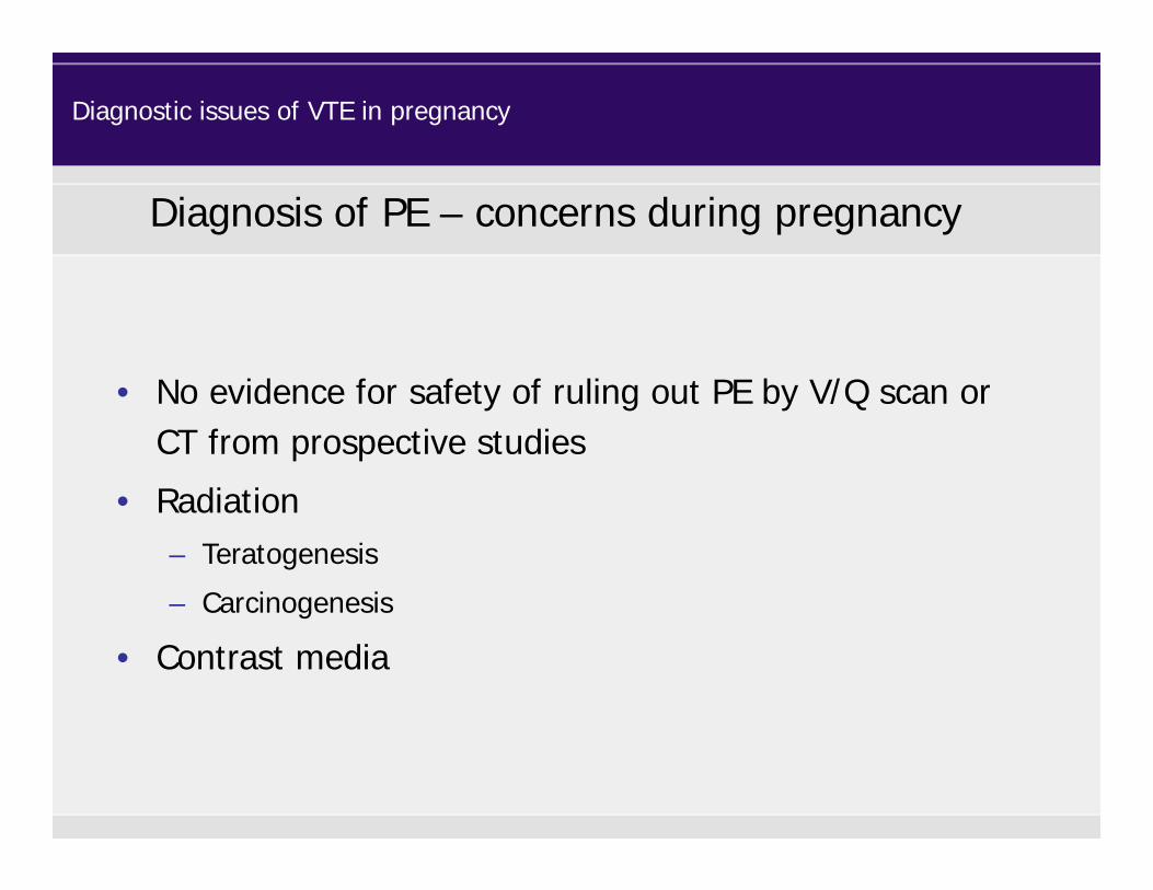
• No evidence for safety of ruling out PE by V/Q scan or CT from prospective studies
• Radiation– Teratogenesis
– Carcinogenesis
• Contrast media
Diagnosis of PE – concerns during pregnancy
Diagnostic issues of VTE in pregnancy

Radiation dose to the fetus
Radiation (mSv)
Unilateral phlebography without shielding 3.14
Unilateral phlebography with shielding < 0.5
Perfusion scintigraphy (99mTc MAA, 200 MBq) 0.2-0.6
Perfusion scintigraphy (99mTc MAA, 40 MBq) 0.11-0.2
Ventilation sintigraphy (99mTc MAA, aerosol) 0.1-0.3
Ventilation scintigraphy (81m Kr, 600 MBq) 0.0001
Single-detector row helical CT 0.026
Multi-detector row helical CT 0.013
Natural radiation exposure 3.8/a
Nijkeuter, J Thromb Haemost 2006
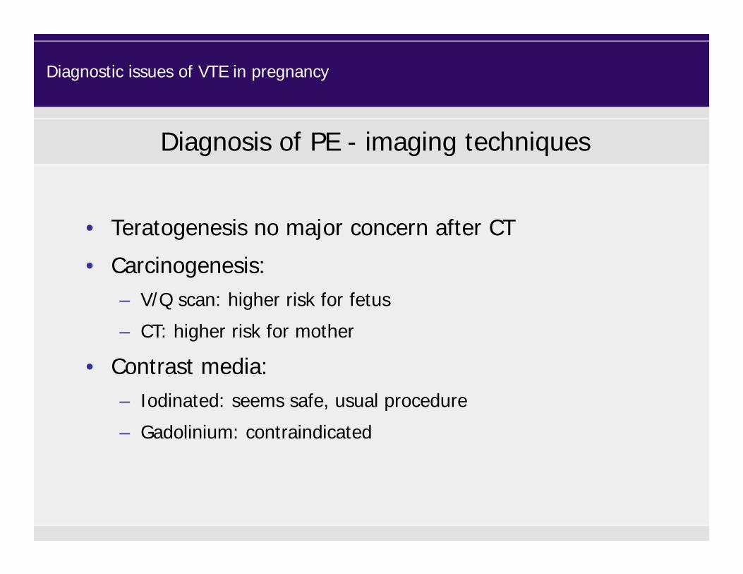
• Teratogenesis no major concern after CT
• Carcinogenesis: – V/Q scan: higher risk for fetus
– CT: higher risk for mother
• Contrast media:– Iodinated: seems safe, usual procedure
– Gadolinium: contraindicated
Diagnosis of PE - imaging techniques
Diagnostic issues of VTE in pregnancy

Suggested algorithm for exclusion of PE during pregnancy
Clinical Suspicion
CUS
no DVT DVT
treat
Multi-slice CT (+shielding) / VQ scan(consider clinic, risks, D-Dimer)
D-Dimer
Diagnostic issues of VTE in pregnancy
consider clinic, risks, D-Dimer

36 year old woman
• Week 7 proximal DVT left leg• LMWH at therapeutic dose (weight adjusted)
• Duration: throughout pregnancy until 6-8 wks after delivery
• LMWH dose reduction before delivery • Outpatient care possible
Treatment of VTE during pregnancy

Bates, Chest 2012
Assisted reproduction
• For women undergoing assisted reproduction, we
recommend against the use of routine thrombosis
prophylaxis (1B).
• For women undergoing assisted reproduction who develop
severe ovarian hyperstimulation syndrome, we suggest
thrombosis prophylaxis (prophylactic LMWH) for 3 months
postresolution of clinical ovarian hyperstimulation syndrome
rather than no prophylaxis (2C) .
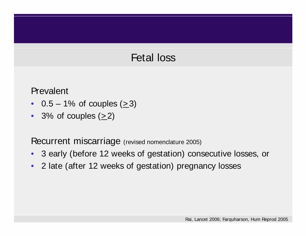
Fetal loss
Prevalent• 0.5 – 1% of couples (>3)• 3% of couples (>2)
Recurrent miscarriage (revised nomenclature 2005)
• 3 early (before 12 weeks of gestation) consecutive losses, or• 2 late (after 12 weeks of gestation) pregnancy losses
Rai, Lancet 2006; Farquharson, Hum Reprod 2005
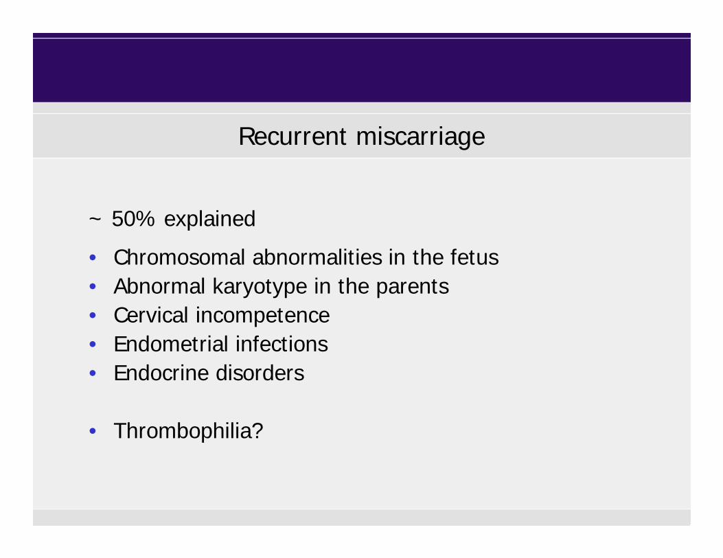
Recurrent miscarriage
~ 50% explained
• Chromosomal abnormalities in the fetus• Abnormal karyotype in the parents• Cervical incompetence• Endometrial infections• Endocrine disorders
• Thrombophilia?

Thrombophilia and pregnancy complications
Rey, Lancet 2003; Robertson, Br J Haematol 2006; Nelen, Fertil Steril 2000
Thrombophilia Sporadic miscarriage OR
Recurrent miscarriageOR
IU fetal deathOR
Lupus anticoagulant 3.0 7.8 2.4
Anticardiolipin antibodies 3.4 3.6 - 5.1 3.3
AT deficiency 1.5 0.97.6 (1.3 - 42.8)
20.1 (3.7 - 109.2)
PC deficiency 1.4 1.6 3.1
PS deficiency heterogeneous data 14.7 (1.0 - 218.0)7.4 (1.3 - 42.8)
20.1 (3.7 - 109.2)
Factor V Leiden 1.7 2.0 2.1 - 3.3
Prothrombin 20210A 2.1 2.3 - 2.7 2.3 - 2.7
Homozygous/ combined defects 2.7 - -
Hyperhomocysteinemia 6.3 2.7 - 4.2 1.0
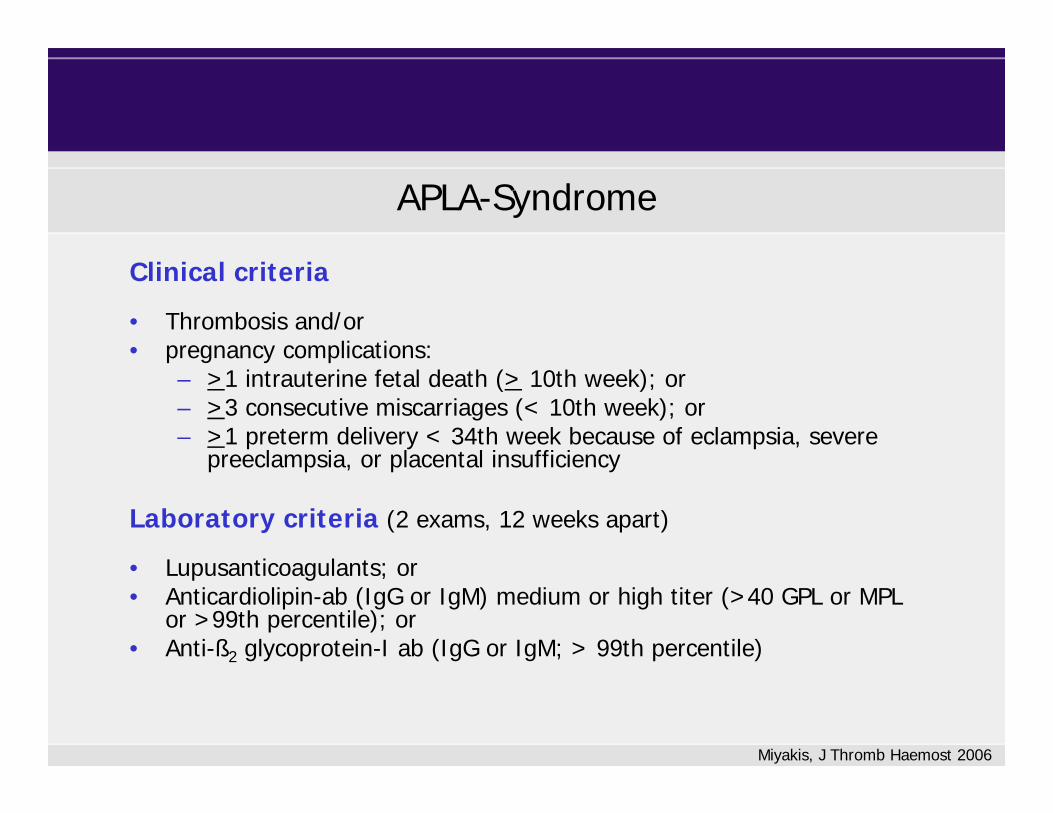
Clinical criteria
• Thrombosis and/or• pregnancy complications:
– >1 intrauterine fetal death (> 10th week); or– >3 consecutive miscarriages (< 10th week); or– >1 preterm delivery < 34th week because of eclampsia, severe
preeclampsia, or placental insufficiency
Laboratory criteria (2 exams, 12 weeks apart)
• Lupusanticoagulants; or • Anticardiolipin-ab (IgG or IgM) medium or high titer (>40 GPL or MPL
or >99th percentile); or • Anti-ß2 glycoprotein-I ab (IgG or IgM; > 99th percentile)
Miyakis, J Thromb Haemost 2006
APLA-Syndrome
Scenario 1: PLA +, previous miscarriage
Phospholipidantibodies during pregnancy

Relevance
LAC ACA ß2GP-AK
Recurrent pregnancy loss ++ + ?
Late pregnancy loss ++ + ?
Preeclampsia +/- +/- ?
Placental abruption +/- +/- ?
IUGR +/- +/- ?
Opatrny, J Rheumatol 2006
Phospholipidantibodies during pregnancy
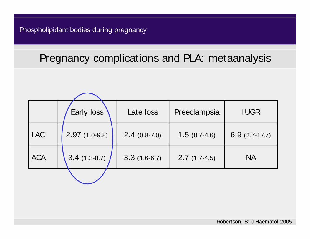
Pregnancy complications and PLA: metaanalysis
Early loss Late loss Preeclampsia IUGR
LAC 2.97 (1.0-9.8) 2.4 (0.8-7.0) 1.5 (0.7-4.6) 6.9 (2.7-17.7)
ACA 3.4 (1.3-8.7) 3.3 (1.6-6.7) 2.7 (1.7-4.5) NA
Robertson, Br J Haematol 2005
Phospholipidantibodies during pregnancy
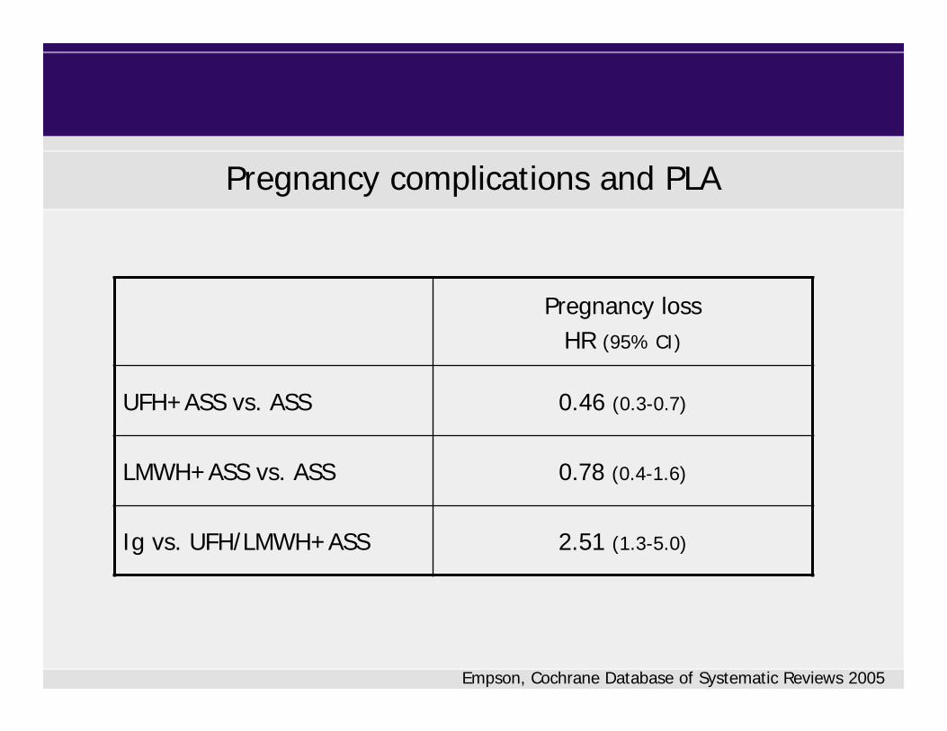
Pregnancy complications and PLA
Pregnancy lossHR (95% CI)
UFH+ASS vs. ASS 0.46 (0.3-0.7)
LMWH+ASS vs. ASS 0.78 (0.4-1.6)
Ig vs. UFH/LMWH+ASS 2.51 (1.3-5.0)
Empson, Cochrane Database of Systematic Reviews 2005
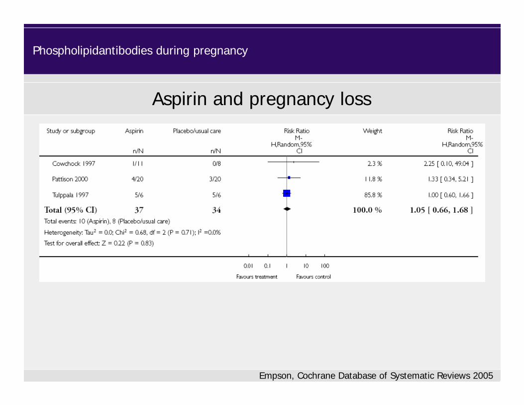
Empson, Cochrane Database of Systematic Reviews 2005
Aspirin and pregnancy loss
Phospholipidantibodies during pregnancy

Empson, Cochrane Database of Systematic Reviews 2005
Heparin and pregnancy loss
Phospholipidantibodies during pregnancy
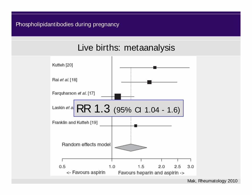
Mak, Rheumatology 2010
RR 1.3 (95% CI 1.04 - 1.6)
Live births: metaanalysis
Phospholipidantibodies during pregnancy

Recommendations
• For women who fulfill the laboratory criteria for PLA syndrome and meet the clinical PLA criteria based on a history of > 3 pregnancy losses, we recommend antepartum administration of prophylactic LMWH combined with low-dose aspirin, 75 to 100 mg/d, over no treatment (1B).
Bates, ACCP Guidelines, Chest 2012Keeling, Br J Haematol 2012
Phospholipidantibodies during pregnancy

ALIFE Study
Kaandorp, N Eng J Med 2010

SPIN Study
Clark, Blood 2010
Enoxaparin 40 mg + ASA 75 mg
Intensive surveillance
> 2 consecutive pregnancy losses 147 147
Pregnancy loss 32 (22%) 29 (20%)OR 0.91 (95% CI 0.5-1.6)

HAPPY Trial
Martinelli, Blood 2012
N=63
N=65
Recommendations
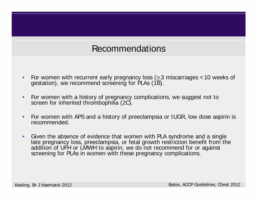
• For women with recurrent early pregnancy loss (>3 miscarriages <10 weeks of gestation), we recommend screening for PLAs (1B).
• For women with a history of pregnancy complications, we suggest not to screen for inherited thrombophilia (2C).
• For women with APS and a history of preeclampsia or IUGR, low dose aspirin is recommended.
• Given the absence of evidence that women with PLA syndrome and a single late pregnancy loss, preeclampsia, or fetal growth restriction benefit from the addition of UFH or LMWH to aspirin, we do not recommend for or against screening for PLAs in women with these pregnancy complications.
Bates, ACCP Guidelines, Chest 2012Keeling, Br J Haematol 2012
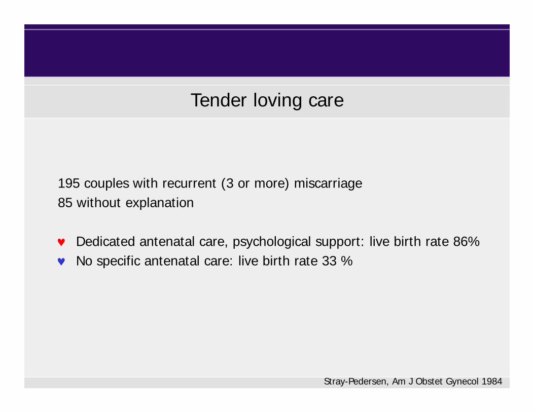
Tender loving care
195 couples with recurrent (3 or more) miscarriage 85 without explanation
Dedicated antenatal care, psychological support: live birth rate 86% No specific antenatal care: live birth rate 33 %
Stray-Pedersen, Am J Obstet Gynecol 1984

Scenario 2: PLA +, previous VTE
Phospholipidantibodies during pregnancy

Vitamin K antagonists and pregnancy
Warfarin-embryopathy- 6th to 9th (12th) gestational week- Dose dependent- Prevalence 5 – 7%- Bone and cartilage malformation
- CNS-defects (opticus atrophy, microcephaly, mental retardation), Pathomechanism unclear
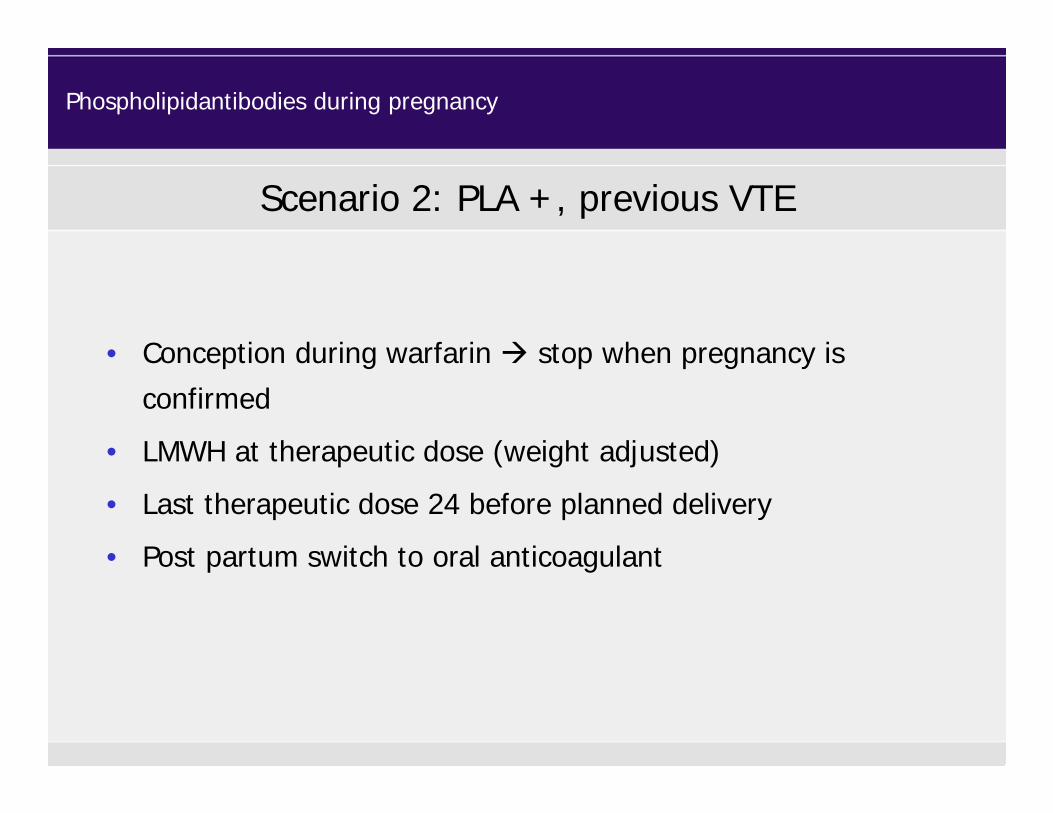
• Conception during warfarin stop when pregnancy is confirmed
• LMWH at therapeutic dose (weight adjusted)
• Last therapeutic dose 24 before planned delivery
• Post partum switch to oral anticoagulant
Scenario 2: PLA +, previous VTE
Phospholipidantibodies during pregnancy

• Sex related differences in hemostasis
• Specific hormone-related issues– Pregnancy
– Hormone use• Oral contraceptives
• Hormone replacement therapy
Women issues and thrombosis

Coagulation factors during oral contraceptives
FibrinogenF II, VII, VIII, X
Lowe, Br J Haematol 1997; Middeldorp, Thromb Haemost 2000
Hormone contraceptives
F V
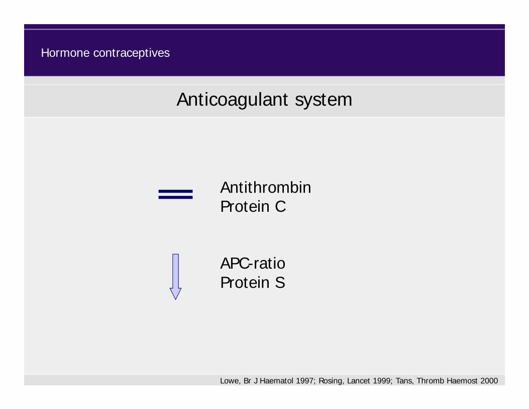
Anticoagulant system
APC-ratioProtein S
Hormone contraceptives
Lowe, Br J Haematol 1997; Rosing, Lancet 1999; Tans, Thromb Haemost 2000
AntithrombinProtein C

Estimated annual incidence of venous thrombosis
OC –
Vandenbroucke, Lancet 1994
all > 70 yrs OC +
1/100
1/10 000 3-4/10 000

Thrombosis Risk According to Duration of OC Use
Van Hylckama Vlieg, BMJ 2009
Multiple Environmental and Genetic Assessment of Risk Factors for Venous Thrombosis Study(MEGA)

Risk of venous thrombosis
Van Hylckama Vlieg, BMJ 2009
Multiple Environmental and Genetic Assessment of Risk Factors for Venous Thrombosis Study(MEGA)

Thrombosis risk according to oestrogen dose
Van Hylckama Vlieg, BMJ 2009
Multiple Environmental and Genetic Assessment of Risk Factors for Venous Thrombosis Study(MEGA)
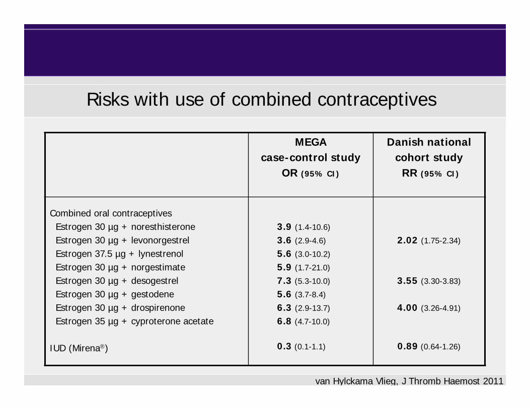
Risks with use of combined contraceptives
van Hylckama Vlieg, J Thromb Haemost 2011
MEGA case-control study
OR (95% CI)
Danish nationalcohort study
RR (95% CI)
Combined oral contraceptivesEstrogen 30 µg + noresthisteroneEstrogen 30 µg + levonorgestrelEstrogen 37.5 µg + lynestrenolEstrogen 30 µg + norgestimateEstrogen 30 µg + desogestrelEstrogen 30 µg + gestodeneEstrogen 30 µg + drospirenoneEstrogen 35 µg + cyproterone acetate
IUD (Mirena)
3.9 (1.4-10.6)
3.6 (2.9-4.6)
5.6 (3.0-10.2)
5.9 (1.7-21.0)
7.3 (5.3-10.0)
5.6 (3.7-8.4)
6.3 (2.9-13.7)
6.8 (4.7-10.0)
0.3 (0.1-1.1)
2.02 (1.75-2.34)
3.55 (3.30-3.83)
4.00 (3.26-4.91)
0.89 (0.64-1.26)
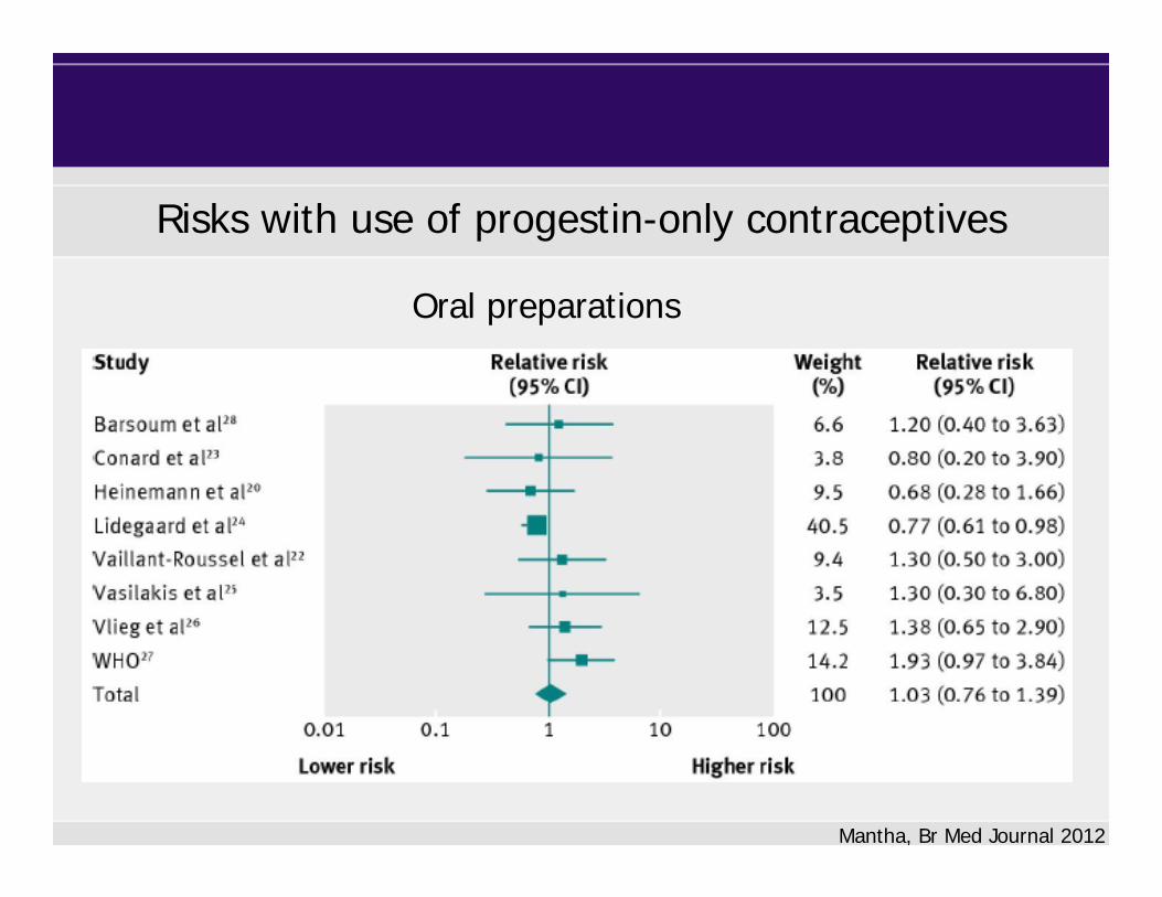
Risks with use of progestin-only contraceptives
Mantha, Br Med Journal 2012
Oral preparations

Risks with use of progestin-only contraceptives
Mantha, Br Med Journal 2012
Injectables

Hormone contraceptives and thrombotic risk
• Female hormone intake increases the thrombotic risk• Combined oral contraceptives:
– risk related to dose of estrogen and type of progestogen– safest option levonorgestrel combined with a low dose of
estrogen
• Progestogen-only contraception:– Limited data – Oral preparations considered as generally safe – No increased risk with IUD-Mirena
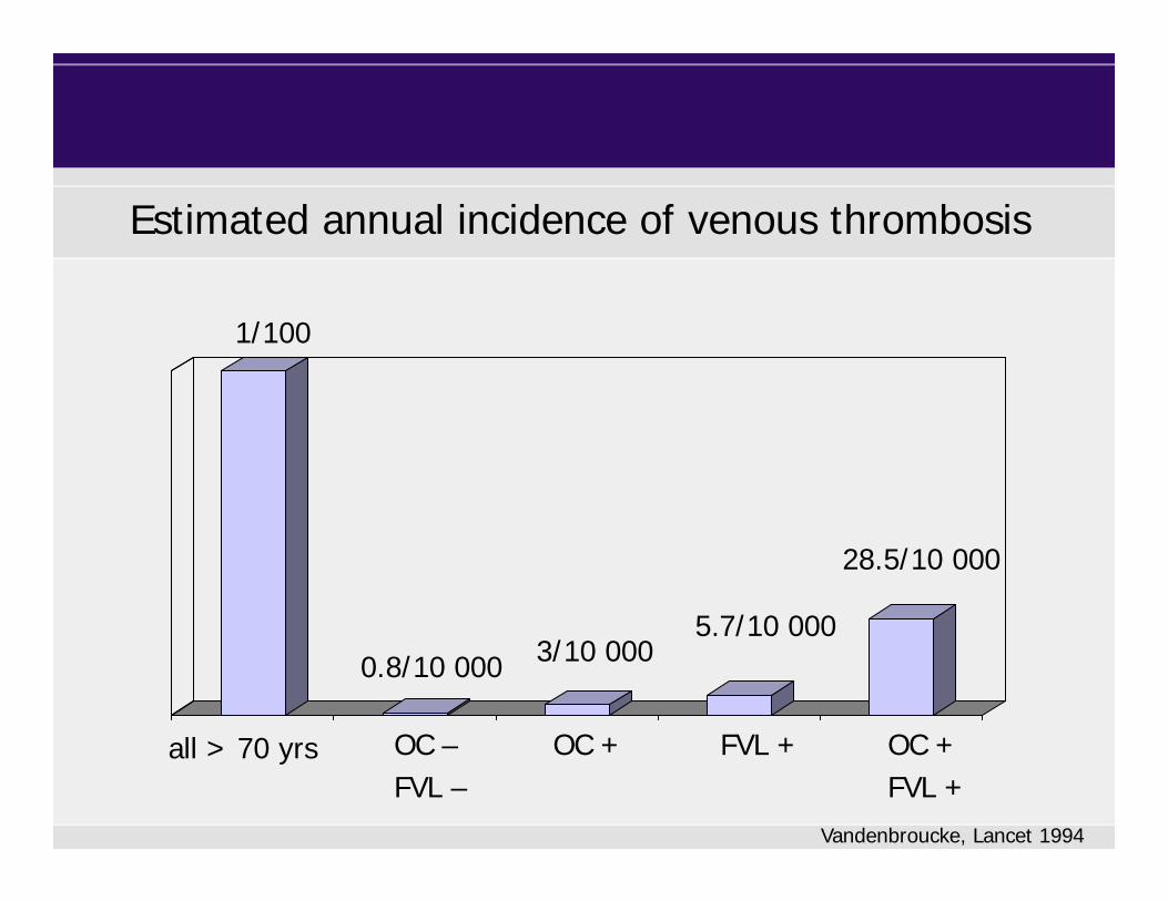
Estimated annual incidence of venous thrombosis
OC –FVL –
Vandenbroucke, Lancet 1994
all > 70 yrs OC + OC +FVL +
FVL +
1/100
0.8/10 000 3/10 0005.7/10 000
28.5/10 000

Family history and the risk of a first VTE
Family history Odds Ratio (95% CI)
negative 1.0 (ref.)
AllAny relative>1 relative
2.5 (1.9 - 3.2)
4.2 (2.4 - 7.4)
Genetic factors*Any relative>1 relative
2.7 (1.7 - 4.4)
4.9 (1.8 - 13.4)
Bezemer, Arch Intern Med 2009
* Low AT/PS/PC, FVL, FII 20210A

Hormone contraception
Recommendations to a woman without VTE
First degree relative + VTE
• Not tested consider alternative contraception• Tested and positive consider alternative contraception• Tested but negative consider alternative contraception

Hormone replacement therapy
• Estrogen (to relief symptoms)– oral, transdermal, intravaginal– daily
± Progestogen (for endometrial protection)– oral, transdermal, IUD– cyclic or daily
• Tibolone– oral synthetic steroid; estrogenic, androgenic, and
progestogenic actions

= FVIII, AT, PC, PS, F1+2, TAT
Fibrinogen, FVII, FIX
Menopause
Effect on hemostatic parameters
Lowe, Br J Haematol 1997

Effect on hemostatic parameters
Hormone replacement therapy
Teede, ATVB 2000
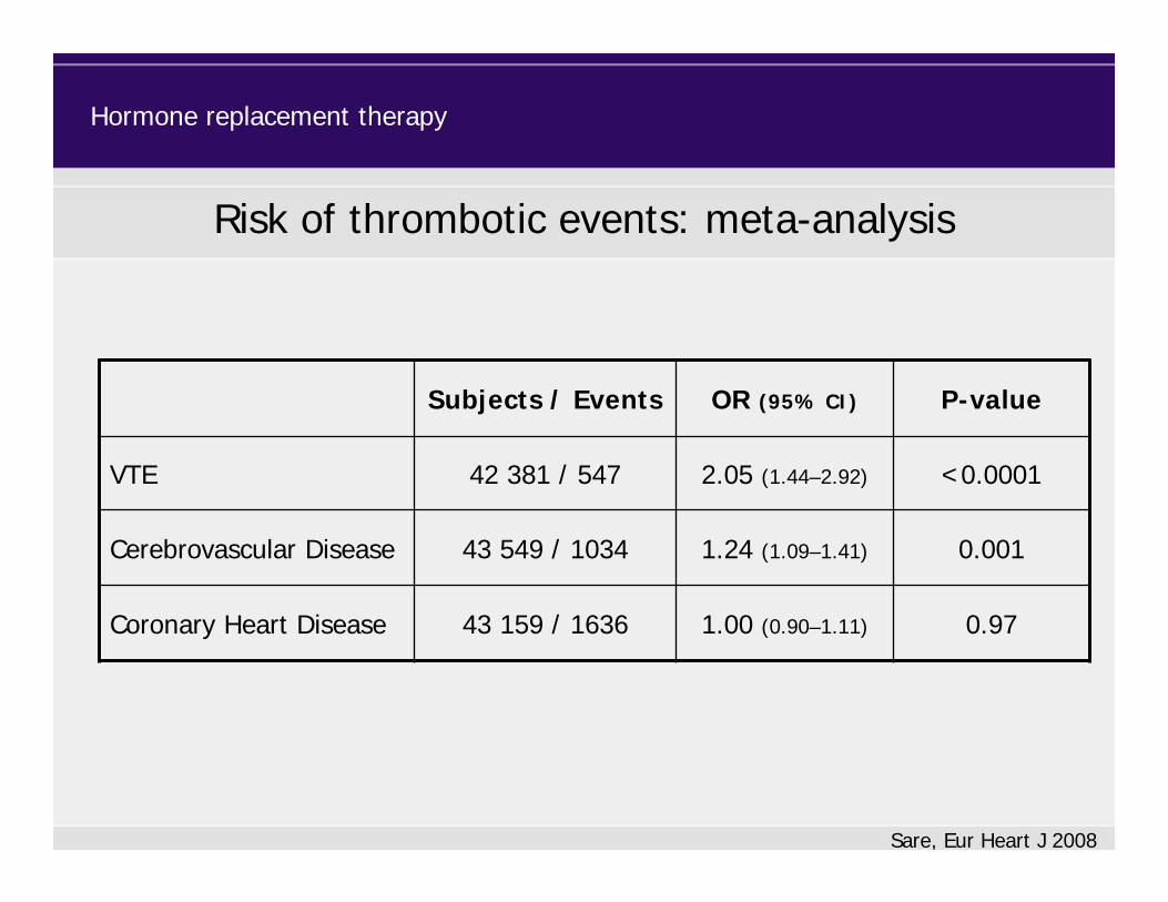
Risk of thrombotic events: meta-analysis
Sare, Eur Heart J 2008
Subjects / Events OR (95% CI) P-value
VTE 42 381 / 547 2.05 (1.44–2.92) <0.0001
Cerebrovascular Disease 43 549 / 1034 1.24 (1.09–1.41) 0.001
Coronary Heart Disease 43 159 / 1636 1.00 (0.90–1.11) 0.97
Hormone replacement therapy

Risk of VTE according to dose
HRT exposure Rate ratio (95% CI)
Oral estrogenLow doseHigh doseVery high dose
1.52 (1.44–1.61)
1.19 (1.04–1.35)
1.55 (1.45–1.65)
1.84 (1.63–2.09)
Hormone replacement therapy
Renoux, J Thromb Haemost 2010

Risk of VTE according to dose
HRT exposure Rate ratio (95% CI)
Oral estrogenLow doseHigh doseVery high dose
1.52 (1.44–1.61)
1.19 (1.04–1.35)
1.55 (1.45–1.65)
1.84 (1.63–2.09)
PatchLow doseHigh dose
1.00 (0.89–1.12)
0.99 (0.87–1.12)
1.05 (0.81–1.36)
Hormone replacement therapy
Renoux, J Thromb Haemost 2010

Cases / Controls OR (95% CI)
Non-users 93 / 261 1.0 (ref.)
Oral estrogen 32 / 27 3.5 (1.8–6.8)
Transdermal estrogen 30 / 93 0.9 (0.5–1.6)
Scarabin, Lancet 2003
Hormone replacement therapy
Risk of VTE during estrogen replacement
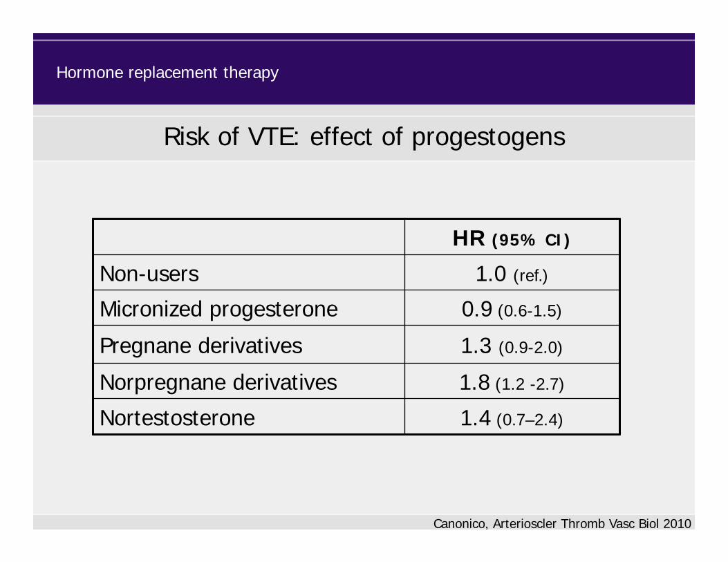
HR (95% CI)
Non-users 1.0 (ref.)
Micronized progesterone 0.9 (0.6-1.5)
Pregnane derivatives 1.3 (0.9-2.0)
Norpregnane derivatives 1.8 (1.2 -2.7)
Nortestosterone 1.4 (0.7–2.4)
Hormone replacement therapy
Risk of VTE: effect of progestogens
Canonico, Arterioscler Thromb Vasc Biol 2010

Hormone replacement therapy
• Female hormone intake increases the risk of VTE
• Risk related to dose and type of estrogen and route of
administration
• Increased by additional progestogens
• Related to type of progestogen
• Risk benefit ratio must be assessed in each woman
0 1 2 3 4 5 6 7
p<0.0001
Men
Women
10
20
30
40
Men vs. Women
Years after anticoagulation
Prob
abili
ty o
f re
curr
ence
%
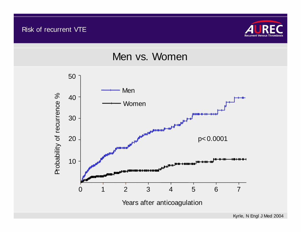
Kyrle, N Engl J Med 2004
50
Risk of recurrent VTE
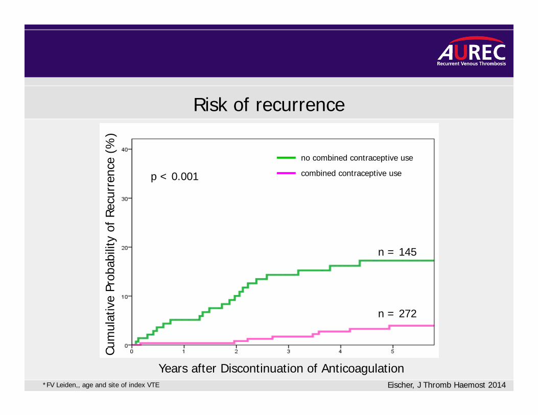
Risk of recurrence
Years after Discontinuation of Anticoagulation
Cum
ulat
ive
Prob
abili
ty o
f Re
curr
ence
(%
)
n = 145
n = 272
no combined contraceptive use
combined contraceptive usep < 0.001
*FV Leiden,, age and site of index VTE Eischer, J Thromb Haemost 2014
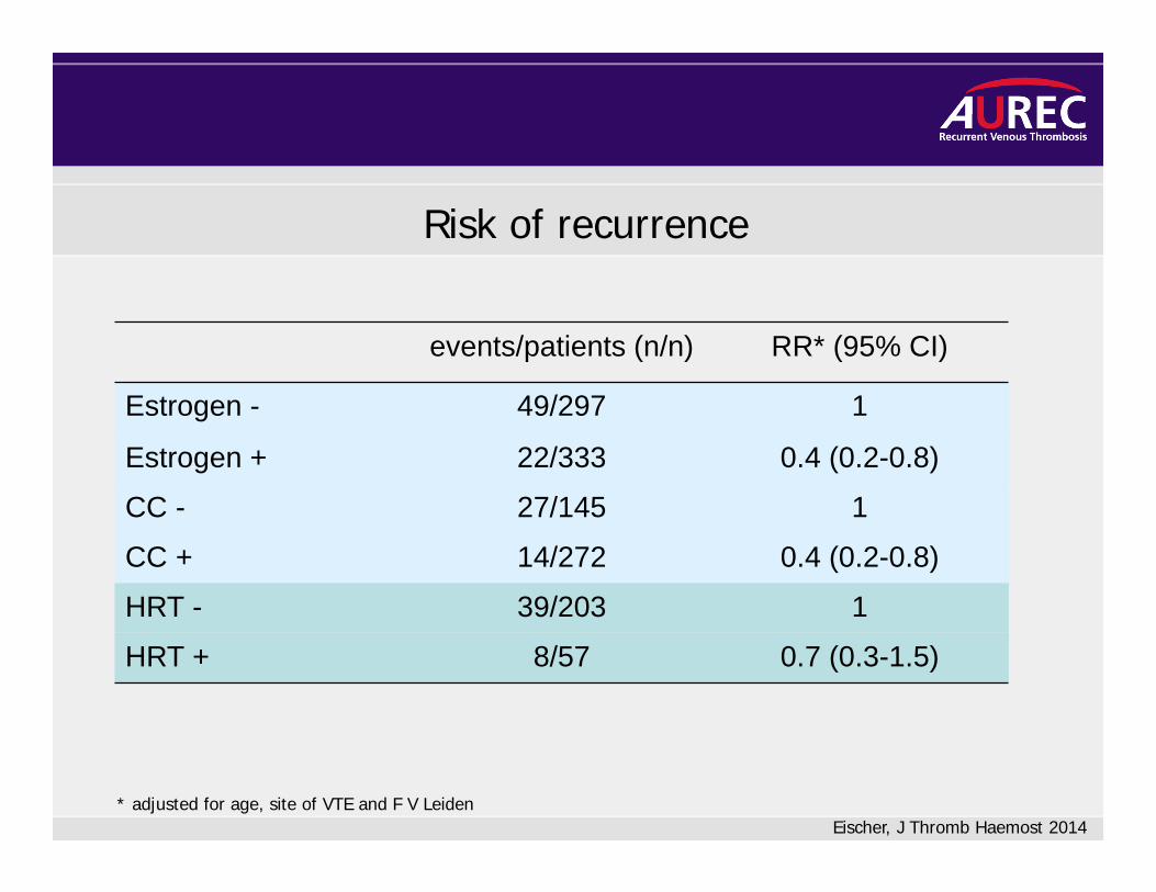
events/patients (n/n) RR* (95% CI)
Estrogen - 49/297 1
Estrogen + 22/333 0.4 (0.2-0.8)
CC - 27/145 1
CC + 14/272 0.4 (0.2-0.8)
HRT - 39/203 1
HRT + 8/57 0.7 (0.3-1.5)
* adjusted for age, site of VTE and F V Leiden
Risk of recurrence
Eischer, J Thromb Haemost 2014
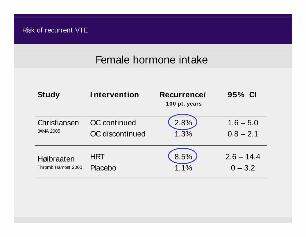
Female hormone intake
Study Intervention Recurrence/100 pt. years
95% CI
ChristiansenJAMA 2005
OC continued OC discontinued
2.8%1.3%
1.6 – 5.00.8 – 2.1
HøibraatenThromb Hamost 2000
HRTPlacebo
8.5%1.1%
2.6 – 14.40 – 3.2
Risk of recurrent VTE
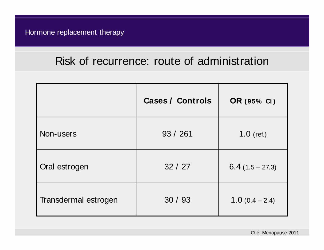
Risk of recurrence: route of administration
Olié, Menopause 2011
Hormone replacement therapy
Cases / Controls OR (95% CI)
Non-users 93 / 261 1.0 (ref.)
Oral estrogen 32 / 27 6.4 (1.5 – 27.3)
Transdermal estrogen 30 / 93 1.0 (0.4 – 2.4)

Risk of recurrent VTE in men and women
• The risk of recurrent VTE is significantly lower in women than in men.• No specific predictor of recurrence with the exception of high FVIII
was found in men.• In women several distinct risk factors of recurrence could be
identified: high D-Dimer, high FVIII, high FIX, FII G20210A, overweight, high hematocrit

The difference between men and women - a conundrum!