worksheet - somerset academy canyons
TRANSCRIPT

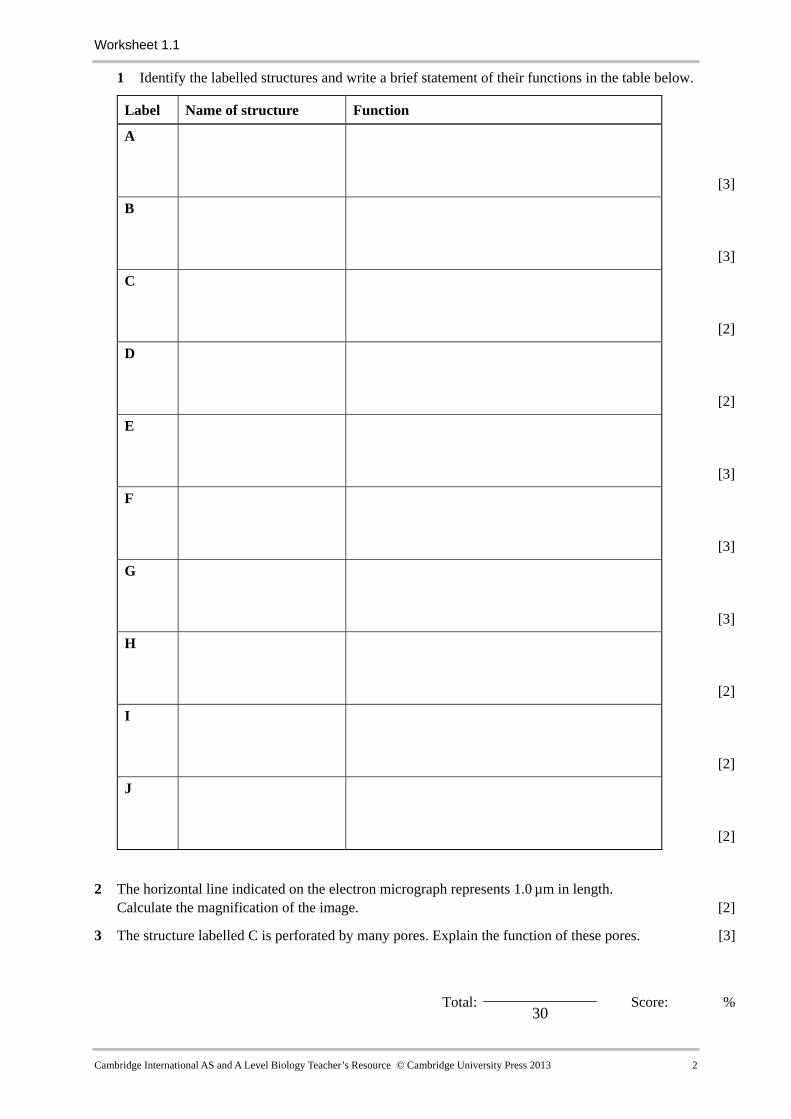
Worksheet 1.1
Interpreting an electron micrograph of a plant cell
This transmission electron micrograph (TEM) shows parts of two palisade cells from a leaf.
J
I
F
B H
G
AD
E 1.0 µm C
© Marilyn Schaller/SPL
Cambridge International AS and A Level Biology Teacher’s Resource © Cambridge University Press 2013 1
Worksheet 1.1
1 Identify the labelled structures and write a brief statement of their functions in the table below.
Label Name of structure Function
A
[3]
B
[3]
C
[2]
D
[2]
E
[3]
F
[3]
G
[3]
H
[2]
I
[2]
J
[2]
2 The horizontal line indicated on the electron micrograph represents 1.0 µm in length.
Calculate the magnification of the image. [2]
3 The structure labelled C is perforated by many pores. Explain the function of these pores. [3]
Total: 30
Score: %
Cambridge International AS and A Level Biology Teacher’s Resource © Cambridge University Press 2013 2