workshop in diagnostic lung cancer: pd-l1 testing, nordiqc eqa · 2019-03-19 · lung cancer: pd-l1...
TRANSCRIPT

Lung cancer:PD-L1 testing, NordiQC EQA
Workshop in Diagnostic Immunohistochemistry Oud St. Jan/ Old St. John – Brugge (Bruges), Belgium June 13th –15nd 2018
Rasmus Røge, MD, NordiQC schemeorganizer

NordiQC external QA PD-L1
• Two pilot runs with 10 labs • All NordiQC participant invited for new ”Companion module”• First run in Spring 2017• Three ”official” runs (C1-C3) and 1 supplementary run (C1x)• Participants from more than 25 countries

Development in pass rates
C1 C2 C3
Participants 68 145 146
Pass rate 50 % 84 % 91 %
Optimal 37 % 59 % 74 %

Development in pass rates
C1 C2 C3
Participants 68 145 146
Pass rate 50 % 84 % 91 %
Optimal 37 % 59 % 74 %

PD-L1 C2, TMA

PD-L1, C2 Participant
scoring

PD-L1 C2, Assessment

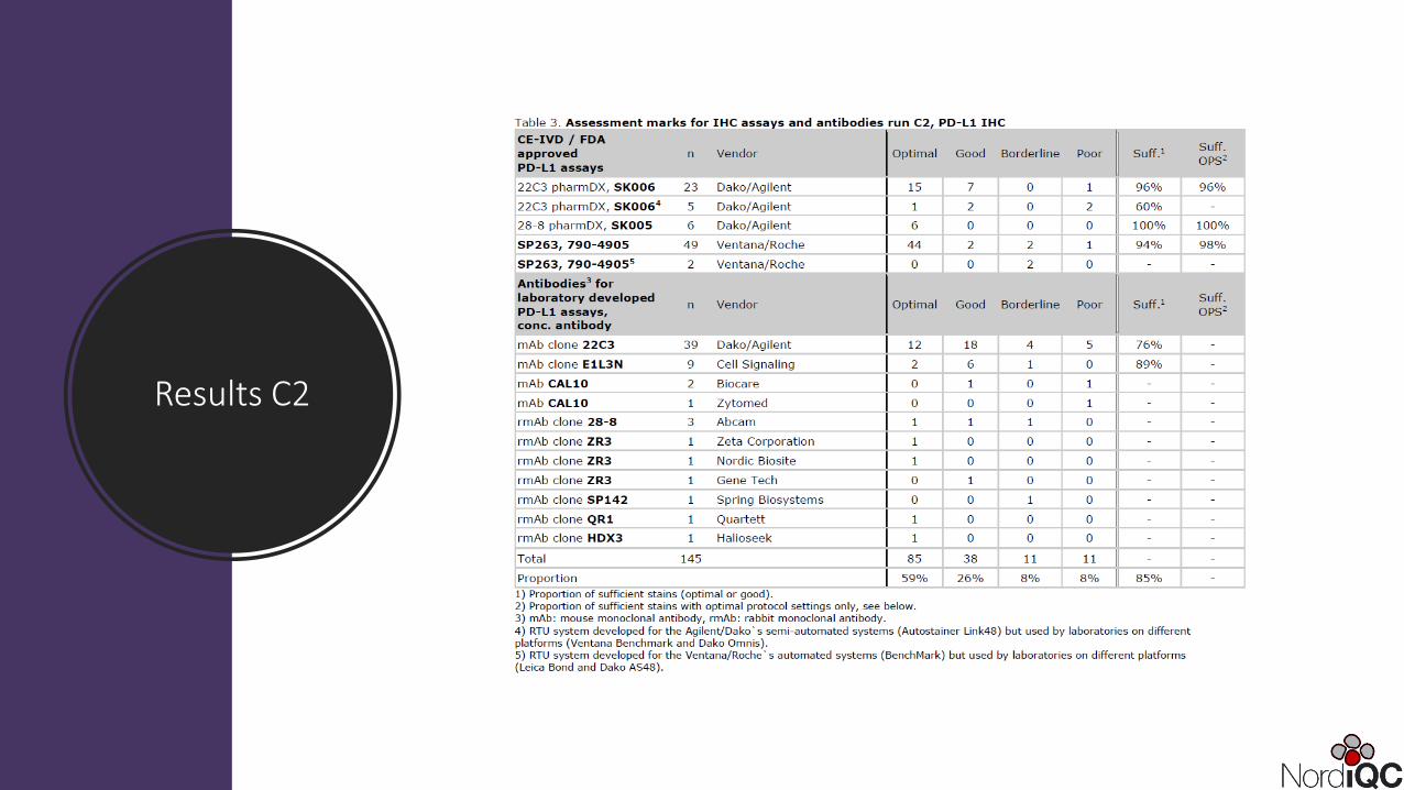
Results C2

Optimal Insufficient
Core 17:TPS: > 50%
Core 19:TPS: > 50%
Core 17:Significantloss of cells
Core 19:Significantloss of cells
pharmDx IHC PD-L1, SK006, mAb 22C3 LDT, mAb 22C3, Benchmark

PD-L1, C2 – Interpretation Excluded
Excluded

PD-L1, C2 –Interpretation
Sufficient vs. insufficient
Excluded

PD-L1 staining with TSA (tonsil)
Optimal staining without amplification Optimal staining with amplification

PD-L1 staining with TSA
Courtesy of O. Nielsen
LDT protocol:mAb 22C3 concVentana BencmarkOptiviewTyramide amp.

Results C3

Optimal Insufficient
Core 9:TPS: 1-49%
Core 10:TPS: > 50%
Core 9:Negative
Core 10:Significantloss of cells
pharmDx IHC PD-L1, SK006, mAb 22C3,Recommended protocol
pharmDx IHC PD-L1, SK006, mAb 22C3, Short HIER, Envion

Controls
PD-L1, C1-C3

Placenta
Optimal Poor

Tonsil
• No staining reaction in the vast majority of lymphocytes including mantle zone and germinal centre B-cells
• A weak to moderate, typically punctuated membranous staining reaction of the majority of germinal centre macrophages
• A moderate to strong staining reaction of the majority of epithelial crypt cells.
• No staining reaction in superficial epithelial cells
Low-level expressor
High-level expressor

Tonsil Tonsil
Optimal Poor
NSCLCTPS: >50%
NSCLCTPS: <50%

Cell lines
PD-L1, C1-C3

PD-L1 IHC 22C3 pharmDx package controls

PD-L1 Horizon cell lines• 1) Strong staining in all cells
• 2) Weak to moderate staining in the majority of cells
• 3) Weak staining in majority of cells
• 4) Negative staining in all cells

Core 2(majorityweak to moderatestainin)
Optimal Poor
Core 2(significantloss of cells)
Core 3(majority, weakstaining)
Core 3(significantloss of cells)

LDT mAb 22C3
Protocols for other platforms
Results C2

22C3 – LDT


TPS:100% TPS:100%
TPS:100% TPS:100%
TPS:40% TPS:40%
TPS:40% TPS:30%
TPS:1% TPS:1%
TPS:1% TPS:0%
Optimized protocols 22C3A) Dako Autostainer (pharmDx)B) Dako OmnisC) Ventana BenchmarkD) Leica Bond


Conclusions
• ”Best practice” PD-L1 22C3 protocols identified• Variations in TPS score between the different stainer platforms• Overall, Leica Bond platform produced slides with marginally lower
TPS• However, concardance in TPS categories was excellent


Questions?