world digestive health day wdhd may 29, 2014 wgo ... handbook on gut microbes world digestive health...
TRANSCRIPT
World Gastroenterology Organisation (WGO)The WGO Foundation (WGO-F)
555 East Wells Street, Suite 1100Milwaukee, WI USA 53202Tel: +1 (414) 918-9798 • Fax: +1 (414) 276-3349Email: [email protected]: www.worldgastroenterology.org • www.wgofoundation.org
World Digestive Health Day WDHD May 29, 2014
WGO Handbook on Gut Microbes
World Digestive Health Day WDHD May 29, 2014 WGO HANDBOOK ON GUT MICROBES 2
Table of ContentsMessage from the Chair ..............................................................................................................................................4Francisco Guarner, MD, Spain
From the Chair and Vice Chair of the WGO Foundation ............................................................................................5Eamonn Quigley MD, FRCP, FACP, FACG, FRCPI, USA, Chair, WGO FoundationRichard Hunt, FRCP, FRCPEd, FRCPC, MACG, AGAF, MWGO, Canada, Vice Chair, WGO Foundation
WDHD 2014 Supporter and Partners .........................................................................................................................6
Microbial Communities ...............................................................................................................................................7Claudia Herrera, MD, SpainFrancisco Guarner, MD, Spain
Functions of the Gut Microbiota ..................................................................................................................................9Francisco Guarner, MD, Spain
Techniques to Characterize the Gut Microbiota ......................................................................................................12Joël Doré, PhD, FranceChaysavanh Manichanh, PhD, Spain
Composition and Structure of the Human Gut Microbiota ......................................................................................16Virginia Robles-Alonso, MD, SpainFrancisco Guarner, MD, SpainDusko Ehrlich, PhD, United Kingdom
Acquisition of the Human Gut Microbiota ................................................................................................................19Yanjiao Zhou, MD, PhD, USABarbara B. Warner, MD, USAPhillip I. Tarr, MD, USA
Impact of Diet on Gut Microbes .................................................................................................................................24Francisco Guarner, MD, SpainLindsey G. Albenberg, DO, USAGary D. Wu, MD, USA
Antibiotics and Gut Microbes ....................................................................................................................................27Chaysavanh Manichanh, PhD, Spain
The Gut Microbiota in Functional Bowel Disorders .................................................................................................31Giovanni Barbara, MD, ItalyCesare Cremon, MD, ItalyVincenzo Stanghellini, MD, Italy
The Gut Microbiota in Inflammatory Bowel Disease ...............................................................................................33Harry Sokol, MD, PhD, France
Gut Microbiota, Obesity and Associated Metabolic Disorders ...............................................................................36Patrice D. Cani, PhD, BelgiumNathalie M. Delzenne, PhD, Belgium
Probiotics: the Concept .............................................................................................................................................39Mary Ellen Sanders, PhD, USA
Probiotics in Diarrheal Diseases ..............................................................................................................................43María Julieta Argüero, MD, ArgentinaJuan Andrés De Paula, MD, Argentina
Probiotics in Functional Bowel Disorders ...............................................................................................................47Eamonn M M Quigley, MD, FRCP, FACP, FACG, FRCPI, USAAmy E Foxx-Orenstein, DO, FACP, FACG, USA
World Digestive Health Day WDHD May 29, 2014 WGO HANDBOOK ON GUT MICROBES 3
Probiotic Therapy for Induction and Remission in Inflammatory Bowel Diseases ..............................................53Richard N Fedorak, MD, Canada
Probiotics in Pediatrics .............................................................................................................................................56Michael D. Cabana, MD, MPH, USA
Prebiotics ...................................................................................................................................................................58GR Gibson, PhD, United Kingdom
Fecal Microbiota Transplant (FMT) ..........................................................................................................................61Elizabeth Broussard, MD, USAChristina Surawicz, MD, USA
Harvesting the Microbiome for the Future ..............................................................................................................64Emma Allen-Vercoe, PhD, Canada
World Digestive Health Day WDHD May 29, 2014 WGO HANDBOOK ON GUT MICROBES 4
Message from the ChairDear Colleagues,
Our knowledge of the microbial communities that inhabit the human gut has grown exponentially over the last few years and there is a profusion of novel information flowing from basic science laboratories into the clinical scenarios. Gut Microbes function like an organ within the gastrointestinal tract, and Gastroenterologists are the health profes-sionals who should bring the new knowledge into practice.
The human host provides a habitat and nutrition to a large and diverse ecosystem of microbial communities and they play key roles in digestion, metabolism and immune function and have a significant impact beyond the gastrointestinal tract. Changes in the diversity and function of those communities are associated with far reaching consequences on host health and have been linked with a number of disorders, including functional bowel disorders, inflammatory bowel diseases and other immune mediated diseases (coeliac disease, allergies), metabolic conditions (type 2 diabetes, NASH), and perhaps, behavioral disorders such as autism and depression. The emerging data on the microbiota and its interaction with the host may provide novel diagnostic and prognostic tests for clinician, and also lead to the development of new and effective therapeutic interventions (functional foods, probiotics, prebiotics, microbiota transplants) to relieve symptoms, as well as treat and prevent illness.
The World Gastroenterology Organisation (WGO) seeks to raise awareness of this novel organ and bring the latest fundamental and clinically relevant knowledge to the Gastroenterologist and, through the Gastroenterologist, to the lay public. The “Gut Microbes - Im-portance in Health and Disease” campaign for World Digestive Health Day 2014 seeks to undertake the challenge of translating science into practice by developing educational and training platforms and materials around the world through a concerted collaboration with WGO Member Societies. Such actions include a WGO Gut Microbes Manual, “Meeting in a Box” tools to share with Member Societies, an update of the Probiotics and Prebiotics WGO Guideline, sponsored meetings and more.
We look forward to a fruitful campaign throughout 2014 and beyond.
Sincerely,
Francisco Guarner Professor Francisco Guarner, MD Chair, WDHD 2014 Barcelona, Spain
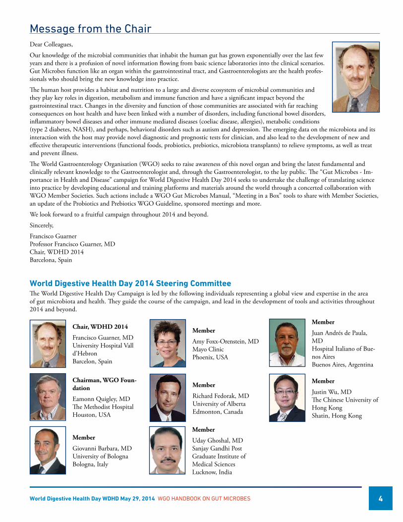
World Digestive Health Day 2014 Steering CommitteeThe World Digestive Health Day Campaign is led by the following individuals representing a global view and expertise in the area of gut microbiota and health. They guide the course of the campaign, and lead in the development of tools and activities throughout 2014 and beyond.
Chair, WDHD 2014
Francisco Guarner, MD University Hospital Vall d’Hebron Barcelon, Spain
Member
Giovanni Barbara, MD University of Bologna Bologna, Italy
Member
Uday Ghoshal, MD Sanjay Gandhi Post Graduate Institute of Medical Sciences Lucknow, India
Member
Amy Foxx-Orenstein, MD Mayo Clinic Phoenix, USA
Chairman, WGO Foun-dation
Eamonn Quigley, MD The Methodist Hospital Houston, USA
Member
Richard Fedorak, MD University of Alberta Edmonton, Canada
Member
Juan Andrés de Paula, MD Hospital Italiano of Bue-nos Aires Buenos Aires, Argentina
Member
Justin Wu, MD The Chinese University of Hong Kong Shatin, Hong Kong
World Digestive Health Day WDHD May 29, 2014 WGO HANDBOOK ON GUT MICROBES 5
World Digestive Health Day (WDHD) was initiated several years ago by the World Gastroenterology Organisation in order to high-light important global issues in digestive diseases. As WDHD has evolved over the years it has developed from a one day event to a year-long campaign which engages with gastroenterologists, doctors, health care professionals and the general public on many aspects of the prevalence, prevention, diagnosis and management of common gastrointestinal and liver symptoms and disorders. Through direct col-laboration with our member societies in 111 countries around the world and with the support of other professional societies with similar interests, non-governmental agencies, governments and industry, we have helped to promote understanding and raise awareness on these issues.
This year we address one of the “hottest” topics in medicine and medical science: gut microbes. Rapid developments in technology have permitted the detailed description of the bugs that normally inhabit our gastrointestinal tracts and are beginning to reveal their many functions in heath and disease. With such progress have come new challenges: in comprehending new terminology, in distinguishing hype from science, in attempting to understand claims for new diagnostic or therapeutic advances based on the assessment or modula-tion of the microbial populations of our guts. A major aim of this year’s WDHD campaign, therefore, is to help everyone from the “man/woman in the street” to the specialist gastroenterologist to make sense of the mass of information on gut microbes that accumulates be-fore our very eyes, and to sift through the claims and counterclaims that are made for medicines, diets, probiotics and prebiotics. To that end Professor Francisco Guarner and his team have assembled some of the most renowned scientists and clinicians in the field to provide an overview of the most important aspects of science and clinical practice related to gut microbes.
On behalf of the WGO Foundation we congratulate Professor Guarner and his team and fellow authors on this wonderful work which we hope that you will not only enjoy but find helpful.
Sincerely,
Eamonn M M Quigley, MD, FRCP, FACP, FACG, FRCPI Chair, WGO Foundation
Richard Hunt, MD Vice Chair, WGO Foundation
From the Chair and Vice Chair of the WGO Foundation
World Digestive Health Day WDHD May 29, 2014 WGO HANDBOOK ON GUT MICROBES 6
The World Gastroenterology Organisation and the WGO Foundation thank the following WDHD 2014 supporter and partners for their generosity and support of the 2014 campaign.
Supporter
Partners
The “Gut Microbiota for Health Experts Exchange” is a community where experts can share news, innovation and information on the topics of gut microbiota.
The content of “The Gut Microbiota For Health Experts Exchange” offers a selection of current topics of conversation organized around the cross-cutting themes of: digestive health, immune function, metabolic conditions, gut brain axis, research tools, trends and discover-ies, nutrition, and probiotics. Each topic is enriched by a selection of articles from scientific literature, traditional media, social media and the best contributions of users. A media room is also included in the platform to help identify key scientific events, important press releases and more. The new content is sent to the members through our Gut Microbiota for Health newsletter twice a month.
The Community encourages contributions from readers, interactions within the website, and beyond. Further sharing and discussions are possible through the Gut Microbiota for Health digital presence on social media. We have a LinkedIn group (“Gut Microbiota for Health“), a Twitter account (@GMFHx), and a Google+ Page (Gut Microbiota for Health on Google+).
The “Gut Microbiota for Health Experts Exchange” is the platform driven by the Gut Microbiota and Health section of the European Society for Neurogastroenterology and Motility (ESNM), with the institutional support of Danone.
WDHD 2014 Supporter and Partners
World Digestive Health Day WDHD May 29, 2014 WGO HANDBOOK ON GUT MICROBES 7
Microbial Communities
Claudia Herrera, MD Digestive System Research Unit Hospital Vall d’Hebron, Ciberehd Barcelona, Spain
Francisco Guarner, MD Digestive System Research Unit Hospital Vall d’Hebron, Ciberehd Barcelona, Spain
Life on Earth
Bacteria have been on Earth for 3.5 billion years, appearing approximately one billion years after the Earth’s crust was
formed. Fossils and associated geochemical markers of biologic activity indicate that microbial organisms inhabited the oceans in Archean times (2.5 to 3.7 billion years ago). The presence early in Earth’s history of morphologically cyanobacterium-like fossils has been widely assumed to be the origin of free oxygen gas in the atmosphere, suggesting that both oxygenic photosynthesis and aerobic respiration of eukaryotic cells are processes derived from microbial biochemistry.
Cyanobacteria are still vastly abundant in modern days, and can be found as planktonic cells in oceans and fresh water. They also occur in damp soil or on moistened rocks. They do not require organic nutrients and can grow on entirely inorganic materials. Cyanobacteria obtain their energy through photosynthesis, and convert solar energy into biomass-stored chemical energy. Like plants, the cyanobacteria release oxygen gas and contribute to carbon fixation by forming carbohydrates from carbon dioxide gas. Some cyanobacteria cell types are able to fix nitrogen gas into ammonia, nitrites or nitrates, which can be absorbed by plants and converted to protein and nucleic acids (nitrogen gas is not bioavailable to plants).
Microbial communities are ubiquitous and truly essential for maintaining life conditions on Earth. As summarized in a report from a colloquium convened by the American Academy of Micro-biology, microbial communities can be found in every corner of the globe, from the permafrost soils of the Arctic Circle to termite guts in sub-Saharan Africa, and on every scale, from microscopic biofilms to massive marine planktonic communities. Because of their enormous global size, microbial communities have a massive impact across the globe. Their diverse contributions affect many aspects of life, not only in relation to human or animal infec-tions, but, more importantly, through their role in cycling the critical elements for maintaining life on Earth. The generation of atmospheric gases, synthesis of organic materials from inorganic sources, corruption of organic to inorganic materials, corrosion,
degradation, bioremediation, etc., are vital ecological functions for global carbon, oxygen and nitrogen cycles, which are the critical cycles relevant to life on Earth.
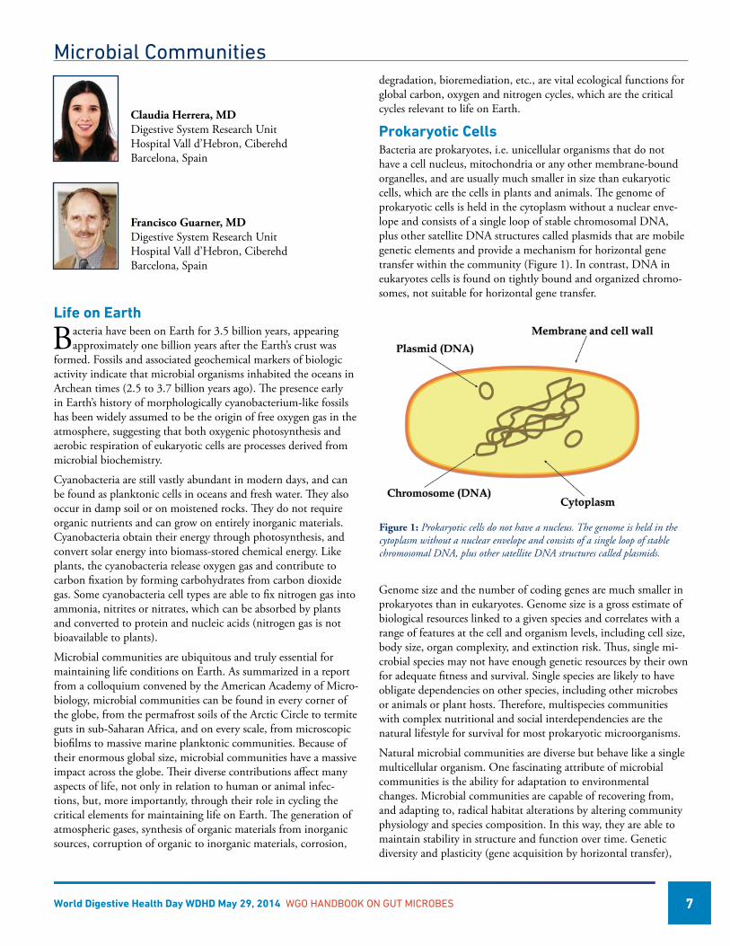
Prokaryotic CellsBacteria are prokaryotes, i.e. unicellular organisms that do not have a cell nucleus, mitochondria or any other membrane-bound organelles, and are usually much smaller in size than eukaryotic cells, which are the cells in plants and animals. The genome of prokaryotic cells is held in the cytoplasm without a nuclear enve-lope and consists of a single loop of stable chromosomal DNA, plus other satellite DNA structures called plasmids that are mobile genetic elements and provide a mechanism for horizontal gene transfer within the community (Figure 1). In contrast, DNA in eukaryotes cells is found on tightly bound and organized chromo-somes, not suitable for horizontal gene transfer.
Genome size and the number of coding genes are much smaller in prokaryotes than in eukaryotes. Genome size is a gross estimate of biological resources linked to a given species and correlates with a range of features at the cell and organism levels, including cell size, body size, organ complexity, and extinction risk. Thus, single mi-crobial species may not have enough genetic resources by their own for adequate fitness and survival. Single species are likely to have obligate dependencies on other species, including other microbes or animals or plant hosts. Therefore, multispecies communities with complex nutritional and social interdependencies are the natural lifestyle for survival for most prokaryotic microorganisms.
Natural microbial communities are diverse but behave like a single multicellular organism. One fascinating attribute of microbial communities is the ability for adaptation to environmental changes. Microbial communities are capable of recovering from, and adapting to, radical habitat alterations by altering community physiology and species composition. In this way, they are able to maintain stability in structure and function over time. Genetic diversity and plasticity (gene acquisition by horizontal transfer),
Figure 1: Prokaryotic cells do not have a nucleus. The genome is held in the cytoplasm without a nuclear envelope and consists of a single loop of stable chromosomal DNA, plus other satellite DNA structures called plasmids.
World Digestive Health Day WDHD May 29, 2014 WGO HANDBOOK ON GUT MICROBES 8
functional redundancy, metabolic cooperation, cell-to-cell signal-ing, and coordinated collective behavior are known attributes of microbial communities. These attributes facilitate community survival by ensuring that they can evolve, adapt and respond to environmental stressors.
The Gut MicrobiotaHuman beings are associated with a large and diverse population of microorganisms that live on body surfaces and in cavities con-nected with the external environment. Associations that benefit the host as well as the microbe are grouped under the term ‘symbiosis’ and the microbial partners called ‘symbionts’. The prevalence of symbiosis has long been recognized on the basis of observa-tions from microscopy, but most aspects of symbiont origins and functions have remained unexplored before the age of molecular techniques because of the difficulties involved in culturing and isolating a large majority of these microbial species.
The skin, mouth, vagina, upper respiratory tract, and gastrointes-tinal tract of humans are inhabited by site-specific microbial com-munities with specialized structures and functions. ‘Microbiota’ is a collective term for the microbial communities in a particular ecological niche, and this expression is preferred over ‘flora’ or ‘microflora’, which perpetuate an outdated classification of bacteria as plants. Thus, the term ‘gut microbiota’ refers to the ecosystem of microorganisms that have adapted to live on the intestinal mucosal surface or within the gut lumen.
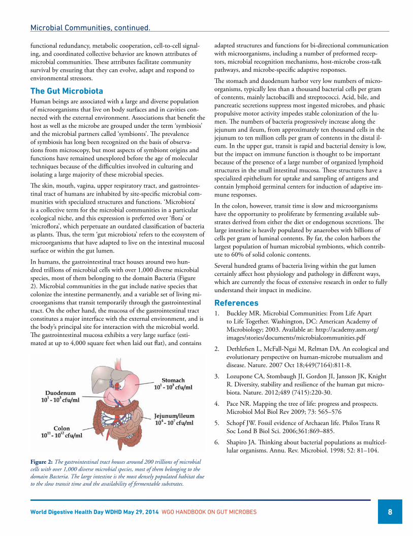
In humans, the gastrointestinal tract houses around two hun-dred trillions of microbial cells with over 1,000 diverse microbial species, most of them belonging to the domain Bacteria (Figure 2). Microbial communities in the gut include native species that colonize the intestine permanently, and a variable set of living mi-croorganisms that transit temporarily through the gastrointestinal tract. On the other hand, the mucosa of the gastrointestinal tract constitutes a major interface with the external environment, and is the body’s principal site for interaction with the microbial world. The gastrointestinal mucosa exhibits a very large surface (esti-mated at up to 4,000 square feet when laid out flat), and contains
adapted structures and functions for bi-directional communication with microorganisms, including a number of preformed recep-tors, microbial recognition mechanisms, host-microbe cross-talk pathways, and microbe-specific adaptive responses.
The stomach and duodenum harbor very low numbers of micro-organisms, typically less than a thousand bacterial cells per gram of contents, mainly lactobacilli and streptococci. Acid, bile, and pancreatic secretions suppress most ingested microbes, and phasic propulsive motor activity impedes stable colonization of the lu-men. The numbers of bacteria progressively increase along the jejunum and ileum, from approximately ten thousand cells in the jejunum to ten million cells per gram of contents in the distal il-eum. In the upper gut, transit is rapid and bacterial density is low, but the impact on immune function is thought to be important because of the presence of a large number of organized lymphoid structures in the small intestinal mucosa. These structures have a specialized epithelium for uptake and sampling of antigens and contain lymphoid germinal centers for induction of adaptive im-mune responses.
In the colon, however, transit time is slow and microorganisms have the opportunity to proliferate by fermenting available sub-strates derived from either the diet or endogenous secretions. The large intestine is heavily populated by anaerobes with billions of cells per gram of luminal contents. By far, the colon harbors the largest population of human microbial symbionts, which contrib-ute to 60% of solid colonic contents.
Several hundred grams of bacteria living within the gut lumen certainly affect host physiology and pathology in different ways, which are currently the focus of extensive research in order to fully understand their impact in medicine.
References1. Buckley MR. Microbial Communities: From Life Apart
to Life Together. Washington, DC: American Academy of Microbiology; 2003. Available at: http://academy.asm.org/images/stories/documents/microbialcommunities.pdf
2. Dethlefsen L, McFall-Ngai M, Relman DA. An ecological and evolutionary perspective on human-microbe mutualism and disease. Nature. 2007 Oct 18;449(7164):811-8.
3. Lozupone CA, Stombaugh JI, Gordon JI, Jansson JK, Knight R. Diversity, stability and resilience of the human gut micro-biota. Nature. 2012;489 (7415):220-30.
4. Pace NR. Mapping the tree of life: progress and prospects. Microbiol Mol Biol Rev 2009; 73: 565–576
5. Schopf JW. Fossil evidence of Archaean life. Philos Trans R Soc Lond B Biol Sci. 2006;361:869–885.
6. Shapiro JA. Thinking about bacterial populations as multicel-lular organisms. Annu. Rev. Microbiol. 1998; 52: 81–104.
Figure 2: The gastrointestinal tract houses around 200 trillions of microbial cells with over 1,000 diverse microbial species, most of them belonging to the domain Bacteria. The large intestine is the most densely populated habitat due to the slow transit time and the availability of fermentable substrates.
Microbial Communities, continued.
World Digestive Health Day WDHD May 29, 2014 WGO HANDBOOK ON GUT MICROBES 9
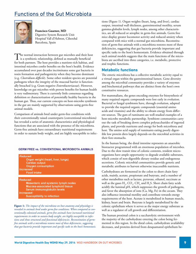
Functions of the Gut Microbiota
The normal interaction between gut microbes and their host is a symbiotic relationship, defined as mutually beneficial
for both partners. The host provides a nutrient-rich habitat, and intestinal microbes confer benefits on the host’s health. Evidence accumulated over past decades incriminates some gut bacteria in toxin formation and pathogenicity when they become dominant (e.g. Clostridium difficile). Some other resident species are potential pathogens when the integrity of the mucosal barrier is function-ally breached (e.g. Gram negative Enterobacteriaceae). However, knowledge on gut microbes with proven benefits for human health is very rudimentary. There is currently little consensus regarding definition or characterization of potentially healthy bacteria in the human gut. Thus, our current concepts on host-microbe symbiosis in the gut are mainly supported by observations using germ-free animal models.
Comparison of animals bred under germ-free conditions with their conventionally raised counterparts (conventional microbiota) has revealed a series of anatomic characteristics and physiological functions that are associated with the presence of the microbiota. Germ-free animals have extraordinary nutritional requirements in order to sustain body weight, and are highly susceptible to infec-
Francisco Guarner, MD Digestive System Research Unit Hospital Vall d’Hebron, Ciberehd Barcelona, Spain
tions (Figure 1). Organ weights (heart, lung, and liver), cardiac output, intestinal wall thickness, gastrointestinal motility, serum gamma-globulin levels, lymph nodes, among other characteris-tics, are all reduced or atrophic in germ-free animals. Germ free mice display greater locomotor activity and reduced anxiety when compared with mice with a normal gut microbiota. Reconstitu-tion of germ-free animals with a microbiota restores most of these deficiencies, suggesting that gut bacteria provide important and specific tasks to the host’s homeostasis. Evidence obtained through such animal models suggests that the main functions of the micro-biota are ascribed into three categories, i.e. metabolic, protective and trophic functions.
Metabolic functionsThe enteric microbiota has a collective metabolic activity equal to a virtual organ within the gastrointestinal lumen. Gene diversity among the microbial community provides a variety of enzymes and biochemical pathways that are distinct from the host’s own constitutive resources.
For mammalians, the genes encoding enzymes for biosynthesis of many required organic compounds were lost early in evolution. Bacterial or fungal symbionts have, through evolution, adapted to provide the required organic compounds (essential amino acids and vitamins) and the ability to obtain energy from differ-ent sources. The guts of ruminants are well-studied examples of a host-microbe metabolic partnership. Symbiont communities carry out the task of breaking down complex polysaccharides of ingested plants, and provide nutrients and energy for both microbiota and host. The amino acid supply of ruminants eating poorly digest-ible low protein diets largely depends on the microbial activities in their fore-stomachs.
In the human being, the distal intestine represents an anaerobic bioreactor programmed with an enormous population of microbes. Due to the slow transit time of colonic contents, resident micro-organisms have ample opportunity to degrade available substrates, which consist of non-digestible dietary residue and endogenous secretions. Colonic microbial communities provide genetic and metabolic attributes to harvest otherwise inaccessible nutrients.
Carbohydrates are fermented in the colon to short chain fatty acids, mainly, acetate, propionate and butyrate, and a number of other metabolites such as lactate, pyruvate, ethanol, succinate as well as the gases H2, CO2, CH4 and H2S. Short chain fatty acids acidify the luminal pH, which suppresses the growth of pathogens, and favor the absorption of ions (Ca, Mg, Fe) in the cecum. They also influence intestinal motility and contribute towards energy requirements of the host. Acetate is metabolized in human muscle, kidney, heart and brain. Butyrate is largely metabolized by the colonic epithelium where it serves as the major energy substrate as well as a regulator of cell growth and differentiation.
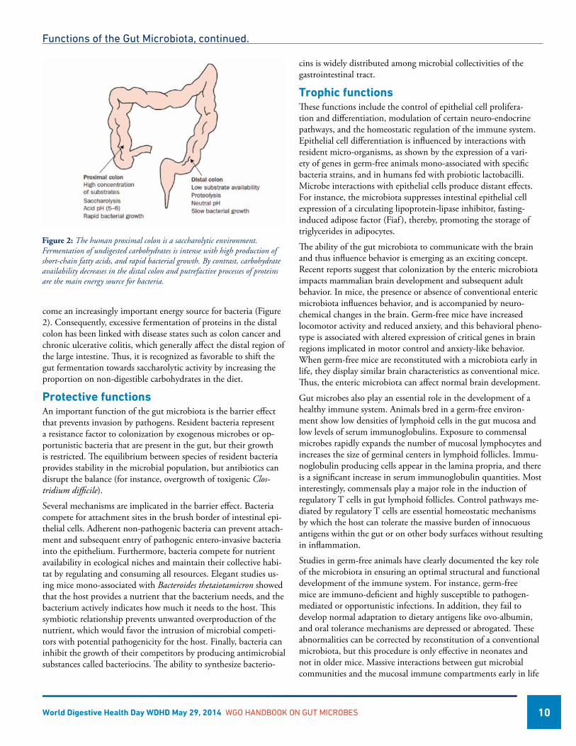
The human proximal colon is a saccharolytic environment with the majority of the carbohydrate entering the colon being fer-mented in this region. In the distal colon, carbohydrate availability decreases, and proteins derived from desquamated epithelium be-
Figure 1: The impact of the microbiota on host anatomy and physiology is revealed in animals bred under germ-free conditions. When compared to con-ventionally colonized animals, germ-free animals have increased nutritional requirements in order to sustain body weight, are highly susceptible to infec-tions and show structural and functional deficiencies. Reconstitution of germ-free animals with a microbiota restores most of these deficiencies, suggesting that gut bacteria provide important and specific tasks to the host’s homeostasis.
World Digestive Health Day WDHD May 29, 2014 WGO HANDBOOK ON GUT MICROBES 10
come an increasingly important energy source for bacteria (Figure 2). Consequently, excessive fermentation of proteins in the distal colon has been linked with disease states such as colon cancer and chronic ulcerative colitis, which generally affect the distal region of the large intestine. Thus, it is recognized as favorable to shift the gut fermentation towards saccharolytic activity by increasing the proportion on non-digestible carbohydrates in the diet.
Protective functionsAn important function of the gut microbiota is the barrier effect that prevents invasion by pathogens. Resident bacteria represent a resistance factor to colonization by exogenous microbes or op-portunistic bacteria that are present in the gut, but their growth is restricted. The equilibrium between species of resident bacteria provides stability in the microbial population, but antibiotics can disrupt the balance (for instance, overgrowth of toxigenic Clos-tridium difficile).
Several mechanisms are implicated in the barrier effect. Bacteria compete for attachment sites in the brush border of intestinal epi-thelial cells. Adherent non-pathogenic bacteria can prevent attach-ment and subsequent entry of pathogenic entero-invasive bacteria into the epithelium. Furthermore, bacteria compete for nutrient availability in ecological niches and maintain their collective habi-tat by regulating and consuming all resources. Elegant studies us-ing mice mono-associated with Bacteroides thetaiotamicron showed that the host provides a nutrient that the bacterium needs, and the bacterium actively indicates how much it needs to the host. This symbiotic relationship prevents unwanted overproduction of the nutrient, which would favor the intrusion of microbial competi-tors with potential pathogenicity for the host. Finally, bacteria can inhibit the growth of their competitors by producing antimicrobial substances called bacteriocins. The ability to synthesize bacterio-
cins is widely distributed among microbial collectivities of the gastrointestinal tract.
Trophic functionsThese functions include the control of epithelial cell prolifera-tion and differentiation, modulation of certain neuro-endocrine pathways, and the homeostatic regulation of the immune system. Epithelial cell differentiation is influenced by interactions with resident micro-organisms, as shown by the expression of a vari-ety of genes in germ-free animals mono-associated with specific bacteria strains, and in humans fed with probiotic lactobacilli. Microbe interactions with epithelial cells produce distant effects. For instance, the microbiota suppresses intestinal epithelial cell expression of a circulating lipoprotein-lipase inhibitor, fasting-induced adipose factor (Fiaf ), thereby, promoting the storage of triglycerides in adipocytes.
The ability of the gut microbiota to communicate with the brain and thus influence behavior is emerging as an exciting concept. Recent reports suggest that colonization by the enteric microbiota impacts mammalian brain development and subsequent adult behavior. In mice, the presence or absence of conventional enteric microbiota influences behavior, and is accompanied by neuro-chemical changes in the brain. Germ-free mice have increased locomotor activity and reduced anxiety, and this behavioral pheno-type is associated with altered expression of critical genes in brain regions implicated in motor control and anxiety-like behavior. When germ-free mice are reconstituted with a microbiota early in life, they display similar brain characteristics as conventional mice. Thus, the enteric microbiota can affect normal brain development.
Gut microbes also play an essential role in the development of a healthy immune system. Animals bred in a germ-free environ-ment show low densities of lymphoid cells in the gut mucosa and low levels of serum immunoglobulins. Exposure to commensal microbes rapidly expands the number of mucosal lymphocytes and increases the size of germinal centers in lymphoid follicles. Immu-noglobulin producing cells appear in the lamina propria, and there is a significant increase in serum immunoglobulin quantities. Most interestingly, commensals play a major role in the induction of regulatory T cells in gut lymphoid follicles. Control pathways me-diated by regulatory T cells are essential homeostatic mechanisms by which the host can tolerate the massive burden of innocuous antigens within the gut or on other body surfaces without resulting in inflammation.
Studies in germ-free animals have clearly documented the key role of the microbiota in ensuring an optimal structural and functional development of the immune system. For instance, germ-free mice are immuno-deficient and highly susceptible to pathogen-mediated or opportunistic infections. In addition, they fail to develop normal adaptation to dietary antigens like ovo-albumin, and oral tolerance mechanisms are depressed or abrogated. These abnormalities can be corrected by reconstitution of a conventional microbiota, but this procedure is only effective in neonates and not in older mice. Massive interactions between gut microbial communities and the mucosal immune compartments early in life
Figure 2: The human proximal colon is a saccharolytic environment. Fermentation of undigested carbohydrates is intense with high production of short-chain fatty acids, and rapid bacterial growth. By contrast, carbohydrate availability decreases in the distal colon and putrefactive processes of proteins are the main energy source for bacteria.
Functions of the Gut Microbiota, continued.
World Digestive Health Day WDHD May 29, 2014 WGO HANDBOOK ON GUT MICROBES 11
seem to be critical for a proper instruction of the immune system. Later in life, multiple and diverse interactions between microbes, epithelium and gut lymphoid tissues are constantly reshaping local and systemic immunity.
In summary, homeostasis of the individual with the external en-vironment seems to be highly influenced by the dynamic balance between microbial communities and the immune system.
References1. Bäckhed F, Ley RE, Sonnenburg JL, Peterson DA, Gordon
JI. Host-bacterial mutualism in the human intestine. Science. 2005 Mar 25;307(5717):1915-20.
2. Cryan JF, O’Mahony SM. The microbiome-gut-brain axis: from bowel to behavior. Neurogastroenterol Motil. 2011 Mar;23(3):187-92.
3. Guarner F, Malagelada JR. Gut flora in health and disease. Lancet. 2003 Feb 8;361(9356):512-9.
Functions of the Gut Microbiota, continued.
4. Hamer HM, Jonkers D, Venema K, Vanhoutvin S, Troost FJ, Brummer RJ. Review article: the role of butyrate on colonic function. Aliment Pharmacol Ther. 2008 Jan 15;27(2):104-19.
5. Hooper LV, Macpherson AJ. Immune adaptations that maintain homeostasis with the intestinal microbiota. Nat Rev Immunol. 2010 Mar;10(3):159-69.
6. Kuhn KA, Stappenbeck TS. Peripheral education of the immune system by the colonic microbiota. Semin Immunol 2013 Nov 30;25(5):364-9.
7. O’Hara AM, Shanahan F. The gut flora as a forgotten organ. EMBO Rep. 2006 Jul;7(7):688-93.
8. Wostmann BS. The germfree animal in nutritional studies. Annu Rev Nutr. 1981;1:257-79.
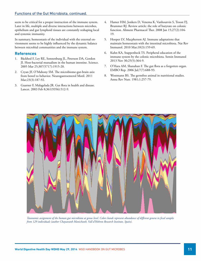
Taxonomic assignment of the human gut microbiota at genus level. Colors bands represent abundance of different genera in fecal samples from 129 individuals (author Chaysavanh Manichanh, Vall d’Hebron Research Institute, Spain).
World Digestive Health Day WDHD May 29, 2014 WGO HANDBOOK ON GUT MICROBES 12
Techniques to Characterize the Gut Microbiota
Joël Doré, Ph.D. Institut National de la Recherche Agronomique (INRA) Jouy en Josas, France
Chaysavanh Manichanh, Ph.D. Digestive System Research Unit, CIBEREHD Vall d’Hebron Research Institute Barcelona, Spain
Introduction
The digestive tract of each human individual hosts microorgan-isms in all its compartments, with especially dense popula-
tions in the colon where concentrations reach 1011 bacteria per gram content. Overall counts of bacteria are 10 times higher than the number of human cells in our body. The current intestinal microbiota, formerly called microflora, stems from a long co-evolution and forms an intimate symbiosis with its human host. Functional interactions between what can be considered as our two genomes ultimately have a major impact on our health.
Our understanding of the microbiota evolved over the years in a fairly chaotic way, markedly influenced by evolutions in method-ologies. Some 20 years ago, our knowledge was restricted to a small number of large studies of the composition of the fecal microbiota, based on the enumeration of culturable microorganisms. The de-velopment of culture-independent molecular approaches since the 1990’s has progressively set the stage for ‘big times’ of conceptual revision.
Culture based microbiota assessmentSince the pioneering description of Bacterium coli communior by Escherich in 1885, successive technological developments allowed stepwise improvements leading to culture and isolation in pure culture intestinal microbes. Major improvements came with the ability to culture bacteria under anaerobic conditions, and yielded by the 1970’s to the recognition of numerous bacterial species of the dominant fecal microbiota, registered according to taxonomic rules into genera such as Bacteroides, Eubacterium, Peptostreptococ-cus, Ruminococcus, Bifidobacterium, Fusobacterium et Clostridium.
Mathematical inference allowed quite early to estimate the expected diversity of the dominant human fecal microbiota to 400 bacterial species. For each individual, 25 to 40 dominant culturable spe-cies could be commonly recovered, reaching population levels of 108 to 1011 per gram of stool. Culture remained for a few decades the only way to access the dominant fecal microbiota and explore its functional contribution. For many reasons this was a major limitation and comparison of microscopic counts and culturable
counts consistently lead to a marked difference known as “the great plate-count anomaly”. It mainly stems from ecological requirements including numerous interactions that cannot be simulated in vitro. Less represented microorganisms are considered sub-dominant. They are still autochtonous and maintain stable levels of populations ranging from 106 to 108 per gram stool. Many of these are facultative anaerobes, tolerating simpler culture conditions such that many are culturable on selective media. Yet even less represented populations are considered transient, and will contain, among others, food-borne microorganisms that will never establish.
The first comparisons of human individuals indicated that each individual harbors his own microbiota, except for twins, suggesting as early as 1983 an impact of the host genetics. Considering that colonization of the gut occurs from the very moment of birth on, it is likely that the neonatal gut is characterized by a fair degree of permissivity up until the immune system becomes fully mature. Numerous factors may hence combine their effect as determinants of the adult microbiota, such as 1) more or less random exposure to microorganisms, from maternal microbiomes or the environ-ment;, 2) ecological selection pressure due to microbial interac-tions; 3) mode of feeding; and 4) host genetics, especially endog-enous receptors and substrates from mucins and epithelial cells.
There is little doubt that special efforts in anaerobic cultiva-tion would allow to identify new species, and yet the major step forward that followed came from the development of molecular phylogenies towards the end of the 1970’s and their further appli-cation to culture-independent microbial ecology towards the end of the 1980’s. This was the first revolution in terms of knowledge gain; a revolution from which we are still enjoying benefits today. Anaerobic culture remains nonetheless the standard for the formal description of new species and their validation by the international committee of systematic.
Phylogenetics of the intestinal microbiota – the ribosomal RNA based approachMethods based on comparative analysis of ribosomal RNAs really warrant a special mention considering the major step forward they allowed. They owe their large and massive application to a few intrinsic characteristics of the target molecule that can be summa-rized in four points:
• rRNA is present in cells of nearly all life-forms on earth
• This molecule is not subject to major lateral transfers of genetic material among contemporary organisms, and point mutations capture the evolutionary history of lineages.
• Its mosaic primary sequence makes it informative in terms of evolutionary relationships from the domain (bacteria, eucarya, archaea) to the species
• The above characteristics permitted the rapid constitution of a large sequence database
Ribosomal RNA methods were structured in two major lines that differed by their respective level of resolution: low resolution, giv-ing access to composition at the level of large groups that compose
World Digestive Health Day WDHD May 29, 2014 WGO HANDBOOK ON GUT MICROBES 13
the dominant microbiota dominated for over a decade while high resolution, informative at the level of species diversity became the method of choice in the mid 1990’s. The major limitation of phy-logenetics is that the question raised can only be “who is there”, giving no functional perspective.
Molecular inventory of species diversity of the intestinal microbiotaComparative analysis of ribosomal RNA sequences allows to infer and represent in a graphic form (tree or dendrogram) the evolu-tionary relatedness of contemporary organisms, which is the basic principle of phylogenetic analysis. Initially applied to isolated microorganisms, it was later applied to ribosomal DNAs obtained by PCR amplification using DNA extracts from natural ecosys-tems. This allows positioning any organism in the tree of life. Since the pioneering work of Ken Wilson who analyzed a few partial ribosomal DNA sequences cloned from a human fecal sample, and with the improvement of high throughput shotgun sequencing, it is thousands of human intestinal samples that have been character-ized. In most studies, an arbitrary threshold of sequence similarity is retained for the clustering of sequences defining Operational Taxonomic Units (OTUs) or “molecular species”. On that basis key observations were made that can be summarized as follows:
• The dominant human fecal microbiota is composed of only very few of the phylogenetic lineages recognized so far, the two dominant ones being the Bacteroidetes and the Firmicutes.
• Mathematical inference gives an estimated average number of species in the dominant fecal microbiota of a healthy adult of ~100, with fairly high inter-individual variations.
• Healthy human adults only share a small number of prevalent species, constituting a phylogenetic core.
• More than 80% of the molecular species have no representa-tive in current international culture collections, hence repre-senting yet non-cultured microorganisms.
Comparative studies and dynamics of species diversity of the intestinal microbiotaIn the late 1990’s Zoetendal and colleagues pioneered the applica-tion of denaturing gradient gel electrophoresis to study dynamics of species diversity of the intestinal microbiota. High throughput sequencing is at present the method of choice. Major lessons from dynamic studies have been that:
• The dominant human fecal microbiota is subject specific, not more similar between siblings or family members than be-tween unrelated individuals except for twins that tend to share similar features of their gut microbiota throughout life.
• The dominant human fecal microbiota is quite stable over time, each person harboring a large set of dominant species that tend to be resistant to change and resilient upon mild stress conditions such as a course of antibiotics.
• The dominant mucosa associated microbiota is also subject-specific and remarkably conserved for a given individual from the ileal to the sigmoid-rectal mucosa.
• The fecal microbiota is less diverse (lower species richness) in numerous conditions of immune-mediated disorders with in-creasing incidence since the middle of the previous century. It is often characterized by dysbiosis, showing specific alterations of its composition.
Phylogenetic profiling of dominant species permitted a major revi-sion of our vision of the human intestinal microbiota. Sequenc-ing costs have become sufficiently low to make it a very popular method. Methodological limitations are nevertheless important, coming mainly from the potential biases introduced in sample col-lection, DNA extraction and amplification. Efforts are still needed to generate guidelines and standards that would raise the degree of confidence in the comparison of diverse studies, a comparison that has been virtually impossible so far and generated inconsistencies in various observations. International efforts such as the European IHMS and American MBQC programs will hopefully bring sig-nificant improvements in that respect.
Metagenomics of the intestinal microbiota – the environmental genome based approachMethods based on whole genomes shotgun sequencing applied to complex ecosystems emerged at the turn of the century. Sequenc-ing the metagenome, also recognized as the second human genome (the combined genes and genomes of dominant human intestinal microbes) lead to yet another major revolution in the field. The requirement for still costly high throughput sequencing technolo-gies and specific bioinformatics has not yet permitted a widespread development but this is essentially a matter of time. Indeed metage-
Techniques to Characterize the Gut Microbiota, continued.
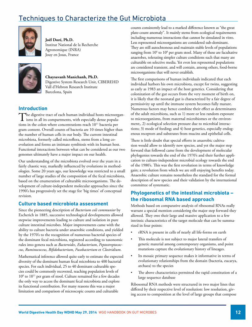
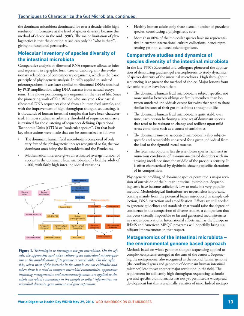
Figure 1. Technologies to investigate the gut microbiota. On the left side, the approaches used when culture of an individual microorgan-ism or the amplification of its genome is conceivable. On the right side, when most of the bacteria in the sample are not cultivable and when there is a need to compare microbial communities, approaches including metagenomics and metatranscriptomics are applied to the whole microbial community in the sample to collect information on microbial diversity, gene content and gene expression.
Human Samples
Genomic DNA
16S rDNA PCR Shotgun (DNA fragmentation)
High‐throughput Sequencing
Community Diversity
Genome Sequencing
Genome functions
Microbial culture
Genome amplification
Total RNA
cDNA synthesis
Gene expression
No cultureNo amplified genome
PhylogeneticMicroarray
High‐throughput Sequencing
rRNA removal
cDNA synthesis
Microarray Gene probes
Extraction
Metagenomics Metatranscriptomics
Gene content
High‐throughput Sequencing
World Digestive Health Day WDHD May 29, 2014 WGO HANDBOOK ON GUT MICROBES 14
nomics represents a unique opening towards addressing the question “who is doing what” beyond simply assessing “who is there”.
As for the human genome, a global effort has been coordinated and steered by the International Human Microbiome Consor-tium (IHMC). It seeks to establish and characterize the human gut microbiome and determine its importance for human health. The Human Microbiome Project (HMP) provided an opportu-nity to study the structure, function and diversity of the healthy human microbiome from samples of around 300 US adults, (HMP Consortium 2012) and the relationships between diet, age, and changes in the microbiome. Similarly, the Metagenom-ics of the Human Intestinal Tract (MetaHIT) project studied the metagenomic profile of faecal samples from an initial 124 healthy European adults (Qin 2010).
Interestingly, the highlights of these studies illustrate how much of a conceptual renewal this approach has stimulated:
• A repertoire of as many as 10 million non redundant micro-bial genes has been built from over a thousand individuals studied so-far.
• Over 99% of the genes of human microbiomes are bacterial and the entire MetaHIT cohort harbours between 1,000 and 1,150 prevalent bacterial species, while each individual hosts at least 160 such species.
• In spite of individual differences, all humans share a common core of prevalent and dominant species (Qin 2010).
• Rather than an even distribution around an average human microbiome, gut microbiota distribute into three densely populated zones within the ecological landscape of all possible compositions. Dominated by specific genera, these composi-tions have been named the Bacteroides-, Prevotella- and Rumi-nococcus- enterotypes.
• As for phylogenetic profiles, metagenomic profiles do show specificities in diseases, that may in turn allow patient stratifi-cation and individualized medicine or preventive nutrition.
• Low gene count is found to be associated with an increased risk of inflammatory comorbidities and an increased tendency to overweight/obese phenotypes.
Early life factors greatly affect the make-up and composition of the human gut microbiota. Profound differences in bacterial species assemblages and functional gene repertoires have been noted between individuals residing in the USA compared to those from Venezuela and Malawi. These distinctive features are evident throughout life after the age of three. Similar observations were reported when comparing infants from Italy and Burkina Faso. Could it be that behavioral, dietary and environmental changes, particularly affecting infant life, over several generations, led to a decrease in microbiome diversity among western world popula-tions, that may have consequences in terms of overall health / disease risk?
The future of microbiome studiesProgress in our understanding of the human intestinal microbiota and its role in health and disease has been over the past decade largely influenced by methodological and technological improve-ments. This is likely to continue in the near future and to conclude we propose a projection into the futures of microbiome studies.
StandardsHuman intestinal metagenomics opened new perspectives consid-ering depth and breadth of its molecular scanning power. Novel concepts emerged such as the core microbiome and the entero-types. Nevertheless, comparing data from different studies has re-mained extremely challenging and possibly hazardous considering that methodologies for sample collection, processing and analysis are neither robust nor concerted. Standard Operating Procedures are still critically awaited and will hopefully derive from ongoing efforts such as MBQC and IHMS.
Large prospective studiesCross-sectional studies have substantiated the concept of dysbio-sis, showing a distortion of microbiota composition in patients compared to healthy individuals. Yet, such observations have systematically and rightfully been criticized as giving no indica-tion of a causal link between observed over- or under-represented bacterial species and the disease condition. Causality is in principle only accessible via a prospective longitudinal study design allow-ing the identification of predictive biomarkers of the microbiota. Large longitudinal studies will also allow identifying predictors of response/non-response to nutritional supplementation or drug therapy. Combined efforts associating clinical teams and academics specialized in metagenomics are hence warranted.
Holistic viewMetagenomics markedly improved our ability to explore the functional potential of the human intestinal microbiota. It is still several steps away from microbe-host interactions on a scale of integrated genomics while metatranscriptomics, metaproteomics and metabolomics are rapidly developing. Their application to intestinal contents will deliver a holistic view of the interactions between the microbiome and host physiology. The main challenge will be the integration of complex data in order to identify mean-ingful relationships.
Ecological understandingUnderstanding what is a ‘healthy state of the microbiota’ will require a strong foundation of knowledge on how it structures after birth as well as what characteristics determine its resistance to change and its resilience, both structural and functional, in re-sponse to various perturbations such as drug therapies, changes in environment and/or nutrition. We really lack the ecological under-standing of the parameters that control composition and change in the microbiota to evolve to a next generation of knowledge-based, scientifically developed strategies of beneficial modulation of the microbiota.
Techniques to Characterize the Gut Microbiota, continued.
World Digestive Health Day WDHD May 29, 2014 WGO HANDBOOK ON GUT MICROBES 15
References1. Arumugam M, Raes J, Pelletier E, Le Paslier D, Yamada T,
Mende DR, Fernandes GR, Tap J, Bruls T, Batto JM, et al. Enterotypes of the human gut microbiome. Nature 2011, 473:174-180.
2. Blottière HM, deVos WM, Ehrlich SD, Doré J. Human intes-tinal metagenomics – state of the art and future. Curr Opin Microbiol, in press.
3. Brown J, de Vos WM, DiStefano PS, Doré J, Huttenhower C, Knight R, Lawley TD, Raes J, Turnbaugh P. Translating the human microbiome. Nat Biotechnol. 2013 Apr; 31(4):304-8. doi: 10.1038/nbt.2543.
4. De Filippo C, Cavalieri D, Di Paola M, Ramazzotti M, Poul-let JB, Massart S, Collini S, Pieraccini G, Lionetti P. Impact of diet in shaping gut microbiota revealed by a comparative study in children from Europe and rural Africa. Proc Natl Acad Sci USA 2010, 107:14691-14696.
5. Doré J, Simrén M, Buttle L, Guarner F. Hot topics in gut microbiota. United Europeen Gastroenterology Journal. 2013 doi: 10.1177/2050640613502477.
Techniques to Characterize the Gut Microbiota, continued.
6. Human Microbiome Project Consortium. Structure, function and diversity of the healthy human microbiome. Nature 2012, 486:207-214.
7. Lepage P, Leclerc MC, Joossens M, Mondot S, Blottière HM, Raes J, Ehrlich D, Doré J. A metagenomic insight into our gut’s microbiome. Gut 2013, 62:146-158.
8. Lozupone CA, Stombaugh JI, Gordon JI, Jansson JK, Knight R. Diversity, stability and resilience of the human gut micro-biota. Nature 2012, 489:220-230.
9. Qin J, Li R, Raes J, Arumugam M, Burgdorf KS, Manichanh C, Nielsen T, Pons N, Levenez F, Yamada T, et al. A human gut microbial gene catalogue established by metagenomic sequencing. Nature 2010, 464:59-65.
10. Yatsunenko T, Rey FE, Manary MJ, et al. Human gut microbiome viewed across age and geography. Nature 2012; 486(7402): 222–227.
World Digestive Health Day WDHD May 29, 2014 WGO HANDBOOK ON GUT MICROBES 16
Composition and Structure of the Human Gut Microbiota
The advent of high-throughput sequencing technologies has lead to a turning point in our understanding of the microbial
colonization of the human gut. Such culture-independent methods allow the characterization of microbial communities as a whole, through the analysis of the genetic material present in an environ-ment. The most common approach consists on the extraction of DNA from a biological sample, followed by the amplification and sequencing of 16S ribosomal RNA genes in the sample. The 16S rRNA gene is present in all bacteria and contains both conserved and variable regions. Thus, similarities and differences in the sequence of nucleotides of the 16S rRNA gene allow taxonomic identification ranging from the domain and phylum level to the species or strain level. Taxonomic identification is based on comparison of 16S rRNA sequences in the sample with reference sequences in the database. In this way, studies on the 16S rRNA gene provide information about bacterial composition and diver-sity of species in a given sample.
The most powerful molecular approach is not limited to 16S rRNA sequencing but it addresses all the genetic material in the sample. The decreasing cost and increasing speed of DNA sequenc-ing, coupled with advances in computational analyses of large datasets, have made it feasible to analyse complex mixtures of en-tire genomes with reasonable coverage. The resulting information describes the collective genetic content of the community from which functional and metabolic networks can be inferred. Impor-tantly, whole genome sequencing provides information about non-bacterial members in the community, including viruses, yeasts and protists. This approach has the advantage of not only providing the phylogenetical characterization of community members but also informing about biological functions present in the community.
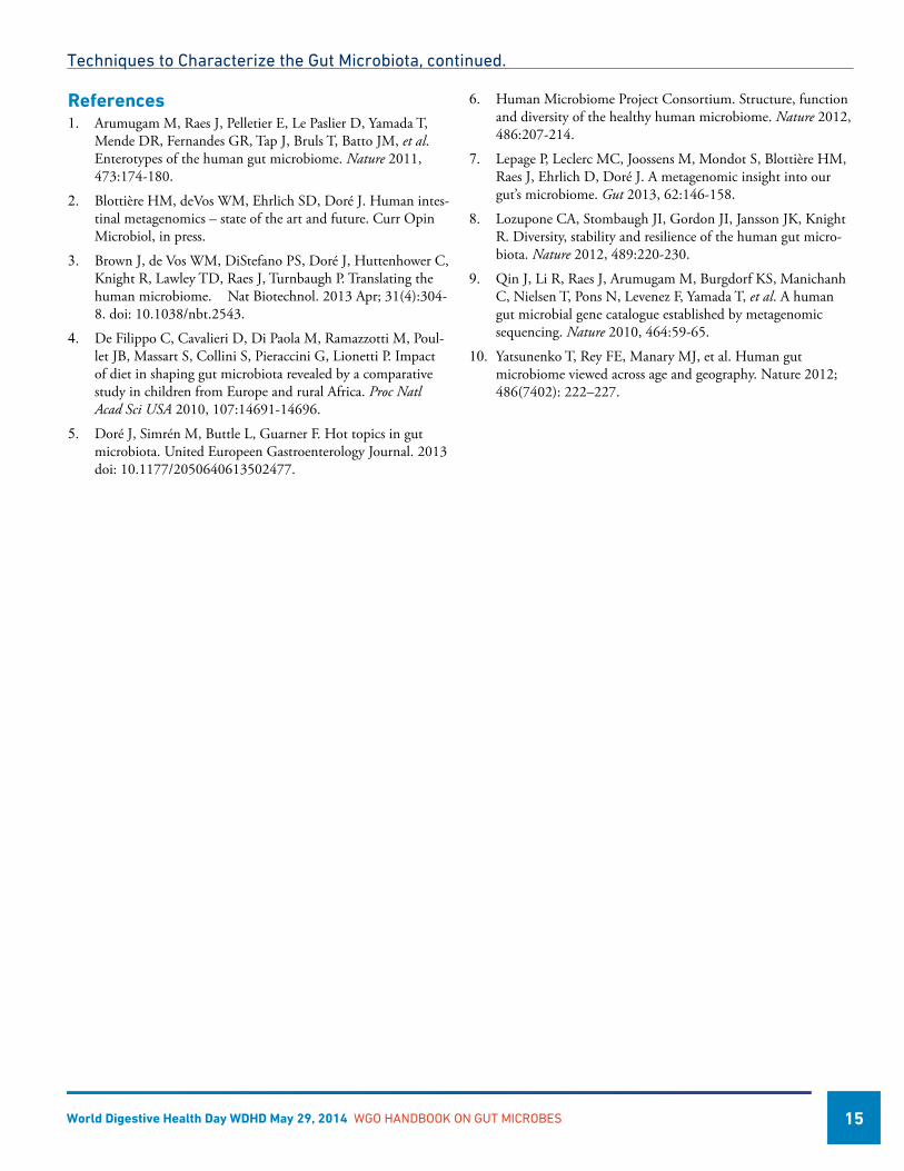
Diversity of the gut microbiotaEstimates suggest that the colon, by far the largest ecological niche for microbial communities in the human body, harbours over 1014 microbial cells, most of them belonging to the domain Bacteria. Molecular studies of faecal samples have highlighted that only 7 to 9 of the 55 known divisions or phyla of the domain Bacteria are detected in faecal or mucosal samples from the human gut. Around 90% of all the bacterial taxa belong to just two divisions: Bacteroidetes and Firmicutes. The other divisions that have been consistently found in samples from the human distal gut are Proteobacteria, Actinobacteria, Fusobacteria, and Verrucomicro-bia. Only very few species of Archea (mostly Methanobrevibacter smithii) seem to be represented in the human distal gut microbio-ta. Eukaryotes (yeasts and protists), and Viruses (phagi and animal viruses) are also present (Figure 1).
Virginia Robles-Alonso, MD Digestive System Research Unit Hospital Vall d’Hebron, Ciberehd Barcelona, Spain
Francisco Guarner, MD Digestive System Research Unit Hospital Vall d’Hebron, Ciberehd Barcelona, Spain
Dusko Ehrlich, Ph.D. Institut National de la Recherche Agronomique, Metagenopolis Unit, Jouy en Josas, France and King’s College London, UK
Figure 1: Phylogenetic classification and abundance (logarithmic scale) of mi-crobial genes identified in faecal samples from European individuals. The vast majority of gene sequences belong to the domain Bacteria or cannot be classi-fied (unknown). Only low percentages were classified as Archea, Eukaryotes or Viruses. Data extracted from supplementary files in Arumugam et al.
Each individual harbours his or her own distinctive pattern of gut microbial communities. Sequencing analysis of 16S rRNA gene indicates that there are differences between faecal and mucosa-associated communities within the same individual. Bacterial composition in the lumen varies from caecum to rectum, and faecal samples may not reproduce luminal contents in proximal segments of the gastrointestinal tract. In contrast, the community of mucosa-associated bacteria is highly stable from terminal ileum to the large bowel in a given individual. However, stool samples are widely accepted as the best approach for investigating gut micro-bial communities due to their accessibility for multiple sampling over time; they should be viewed as a proxy for other, less acces-sible, anatomic sites.
Factors such as diet, drug intake, travelling or simply colonic transit time, have an impact on microbial composition in faecal samples over time in a unique host. Thus, intra-individual fluctua-tions in the composition of the microbiota can be remarkable, but the microbial ecosystem tends to return to their typical compo-
World Digestive Health Day WDHD May 29, 2014 WGO HANDBOOK ON GUT MICROBES 17
sitional pattern and most strains are resident in an individual for decades. This phenomenon is called resilience.
There are striking differences in composition and diversity between westernized and non-westernized populations. Microbial diversity changes with age, but the faecal microbiota of adults is less diverse in metropolitan areas of North America than in rural non-western-ized populations of Africa and South America.
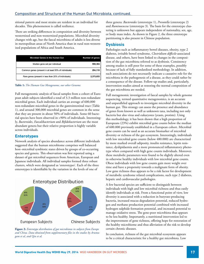
three genera: Bacteroides (enterotype 1), Prevotella (enterotype 2) and Ruminococcus (enterotype 3). The basis for the enterotype clus-tering is unknown but appears independent of nationality, sex, age, or body mass index. As shown in Figure 2, the three enterotype partitioning is also present in Chinese population.
DysbiosisPathologies such as inflammatory bowel diseases, obesity, type 2 diabetes, irritable bowel syndrome, Clostridium difficile-associated disease, and others, have been linked to changes in the composi-tion of the gut microbiota referred to as dysbiosis. Consistency among studies is still poor for some of these examples, possibly because of lack of fully standardized methodology. In addition, such associations do not necessarily indicate a causative role for the microbiota in the pathogenesis of a disease, as they could rather be a consequence of the disease. Follow-up studies and, particularly, intervention studies aimed at restoring the normal composition of the gut microbiota are needed.
Full metagenomic investigation of faecal samples by whole genome sequencing, termed quantitative metagenomics, is an accurate and unparalleled approach to investigate microbial diversity in the human gut. This strategy can assess the presence and abundance of genes from known as well as unknown taxa, including not only bacteria but also virus and eukaryotes (yeasts, protists). Using this methodology, it has been shown that a high proportion of Europeans (23%) exhibit microbial gene counts below the median of 600.000 previously established in a European cohort. Microbial gene counts can be used as an accurate biomarker of microbial diversity or richness of the gut ecosystem. Interestingly, individuals with low microbial gene counts (below 480.000) are characterized by more marked overall adiposity, insulin resistance, leptin resis-tance, dyslipidaemia and a more pronounced inflammatory pheno-type when compared with high gene counts individuals. Moreover, these metabolic parameters were found to be slightly altered even in otherwise healthy individuals with low microbial gene counts. Obese individuals with low gene counts gain more weight over time and have a propensity towards a malignant form of obesity. Low gene richness thus appears to be a risk factor for development of metabolic syndrome related complications, such type 2 diabetes, hepatic and cardiovascular pathologies.
A few bacterial species are sufficient to distinguish between individuals with high and low microbial richness and thus easily identify individuals at risk. From a functional point of view, low diversity is associated with a reduction in butyrate-producing bacteria, increased mucus degradation potential, reduced hydro-gen and methane production potential combined with increased hydrogen sulphide formation potential, and increased potential to manage oxidative stress. The gene-poor microbiota thus appears to be less healthy. Importantly, a nutritional intervention led to the improvement of gene richness, offering hope for restoration of the healthy microbiome and thus alleviation of the risk to develop certain chronic diseases.
In conclusion, richness of the gut microbial ecosystem appears to be a critical characteristic for a healthy gut microbiota. Low
Figure 2: Enterotype distribution of gut microbiotas in subjects from Europe and China. Data obtained from supplementary files in the studies by Arumu-gam et al, and Qin et al.
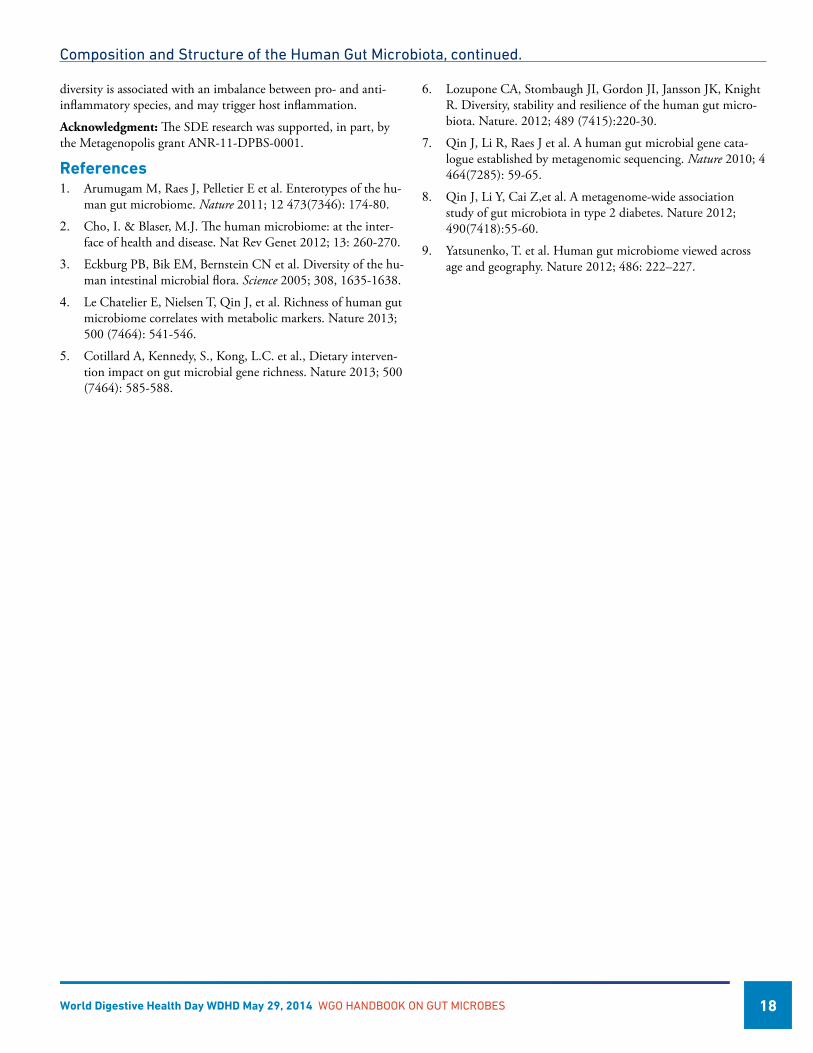
Table 1: The Human Gut Metagenome, our other Genome.
Composition and Structure of the Human Gut Microbiota, continued.
Full metagenomic analysis of faecal samples from a cohort of Euro-pean adult subjects identified a total of 3.3 million non-redundant microbial genes. Each individual carries an average of 600,000 non-redundant microbial genes in the gastrointestinal tract (Table 1), and around 300,000 microbial genes are common in the sense that they are present in about 50% of individuals. Some 60 bacte-rial species have been observed in >90% of individuals. Interesting-ly, Bacteroides, Faecalibacterium and Bifidobacterium are the most abundant genera but their relative proportion is highly variable across individuals.
EnterotypesNetwork analysis of species abundance across different individuals suggested that the human microbiome comprises well balanced host–microbial symbiotic states driven by groups of co-occurring species and genera. This observation was first reported using a dataset of gut microbial sequences from American, European and Japanese individuals. All individual samples formed three robust clusters, which were designated as ‘enterotypes’. Each of the three enterotypes is identifiable by the variation in the levels of one of
World Digestive Health Day WDHD May 29, 2014 WGO HANDBOOK ON GUT MICROBES 18
Composition and Structure of the Human Gut Microbiota, continued.
diversity is associated with an imbalance between pro- and anti-inflammatory species, and may trigger host inflammation.
Acknowledgment: The SDE research was supported, in part, by the Metagenopolis grant ANR-11-DPBS-0001.
References1. Arumugam M, Raes J, Pelletier E et al. Enterotypes of the hu-
man gut microbiome. Nature 2011; 12 473(7346): 174-80.
2. Cho, I. & Blaser, M.J. The human microbiome: at the inter-face of health and disease. Nat Rev Genet 2012; 13: 260-270.
3. Eckburg PB, Bik EM, Bernstein CN et al. Diversity of the hu-man intestinal microbial flora. Science 2005; 308, 1635-1638.
4. Le Chatelier E, Nielsen T, Qin J, et al. Richness of human gut microbiome correlates with metabolic markers. Nature 2013; 500 (7464): 541-546.
5. Cotillard A, Kennedy, S., Kong, L.C. et al., Dietary interven-tion impact on gut microbial gene richness. Nature 2013; 500 (7464): 585-588.
6. Lozupone CA, Stombaugh JI, Gordon JI, Jansson JK, Knight R. Diversity, stability and resilience of the human gut micro-biota. Nature. 2012; 489 (7415):220-30.
7. Qin J, Li R, Raes J et al. A human gut microbial gene cata-logue established by metagenomic sequencing. Nature 2010; 4 464(7285): 59-65.
8. Qin J, Li Y, Cai Z,et al. A metagenome-wide association study of gut microbiota in type 2 diabetes. Nature 2012; 490(7418):55-60.
9. Yatsunenko, T. et al. Human gut microbiome viewed across age and geography. Nature 2012; 486: 222–227.
World Digestive Health Day WDHD May 29, 2014 WGO HANDBOOK ON GUT MICROBES 19
Acquisition of the Human Gut MicrobiotaYanjiao Zhou, MD, Ph.D. Department of Pediatrics The Microbial Genomics Group at The Genome Institute Washington University School of Medicine St. Louis, USA
Barbara B. Warner, MD Department of Pediatrics The Microbial Genomics Group at The Genome Institute Washington University School of Medicine St. Louis, USA
Phillip I. Tarr, MD Department of Pediatrics The Microbial Genomics Group at The Genome Institute Washington University School of Medicine St. Louis, USA
Introduction
In the days and weeks following parturition, the human infant gut acquires its own microbiome, and the transition to bacte-
rial population equilibrium begins. This early-in-life microbial population quite likely influences later-in-life host biology. The process by which the human gastrointestinal tract is colonized after birth is a fascinating example of ecological succession, but it is also a process that is very poorly studied. By delineating the dynamics of the de novo assembly of this microbial community we could gain a better understanding as to how the gut acquires its founding microbiome, the first step in the process to population equilibrium (Yatsunenko, Rey et al. 2012, Faith, Guruge et al. 2013, Zhou, Gao et al. 2013). Here, we review the potential implications of this colonization and succession, and discuss its importance for later-in-life events.
MeconiumClassic theory teaches that the meconium is devoid of microorgan-isms at birth (Tissier 1900). However, several reports over the past decade have prompted us to reconsider this dogma (Funkhouser and Bordenstein 2013). Specifically, there are bacterial sequences
Setting (Reference) No. of Sub-jects (ages at sampling
Samples per subject
Enumeration technology(16S rRNA region sequenced)*
Conclusions Comments
USA(Palmer, Bik et al. 2007)
14 (0-1 yr)
26 Microarray Colonization process of the gut flora is individual specific; Gut microbiota converges to adult-like profile at age 1 year
Comprehensive characterization of the progression of gut microbiota in term babies
Finland, Spain(Collado, Isolauri et al. 2010)
42 (1 and 6 mo)
2 qPCR Infant gut microbes are affected by maternal BMI and BMI gain in pregnancy
Mother’s microbiota is an important factor for infant health
USA(Koenig, Spor et al. 2011)
1 (0-2.5 yr)
60 454 FLX pyrosequencing (V1-2)
Microbial succession associated with diet and other life events; Gut bacteria start to stabilize at 1 year of age
Fine-scale temporal sampling of one infant
Africa, USA, Amer-indians(Yatsunenko, Rey et al. 2012)
146 (0-3 yr)
1 Illumina HiSeq 2000 (V4)
Gut microbiome varies by age and geography, but becomes adult-like at the age of 3.
Multinational survey of microbiome changes at population level within a wide range of ages
Switzerland(Jost, Lacroix et al. 2012)
7 (4-30 d)
3 Sanger (V1-9), culture and 454 pyrosequencing (V4-5)
Anaerobes are pioneer colonizers, and their abundances are similar as adults in the first week of life.
Used complementary techniques to study gut colonization in early infancy
Sweden 65 (1-8 wk)
4 Culture Early gut microbiota including E. coli and Bifidobacteria contribute to B cell activation and memory differentiation
Associate gut colonization pattern with development of immune system in humans
USA(Song, Lauber et al. 2013)
12 (<1 yr)
1 Illumina GAIIx (V2) Pronounced changes in gut microbi-ome occur in a protracted timeframe;
Exogenous factors shape gut bacterial community structure.
Canada(Azad, Konya et al. 2013)
24 (4 mo)
1 High throughput se-quencing (V5-7)
Formula-fed infants have higher rich-ness than breastfed infants. C. difficile is more abundant in formula-fed babies. Escherichia and Bacteroides were less abundant in babies born by Caesarian section
A small cross sectional study on how diet and mode of delivery could affect microbial community structure in early day of life
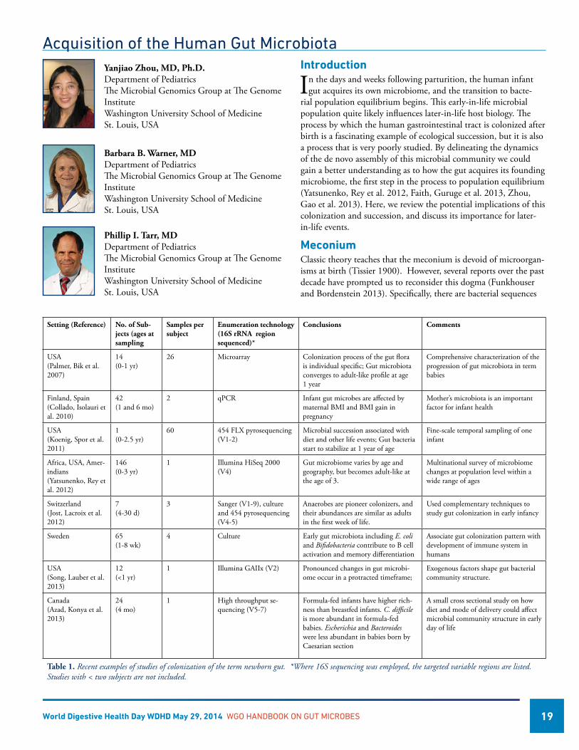
Table 1. Recent examples of studies of colonization of the term newborn gut. *Where 16S sequencing was employed, the targeted variable regions are listed. Studies with < two subjects are not included.
World Digestive Health Day WDHD May 29, 2014 WGO HANDBOOK ON GUT MICROBES 20
in freshly passed meconium (Mshvildadze, Neu et al. 2010), and cord blood can contain viable bacteria (Jimenez, Fernandez et al. 2005). The amniotic fluid and placenta also have evidence of microbial colonization, even in the absence of premature rupture of the membranes. However, it is important to note that DNA sequences are more commonly identified than are viable bacteria (Jimenez, Fernandez et al. 2005); (DiGiulio, Romero et al. 2008); (Rautava, Collado et al. 2012). Moreover, decades of gnotobiotic animal research have been performed in which viable bacteria are not transmitted vertically from mothers to offspring. If the first (or any) generation following derivation retained viable bacteria, then one would expect that gnotobiotic techniques would be unsuccess-ful. There has also been considerable attention paid to amniotic infection/colonization and preterm labor, but the role of bacteria within fetal membranes in causing preterm labor needs further work before this association should be considered to be established (Steel, Malatos et al. 2005); (Stout, Conlon et al. 2013).
First colonizers in term infantsSeveral studies have presented the sequential phases of bacterial colonization in term infants. These studies pose logistic challenges, particularly because it is very difficult to obtain stools at high fre-quencies from infants residing with their families in the communi-ty, and sampling and sequencing methodologies differ considerably between studies. Table 1 summarizes eight series published since 2007. A convergence to an adult population of gut microbes does not occur until about three years of age. Gram-negative bacteria are present at concentrations that are greater than in the stools of older children and adults (Zhang, DiBaise et al. 2009, Saulnier, Riehle et al. 2011). Interestingly, antibiotic administrations are not uniformly correlated with alterations in microbial content, with some individuals showing perturbations and others not (Palmer, Bik et al. 2007). Anaerobes are well represented members of the gut microbiota within several days of birth (Jost, Lacroix et al. 2012). (Karlsson, Molin et al. 2011). Dietary changes precede gut microbial population shifts (Koenig, Spor et al. 2011), and initial feeding choice (breast milk or formula) had persistent ef-fects (Fallani, Amarri et al. 2011). Maternal body habitus might also play a role in infant microbial gut content: elevated maternal BMI is associated with higher concentrations of fecal Bacteroides, Clostridium, and Staphylococcus genera, and lower densities of Bifidobacteria. Akkermansia muciniphila, Staphylococcus spp. and Clostridium difficile were lower in infants of mothers who had normal body mass indices (Collado, Isolauri et al. 2010).
Published data from premature infants are also limited. Table 2 represents the current state of sequencing of premature infant cohorts without substantial overt gut pathology. As with term infants, the studies published so far have utilized different frequen-cies of sampling and a diversity of bacterial enumeration strategies, including culturing, gel-based methods, and sequencing. Because the stools of few such premature infants have been sequenced, and because sampling is generally limited, it is difficult to draw firm conclusions about the earliest in life colonization events in preterm infants. However, Gammaproteobacteria are exceptionally abun-
dant, and present in higher proportions than in older children and adults (Zhang, DiBaise et al. 2009, Saulnier, Riehle et al. 2011).
Study reference Subjects Sampling Sample n
Method(s)*
(de la Cochetiere, Piloquet et al. 2004)
9 Weekly 23 TTGE
(Wang, Hoenig et al. 2009)
10 Once 10 16S (V1-9)
(Chang, Shin et al. 2011)
10 <72h, 2 wks, 1 mo
30 16S (V2)
(Mshvildadze, Neu et al. 2010)
23 Weekly 1-15/subject
DGGE,16S (V1-2)
(Jacquot, Neveu et al. 2011)
29 Every 3rd day, to DOL 56 and at discharge
342 TTGE
(LaTuga, Ellis et al. 2011)
11 DOL 9 to 35 20 16S
(Mai, Young et al. 2011)
9 Weekly 18 16S (V1-2)
(Smith, Bode et al. 2012)
142 Day 0 to 5, days 10 and 30
423 Culture, DGGE
(Stewart, Marrs et al. 2012)
30 Weekly 76 Culture,DGGE
(Claud, Keegan et al. 2013)
5 Weekly to 10 wk
30 16S (V3-4), shotgun sequencing
(Morrow, Lagomar-cino et al. 2013)
21 Up to DOL 16
40 16S (V3-5)
(Torrazza, Ukhanova et al. 2013)
35 Weekly 77 16S (V1-3) and qPCR for Bifidobacter
(Mai, Torrazza et al. 2013)
28 Weekly 71 16S (V1-3)
(Stewart, Marrs et al. 2013)
22 Not specified 134 16S (V3-5), DGGE
(Normann, Fahlen et al. 2013)
10 Weekly 36 16S (V3-4)
Table 2. First colonizers in pre-term infants. *Where 16S sequencing was employed, the targeted variable regions are listed. Abbreviations: DOL = Day of life; TTGE = temporal temperature gradient gel electrophoresis; DGGE = denaturing gradient gel electrophoresis
Mode of delivery and infant gut microbiotaSome literature suggests effects of mode of birth on infant micro-biota. Infants born vaginally or via Caesarian section are generally colonized at extra-intestinal sites with bacteria of vaginal or skin origian, respectively, but the number of mother-infant dyads stud-ied is still quite limited (Dominguez-Bello, Costello et al. 2010). When gut microbial populations are examined, infants born via vaginal delivery have a more rapid in-flux of Proteobacteria (Gram-negative organisms), and a higher proportion of Bifidobacteria
Acquisition of the Human Gut Microbiota, continued
World Digestive Health Day WDHD May 29, 2014 WGO HANDBOOK ON GUT MICROBES 21
(multiple species, but particularly catenulatum and longum) than those born via Caesarian section (Biasucci, Rubini et al. 2010). By four months of age, the gut microbial concentrations of infants who had been born via Caesarian are under-represented in Esch-erichia coli and Bacteroides spp. (Azad, Konya et al. 2013). Again, the number of subjects is small, and the number of samplings per child in these studies is limited.
Effects of early-in-life colonzation on subseqent well-being of the hostBecause the members of the gut microbiome in later life are associ-ated with various states of health and disease (Turnbaugh, Hamady et al. 2009) (Karlsson, Fak et al. 2012) (Nieuwdorp, Gilijamse et al. 2014), it is logical to consider the durable effects of early-in-life colonization of the gut with later-in-life events. Animal data are intriguing: germ free Swiss-Webster mice introduced to specific pathogen free (but colonized) mice at 1-3 weeks of age have higher concentrations of circulating regulatory cytokines than do germ-free mice not exposed to colonized cage mates (Hansen, Nielsen et al. 2012). Mice raised in germ free conditions also have an exag-erated nueroendocrine response to stress, which can be mitigated by early expsoure to specific comensal bacteria (Sudo, Chida et al. 2004); (Clarke, Grenham et al. 2013). Mice exposed to bacteria early-in-life are also protected from oxazolone-induced colitis in an invariant natural killer cell model (Olszak, An et al. 2012). In humans, epidemiologic evidence suggests that early-in-life expo-sures to microorganisms influences the subsequent development of asthma and inflammatory bowel disease (Lopez-Serrano, Perez-Calle et al. 2010); (Ege, Mayer et al. 2011).
Some preliminary prospective human data suggest that early-in-life colonization does, indeed, affect later clinical outcomes. Children with allergies at age five years had lower densities of Lactobacilli in their stools in early infancy than children without allergies (Jo-hansson, Sjogren et al. 2011). Colonization with C. difficile in the first month of life is related to atopy and asthma at age six years (van Nimwegen, Penders et al. 2011). Staphylococcus species and E. coli and Bacteroides in stools in the first several months of life were associated with expected childhood body mass (weight for age Z-scores) at up to 24 months of age in a Norwegian study (White, Bjornholt et al. 2013).
ConclusionsAnimal and circumstantial human data continue to accrue and implicate the enteric microbiome in infants and children with later disorders in childhood and adulthood. In future studies, it will be important to seek the presence or absence of harmful or protective microbial drivers of host phenotypes before the outcomes are ap-parent. Temporality, i.e., the exposure to a factor occurs before an outcome is apparent, is a necessary criterion in building a case that a microbe or microbial community ordains a disease process (or protection from that process) (Bradford-Hill 1965). This task of determining the “normal” pattern of colonization will be challeng-ing, because there are likely to be geographic (Yatsunenko, Rey et al. 2012) and seasonal variations (Davenport, Mizrahi-Man et al.
2014), as well as abrupt changes in community structure (Zoe-tendal, Akkermans et al. 1998, Palmer, Bik et al. 2007, Costello, Lauber et al. 2009, Caporaso, Lauber et al. 2011, Human Mi-crobiome Project 2012). It will be important to also incorporate computational considerations that are able to address community changes over time(Stein, Bucci et al. 2013) in dynamic popula-tions (Marino, Baxter et al. 2014).
References1. Azad, M. B., et al. (2013). “Gut microbiota of healthy Cana-
dian infants: profiles by mode of delivery and infant diet at 4 months.” CMAJ 185(5): 385-394.
2. Biasucci, G., et al. (2010). “Mode of delivery affects the bacterial community in the newborn gut.” Early Hum Dev 86 Suppl 1: 13-15.
3. Bradford-Hill, A. (1965). “The Environment and Disease: As-sociation or Causation?” Proc R Soc Med 58: 295-300.
4. Caporaso, J. G., et al. (2011). “Moving pictures of the human microbiome.” Genome Biology 12(5): R50.
5. Chang, J. Y., et al. (2011). „Pyrosequencing-based molecular monitoring of the intestinal bacterial colonization in preterm infants.“ J Pediatr Gastroenterol Nutr 53(5): 512-519.
6. Clarke, G., et al. (2013). „The microbiome-gut-brain axis dur-ing early life regulates the hippocampal serotonergic system in a sex-dependent manner.“ Mol Psychiatry 18(6): 666-673.
7. Claud, E. C., et al. (2013). „Bacterial community structure and functional contributions to emergence of health or necro-tizing enterocolitis in preterm infants.“ Microbiome 1(1): 20.
8. Collado, M. C., et al. (2010). „Effect of mother‘s weight on infant‘s microbiota acquisition, composition, and activity during early infancy: a prospective follow-up study initiated in early pregnancy.“ Am J Clin Nutr 92(5): 1023-1030.
9. Costello, E. K., et al. (2009). „Bacterial community varia-tion in human body habitats across space and time.“ Science 326(5960): 1694-1697.
10. Davenport, E. R., et al. (2014). „Seasonal variation in human gut microbiome composition.“ PLoS One 9(3): e90731.
11. DiGiulio, D. B., et al. (2008). “Microbial prevalence, diver-sity and abundance in amniotic fluid during preterm labor: a molecular and culture-based investigation.” PLoS One 3(8): e3056.
12. Dominguez-Bello, M. G., et al. (2010). “Delivery mode shapes the acquisition and structure of the initial microbiota across multiple body habitats in newborns.” Proc Natl Acad Sci U S A 107(26): 11971-11975.
13. Ege, M. J., et al. (2011). “Exposure to environmental micro-organisms and childhood asthma.” N Engl J Med 364(8): 701-709.
14. Faith, J. J., et al. (2013). “The long-term stability of the hu-man gut microbiota.” Science 341(6141): 1237439.
Acquisition of the Human Gut Microbiota, continued
World Digestive Health Day WDHD May 29, 2014 WGO HANDBOOK ON GUT MICROBES 22
15. Fallani, M., et al. (2011). “Determinants of the human infant intestinal microbiota after the introduction of first comple-mentary foods in infant samples from five European centres.” Microbiology 157(Pt 5): 1385-1392.
16. Funkhouser, L. J. and S. R. Bordenstein (2013). „Mom knows best: the universality of maternal microbial transmission.“ PLoS Biol 11(8): e1001631.
17. Hansen, C. H., et al. (2012). „Patterns of early gut coloniza-tion shape future immune responses of the host.“ PLoS One 7(3): e34043.
18. Human Microbiome Project, C. (2012). „Structure, function and diversity of the healthy human microbiome.“ Nature 486(7402): 207-214.
19. Jacquot, A., et al. (2011). “Dynamics and clinical evolution of bacterial gut microflora in extremely premature patients.” J Pediatr 158(3): 390-396.
20. Jimenez, E., et al. (2005). “Isolation of commensal bacteria from umbilical cord blood of healthy neonates born by cesar-ean section.” Curr Microbiol 51(4): 270-274.
21. Johansson, M. A., et al. (2011). “Early colonization with a group of Lactobacilli decreases the risk for allergy at five years of age despite allergic heredity.” PLoS One 6(8): e23031.
22. Jost, T., et al. (2012). “New insights in gut microbiota es-tablishment in healthy breast fed neonates.” PLoS One 7(8): e44595.
23. Karlsson, C. L., et al. (2011). “The pioneer gut microbiota in human neonates vaginally born at term-a pilot study.” Pediatr Res 70(3): 282-286.
24. Karlsson, F. H., et al. (2012). “Symptomatic atherosclerosis is associated with an altered gut metagenome.” Nat Commun 3: 1245.
25. Koenig, J. E., et al. (2011). „Succession of microbial consortia in the developing infant gut microbiome.“ Proc Natl Acad Sci U S A 108 Suppl 1: 4578-4585.
26. LaTuga, M. S., et al. (2011). “Beyond bacteria: a study of the enteric microbial consortium in extremely low birth weight infants.” PLoS One 6(12): e27858.
27. Lopez-Serrano, P., et al. (2010). “Environmental risk factors in inflammatory bowel diseases. Investigating the hygiene hy-pothesis: a Spanish case-control study.” Scand J Gastroenterol 45(12): 1464-1471.
28. Mai, V., et al. (2013). “Distortions in development of intes-tinal microbiota associated with late onset sepsis in preterm infants.” PLoS One 8(1): e52876.
29. Mai, V., et al. (2011). “Fecal microbiota in premature infants prior to necrotizing enterocolitis.” PLoS One 6(6): e20647.
30. Marino, S., et al. (2014). “Mathematical modeling of primary succession of murine intestinal microbiota.” Proceedings of
the National Academy of Sciences of the United States of America 111(1): 439-444.
31. Morrow, A. L., et al. (2013). “Early microbial and metabolo-mic signatures predict later onset of necrotizing enterocolitis in preterm infants.” Microbiome 1(1): 13.
32. Mshvildadze, M., et al. (2010). “Intestinal microbial ecology in premature infants assessed with non-culture-based tech-niques.” J Pediatr 156(1): 20-25.
33. Nieuwdorp, M., et al. (2014). “Role of the Microbiome in Energy Regulation and Metabolism.” Gastroenterology.
34. Normann, E., et al. (2013). “Intestinal microbial profiles in extremely preterm infants with and without necrotizing enterocolitis.” Acta Paediatr 102(2): 129-136.
35. Olszak, T., et al. (2012). “Microbial exposure during early life has persistent effects on natural killer T cell function.” Science 336(6080): 489-493.
36. Palmer, C., et al. (2007). “Development of the human infant intestinal microbiota.” PLoS Biol 5(7): e177.
37. Rautava, S., et al. (2012). “Probiotics modulate host-microbe interaction in the placenta and fetal gut: a randomized, double-blind, placebo-controlled trial.” Neonatology 102(3): 178-184.
38. Saulnier, D. M., et al. (2011). “Gastrointestinal microbiome signatures of pediatric patients with irritable bowel syn-drome.” Gastroenterology 141(5): 1782-1791.
39. Smith, B., et al. (2012). “Investigation of the early intestinal microflora in premature infants with/without necrotizing enterocolitis using two different methods.” Pediatr Res 71(1): 115-120.
40. Song, S. J., et al. (2013). “Cohabiting family members share microbiota with one another and with their dogs.” Elife 2: e00458.
41. Steel, J. H., et al. (2005). “Bacteria and inflammatory cells in fetal membranes do not always cause preterm labor.” Pediatr Res 57(3): 404-411.
42. Stein, R. R., et al. (2013). “Ecological modeling from time-se-ries inference: insight into dynamics and stability of intestinal microbiota.” PLoS Computational Biology 9(12): e1003388.
43. Stewart, C. J., et al. (2012). “The preterm gut microbiota: changes associated with necrotizing enterocolitis and infec-tion.” Acta Paediatr 101(11): 1121-1127.
44. Stewart, C. J., et al. (2013). “Development of the preterm gut microbiome in twins at risk of necrotising enterocolitis and sepsis.” PLoS One 8(8): e73465.
45. Stout, M. J., et al. (2013). “Identification of intracellular bacteria in the basal plate of the human placenta in term and preterm gestations.” Am J Obstet Gynecol 208(3): 226 e221-227.
Acquisition of the Human Gut Microbiota, continued
World Digestive Health Day WDHD May 29, 2014 WGO HANDBOOK ON GUT MICROBES 23
46. Sudo, N., et al. (2004). “Postnatal microbial colonization programs the hypothalamic-pituitary-adrenal system for stress response in mice.” J Physiol 558(Pt 1): 263-275.
47. Tissier, H. (1900). Recherches sur la flore intestinale des nour-rissons (état normal et pathologique). Paris, G Carre and C Naud.
48. Torrazza, R. M., et al. (2013). “Intestinal microbial ecology and environmental factors affecting necrotizing enterocolitis.” PLoS One 8(12): e83304.
49. Turnbaugh, P. J., et al. (2009). “A core gut microbiome in obese and lean twins.” Nature 457(7228): 480-484.
50. van Nimwegen, F. A., et al. (2011). “Mode and place of delivery, gastrointestinal microbiota, and their influence on asthma and atopy.” J Allergy Clin Immunol 128(5): 948-955 e941-943.
51. Wang, Y., et al. (2009). “16S rRNA gene-based analysis of fecal microbiota from preterm infants with and without nec-rotizing enterocolitis.” ISME J 3(8): 944-954.
52. White, R. A., et al. (2013). “Novel developmental analyses identify longitudinal patterns of early gut microbiota that af-fect infant growth.” PLoS Comput Biol 9(5): e1003042.
53. Yatsunenko, T., et al. (2012). “Human gut microbiome viewed across age and geography.” Nature 486(7402): 222-227.
54. Zhang, H., et al. (2009). “Human gut microbiota in obe-sity and after gastric bypass.” Proceedings of the National Academy of Sciences of the United States of America 106(7): 2365-2370.
55. Zhou, Y., et al. (2013). “Biogeography of the ecosystems of the healthy human body.” Genome Biology 14(1): R1.
56. Zoetendal, E. G., et al. (1998). “Temperature gradient gel electrophoresis analysis of 16S rRNA from human fecal samples reveals stable and host-specific communities of active bacteria.” Applied and Environmental Microbiology 64(10): 3854-3859.
Acquisition of the Human Gut Microbiota, continued
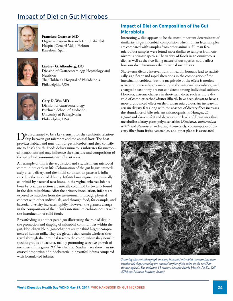
World Digestive Health Day WDHD May 29, 2014 WGO HANDBOOK ON GUT MICROBES 24
Impact of Diet on Gut Microbes
Scanning electron micrograph showing intestinal microbial communities with bacillar cell shape covering the mucosal surface of the colon in the rat (Rat-tus norvegicus). Bar indicates 15 microns (author Maria Vicario, Ph.D., Vall d’Hebron Research Institute, Spain).
Francisco Guarner, MD Digestive System Research Unit, Ciberehd Hospital General Vall d’Hebron Barcelona, Spain
Lindsey G. Albenberg, DO Division of Gastroenterology, Hepatology and Nutrition The Children’s Hospital of Philadelphia Philadelphia, USA
Gary D. Wu, MD Division of Gastroenterology Perelman School of Medicine University of Pennsylvania Philadelphia, USA
Diet is assumed to be a key element for the symbiotic relation-ship between gut microbes and the animal host. The host
provides habitat and nutrition for gut microbes, and they contrib-ute to host’s health. Foods deliver numerous substrates for microbi-al metabolism and may influence the structure and composition of the microbial community in different ways.
An example of this is the acquisition and establishment microbial communities early in life. Colonization of the gut begins immedi-ately after delivery, and the initial colonization pattern is influ-enced by the mode of delivery. Infants born vaginally are initially colonized by bacterial taxa found in the vagina, whereas infants born by cesarean section are initially colonized by bacteria found in the skin microbiota. After the primary inoculation, infants are exposed to microbes from the environment, through physical contact with other individuals, and through food, for example, and bacterial diversity increases rapidly. However, the greatest change in the composition of the infant’s intestinal microbiota occurs with the introduction of solid foods.
Breastfeeding is another paradigm illustrating the role of diet in the promotion and shaping of microbial communities within the gut. Non-digestible oligosaccharides are the third largest compo-nent of human milk. They are glycans that remain whole as they travel through the intestinal tract to the colon, where they nourish specific groups of bacteria, mainly promoting selective growth of members of the genus Bifidobacterium. Studies have shown an in-creased proportion of bifidobacteria in breastfed infants compared with formula-fed infants.
Impact of Diet on Composition of the Gut Microbiota Interestingly, diet appears to be the most important determinant of similarity in gut microbial composition when human fecal samples are compared with samples from other animals. Human fecal microbiota samples were found most similar to samples from om-nivorous primate species. The variety of foods in an omnivorous diet, as well as the free-living nature of our species, could affect how our diet determines the intestinal microbiota.
Short-term dietary interventions in healthy humans lead to statisti-cally significant and rapid alterations in the composition of the intestinal microbiota, but the magnitude of the effect is modest relative to inter-subject variability in the intestinal microbiota, and changes in taxonomy are not consistent among individual subjects. However, extreme changes in short-term diets, such as those de-void of complex carbohydrates (fibers), have been shown to have a more pronounced effect on the human microbiota. An increase in certain dietary fats along with the absence of dietary fiber increases the abundance of bile-tolerant microorganisms (Alistipes, Bi-lophila and Bacteroides) and decreases the levels of Firmicutes that metabolize dietary plant polysaccharides (Roseburia, Eubacterium rectale and Ruminococcus bromii). Conversely, consumption of di-etary fiber from fruits, vegetables, and other plants is associated
World Digestive Health Day WDHD May 29, 2014 WGO HANDBOOK ON GUT MICROBES 25
with significant and meaningful alterations in the gut microbiota. In controlled dietary experiments in humans, variations in intake of resistant starch or non-starch polysaccharide altered levels of specific bacterial taxa such as Ruminococcus bromii and Eubacteri-um rectale. These taxa were shown to selectively metabolize specific insoluble carbohydrate substrates based on in vitro analyses of human fecal samples.
Different diets driven among different populations help shape the taxonomy of their gut microbial ecosystem. In a landmark study, De Filippo et al showed that microbiota of children in Burkina Faso had greater amounts of Prevotella, lower amounts of Bacteroi-des, greater microbial richness and produced higher levels of short-chain fatty acids than the microbiota of European children. It would be reasonable to speculate that the agrarian diet of Burkina Faso (rich in carbohydrate content, fiber, and non-animal protein) compared with the Western diet (high in animal protein, sugar, starch, and fat and low in fiber) has a predominant role in these observed differences.
The inverse relationship between Prevotella and Bacteroides has been reproduced in studies comparing the intestinal microbiota of residents of agrarian societies with that of residents of industrial-ized societies. The MetaHIT Consortium proposed that people can be classified as having intestinal microbiota predominantly com-posed of Prevotella or Bacteroides; a third group has higher propor-tions of Ruminococcus compared with the others. These categories were named “enterotypes.” There has been considerable discussion about the integrity of enterotypes; some data sets support the existence of these categories, whereas others do not. Nonetheless, a greater proportion of Prevotella in the human intestinal micro-biota is a marker of residence in an agrarian culture, whereas a greater proportion of Bacteroides is associated with residence in more industrialized regions. Associations between diet and bacte-rial taxonomy, based on answers to dietary questionnaires collected over long periods, indicate that diet affects the proportions of Prevotella vs Bacteroides in US populations. Thus, the presence of stable gut microbial communities can be linked with long-term dietary patterns.
A number of studies have associated increased microbial richness, at either the taxonomic or gene level, with diets higher in fruits, vegetables, and fiber. In elderly subjects, differences in the tax-onomy of the intestinal microbiota were associated with residence in different environments. The most extreme differences were ob-served between those living independently in the community resi-dence and those in long-term residential care and were attributed to differences in diet; community residents typically consume diets higher in fiber and lower in fat. Moreover, diets higher in fruits, vegetables, and fiber (associated with community residence) were linked to lower levels of frailty. Interestingly, only long-term altera-tions in environment and diet were associated with the composi-tion of the microbiota, supporting previous observations in studies performed with dietary questionnaires. Some other studies have associated low microbial gene richness with obesity, insulin resis-tance, dyslipidemia, and low-grade inflammation.
Diet and Microbial MetabolomeDiet can alter the functional metabolism of the intestinal microbi-ome. Many molecules in foods are substrates for the intestinal mi-crobiota, which then produce small molecules that, after metabo-lism in the liver, affect host physiology. For example, indigestible carbohydrates in the diet are fermented by the intestinal microbi-ota to produce short-chain fatty acids, with a number of beneficial functions for the host.
The intestinal microbiota may also contribute to the development of atherosclerosis by producing metabolites of the dietary lipid phosphatidylcholine that are associated with the risk of coronary vascular disease. Foods rich in phosphatidyl-choline are a major source of choline. Catabolism of choline by the intestinal microbi-ota results in the formation of the trimethylamine (TMA), which is metabolized by the liver into trimethylamine oxide (TMAO). This small molecule that is strongly associated with an increased risk of coronary vascular disease in humans. A similar pathway has been identified for conversion of dietary carnitine, which is high in red meat, into TMAO.
Researchers have identified the bacterial gene family responsible for the conversion of choline into TMA. These genes are choline TMA lyases. Using this information, it might be possible to de-velop technologies to quantify patients’ risk of heart disease related to consumption of choline, based on proportions of bacteria in the gut that carry choline TMA lyase genes. Eventually, it might also be possible to reduce or remove bacteria that express TMA lyases from the intestine.
ConclusionsLarge amount of data indicate the importance of diet in establish-ing the composition and function of the human intestinal microbi-ota. Functional studies in animal models, together with descriptive association studies in humans, provide evidence for a role of diet in disease pathogenesis through its effects on intestinal microbes. The challenge moving forward will be to provide evidence for dietary influences on the intestinal microbiome that have meaningful ef-fects on human physiology.
References1. Albenberg LG, Wu GD. Diet and the intestinal microbiome:
associations, functions, and implications for health and dis-ease. Gastroenterology 2014;146(6):1564-72.
2. Claesson MJ, Jeffery IB, Conde S, et al. Gut microbiota com-position correlates with diet and health in the elderly. Nature 2012;488:178–184.
3. David LA, Maurice CF, Carmody RN, et al. Diet rapidly and reproducibly alters the human gut microbiome. Nature 2014;505:559–563.
4. De Filippo C, Cavalieri D, Di Paola M, et al. Impact of diet in shaping gut microbiota revealed by a comparative study in children from Europe and rural Africa. Proc Natl Acad Sci U S A 2010;107:14691–14696.
Impact of Diet on Gut Microbes, continued.
World Digestive Health Day WDHD May 29, 2014 WGO HANDBOOK ON GUT MICROBES 26
5. Le Chatelier E, Nielsen T, Qin J, et al. Richness of human gut microbiome correlates with metabolic markers. Nature 2013;500:541–546.
6. Walker AW, Ince J, Duncan SH, et al. Dominant and dietresponsive groups of bacteria within the human colonic microbiota. ISME J 2011;5:220–230.
7. Wu GD, Chen J, Hoffmann C, et al. Linking long-term dietary patterns with gut microbial enterotypes. Science 2011;334:105–108.
Impact of Diet on Gut Microbes, continued.
World Digestive Health Day WDHD May 29, 2014 WGO HANDBOOK ON GUT MICROBES 27
Antibiotics and Gut Microbes
Chaysavanh Manichanh, Ph.D. Digestive System Research Unit, CIBEREHD Vall d’Hebron Research Institute Barcelona, Spain
Antibiotic use
In most developed and developing countries, antibiotics are misused and over-used. Data from the Centers for Disease
Control and Prevention indicate that the average child in the U.S. receives about 3 antibiotic treatments in the first 2 years of life and approximately 11 by the age of 10 [1]. Repeated exposure to anti-biotics for the treatment of ear, sinus, and throat infections is com-mon during early childhood (before the age of 3). Most antibiotics prescribed include penicillins, cephalosporins, or fluoroquinolones. In this regard, inappropriate prescription by doctors, the use of antibiotics without prescription, and low adherence levels on the part of patients are leading to a dramatic increase in antimicrobial resistance worldwide.
Antibiotics are among the most prescribed medications during early life. This is also a period during which the gut microbiota is shaped –to a large degree– by the external environment [2]. Indeed, from birth to 3 years of age, the composition of the gut community undergoes continuous changes, with a gradual increase in phylogenetic diversity. The introduction of solid meals is as-sociated with an increase in the abundance of Bacteroidetes and a switch from genes facilitating lactate utilization to those linked to carbohydrate utilization, vitamin biosynthesis, and xenobiotic degradation. Superimposed on these patterns of gradual change are the effects of antibiotics, which result in large shifts in the relative abundance of taxonomic groups and a decrease in phylogenetic diversity. A recent study showed a significant rise in the propor-tion of several unknown taxa belonging to the Bacteroides genus, a Gram-negative group of bacteria, during a seven-day course of fluoroquinolones or β-lactams [3]. Unexpectedly, the total number of microbial cells per gram of sample increased during antibiotic treatment due to the rise in Bacteroides. Thus, use of antibiotics induces a decrease in microbial diversity (loss of richness in the ecosystem) and overgrowth of resistant species, which may even result in an overall increase of microbial load.
The gut microbiota emerges as an individual signature in the first year of life. Therefore, it is plausible that perturbations during this period of development combined with genetic susceptibility may have a long-lasting impact on the immune system leading to disease or predisposition to disease later in life. Indeed, it has also been shown that inflammatory bowel diseases (IBD), metabolic disorders such as obesity and atopic diseases are associated with an alteration of the gut community composition.
Antibiotic use in infancy and risk of IBD The major forms of IBD are Crohn’s disease (CD) and ulcerative colitis (UC). Both entities are chronic intestinal conditions with-out a clear etiology. Intestinal lesions are believed to result from an inappropriate mucosal immune response to abnormal microbial colonization of the gastrointestinal tract in individuals with genetic susceptibility. The incidence and prevalence of childhood IBD have doubled over the last decade. A leading hypothesis regarding their pathogenesis is that alterations of the gut microbial community caused by repeated exposure to antibiotics trigger inflammation.
Several retrospective and nationwide cohort studies have examined the potential correlation between the use of antibiotics and IBD in childhood. Those infants receiving antibiotics before one year of age were found to be more likely to be diagnosed with IBD than non-users [4]. This association appeared to be strongest in the first 3 months after use and among children with more than 7 courses of antibiotic treatment [5,6]. No definitive link between the type of antibiotic used and IBD was made in any of the studies. In this regard, they all showed that antibiotic exposure was associated only with CD and not UC. Finally, this effect was markedly stronger in boys than in girls.
Antibiotic use and obesity Although it has been demonstrated that human genetics and diet play an important role in determining body weight, it is now widely accepted that the increase in the prevalence of obesity over the past 30 years is also attributable to the alteration of the gut microbial community composition. The demonstration that the obesity phenotype can be transferred to germ-free recipient mice via microbiome transplantation provided evidence that the gut microbial community contributes to obesity, perhaps by increasing caloric recovery from consumed foods.
Germ-free mice, despite eating more food than conventional mice, have a significantly lower weight and body fat percentage, demon-strating the capacity of the gut mictobiota to extract energy from otherwise indigestible components of the diet. It is estimated that 4 to 10% of the energy intake from food in human diets is derived from the short-chain fatty acids produced by colonic bacterial fermentation.
Indeed, obesity has been associated with an alteration of the com-position and function of the gut microbial community. Although not found consistently, differences at the phylum level have been described in obese compared with lean individuals [7]. Interest-ingly, reduced diversity and lower gene counts in the microbial gut community has been associated with increased adiposity, insulin and leptin resistance, and a more pronounced inflammatory phenotype [8]. These traits are also found after repeated antibiotic treatments. For instance, antibiotic exposure in early life, when host adipocyte populations are developing, has been associated with the development of adiposity in humans.
A recent study using metaproteomic approaches has shown that an alteration of the active fraction of enzymes controlling the thickness, composition, and consistency of mucin glycans can occur during an-
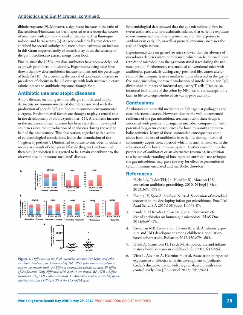
World Digestive Health Day WDHD May 29, 2014 WGO HANDBOOK ON GUT MICROBES 28
tibiotic exposure [9]. Moreover, a significant increase in the ratio of Bacteroidetes/Firmicutes has been reported over a seven-day course of treatment with commonly used antibiotics such as fluoroqui-nolones and beta-lactams [3]. As genes coded by Bacteroidetes are enriched for several carbohydrate metabolism pathways, an increase in this Gram-negative family of bacteria may boost the capacity of the gut microbiota to extract energy from food.
Finally, since the 1950s, low dose antibiotics have been widely used as growth promoters in husbandry. Experiments using mice have shown that low dose antibiotics increase fat mass and the percentage of body fat [10]. As a curiosity, the period of accelerated increase in prevalence of obesity in the US overlaps with both increased dietary caloric intake and antibiotic exposure through food.
Antibiotic use and atopic diseases Atopic diseases including asthma, allergic rhinitis, and atopic dermatitis are immune-mediated disorders associated with the production of specific IgE antibodies to common environmental allergens. Environmental factors are thought to play a crucial role in the development of atopic syndromes [11]. A dramatic increase in the incidence of such diseases has been recorded in developed countries since the introduction of antibiotics during the second half of the past century. This observation, together with a series of epidemiological associations, led to the formulation of the “hygiene hypothesis”. Diminished exposure to microbes in modern society as a result of changes in lifestyle (hygiene) and medical therapies (antibiotics) is suggested to be a main contributor to the observed rise in ‘immune-mediated’ diseases.
Epidemiological data showed that the gut microbiota differs be-tween asthmatic and non-asthmatic infants, that early life exposure to environmental microbes is protective, and that exposure to antibiotics in early life, as well as prenatal exposure, increases the risk of allergic asthma.
Experimental data on germ-free mice showed that the absence of microbiota depletes immunotolerance, which can be restored upon transfer of microbes into the gastrointestinal tract during the neo-natal period. Furthermore, treatment of conventional mice with antibiotics, particularly during early postnatal life, causes altera-tions of the immune system similar to those observed in the germ-free mice, including increased production of interleukin 4 and IgE, diminished numbers of intestinal regulatory T cells (Treg cells), increased infiltration of the colon by NKT cells, and susceptibility later in life to allergen-induced airway hyper-reactivity.
ConclusionsAntibiotics are powerful medicines to fight against pathogens and cure infectious diseases. However, despite the well-documented resilience of the gut microbiota, treatment with these drugs is associated with persistent changes in microbial composition with potential long-term consequences for host immunity and meta-bolic activities. Many of these unintended consequences come about from the use of antibiotics in early life, during microbial community acquisition, a period which, in turn, is involved in the education of the host’s immune system. Further research into the proper use of antibiotics or an alternative treatment, in addition to a better understanding of how repeated antibiotic use reshapes the gut microbiota, may pave the way for effective prevention of certain immune-mediated and metabolic disorders.
References1. Hicks LA, Taylor TH, Jr., Hunkler RJ. More on U.S.
outpatient antibiotic prescribing, 2010. N Engl J Med 2013;369:1175-6.
2. Koenig JE, Spor A, Scalfone N, et al. Succession of microbial consortia in the developing infant gut microbiome. Proc Natl Acad Sci U S A 2011;108 Suppl 1:4578-85.
3. Panda S, El Khader I, Casellas F, et al. Short-term ef-fect of antibiotics on human gut microbiota. PLoS One 2014;9:e95476.
4. Kronman MP, Zaoutis TE, Haynes K, et al. Antibiotic expo-sure and IBD development among children: a population-based cohort study. Pediatrics 2012;130:e794-803.
5. Hviid A, Svanstrom H, Frisch M. Antibiotic use and inflam-matory bowel diseases in childhood. Gut 2011;60:49-54.
6. Virta L, Auvinen A, Helenius H, et al. Association of repeated exposure to antibiotics with the development of pediatric Crohn’s disease--a nationwide, register-based finnish case-control study. Am J Epidemiol 2012;175:775-84.
Figure 1. Differences in the fecal microbial communities before and after antibiotic treatment as determined by 16S rRNA gene sequence analysis at various taxonomic levels. A) Effect of amoxicillin-clavulanic acid. B) Effect of levofloxacin. Only differences with q<0.01 are shown. BF_ATB = before treatment; AF_ATB = after treatment. C) Microbial load as assessed by quan-titative real-time PCR (qPCR) of the 16S rRNA gene.
Antibiotics and Gut Microbes, continued.
World Digestive Health Day WDHD May 29, 2014 WGO HANDBOOK ON GUT MICROBES 29
7. Turnbaugh PJ, Hamady M, Yatsunenko T, et al. A core gut microbiome in obese and lean twins. Nature 2009;457:480-4.
8. Le Chatelier E, Nielsen T, Qin J, et al. Richness of human gut microbiome correlates with metabolic markers. Nature 2013;500:541-6.
9. Hernandez E, Bargiela R, Diez MS, et al. Functional con-sequences of microbial shifts in the human gastrointestinal tract linked to antibiotic treatment and obesity. Gut Microbes 2013;4:306-15.
10. Cho I, Yamanishi S, Cox L, et al. Antibiotics in early life alter the murine colonic microbiome and adiposity. Nature 2012;488:621-6.
11. Zeissig S, Blumberg RS. Life at the beginning: perturbation of the microbiota by antibiotics in early life and its role in health and disease. Nat Immunol 2014;15:307-10.
Antibiotics and Gut Microbes, continued.
WGO’s Newest Educational Offering is Now Available!
The World Gastroenterology Organisation is pleased to announce the release of its newest educational program:
Irritable Bowel Syndrome (IBS): What is it, what causes it and can I do anything about it?
A Web-Based Educational Program for the General Public
This webcast, which was developed from the World Digestive Health Day 2012 Campaign “From Heartburn to Constipation - Common GI Symptoms in the Community: Impact and Interpretation”, will target those common symptoms most associated with irritable bowel syndrome (IBS) and will focus, in particular, on an approach to educate the general public on issues related to this condition. It is led by Professors Eamonn Quigley, USA, WGO Foundation Chair, Richard Hunt, UK, WGO Foundation Vice Chair, Pali Hungin, UK, and Anton Emmanuel, UK. The webcast is available as a full program, as well as individual segments, so that you may choose which topics you would like to view. Segment 1 focuses on “What is IBS?” and “How to communicate symptoms to help the doctor make the right diagnosis.” Here the focus is on the various symptoms that may be experienced by the IBS sufferer and the various definitions of IBS used in clinical practice and in research are reviewed. Strategies that facilitate the best interaction between the sufferer and their doctor are discussed. In Segment 2 you will learn about “Progress in IBS” and “Could it be something else?” The various factors that might contribute to the development of symptoms are reviewed and the panel addresses what is often a major concern for the sufferer and their doctor: the fear of missing other diagnoses. Segment 3 will look at “What can I do to deal with my symptoms?” and “How about diet and dietary supplements?” The role of diet in IBS is a “hot” topic at present and the various ways that constituents of the diet might relate to symptoms are evaluated. And finally in Segment 4 “Managing IBS” and “Living with IBS” is discussed. Here there is good news for IBS sufferers both in terms of new, effective treatments and ongoing research for new approaches to managing IBS. We hope that you will share this information with your colleagues, patients, followers on social media, and anyone else who might benefit from this important information. We thank you for your support of this program!
This webcast was created thanks to an unrestricted educational grant from
Click Here to Begin Viewing the Webcast!
World Digestive Health Day WDHD May 29, 2014 WGO HANDBOOK ON GUT MICROBES 31
The Gut Microbiota in Functional Bowel Disorders
Giovanni Barbara, MD Department of Medical and Surgical Sciences University of Bologna Bologna, Italy
Cesare Cremon, MD Department of Medical and Surgical Sciences University of Bologna Bologna, Italy
Vincenzo Stanghellini, MD Department of Medical and Surgical Sciences University of Bologna Bologna, Italy
The functional bowel disorders (FBDs) are a group of highly prevalent digestive disorders in which symptoms are attrib-
utable to the mid and lower intestinal tract. The most common FBD are the irritable bowel syndrome (IBS), functional bloating, functional diarrhea, and functional constipation. FBDs are associ-ated with reduced quality of life in affected patients and high social costs. The term functional was originally introduced to support the concept that these disorders are characterized by gut dysfunction in the absence of organic causes identifiable by common investiga-tions. Psychological factors (e.g., anxiety, depression) abnormal gut motor patterns and increased visceral sensitivity are key elements in the pathophysiology of most FBD. Recent growing evidence indicates that subtle, organic abnormalities can be detected along the brain-gut axis, including the intestinal tract, the central nervous and the neural-hormonal system connecting them. These abnormalities include altered composition of the luminal milieu, dysfunction of epithelial tight junctions leading to increased intes-tinal permeability, abnormal gut endocrine system signaling and low-grade mucosal inflammation. In this scenario, the putative role of gut microbiota is receiving increasing interest.
Most data linking microbiota to FBD have been obtained in patients with IBS. A current working hypothesis suggests that changes in gut microbiota participate in abnormal fermentation of dietary substrates, predominantly carbohydrates. Microbiota would also elicit excessive stimulation of the mucosal immune system through a leaky gut. The consequent low-grade inflamma-tion and release of inflammatory mediators would ultimately affect gut motor responses and elicit visceral hypersensitivity. This view and the participation of microbial factors in the pathogenesis of FBD are supported by the following evidence. First, about 10%
of episodes of acute infectious gastroenteritis lead to the onset of IBS, (so called post-infectious IBS). Second, subsets of patients show both qualitative and quantitative changes and instability over time of the microbiota. Third, patients with IBS have increased circulating antibodies against flagellin, a component of indig-enous bacteria inhabiting the human gut. They also have increased fecal levels of human beta-defensin-2, an antimicrobial protein produced by the gut mucosa (i.e., by Paneth cells) and mucosal expression of specific microbial receptors (called Toll-like receptors, TLRs). Fourth, the modulation of gut microbiota with probiot-ics and non-absorbable antibiotics has been shown to improve symptoms over placebo, at least in subgroups of patients with IBS. Conversely, systemic antibiotics may induce or worsen IBS symptoms. Taken together, these data provide a proof of concept implicating intestinal bacteria-host interactions in pathophysiology and symptom generation in patients with IBS.
The most compelling evidence linking micro-organisms with FBD is represented by the post-infectious IBS paradigm. Acute infec-tious gastroenteritis is currently the strongest known risk factor (mean OR increase of 6 fold) for the development of IBS. Chronic IBS symptoms occur in 3.7%-36.2% of subjects after Salmonella, Shigella, Campylobacter, or Norovirus gastroenteritis. It is esti-mated that this post-infectious group accounts for 6%-17% of all IBS patients. Multiple mechanisms contribute to persistence of ab-normal bowel physiology and symptoms post-infection. These in-clude genetic factors (genes involved in epithelial barrier function and the innate immune response to enteric bacteria), psychological factors (e.g., adverse life events, depression and hypochondriasis), increased epithelial permeability, low-grade mucosal inflammation, severity of the initial illness, bacterial toxicity and antibiotic treat-ment during the acute infection. Compared to classical IBS, the prognosis for post-infectious forms seems to be more favorable in the long term. However, recent evidence indicates that children are more susceptible to post infectious IBS than adults and symptoms may last for many years.
Although most bacteria reside in the colon, it has been suggested that some patients with IBS have an excessive growth of bacteria in the small intestine (i.e., small intestinal bacterial overgrowth, SIBO). This is based on evidence that compared with controls, a higher proportion of patients with IBS have a positive lactulose or glucose breath test. Nonetheless, the role of SIBO in IBS remains very controversial. This is because the breath tests employed to establish this condition have not been validated. Also, they are flawed by poor diagnostic performance. The lactulose breath test is highly influenced by gut transit, hence leading to erroneous interpretations. The glucose breath performance is slightly better. However, as glucose is rapidly absorbed in the upper small intes-tine, it fails to detect distal intestinal overgrowth.
Earlier culture-based microbiological studies showed decreased numbers of Lactobacilli, Bifidobacteria, and anaerobic bacteria in IBS. However, only about 20% of the bacterial species and strains that inhabit the gut are have been identified by conventional culture techniques. Thus culture-based studies provide limited information. The advent of culture-independent, high-throughput
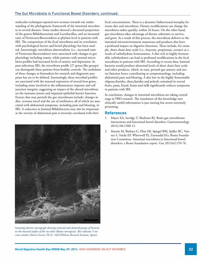
World Digestive Health Day WDHD May 29, 2014 WGO HANDBOOK ON GUT MICROBES 32
molecular techniques opened new avenues towards our under-standing of the phylogenetic framework of the intestinal microbio-ta in several diseases. Some studies showed a decreased proportion of the genera Bifidobacterium and Lactobacillus, and an increased ratio of Firmicutes:Bacteroidetes at phylum level in patients with IBS. The composition of the fecal microbiota and its correlation with psychological factors and bowel physiology has been stud-ied. Interestingly, microbiota abnormalities (i.e., increased ratio of Firmicutes:Bacteroidetes) were associated with changes in gut physiology including transit, while patients with normal micro-biota profiles had increased levels of anxiety and depression. In post-infectious IBS, the microbiota profile (27 genus-like groups) can distinguish these patients from healthy controls. The usefulness of these changes as biomarkers for research and diagnostic pur-poses has yet to be defined. Interestingly, these microbial profiles are associated with the mucosal expression of several host genes, including some involved in the inflammatory response and cell junction integrity, suggesting an impact of the altered microbiota on the immune system and impaired epithelial barrier function. Factors that may perturb the gut microbioata include: changes in diet, overseas travel and the use of antibiotics; all of which are asso-ciated with abdominal symptoms, including pain and bloating, in IBS. A reduction in luminal Bifidobacteria may also be important as the severity of abdominal pain is inversely correlated with their
fecal concentrations. There is a dynamic bidirectional interplay be-tween diet and microbiota. Dietary modifications can change the microbiota rather quickly, within 24 hours. On the other hand, gut microbiota takes advantage of dietary substrates to survive, and grow. As a result of this process, the microbiota delivers to the intestinal microenvironment numerous end-products that have a profound impact on digestive functions. These include, for exam-ple, short-chain fatty acids (i.e., butyrate, propionate, acetate) as a result of carbohydrate fermentation. A diet rich in highly ferment-able carbohydrates can lead to profound modifications in the fecal microbiota in patients with IBS. According to recent data, luminal bacteria would produce abnormal levels of short chain fatty acids and other products, which, in turn, perturb gut sensory and mo-tor function hence contributing to symptomatology, including abdominal pain and bloating. A diet low in the highly fermentable oligosaccharides, disaccharides and polyols contained in several fruits, pasta, bread, beans and milk significantly reduces symptoms in patients with IBS.
In conclusion, changes in intestinal microbiota are taking central stage in FBD research. The translation of this knowledge into clinically useful information is just starting but seems extremely promising.
References1. Mayer EA, Savidge T, Shulman RJ. Brain-gut microbiome
interactions and functional bowel disorders. Gastroenterology 2014;146:1500-12.
2. Simrén M, Barbara G, Flint HJ, Spiegel BM, Spiller RC, Van-ner S, Verdu EF, Whorwell PJ, Zoetendal EG; Rome Founda-tion Committee. Intestinal microbiota in functional bowel disorders: a Rome foundation report. Gut 2013;62:159-76.
The Gut Microbiota in Functional Bowel Disorders, continued.
Scanning electron micrograph showing scattered and clustered groups of bacteria on the mucosal surface of the rat colon (Rattus norvegicus). Bar indicates 5 mi-crons (author Maria Vicario, Ph.D., Vall d’Hebron Research Institute, Spain).
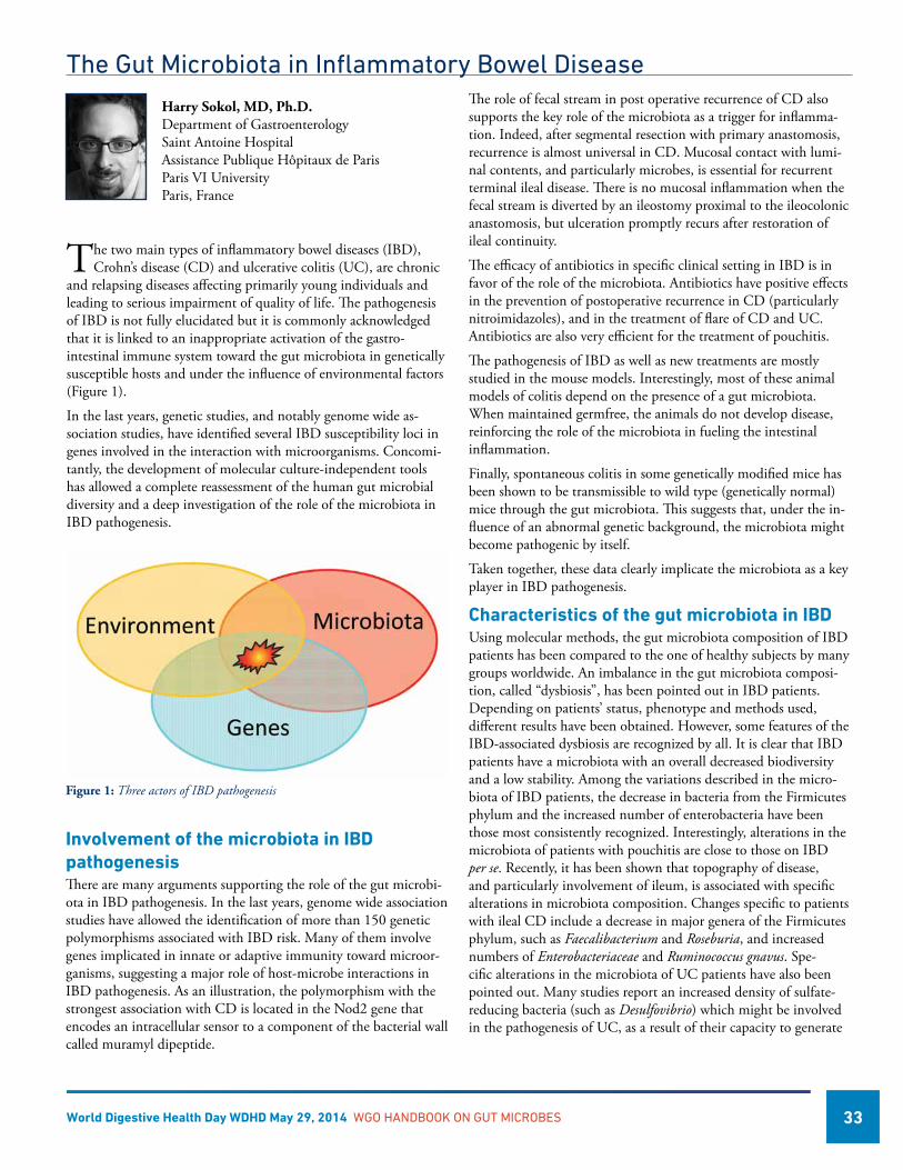
World Digestive Health Day WDHD May 29, 2014 WGO HANDBOOK ON GUT MICROBES 33
The Gut Microbiota in Inflammatory Bowel Disease
The two main types of inflammatory bowel diseases (IBD), Crohn’s disease (CD) and ulcerative colitis (UC), are chronic
and relapsing diseases affecting primarily young individuals and leading to serious impairment of quality of life. The pathogenesis of IBD is not fully elucidated but it is commonly acknowledged that it is linked to an inappropriate activation of the gastro-intestinal immune system toward the gut microbiota in genetically susceptible hosts and under the influence of environmental factors (Figure 1).
In the last years, genetic studies, and notably genome wide as-sociation studies, have identified several IBD susceptibility loci in genes involved in the interaction with microorganisms. Concomi-tantly, the development of molecular culture-independent tools has allowed a complete reassessment of the human gut microbial diversity and a deep investigation of the role of the microbiota in IBD pathogenesis.
Harry Sokol, MD, Ph.D. Department of Gastroenterology Saint Antoine Hospital Assistance Publique Hôpitaux de Paris Paris VI University Paris, France
The role of fecal stream in post operative recurrence of CD also supports the key role of the microbiota as a trigger for inflamma-tion. Indeed, after segmental resection with primary anastomosis, recurrence is almost universal in CD. Mucosal contact with lumi-nal contents, and particularly microbes, is essential for recurrent terminal ileal disease. There is no mucosal inflammation when the fecal stream is diverted by an ileostomy proximal to the ileocolonic anastomosis, but ulceration promptly recurs after restoration of ileal continuity.
The efficacy of antibiotics in specific clinical setting in IBD is in favor of the role of the microbiota. Antibiotics have positive effects in the prevention of postoperative recurrence in CD (particularly nitroimidazoles), and in the treatment of flare of CD and UC. Antibiotics are also very efficient for the treatment of pouchitis.
The pathogenesis of IBD as well as new treatments are mostly studied in the mouse models. Interestingly, most of these animal models of colitis depend on the presence of a gut microbiota. When maintained germfree, the animals do not develop disease, reinforcing the role of the microbiota in fueling the intestinal inflammation.
Finally, spontaneous colitis in some genetically modified mice has been shown to be transmissible to wild type (genetically normal) mice through the gut microbiota. This suggests that, under the in-fluence of an abnormal genetic background, the microbiota might become pathogenic by itself.
Taken together, these data clearly implicate the microbiota as a key player in IBD pathogenesis.
Characteristics of the gut microbiota in IBDUsing molecular methods, the gut microbiota composition of IBD patients has been compared to the one of healthy subjects by many groups worldwide. An imbalance in the gut microbiota composi-tion, called “dysbiosis”, has been pointed out in IBD patients. Depending on patients’ status, phenotype and methods used, different results have been obtained. However, some features of the IBD-associated dysbiosis are recognized by all. It is clear that IBD patients have a microbiota with an overall decreased biodiversity and a low stability. Among the variations described in the micro-biota of IBD patients, the decrease in bacteria from the Firmicutes phylum and the increased number of enterobacteria have been those most consistently recognized. Interestingly, alterations in the microbiota of patients with pouchitis are close to those on IBD per se. Recently, it has been shown that topography of disease, and particularly involvement of ileum, is associated with specific alterations in microbiota composition. Changes specific to patients with ileal CD include a decrease in major genera of the Firmicutes phylum, such as Faecalibacterium and Roseburia, and increased numbers of Enterobacteriaceae and Ruminococcus gnavus. Spe-cific alterations in the microbiota of UC patients have also been pointed out. Many studies report an increased density of sulfate-reducing bacteria (such as Desulfovibrio) which might be involved in the pathogenesis of UC, as a result of their capacity to generate
Figure 1: Three actors of IBD pathogenesis
Involvement of the microbiota in IBD pathogenesisThere are many arguments supporting the role of the gut microbi-ota in IBD pathogenesis. In the last years, genome wide association studies have allowed the identification of more than 150 genetic polymorphisms associated with IBD risk. Many of them involve genes implicated in innate or adaptive immunity toward microor-ganisms, suggesting a major role of host-microbe interactions in IBD pathogenesis. As an illustration, the polymorphism with the strongest association with CD is located in the Nod2 gene that encodes an intracellular sensor to a component of the bacterial wall called muramyl dipeptide.
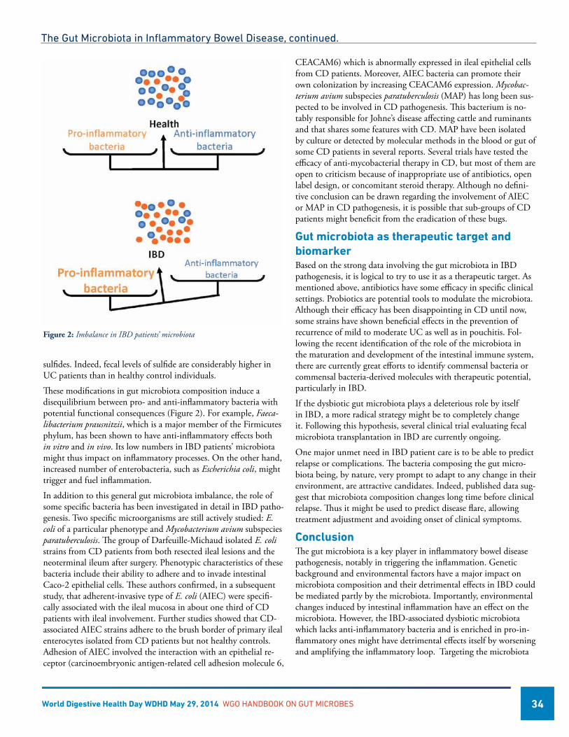
World Digestive Health Day WDHD May 29, 2014 WGO HANDBOOK ON GUT MICROBES 34
sulfides. Indeed, fecal levels of sulfide are considerably higher in UC patients than in healthy control individuals.
These modifications in gut microbiota composition induce a disequilibrium between pro- and anti-inflammatory bacteria with potential functional consequences (Figure 2). For example, Faeca-libacterium prausnitzii, which is a major member of the Firmicutes phylum, has been shown to have anti-inflammatory effects both in vitro and in vivo. Its low numbers in IBD patients’ microbiota might thus impact on inflammatory processes. On the other hand, increased number of enterobacteria, such as Escherichia coli, might trigger and fuel inflammation.
In addition to this general gut microbiota imbalance, the role of some specific bacteria has been investigated in detail in IBD patho-genesis. Two specific microorganisms are still actively studied: E. coli of a particular phenotype and Mycobacterium avium subspecies paratuberculosis. The group of Darfeuille-Michaud isolated E. coli strains from CD patients from both resected ileal lesions and the neoterminal ileum after surgery. Phenotypic characteristics of these bacteria include their ability to adhere and to invade intestinal Caco-2 epithelial cells. These authors confirmed, in a subsequent study, that adherent-invasive type of E. coli (AIEC) were specifi-cally associated with the ileal mucosa in about one third of CD patients with ileal involvement. Further studies showed that CD-associated AIEC strains adhere to the brush border of primary ileal enterocytes isolated from CD patients but not healthy controls. Adhesion of AIEC involved the interaction with an epithelial re-ceptor (carcinoembryonic antigen-related cell adhesion molecule 6,
CEACAM6) which is abnormally expressed in ileal epithelial cells from CD patients. Moreover, AIEC bacteria can promote their own colonization by increasing CEACAM6 expression. Mycobac-terium avium subspecies paratuberculosis (MAP) has long been sus-pected to be involved in CD pathogenesis. This bacterium is no-tably responsible for Johne’s disease affecting cattle and ruminants and that shares some features with CD. MAP have been isolated by culture or detected by molecular methods in the blood or gut of some CD patients in several reports. Several trials have tested the efficacy of anti-mycobacterial therapy in CD, but most of them are open to criticism because of inappropriate use of antibiotics, open label design, or concomitant steroid therapy. Although no defini-tive conclusion can be drawn regarding the involvement of AIEC or MAP in CD pathogenesis, it is possible that sub-groups of CD patients might beneficit from the eradication of these bugs.
Gut microbiota as therapeutic target and biomarkerBased on the strong data involving the gut microbiota in IBD pathogenesis, it is logical to try to use it as a therapeutic target. As mentioned above, antibiotics have some efficacy in specific clinical settings. Probiotics are potential tools to modulate the microbiota. Although their efficacy has been disappointing in CD until now, some strains have shown beneficial effects in the prevention of recurrence of mild to moderate UC as well as in pouchitis. Fol-lowing the recent identification of the role of the microbiota in the maturation and development of the intestinal immune system, there are currently great efforts to identify commensal bacteria or commensal bacteria-derived molecules with therapeutic potential, particularly in IBD.
If the dysbiotic gut microbiota plays a deleterious role by itself in IBD, a more radical strategy might be to completely change it. Following this hypothesis, several clinical trial evaluating fecal microbiota transplantation in IBD are currently ongoing.
One major unmet need in IBD patient care is to be able to predict relapse or complications. The bacteria composing the gut micro-biota being, by nature, very prompt to adapt to any change in their environment, are attractive candidates. Indeed, published data sug-gest that microbiota composition changes long time before clinical relapse. Thus it might be used to predict disease flare, allowing treatment adjustment and avoiding onset of clinical symptoms.
ConclusionThe gut microbiota is a key player in inflammatory bowel disease pathogenesis, notably in triggering the inflammation. Genetic background and environmental factors have a major impact on microbiota composition and their detrimental effects in IBD could be mediated partly by the microbiota. Importantly, environmental changes induced by intestinal inflammation have an effect on the microbiota. However, the IBD-associated dysbiotic microbiota which lacks anti-inflammatory bacteria and is enriched in pro-in-flammatory ones might have detrimental effects itself by worsening and amplifying the inflammatory loop. Targeting the microbiota
The Gut Microbiota in Inflammatory Bowel Disease, continued.
Figure 2: Imbalance in IBD patients’ microbiota
World Digestive Health Day WDHD May 29, 2014 WGO HANDBOOK ON GUT MICROBES 35
or using its components as therapeutic targets or tools is thus a promising strategy in IBD.
References1. Sokol H, Seksik P. The intestinal microbiota in inflamma-
tory bowel diseases: time to connect with the host. Curr Opin Gastroenterol. 2010 Jul;26(4):327-31. doi: 10.1097/MOG.0b013e328339536b. Review. PubMed PMID: 20445446.
2. Sokol H, Lay C, Seksik P, Tannock GW. Analysis of bacterial bowel communities of IBD patients: what has it revealed? Inflamm Bowel Dis. 2008 Jun;14(6):858-67. doi: 10.1002/ibd.20392. Review. PubMed PMID: 18275077.
3. Manichanh C, Borruel N, Casellas F, Guarner F. The gut microbiota in IBD. Nat Rev Gastroenterol Hepatol. 2012 Oct;9(10):599-608. doi: 10.1038/nrgastro.2012.152. Epub 2012 Aug 21. Review. PubMed PMID: 22907164.
4. Jostins L, Ripke S, Weersma RK, Duerr RH, McGovern DP, Hui KY, Lee JC, Schumm LP, Sharma Y, Anderson CA, Es-sers J, Mitrovic M, Ning K, Cleynen I, Theatre E, Spain SL, Raychaudhuri S, Goyette P, Wei Z, Abraham C, Achkar JP, Ahmad T, Amininejad L, Ananthakrishnan AN, Andersen V, Andrews JM, Baidoo L, Balschun T, Bampton PA, Bit-ton A, Boucher G, Brand S, Büning C, Cohain A, Cichon S, D’Amato M, De Jong D, Devaney KL, Dubinsky M, Ed-wards C, Ellinghaus D, Ferguson LR, Franchimont D, Fran-sen K, Gearry R, Georges M, Gieger C, Glas J, Haritunians T, Hart A, Hawkey C, Hedl M, Hu X, Karlsen TH, Kupcinskas L, Kugathasan S, Latiano A, Laukens D, Lawrance IC, Lees CW, Louis E, Mahy G, Mansfield J, Morgan AR, Mowat C, Newman W, Palmieri O, Ponsioen CY, Potocnik U, Prescott NJ, Regueiro M, Rotter JI, Russell RK, Sanderson JD, Sans M, Satsangi J, Schreiber S, Simms LA, Sventoraityte J, Targan SR, Taylor KD, Tremelling M, Verspaget HW, De Vos M, Wijmenga C, Wilson DC, Winkelmann J, Xavier RJ, Zeissig S, Zhang B, Zhang CK, Zhao H; International IBD Genetics Consortium (IIBDGC), Silverberg MS, Annese V, Hakonar-son H, Brant SR, Radford-Smith G, Mathew CG, Rioux JD, Schadt EE, Daly MJ, Franke A, Parkes M, Vermeire S, Barrett JC, Cho JH. Host-microbe interactions have shaped the genetic architecture of inflammatory bowel disease. Nature. 2012 Nov 1;491(7422):119-24. doi: 10.1038/nature11582. PubMed PMID: 23128233; PubMed Central PMCID: PMC3491803.
The Gut Microbiota in Inflammatory Bowel Disease, continued.

World Digestive Health Day WDHD May 29, 2014 WGO HANDBOOK ON GUT MICROBES 36
Gut Microbiota, Obesity and Associated Metabolic Disorders
Introduction
Obesity is defined as a massive expansion of the adipose tissue and is typically associated with a wide cluster of metabolic
alterations, including glucose homeostasis disorders (e.g., glucose intolerance, insulin resistance and type 2 diabetes), cardiovascular diseases or risk factors (e.g., hypertension, dyslipidaemia, and fibri-nolysis disorders) and non-alcoholic fatty liver disease (NAFLD) [1]. The majority of these alterations likely results from a combina-tion of variable associations between genetic and environmental factors. Low-grade chronic inflammation appears to be a common feature that may contribute to the development of insulin resis-tance, type 2 diabetes and cardiovascular diseases [2]. However, the mechanisms underlying obesity, fat mass development and the development of inflammation are not fully defined.
The gut microbiota may be a key exteriorised organ that can con-tribute to the onset of these metabolic dysregulations (for review, see references [3-5]. The gut microbiota is now considered a full organ that is involved in the regulation of numerous physiological pathways by impacting different functions of the host [6]. Among these regulations, the influence of gut microbes on energy metabo-lism is of particular interest because it has been suggested to be a driving force in the pathogenesis of metabolic diseases, particularly obesity. In this chapter, we will shortly discuss recent evidence sup-porting the hypothesis that the gut microbiota can influence host metabolism using various mechanisms and that changes in micro-biota composition trigger modifications of metabolic behaviour.
Gut microbiota composition and metabolic disordersSeveral papers and reviews support the idea that a “dysbiosis” (altered gut microbiota composition and/or activity related to host disease) characterizes overweight, obese or diabetic individuals [7, 8]. Regarding the inadequate composition of the gut microbiota, obese and overweight people were initially characterized by a change in the Firmicutes/Bacteroidetes ratio. However, a number of studies, including very recent human cohorts, reported no varia-tions in this ratio between diabetic or obese patients and controls.
Patrice D. Cani, Ph.D. Université catholique de Louvain Louvain Drug Research Institute; WELBIO (Wal-loon Excellence in Life sciences and BIOtechnol-ogy) Metabolism and Nutrition research group Brussels, Belgium
Nathalie M. Delzenne, Ph.D. Université catholique de Louvain Louvain Drug Research Institute; WELBIO (Wal-loon Excellence in Life sciences and BIOtechnol-ogy) Metabolism and Nutrition research group Brussels, Belgium
Thus a concept of dysbiosis which extends to other bacterial phyla, genera or species seems more appropriate to the characterization of obesity and associated disorders (reviewed in [9]). In addition and more recently, the concept of “enterotypes” has been proposed: the analysis of the microbial composition of human fecal samples revealed that their bacterial population can be stratified into three robust clusters. Abundance is a measure of the relative proportion of each bacterial phyla inside an ecosystem, while diversity takes into account the number of bacterial phyla identified (richness) in addition to their relative abundance. Despite being very useful descriptors of the bacterial ecosystem in general, neither of these seems to be reliable indicators of the diabetic status of the host [10]. Although animal experiments show a clear separation be-tween diabetic and non-diabetic subjects based on their microbiota profiles, the inter-individual variability in human subjects most likely masks these wide scale differences. Therefore, it appears that, in addition to these quantitative modifications of microbial phyla, obesity and some related metabolic diseases might be associated with modifications of microbial genes expression and, therefore, to the modulation of metabolic functions of the gut microbiota. The microbiome is now considered as a new therapeutic target against obesity and its linked diseases [11]. In fact, changes in dietary habits and, especially, an enrichment in some bioactive food com-ponents present in whole grain cereals are able to modify the com-position of gut microbiota and could be helpful in the prevention of chronic diseases, including obesity and related disorders such as type 2 diabetes [12]. Wu et al have shown that microbiome com-position may change 24 hours after initiating a high fat/low fiber or a high fiber/low fat diet, but that enterotype identity remained stable during a ten day nutritional intervention [13]. They suggest that nutrients like dietary fibers, which are not digested by host enzymes but are fermented by gut bacteria, could modulate the gut microbiome in a relatively short period of time, independent of the effect of their effect on gut transit time.
Gut microbiota, low grade inflammation, obesity and type 2 diabetes: evidence for altered gut barrier function We recently defined gut-microbiota-derived lipopolysaccharide (LPS) as a factor involved in the early development of inflamma-tion and metabolic diseases. Briefly, intake of excess dietary fat increases systemic exposure to potentially pro-inflammatory free fatty acids and their derivatives and increases plasma LPS levels, a state referred to as metabolic endotoxaemia (reviewed in [3, 14]). Because LPS can affect inflammation throughout the body and interfere with metabolism and the function of the immune system, this major breakthrough provides new insights into the role of gut-microbiota-derived products and metabolism. Among the mechanisms explaining the development of low grade inflamma-tion, we identified several links between the gut barrier and the gut microbiota (for review see [6, 15].
We have previously shown that the beneficial effects of prebiotics on gut barrier function, inflammation and insulin resistance in obesity requires a functional GLP-1 receptor and is associated with
World Digestive Health Day WDHD May 29, 2014 WGO HANDBOOK ON GUT MICROBES 37
obesity by increasing the release of gut hormones, such as gluca-gon-like peptide-1 and -2 (GLP-1 and GLP-2) [16-18]. These en-docrine peptides represent an interesting pathway involved in the cross-talk between gut microbes and host cell. More importantly, they are considered as potential targets regulate endocrine peptides through the gut microbiota. Using complementary approaches involving specific modulation of the gut microbiota (antibiotics, probiotics, prebiotics) and pharmacological inhibition or activa-tion of the GLP-2 receptor, we discovered that gut microbiota participate in the modulation of gut barrier function and the con-sequent systemic inflammatory phenotype (for review [15, 19]). These findings indicate that targeting enteroendocrine function may be a novel therapeutic approach for treating the inflammatory phenotype associated with obesity and type 2 diabetes. Although the enteroendocrine function of L-cells is an important mechanism in regulating gut barrier function, molecular links between the gut microbiota and enteroendocrine function of the gut remain unknown.
Among the most studied bacterial metabolites that may interfere with host metabolism are the SCFAs. These products of microbi-ota-mediated fermentation of polysaccharides modulate the levels of several gut hormones involved in glucose and energy homeo-stasis, such as GLP-1 and ghrelin. Additionally, these metabolites can circulate in the blood and act thus on peripheral targets to modulate insulin sensitivity and whole host energy metabolism. Unfortunately, most of pathways underlying these effects remain largely unknown, but several studies have suggested that they are linked to the members of a recently identified G-protein coupled receptor family that includes G-protein coupled receptor 43 and 41 (GPR43 and GPR41) [20]. Additionally, there is a growing interest in the study of the intestinal mucus layer and its interac-tions with microbiota. We have recently demonstrated the key role played by the gut microbiota and its interaction with the mucus layer in the context of diet-induced obesity and type 2 diabetes. We demonstrated that Akkermansia muciniphila, a mucin-degrad-ing bacterium that resides in, and abundantly colonises, the mucus layer, negatively correlates with body weight and is decreased under HFD conditions [21]. Moreover, daily administration of A. muciniphila to HFD-induced obese mice for 4 weeks improved their metabolic profile by decreasing weight gain, restoring mucus layer thickness, and counteracting metabolic endotoxemia and insulin resistance [21].
Gut microbiota and type 2 diabetes: a lack of consensusBecause the taxonomy of the microbiota alone cannot explain its influence on the onset of the metabolic syndrome, the descrip-tion of the bacterial metabolic functions (at the genetic level) using metagenomic shotgun sequencing in humans and mice has been shown to be a reliable complementary tool and has revealed shifts in metabolic functions related to obesity and type 2 diabetes [22]. The most prominent features in type 2 diabetes-associated metagenomes are enrichments in pathways related to carbohydrate metabolism and transport, branched-chain amino acid transport
and the response to oxidative stress. On the other hand, pathways related to flagellar assembly, butyrate biosynthesis and vitamin metabolism were reduced in Danish and Chinese human cohorts [23, 24].
Following the same reasoning, several studies have identified individual taxa as important markers for the onset of obesity and diabetes, although the exact roles of some of these species are not currently known. The genera Bacteroides, Roseburia and Akker-mansia, as well as Faecalibacterium prausnitzii, were depleted in type 2 diabetic Chinese subjects, whereas Dorea, Prevotella and Collinsella had relatively higher abundance [10]. In humans and mice Prevotella, Akkermansia and enterobacteria have previously been shown to significantly vary between obese and lean subjects (reviewed in [19]). A reduction in a cluster of genes belonging to Roseburia and F. prausnitzii was identified as a discriminant marker for the prediction of diabetic status in European women [23]. In an obese French cohort, F. prausnitzii was lower than in control subjects, and an increase in the abundance of this bacterium cor-related with improved inflammatory status [25]. However, this bacterium was increased in obese Indian children compared to the lean controls, highlighting once again the specificity of findings to populations, age groups and diets in phenotype-taxonomy associa-tions [26].
Thus, finding a treatment for type 2 diabetes is currently challeng-ing. Exercise and dietary intervention yield only modest results, while pharmacological treatments are often associated with delete-rious side effects.
ConclusionsTaken together, these data demonstrate that several relationships exist between the gut microbiota, glucose and energy homeosta-sis. However, the causality between the observed variations in the composition of the gut microbiota and metabolic symptoms are still unclear. Thus, investigating the gut microbiota–host interac-tions and deciphering their symbiotic interactions constitute an important area of research.
AcknowledgmentsP. D. Cani is a research associate from the FRS-FNRS (Fonds de la Recherche Scientifique) and a recipient of grants from the FNRS and PDR (Projet de recherche FRS-FNRS, Belgium), from ARC (Actions de Recherche Concertées - Communauté française de Belgique convention :12/17-047). Moreover, P. D. Cani is a recipi-ent of the ERC Starting Grant 2013 (European Research Council, Starting grant 336452-ENIGMO).
References[1] Ogden CL, Yanovski SZ, Carroll MD, Flegal KM. The epide-
miology of obesity. Gastroenterology 2007;132:2087-102.
[2] Shoelson SE, Goldfine AB. Getting away from glucose: fan-ning the flames of obesity-induced inflammation. NatMed 2009;15:373-74.
Gut microbiota, obesity and associated metabolic disorders, continued.
World Digestive Health Day WDHD May 29, 2014 WGO HANDBOOK ON GUT MICROBES 38
[3] Cani PD, Delzenne NM. The role of the gut microbiota in energy metabolism and metabolic disease. CurrPharmDes 2009;15:1546-58.
[4] Cani PD, Osto M, Geurts L, Everard A. Involvement of gut microbiota in the development of low-grade inflammation and type 2 diabetes associated with obesity. Gut microbes 2012 Jul 1;3.
[5] Delzenne NM, Neyrinck AM, Backhed F, Cani PD. Targeting gut microbiota in obesity: effects of prebiotics and probiotics. Nature reviews Endocrinology 2011;7:639-46.
[6] Cani PD. Metabolism in 2013: The gut microbiota man-ages host metabolism. Nature reviews Endocrinology 2014 Feb;10:74-6.
[7] Everard A, Cani PD. Diabetes, obesity and gut microbiota. Best practice & research Clinical gastroenterology 2013 Feb;27:73-83.
[8] Petschow B, Dore J, Hibberd P, Dinan T, Reid G, Blaser M, et al. Probiotics, prebiotics, and the host microbiome: the science of translation. Annals of the New York Academy of Sciences 2013;22:12303.
[9] Zhao L. The gut microbiota and obesity: from correlation to causality. Nature reviews Microbiology 2013 Sep;11:639-47.
[10] Zhang X, Shen D, Fang Z, Jie Z, Qiu X, Zhang C, et al. Hu-man gut microbiota changes reveal the progression of glucose intolerance. PLoS One 2013;8:e71108.
[11] Cani PD, Delzenne NM. The gut microbiome as therapeutic target. Pharmacology & therapeutics 2011 May;130:202-12.
[12] Gil A, Ortega RM, Maldonado J. Wholegrain cereals and bread: a duet of the Mediterranean diet for the prevention of chronic diseases. Public health nutrition 2011 Dec;14:2316-22.
[13] Wu GD, Chen J, Hoffmann C, Bittinger K, Chen YY, Keil-baugh SA, et al. Linking long-term dietary patterns with gut microbial enterotypes. Science 2011 Oct 7;334:105-8.
[14] Cani PD, Amar J, Iglesias MA, Poggi M, Knauf C, Bastelica D, et al. Metabolic endotoxemia initiates obesity and insulin resistance. Diabetes 2007;56:1761-72.
[15] Geurts L, Neyrinck AM, Delzenne NM, Knauf C, Cani PD. Gut microbiota controls adipose tissue expansion, gut barrier and glucose metabolism: novel insights into molecular targets and interventions using prebiotics. Beneficial microbes 2014 Mar;5:3-17.
[16] Cani PD, Knauf C, Iglesias MA, Drucker DJ, Delzenne NM, Burcelin R. Improvement of glucose tolerance and hepatic insulin sensitivity by oligofructose requires a functional glucagon-like peptide 1 receptor. Diabetes 2006;55:1484-90.
[17] Cani PD, Possemiers S, Van de WT, Guiot Y, Everard A, Rot-tier O, et al. Changes in gut microbiota control inflammation in obese mice through a mechanism involving GLP-2-driven improvement of gut permeability. Gut 2009;58:1091-103.
[18] Delzenne NM, Neyrinck AM, Cani PD. Gut microbiota and metabolic disorders: How prebiotic can work? The British journal of nutrition 2013 Jan;109 Suppl 2:S81-5.
[19] Cani PD, Geurts L, Matamoros S, Plovier H, Duparc T. Glucose metabolism: Focus on gut microbiota, the endocan-nabinoid system and beyond. Diabetes Metab 2014 Mar 13.
[20] Cani PD, Everard A, Duparc T. Gut microbiota, enteroendo-crine functions and metabolism. Current opinion in pharma-cology 2013 Dec;13:935-40.
[21] Everard A, Belzer C, Geurts L, Ouwerkerk JP, Druart C, Bin-dels LB, et al. Cross-talk between Akkermansia muciniphila and intestinal epithelium controls diet-induced obesity. Proc Natl Acad Sci U S A 2013 May 28;110:9066-71.
[22] Turnbaugh PJ, Ley RE, Mahowald MA, Magrini V, Mardis ER, Gordon JI. An obesity-associated gut microbiome with increased capacity for energy harvest. Nature 2006;444:1027-31.
[23] Karlsson FH, Tremaroli V, Nookaew I, Bergstrom G, Behre CJ, Fagerberg B, et al. Gut metagenome in European women with normal, impaired and diabetic glucose control. Nature 2013 Jun 6;498:99-103.
[24] Qin J, Li Y, Cai Z, Li S, Zhu J, Zhang F, et al. A metage-nome-wide association study of gut microbiota in type 2 diabetes. Nature 2012 Oct 4;490:55-60.
[25] Furet JP, Kong LC, Tap J, Poitou C, Basdevant A, Bouillot JL, et al. Differential adaptation of human gut microbiota to bar-iatric surgery-induced weight loss: links with metabolic and low-grade inflammation markers. Diabetes 2010;59:3049-57.
[26] Balamurugan R, George G, Kabeerdoss J, Hepsiba J, Chan-dragunasekaran AM, Ramakrishna BS. Quantitative differenc-es in intestinal Faecalibacterium prausnitzii in obese Indian children. BrJNutr 2010;103:335-38.
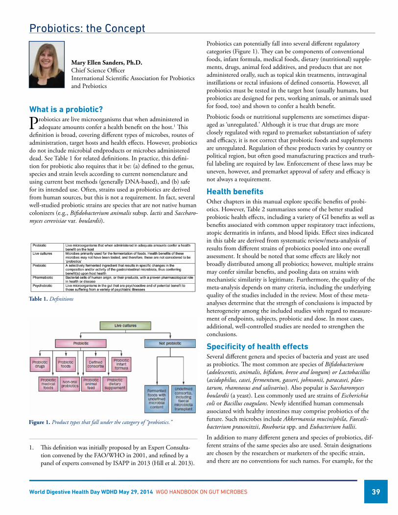
World Digestive Health Day WDHD May 29, 2014 WGO HANDBOOK ON GUT MICROBES 39
Probiotics: the Concept
What is a probiotic?
Probiotics are live microorganisms that when administered in adequate amounts confer a health benefit on the host.1 This
definition is broad, covering different types of microbes, routes of administration, target hosts and health effects. However, probiotics do not include microbial endproducts or microbes administered dead. See Table 1 for related definitions. In practice, this defini-tion for probiotic also requires that it be: (a) defined to the genus, species and strain levels according to current nomenclature and using current best methods (generally DNA-based), and (b) safe for its intended use. Often, strains used as probiotics are derived from human sources, but this is not a requirement. In fact, several well-studied probiotic strains are species that are not native human colonizers (e.g., Bifidobacterium animalis subsp. lactis and Saccharo-myces cerevisiae var. boulardii).
Probiotics can potentially fall into several different regulatory categories (Figure 1). They can be components of conventional foods, infant formula, medical foods, dietary (nutritional) supple-ments, drugs, animal feed additives, and products that are not administered orally, such as topical skin treatments, intravaginal instillations or rectal infusions of defined consortia. However, all probiotics must be tested in the target host (usually humans, but probiotics are designed for pets, working animals, or animals used for food, too) and shown to confer a health benefit.
Probiotic foods or nutritional supplements are sometimes dispar-aged as ‘unregulated.’ Although it is true that drugs are more closely regulated with regard to premarket substantiation of safety and efficacy, it is not correct that probiotic foods and supplements are unregulated. Regulation of these products varies by country or political region, but often good manufacturing practices and truth-ful labeling are required by law. Enforcement of these laws may be uneven, however, and premarket approval of safety and efficacy is not always a requirement.
Health benefits Other chapters in this manual explore specific benefits of probi-otics. However, Table 2 summarizes some of the better studied probiotic health effects, including a variety of GI benefits as well as benefits associated with common upper respiratory tract infections, atopic dermatitis in infants, and blood lipids. Effect sizes indicated in this table are derived from systematic review/meta-analysis of results from different strains of probiotics pooled into one overall assessment. It should be noted that some effects are likely not broadly distributed among all probiotics; however, multiple strains may confer similar benefits, and pooling data on strains with mechanistic similarity is legitimate. Furthermore, the quality of the meta-analysis depends on many criteria, including the underlying quality of the studies included in the review. Most of these meta-analyses determine that the strength of conclusions is impacted by heterogeneity among the included studies with regard to measure-ment of endpoints, subjects, probiotic and dose. In most cases, additional, well-controlled studies are needed to strengthen the conclusions.
Specificity of health effectsSeveral different genera and species of bacteria and yeast are used as probiotics. The most common are species of Bifidobacterium (adolescentis, animalis, bifidum, breve and longum) or Lactobacillus (acidophilus, casei, fermentum, gasseri, johnsonii, paracasei, plan-tarum, rhamnosus and salivarius). Also popular is Saccharomyces boulardii (a yeast). Less commonly used are strains of Escherichia coli or Bacillus coagulans. Newly identified human commensals associated with healthy intestines may comprise probiotics of the future. Such microbes include Akkermansia muciniphila, Faecali-bacterium prausnitzii, Roseburia spp. and Eubacterium hallii.
In addition to many different genera and species of probiotics, dif-ferent strains of the same species also are used. Strain designations are chosen by the researchers or marketers of the specific strain, and there are no conventions for such names. For example, for the
Mary Ellen Sanders, Ph.D. Chief Science Officer International Scientific Association for Probiotics and Prebiotics
1. This definition was initially proposed by an Expert Consulta-tion convened by the FAO/WHO in 2001, and refined by a panel of experts convened by ISAPP in 2013 (Hill et al. 2013).
Figure 1. Product types that fall under the category of “probiotics.”
Table 1. Definitions

World Digestive Health Day WDHD May 29, 2014 WGO HANDBOOK ON GUT MICROBES 40
probiotic strain Lactobacillus rhamnosus GG, the genus is “Lacto-bacillus”, the species is “rhamnosus” and the strain designation is “GG.”
From a clinical perspective, it’s important to realize that not all preparations called “probiotic” will have the same health effects. One clinical example (demonstrated by O’Mahony et al. in 2005) compared the ability of Lactobacillus salivarius UCC4331 or Bifi-dobacterium infantis 35624 to alleviate symptoms of irritable bowel syndrome; only B. infantis 35624 was effective. Therefore, it is best to recommend probiotics that have been specifically tested and shown to have the desired benefits.
Product effects are also dose-specific. Few dose response studies have been conducted on clinical endpoints, but some products are effective at 50 million colony-forming units (CFUs)/day whereas others are used at more than 1 trillion CFU/day. This huge range
in effective doses likely reflects differences in strains, clinical end points, and perhaps the best guess of the researcher of what level would be sufficient. Therefore, it is best to recommend the dose of a specific probiotic that has been tested and shown to have the desired benefits.
SafetyProbiotics must be safe under the intended conditions of use. For different types of foods, including infant formula, and dietary supplements, probiotics must be safe when consumed by the generally healthy target population. For drugs, safety considers a risk/benefit assessment. It is important for clinicians to consider safety for off-label uses for probiotics, especially if administering to severely ill or immunocompromised patients. Probiotics are NOT recommended for patients with short bowel. Use of probiotics in either diseased or immunocompromised individuals must be done
Table 2. Different health benefits researched for probiotics. Effect sizes are estimates from indicated systematic review/meta-analyses. Adapted from Sanders et al. 2013. Probiotics and Prebiotics: Prospects for Public Health and Nutritional Recommendations. Ann N Y Acad Sci. 2014 Feb;1309(1):19-29.
Probiotics: the Concept, continued
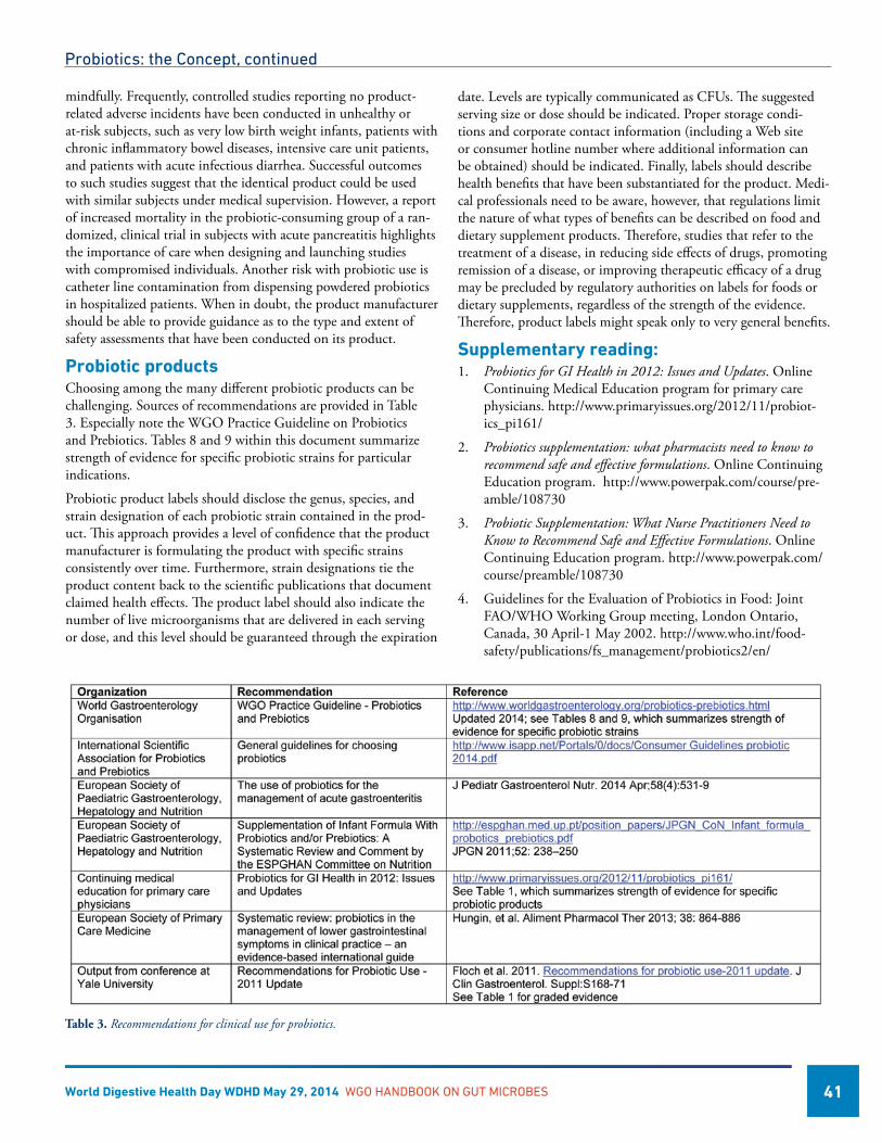
World Digestive Health Day WDHD May 29, 2014 WGO HANDBOOK ON GUT MICROBES 41
mindfully. Frequently, controlled studies reporting no product-related adverse incidents have been conducted in unhealthy or at-risk subjects, such as very low birth weight infants, patients with chronic inflammatory bowel diseases, intensive care unit patients, and patients with acute infectious diarrhea. Successful outcomes to such studies suggest that the identical product could be used with similar subjects under medical supervision. However, a report of increased mortality in the probiotic-consuming group of a ran-domized, clinical trial in subjects with acute pancreatitis highlights the importance of care when designing and launching studies with compromised individuals. Another risk with probiotic use is catheter line contamination from dispensing powdered probiotics in hospitalized patients. When in doubt, the product manufacturer should be able to provide guidance as to the type and extent of safety assessments that have been conducted on its product.
Probiotic productsChoosing among the many different probiotic products can be challenging. Sources of recommendations are provided in Table 3. Especially note the WGO Practice Guideline on Probiotics and Prebiotics. Tables 8 and 9 within this document summarize strength of evidence for specific probiotic strains for particular indications.
Probiotic product labels should disclose the genus, species, and strain designation of each probiotic strain contained in the prod-uct. This approach provides a level of confidence that the product manufacturer is formulating the product with specific strains consistently over time. Furthermore, strain designations tie the product content back to the scientific publications that document claimed health effects. The product label should also indicate the number of live microorganisms that are delivered in each serving or dose, and this level should be guaranteed through the expiration
Probiotics: the Concept, continued
date. Levels are typically communicated as CFUs. The suggested serving size or dose should be indicated. Proper storage condi-tions and corporate contact information (including a Web site or consumer hotline number where additional information can be obtained) should be indicated. Finally, labels should describe health benefits that have been substantiated for the product. Medi-cal professionals need to be aware, however, that regulations limit the nature of what types of benefits can be described on food and dietary supplement products. Therefore, studies that refer to the treatment of a disease, in reducing side effects of drugs, promoting remission of a disease, or improving therapeutic efficacy of a drug may be precluded by regulatory authorities on labels for foods or dietary supplements, regardless of the strength of the evidence. Therefore, product labels might speak only to very general benefits.
Supplementary reading:1. Probiotics for GI Health in 2012: Issues and Updates. Online
Continuing Medical Education program for primary care physicians. http://www.primaryissues.org/2012/11/probiot-ics_pi161/
2. Probiotics supplementation: what pharmacists need to know to recommend safe and effective formulations. Online Continuing Education program. http://www.powerpak.com/course/pre-amble/108730
3. Probiotic Supplementation: What Nurse Practitioners Need to Know to Recommend Safe and Effective Formulations. Online Continuing Education program. http://www.powerpak.com/course/preamble/108730
4. Guidelines for the Evaluation of Probiotics in Food: Joint FAO/WHO Working Group meeting, London Ontario, Canada, 30 April-1 May 2002. http://www.who.int/food-safety/publications/fs_management/probiotics2/en/
Table 3. Recommendations for clinical use for probiotics.
World Digestive Health Day WDHD May 29, 2014 WGO HANDBOOK ON GUT MICROBES 42
Probiotics: the Concept, continued
5. Probiotics, Prebiotics and the Gut Microbiota, 2013, Concise Monograph, International Life Sciences Institute – Europe. http://www.ilsi.org/Europe/Publications/Prebiotics-Probiotics.pdf
6. Sanders ME, Lenoir-Wijnkoop I, Salminen S, Merenstein DJ, Gibson GR, Petschow BW, Nieuwdorp M, Tancredi D, Cifelli C, Jacques P, Pot B. Probiotics and Prebiotics: Prospects for Public Health and Nutritional Recommendations. Ann N Y Acad Sci. 2014 Feb;1309(1):19-29. http://www.ncbi.nlm.nih.gov/pubmed/24571254
7. Hill C, Guarner F, Reid G, Gibson GR, Merenstein DJ, Pot B, Morelli L, Canani RB, Flint, HJ, Salminen S, Calder PC, Sanders ME. The International Scientific Association for Probiotics and Prebiotics consensus statement on the scope of probiotics. In Press, Nature Rev Gastro Hepatol.
8. Hill. Probiotics and pharmabiotics. Alternative medicine or an evidence-based alternative? Bioeng Bugs. 2010 Mar-Apr; 1(2): 79–84. http://www.ilsi.org/Europe/Publications/Prebiot-ics-Probiotics.pdf
9. Sanders, M.E. 2009. How do we know when something called “probiotic” is really a probiotic? A guideline for con-sumers and healthcare professionals. Functional Food Rev 1:3-12. http://journals.bcdecker.com/pubs/FFR/volume 01, 2009/issue 01, Spring/FFR_2009_00002/FFR_2009_00002.pdf.
10. Sanders M.E., Akkermans LMA., Haller D, Hammerman C, Heimbach J, Huys G, Levy D, Mack D, Phothirath P, Constable A, Solano-Aguilar G, Vaughan E. 2010. Assessment of probiotic safety for human use. Gut Microbes 1 (3):1-22. http://www.landesbioscience.com/journals/gutmicrobes/ar-ticle/12127/
World Digestive Health Day WDHD May 29, 2014 WGO HANDBOOK ON GUT MICROBES 43
Probiotics in Diarrheal Diseases
Introduction
Microbes living inside the gut, usually referred to as the gut microbiota, are about 10 times more numerous that the hu-
man cells in the body. Half a million of genes from approximately 300-500 species comprise the microbiome (collective assembly of microbial genomes) of a human individual. In utero, the entire intestinal tract is sterile, and bacteria enter the gut at birth and with foods. The gut microbial ecosystem is subjected to signifi-cant fluctuations for up to three years but, thereafter, the overall structure of the ecosystem remains relatively stable. Indeed, each individual’s microbiota is so distinctive that it could be used as an alternative of fingerprinting. However, the microbial composition is influenced by diet, socio-economic conditions and, above all, by the use of antibiotics [1].
The normal microbiota influences a variety of intestinal functions and plays a key role in nutrition, in maintaining the integrity of the epithelial barrier and in the development of mucosal im-munity. The relationship between the host’s immune system and nonpathogenic constituents of the microbiota is important in protecting the host from colonization by pathogenic species [2].
Probiotics are defined as live microorganisms that, when ingested in adequate amounts, exert a health benefit to the host.
There are 3 main mechanisms whereby probiotics can assist in the defense against pathogens:
• Directantagonism: certain probiotics secrete small molecules or bioactive peptides that have antimicrobial activities. For example, Lactobacilus salivarius UCC118 produces an antimi-crobial peptide that kills Listeria monocytogenes in the lumen of the mouse gastrointestinal tract 30 minutes after oral administration. Saccharomyces boulardii (SB) secretes a serine protease that hydrolyzes toxin A, a virulence factor produced by C. difficile.
• Immunomodulation: probiotics elicit a variety of responses from immune cells in vitro and in vivo. Differential immune regulation may prime the immune system to limit infections, inflammation and pathogen-mediated damage.
María Julieta Argüero, MD Gastroenterological Division of the Hospital Italiano de Buenos Aires Buenos Aires, Argentina
Juan Andrés De Paula, MD Chairman of the Gastroenterological Division of the Hospital Italiano de Buenos Aires Associate Professor of Medicine of the University Institute of the Hospital Italiano de Buenos Aires Buenos Aires, Argentina
• Exclusion: probiotics can make the GI environment less hospitable for pathogens. This mechanism includes decreasing luminal pH, improving epithelial barrier function, interfering with pathogen binding site by down-regulating host receptors, as well as stimulating release of mucins and defensins. [2]. (See Figure 1)
The efficacy of a given probiotic treatment has been shown to be highly dependent on the genus, species, and even the strain. For example, not all lactic acid bacteria have a probiotic effect [3]. The effects of probiotics may be revealed through changes in either microbial populations or their metabolic activity. A recent study demonstrated that a probiotic yogurt changes urinary bacterial me-tabolites, but not fecal bacterial communities. Such results suggest that probiotics have the capability of influencing function rather than composition of the microbiota [3]. While experimental data suggest potential benefits for probiotics in a variety of gastrointes-tinal, pancreatic and liver disorders, as well as in the modulation body weight, solid data are mainly confined to three areas: infec-tions, inflammatory bowel diseases and irritable bowel syndrome. Regarding diarrheal diseases, there are three entities with con-firmed efficacy for probiotics: infectious diarrhea (ID) especially rotavirus-associated diarrhea, antibiotic-associated diarrhea (AAD) and Clostridium difficile-associated disease (CDAD) [1].
Acute DiarrheaDiarrhea is defined by the World Health Organization as three or more loose or liquid stools in a 24-hour period or more frequent stool than normal for the individual [4]. Enteric and diarrheal
1. Direct antagonism of the resident microbiota and transitory microbiota to pathogens;
2. Immunomodulation of the host defenses enhancing the functionality of innate and/or adaptive immunity, or limiting the ability of the pathogen to initiate or facilitate an immune response; and,
3. Excluding pathogens from the mucosal surface by altering the microenvi-ronment to prevent pathogens from gaining access to appropriate recep-tors, limiting pathogen attachment, entry, or translocation, or improving barrier function.
IEC: intestinal enteric cell; M: M cell; T: T lymphocyte; B: B lymphocyte; MAC: macrophage; DC: dendritic cell; TJ: Tight junctionAdapted from Preidis GA, Gastroenterology 2011; 140: 8.
Figure 1: Proposed mechanisms by which probiotics exclude pathogenic microorganisms.
These include:
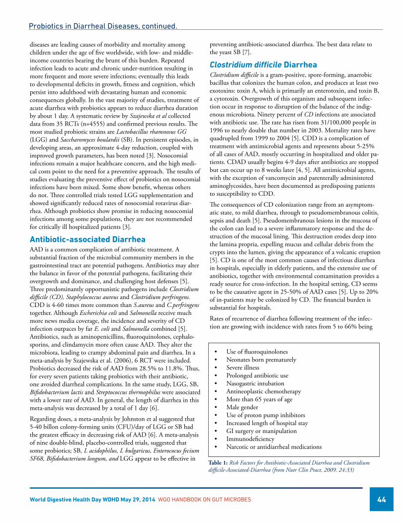
World Digestive Health Day WDHD May 29, 2014 WGO HANDBOOK ON GUT MICROBES 44
diseases are leading causes of morbidity and mortality among children under the age of five worldwide, with low- and middle-income countries bearing the brunt of this burden. Repeated infection leads to acute and chronic under-nutrition resulting in more frequent and more severe infections; eventually this leads to developmental deficits in growth, fitness and cognition, which persist into adulthood with devastating human and economic consequences globally. In the vast majority of studies, treatment of acute diarrhea with probiotics appears to reduce diarrhea duration by about 1 day. A systematic review by Szajewska et al collected data from 35 RCTs (n=4555) and confirmed previous results. The most studied probiotic strains are Lactobacillus rhamnosus GG (LGG) and Saccharomyces boulardii (SB). In persistent episodes, in developing areas, an approximate 4-day reduction, coupled with improved growth parameters, has been noted [3]. Nosocomial infections remain a major healthcare concern, and the high medi-cal costs point to the need for a preventive approach. The results of studies evaluating the preventive effect of probiotics on nosocomial infections have been mixed. Some show benefit, whereas others do not. Three controlled trials tested LGG supplementation and showed significantly reduced rates of nosocomial rotavirus diar-rhea. Although probiotics show promise in reducing nosocomial infections among some populations, they are not recommended for critically ill hospitalized patients [3].
Antibiotic-associated DiarrheaAAD is a common complication of antibiotic treatment. A substantial fraction of the microbial community members in the gastrointestinal tract are potential pathogens. Antibiotics may alter the balance in favor of the potential pathogens, facilitating their overgrowth and dominance, and challenging host defenses [5]. Three predominantly opportunistic pathogens include Clostridium difficile (CD), Staphylococcus aureus and Clostridium perfringens. CDD is 4-60 times more common than S.aureus and C.perfringens together. Although Escherichia coli and Salmonella receive much more news media coverage, the incidence and severity of CD infection outpaces by far E. coli and Salmonella combined [5]. Antibiotics, such as aminopenicillins, fluoroquinolones, cephalo-sporins, and clindamycin more often cause AAD. They alter the microbiota, leading to crampy abdominal pain and diarrhea. In a meta-analysis by Szajewska et al. (2006), 6 RCT were included. Probiotics decreased the risk of AAD from 28.5% to 11.8%. Thus, for every seven patients taking probiotics with their antibiotic, one avoided diarrheal complications. In the same study, LGG, SB, Bifidobacterium lactis and Streptococcus thermophilus were associated with a lower rate of AAD. In general, the length of diarrhea in this meta-analysis was decreased by a total of 1 day [6].
Regarding doses, a meta-analysis by Johnston et al suggested that 5-40 billon colony-forming units (CFU)/day of LGG or SB had the greatest efficacy in decreasing risk of AAD [6]. A meta-analysis of nine double-blind, placebo-controlled trials, suggested that some probiotics; SB, L acidophilus, L bulgaricus, Enterococus fecium SF68, Bifidobacterium longum, and LGG appear to be effective in
preventing antibiotic-associated diarrhea. The best data relate to the yeast SB [7].
Clostridium difficile DiarrheaClostridium difficile is a gram-positive, spore-forming, anaerobic bacillus that colonizes the human colon, and produces at least two exotoxins: toxin A, which is primarily an enterotoxin, and toxin B, a cytotoxin. Overgrowth of this organism and subsequent infec-tion occur in response to disruption of the balance of the indig-enous microbiota. Ninety percent of CD infections are associated with antibiotic use. The rate has risen from 31/100,000 people in 1996 to nearly double that number in 2003. Mortality rates have quadrupled from 1999 to 2004 [5]. CDD is a complication of treatment with antimicrobial agents and represents about 5-25% of all cases of AAD, mostly occurring in hospitalized and older pa-tients. CDAD usually begins 4-9 days after antibiotics are stopped but can occur up to 8 weeks later [4, 5]. All antimicrobial agents, with the exception of vancomycin and parenterally administered aminoglycosides, have been documented as predisposing patients to susceptibility to CDD.
The consequences of CD colonization range from an asymptom-atic state, to mild diarrhea, through to pseudomembranous colitis, sepsis and death [5]. Pseudomembranous lesions in the mucosa of the colon can lead to a severe inflammatory response and the de-struction of the mucosal lining. This destruction erodes deep into the lamina propria, expelling mucus and cellular debris from the crypts into the lumen, giving the appearance of a volcanic eruption [5]. CD is one of the most common causes of infectious diarrhea in hospitals, especially in elderly patients, and the extensive use of antibiotics, together with environmental contamination provides a ready source for cross-infection. In the hospital setting, CD seems to be the causative agent in 25-50% of AAD cases [5]. Up to 20% of in-patients may be colonized by CD. The financial burden is substantial for hospitals.
Rates of recurrence of diarrhea following treatment of the infec-tion are growing with incidence with rates from 5 to 66% being
• Useoffluoroquinolones• Neonatesbornprematurely• Severeillness• Prolongedantibioticuse• Nasogastricintubation• Antineoplasticchemotherapy• Morethan65yearsofage• Malegender• Useofprotonpumpinhibitors• Increasedlengthofhospitalstay• GIsurgeryormanipulation• Immunodeficiency• Narcoticorantidiarrhealmedications
Table 1: Risk Factors for Antibiotic-Associated Diarrhea and Clostridium difficile-Associated-Diarrhea (from Nutr Clin Pract, 2009. 24:33)
Probiotics in Diarrheal Diseases, continued.

World Digestive Health Day WDHD May 29, 2014 WGO HANDBOOK ON GUT MICROBES 45
reported. CD spores often remain in the gut even after aggressive treatment. Spores can stay tucked away in colonic diverticula, avoiding peristalsis and exposure to antibiotics. The risk of recur-rence increases to 50-65% in patients with 2 or more previous episodes [5]. Besides taking antibiotics, there are other risk factors for the development of AAD/CDAD (see table 1). Life-threaten-ing complications may occur when patients with CDAD receive narcotics and antidiarrheals that may have an antiperistaltic effect, leading to toxic mega-colon. This complication results in extended hospital stay, increased rate of unrelated infections, and 2-3 fold increase in mortality.
Highly virulent CD strains have been reported recently. In Quebec, Canada the percentage of patients who die within 30 days increased from 4.7 to 13.8%. This seems to be related to a virulent strain of the bacteria. Severe outbreaks of the disease have led to the recognition of the emergence of NAP 1/027, an epidemic strain of CD associated with increase severity and death. The increase in the incidence and severity of CDD has prompted much interest in the use of probiotics, in combination with cur-rent antibiotic therapies, to prevent and treat AAD and reduce the prevalence of CDAD [5].
The most effective therapy for AAD is cessation of the responsible antibiotic(s). CDAD is diagnosed by a positive stool toxin assay in the context of the clinical manifestations of the infection: diarrhea, leukocytosis, fever and abdominal pain. Empirical antibiotic treat-ment is appropriate if suspicion is high, even if the diagnostic assay is not positive. Oral metronidazole, for a minimum of 10 days, is the drug of choice for an initial episode. Patients with moderate to severe infection or those who fail treatment with metronidazole should receive oral vancomycin for 10 days. Of concern is the potential ability of CD spores to survive and cause recurrence. Intermittent antibiotics may be given using pulsed protocols. A pulsed regimen consists of administering the same drug for the original infection every few days. A tapered process is a stepwise decrease in dose over a period of time [5].
Some authors suggested that the prophylactic use of probiotics might be necessary to bolster the colonization resistance of the normal microbiota, disrupted by the effects of antibiotic therapy. A double-blind, placebo-controlled study examined the role of probiotic administration in the prevention of CDAD in elderly patients receiving antibiotics. The incidence of samples positive for CD toxins was 2.9% in the probiotic group compared with 7.2% in the placebo-control group. When samples from all patients were tested (rather than just of those developing diarrhea) 46% of probiotic group were toxin-positive compared with 78% of the placebo group [8].
Several studies with probiotics report reductions in nosocomial diarrhea rates, as well as reductions in AAD and recurrence of CDAD. These effects include a 40-60% reduction in the frequency of AAD, but studies documenting a reduction in CDAD are far fewer and remain the subject of controversy. Indeed, Floch et al considered evidence insufficient for an ¨A¨ recommendation for this indication [3]. McFarland et al showed that SB and LGG
significantly reduce the rate of development of AAD and the com-bination of L. acidophilus and Bifidobacterium reduced fecal counts of CD [3]. A randomized double blind placebo controlled study concluded that twice daily intake of a probiotic drink containing L casei, L bulgaricus and S thermophiles for one week longer than the duration of antibiotic treatment can prevent AAD and CDD [7]. A meta-analysis of 35 randomized, controlled trials supported the efficacy of probiotics in the prevention of AAD but not necessarily CDAD. A total of 2810 patients with ADD were included. Sixty-four percent of the studies consisted of adults and 36% children; all received antibiotics. Significant efficacy for probiotics was seen in 44% of adult studies and 67% of the trials involving children. It was concluded that SB, LGG and probiotics mixtures showed the most potential for a protective effect from AAD [5].
As mentioned before, CDAD has a high risk of recurrence on completion of a course of metronidazole or vancomycin. In six tri-als involving 354 adults with CDAD, only SB achieved a signifi-cant reduction in recurrence rate in patients who were receiving high-dose oral vancomycin. Another study corroborated these results in a subgroup of individuals who received high-dose vanco-mycin to treat recurrent, moderate to severe, CDAD [5]. In a sys-tematic review conducted by McFarland, that collected data from 31 RCTs (n=5029), SB was safe and effective in 84% of treatments in the prevention of AAD, so this probiotic can be recommended for the prevention of AAD (figure 2).
Discrepancies between studies may be due to variations in CD strain virulence, dosing of the probiotic, laboratory tests used for CDAD diagnosis, and even in the definition. More studies are needed to analyze how many CFU/day are needed to ensure a beneficial outcome. Biomarkers of intestinal pathology must be
Copy from McFarland LV, World J Gastroenterol 2010; 16: 2202.
Figure 2: Schematic of intestinal tract, illustrating the different potential mechanisms of action of Saccharomyces boulardii (Sb). On the left, effects of different pathogenic microbes are depicted. On the right, seven different protective effects of Sb are depicted. Within the lumen of the intestine, Sb may degrade toxins of pathogens, interfere with pathogenic adherence, modulate normal microbiota and preserve normal intestinal physiology. Sb may also indirectly restore normal short chain fatty acid (SCFA) balance. Sb may also increase secretory IgA (sIgA) levels or act as an immune regulator by influenc-ing cytokine levels.
Probiotics in Diarrheal Diseases, continued.
World Digestive Health Day WDHD May 29, 2014 WGO HANDBOOK ON GUT MICROBES 46
developed to determine therapeutic efficacy of existing and new probiotics [2].
In summary, the two most promising probiotics are SB and LGG. Prevention remains the best defense against the rising incidence of AAD and CDAD. Although judicious use of antibiotics is of primary importance, probiotics can effectively reduce the inci-dence of AAD and CDD with minimal or no secondary effects. In human biology and medicine we have entered into the era of the microbiome, and its implication in human health and disease is just beginning to be revealed.
References 1. Quigley, E.M., Prebiotics and probiotics; modifying and mining
the microbiota. Pharmacol Res, 2010. 61(3): p. 213-8.
2. Preidis, G.A., et al., Probiotics, enteric and diarrheal diseases, and global health. Gastroenterology, 2011. 140(1): p. 8-14.
3. Sanders, M.E., et al., An update on the use and investigation of probiotics in health and disease. Gut, 2013. 62(5): p. 787-96.
4. Hickson, M., Use of probiotic Lactobacillus preparation to prevent diarrhea associated with antibiotics: randomised double blind placebo controlled trial. BMJ, 2007: p. 1-5.
5. Rohde, C.L., V. Bartolini, and N. Jones, The use of probiotics in the prevention and treatment of antibiotic-associated diarrhea with special interest in Clostridium difficile-associated diarrhea. Nutr Clin Pract, 2009. 24(1): p. 33-40.
6. Jones, K., Probiotics: preventing antibiotic-associated diarrhea. J Spec Pediatr Nurs, 2010. 15(2): p. 160-2.
7. D’Souza, A.L., et al., Probiotics in prevention of antibiotic as-sociated diarrhoea: meta-analysis. BMJ, 2002. 324(7350): p. 1361.
8. Plummer, S., et al., Clostridium difficile pilot study: effects of probiotic supplementation on the incidence of C. difficile diar-rhoea. Int Microbiol, 2004. 7(1): p. 59-62.
9. Ritchie, M.L. and T.N. Romanuk, A meta-analysis of probiotic efficacy for gastrointestinal diseases. PLoS One, 2012. 7(4): p. e34938.
10. McFarland, L.V., Systematic review and meta-analysis of Sac-charomyces boulardii in adult patients. World J Gastroenterol, 2010. 16(18): p. 2202-22.
Probiotics in Diarrheal Diseases, continued.
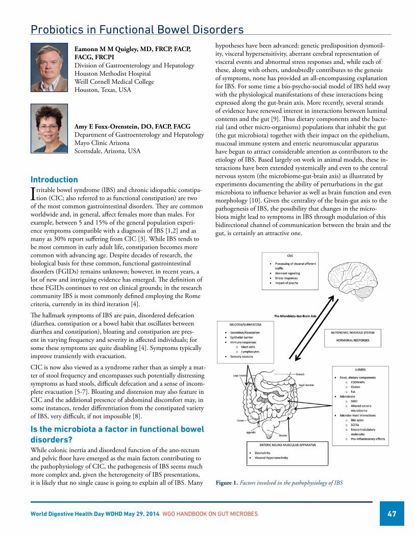
World Digestive Health Day WDHD May 29, 2014 WGO HANDBOOK ON GUT MICROBES 47
Probiotics in Functional Bowel Disorders
Introduction
Irritable bowel syndrome (IBS) and chronic idiopathic constipa-tion (CIC; also referred to as functional constipation) are two
of the most common gastrointestinal disorders. They are common worldwide and, in general, affect females more than males. For example, between 5 and 15% of the general population experi-ence symptoms compatible with a diagnosis of IBS [1,2] and as many as 30% report suffering from CIC [3]. While IBS tends to be most common in early adult life, constipation becomes more common with advancing age. Despite decades of research, the biological basis for these common, functional gastrointestinal disorders (FGIDs) remains unknown; however, in recent years, a lot of new and intriguing evidence has emerged. The definition of these FGIDs continues to rest on clinical grounds; in the research community IBS is most commonly defined employing the Rome criteria, currently in its third iteration [4].
The hallmark symptoms of IBS are pain, disordered defecation (diarrhea, constipation or a bowel habit that oscillates between diarrhea and constipation), bloating and constipation are pres-ent in varying frequency and severity in affected individuals; for some these symptoms are quite disabling [4]. Symptoms typically improve transiently with evacuation.
CIC is now also viewed as a syndrome rather than as simply a mat-ter of stool frequency and encompasses such potentially distressing symptoms as hard stools, difficult defecation and a sense of incom-plete evacuation [5-7]. Bloating and distension may also feature in CIC and the additional presence of abdominal discomfort may, in some instances, render differentiation from the constipated variety of IBS, very difficult, if not impossible [8].
Is the microbiota a factor in functional bowel disorders?While colonic inertia and disordered function of the ano-rectum and pelvic floor have emerged as the main factors contributing to the pathophysiology of CIC, the pathogenesis of IBS seems much more complex and, given the heterogeneity of IBS presentations, it is likely that no single cause is going to explain all of IBS. Many
Eamonn M M Quigley, MD, FRCP, FACP, FACG, FRCPI Division of Gastroenterology and Hepatology Houston Methodist Hospital Weill Cornell Medical College Houston, Texas, USA
AmyEFoxx-Orenstein,DO,FACP,FACG Department of Gastroenterology and Hepatology Mayo Clinic Arizona Scottsdale, Arizona, USA
hypotheses have been advanced: genetic predisposition dysmotil-ity, visceral hypersensitivity, aberrant cerebral representation of visceral events and abnormal stress responses and, while each of these, along with others, undoubtedly contributes to the genesis of symptoms, none has provided an all-encompassing explanation for IBS. For some time a bio-psycho-social model of IBS held sway with the physiological manifestations of these interactions being expressed along the gut-brain axis. More recently, several strands of evidence have renewed interest in interactions between luminal contents and the gut [9]. Thus dietary components and the bacte-rial (and other micro-organisms) populations that inhabit the gut (the gut microbiota) together with their impact on the epithelium, mucosal immune system and enteric neuromuscular apparatus have begun to attract considerable attention as contributors to the etiology of IBS. Based largely on work in animal models, these in-teractions have been extended systemically and even to the central nervous system (the microbiome-gut-brain axis) as illustrated by experiments documenting the ability of perturbations in the gut microbiota to influence behavior as well as brain function and even morphology [10]. Given the centrality of the brain-gut axis to the pathogenesis of IBS, the possibility that changes in the micro-biota might lead to symptoms in IBS through modulation of this bidirectional channel of communication between the brain and the gut, is certainly an attractive one.
Figure 1. Factors involved in the pathophysiology of IBS
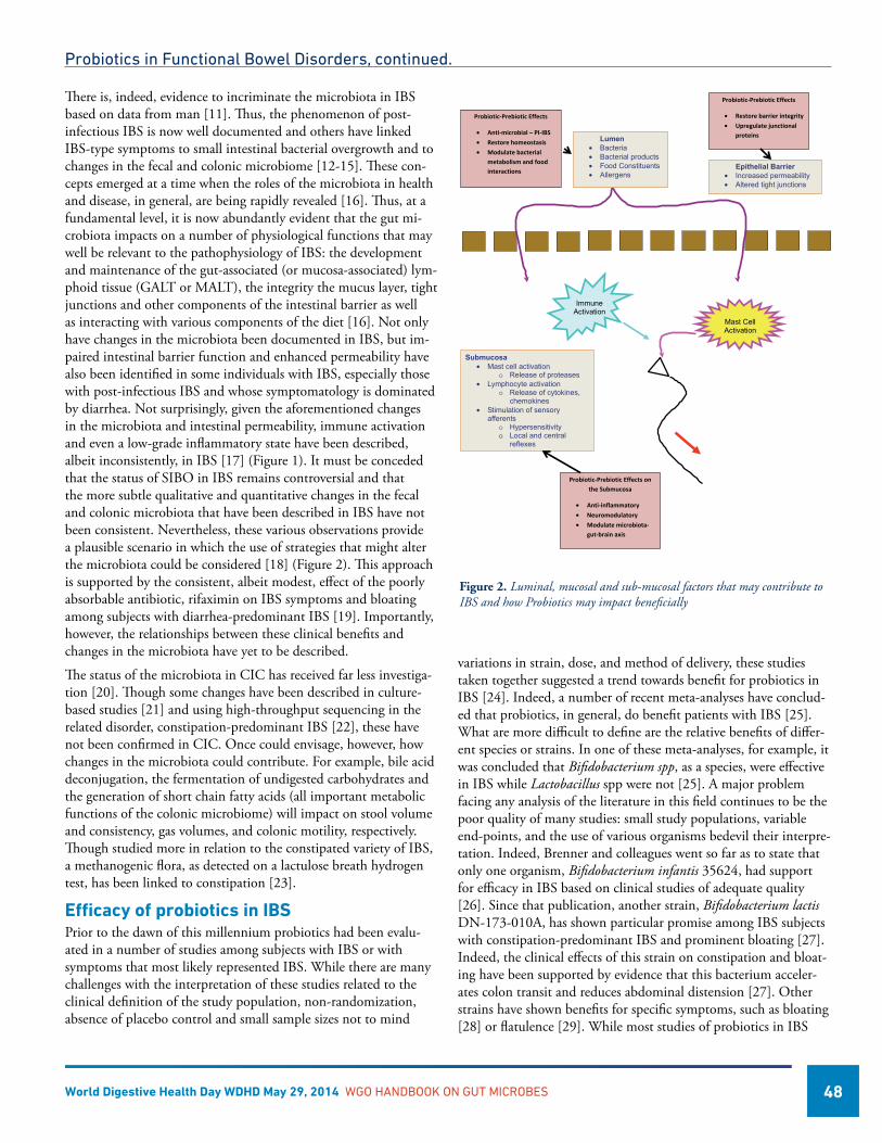
World Digestive Health Day WDHD May 29, 2014 WGO HANDBOOK ON GUT MICROBES 48
variations in strain, dose, and method of delivery, these studies taken together suggested a trend towards benefit for probiotics in IBS [24]. Indeed, a number of recent meta-analyses have conclud-ed that probiotics, in general, do benefit patients with IBS [25]. What are more difficult to define are the relative benefits of differ-ent species or strains. In one of these meta-analyses, for example, it was concluded that Bifidobacterium spp, as a species, were effective in IBS while Lactobacillus spp were not [25]. A major problem facing any analysis of the literature in this field continues to be the poor quality of many studies: small study populations, variable end-points, and the use of vari ous organisms bedevil their interpre-tation. Indeed, Brenner and colleagues went so far as to state that only one organism, Bifidobacterium infantis 35624, had support for efficacy in IBS based on clinical studies of adequate quality [26]. Since that publication, another strain, Bifidobacterium lactis DN-173-010A, has shown particular promise among IBS subjects with constipation-predominant IBS and prominent bloating [27]. Indeed, the clinical effects of this strain on constipation and bloat-ing have been supported by evidence that this bac terium acceler-ates colon transit and reduces abdominal distension [27]. Other strains have shown benefits for specific symptoms, such as bloating [28] or flatulence [29]. While most studies of probiotics in IBS
There is, indeed, evidence to incriminate the microbiota in IBS based on data from man [11]. Thus, the phenomenon of post-infectious IBS is now well documented and others have linked IBS-type symptoms to small intestinal bacterial overgrowth and to changes in the fecal and colonic microbiome [12-15]. These con-cepts emerged at a time when the roles of the microbiota in health and disease, in general, are being rapidly revealed [16]. Thus, at a fundamental level, it is now abundantly evident that the gut mi-crobiota impacts on a number of physiological functions that may well be relevant to the pathophysiology of IBS: the development and maintenance of the gut-associated (or mucosa-associated) lym-phoid tissue (GALT or MALT), the integrity the mucus layer, tight junctions and other components of the intestinal barrier as well as interacting with various components of the diet [16]. Not only have changes in the microbiota been documented in IBS, but im-paired intestinal barrier function and enhanced permeability have also been identified in some individuals with IBS, especially those with post-infectious IBS and whose symptomatology is dominated by diarrhea. Not surprisingly, given the aforementioned changes in the microbiota and intestinal permeability, immune activation and even a low-grade inflammatory state have been described, albeit inconsistently, in IBS [17] (Figure 1). It must be conceded that the status of SIBO in IBS remains controversial and that the more subtle qualitative and quantitative changes in the fecal and colonic microbiota that have been described in IBS have not been consistent. Nevertheless, these various observations provide a plausible scenario in which the use of strategies that might alter the microbiota could be considered [18] (Figure 2). This approach is supported by the consistent, albeit modest, effect of the poorly absorbable antibiotic, rifaximin on IBS symptoms and bloating among subjects with diarrhea-predominant IBS [19]. Importantly, however, the relationships between these clinical benefits and changes in the microbiota have yet to be described.
The status of the microbiota in CIC has received far less investiga-tion [20]. Though some changes have been described in culture-based studies [21] and using high-throughput sequencing in the related disorder, constipation-predominant IBS [22], these have not been confirmed in CIC. Once could envisage, however, how changes in the microbiota could contribute. For example, bile acid deconjugation, the fermentation of undigested carbohydrates and the generation of short chain fatty acids (all important metabolic functions of the colonic microbiome) will impact on stool volume and consistency, gas volumes, and colonic motility, respectively. Though studied more in relation to the constipated variety of IBS, a methanogenic flora, as detected on a lactulose breath hydrogen test, has been linked to constipation [23].
Efficacy of probiotics in IBS Prior to the dawn of this millennium probiotics had been evalu-ated in a number of studies among subjects with IBS or with symptoms that most likely represented IBS. While there are many challenges with the interpretation of these studies related to the clinical definition of the study population, non-randomization, absence of placebo control and small sample sizes not to mind
Probiotics in Functional Bowel Disorders, continued.
1
Immune Activation
Mast Cell Activation
Lumen • Bacteria • Bacterial products • Food Constituents • Allergens
Epithelial Barrier • Increased permeability • Altered tight junctions
Submucosa • Mast cell activation
o Release of proteases • Lymphocyte activation
o Release of cytokines, chemokines
• Stimulation of sensory afferents
o Hypersensitivity o Local and central
reflexes
Probiotic-Prebiotic Effects
• Anti-microbial – PI-IBS • Restore homeostasis • Modulate bacterial
metabolism and food interactions
Probiotic-Prebiotic Effects
• Restore barrier integrity • Upregulate junctional
proteins
Probiotic-Prebiotic Effects on the Submucosa
• Anti-inflammatory • Neuromodulatory • Modulate microbiota-
gut-brain axis
Figure 2. Luminal, mucosal and sub-mucosal factors that may contribute to IBS and how Probiotics may impact beneficially
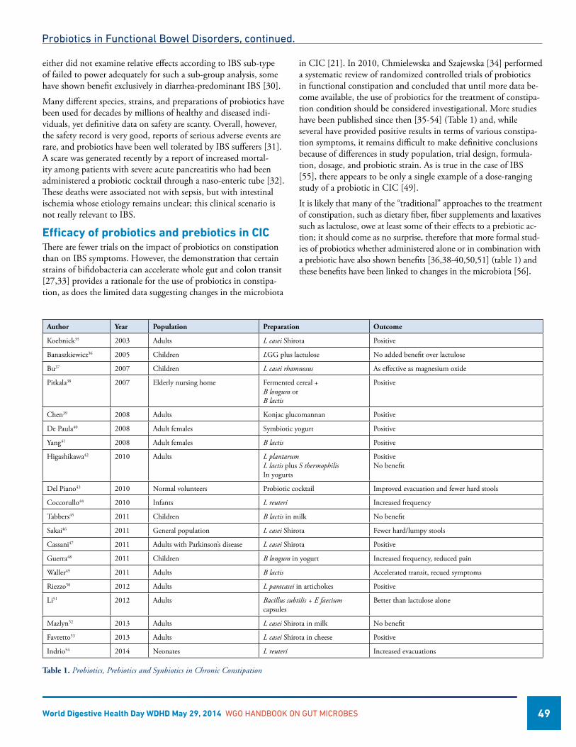
World Digestive Health Day WDHD May 29, 2014 WGO HANDBOOK ON GUT MICROBES 49
either did not examine relative effects according to IBS sub-type of failed to power adequately for such a sub-group analysis, some have shown benefit exclusively in diarrhea-predominant IBS [30].
Many different species, strains, and preparations of probiotics have been used for decades by millions of healthy and diseased indi-viduals, yet definitive data on safety are scanty. Overall, however, the safety record is very good, reports of serious adverse events are rare, and probiotics have been well tolerated by IBS sufferers [31]. A scare was generated recently by a report of increased mortal-ity among patients with severe acute pancreatitis who had been administered a probiotic cocktail through a naso-enteric tube [32]. These deaths were associated not with sepsis, but with intestinal ischemia whose etiology remains unclear; this clinical scenario is not really relevant to IBS.
Efficacy of probiotics and prebiotics in CICThere are fewer trials on the impact of probiotics on constipation than on IBS symptoms. However, the demonstration that certain strains of bifidobacteria can accelerate whole gut and colon transit [27,33] provides a rationale for the use of probiotics in constipa-tion, as does the limited data suggesting changes in the microbiota
in CIC [21]. In 2010, Chmielewska and Szajewska [34] performed a systematic review of randomized controlled trials of probiotics in functional constipation and concluded that until more data be-come available, the use of probiotics for the treatment of constipa-tion condition should be considered investigational. More studies have been published since then [35-54] (Table 1) and, while several have provided positive results in terms of various constipa-tion symptoms, it remains difficult to make definitive conclusions because of differences in study population, trial design, formula-tion, dosage, and probiotic strain. As is true in the case of IBS [55], there appears to be only a single example of a dose-ranging study of a probiotic in CIC [49].
It is likely that many of the “traditional” approaches to the treatment of constipation, such as dietary fiber, fiber supplements and laxatives such as lactulose, owe at least some of their effects to a prebiotic ac-tion; it should come as no surprise, therefore that more formal stud-ies of probiotics whether administered alone or in combination with a prebiotic have also shown benefits [36,38-40,50,51] (table 1) and these benefits have been linked to changes in the microbiota [56].
Author Year Population Preparation Outcome
Koebnick35 2003 Adults L casei Shirota Positive
Banaszkiewicz36 2005 Children LGG plus lactulose No added benefit over lactulose
Bu37 2007 Children L casei rhamnosus As effective as magnesium oxide
Pitkala38 2007 Elderly nursing home Fermented cereal +B longum orB lactis
Positive
Chen39 2008 Adults Konjac glucomannan Positive
De Paula40 2008 Adult females Symbiotic yogurt Positive
Yang41 2008 Adult females B lactis Positive
Higashikawa42 2010 Adults L plantarumL lactis plus S thermophilisIn yogurts
PositiveNo benefit
Del Piano43 2010 Normal volunteers Probiotic cocktail Improved evacuation and fewer hard stools
Coccorullo44 2010 Infants L reuteri Increased frequency
Tabbers45 2011 Children B lactis in milk No benefit
Sakai46 2011 General population L casei Shirota Fewer hard/lumpy stools
Cassani47 2011 Adults with Parkinson’s disease L casei Shirota Positive
Guerra48 2011 Children B longum in yogurt Increased frequency, reduced pain
Waller49 2011 Adults B lactis Accelerated transit, recued symptoms
Riezzo50 2012 Adults L paracasei in artichokes Positive
Li51 2012 Adults Bacillus subtilis + E faecium capsules
Better than lactulose alone
Mazlyn52 2013 Adults L casei Shirota in milk No benefit
Favretto53 2013 Adults L casei Shirota in cheese Positive
Indrio54 2014 Neonates L reuteri Increased evacuations
Table 1. Probiotics, Prebiotics and Synbiotics in Chronic Constipation
Probiotics in Functional Bowel Disorders, continued.
World Digestive Health Day WDHD May 29, 2014 WGO HANDBOOK ON GUT MICROBES 50
Prebiotics and synbiotics in IBS Silk and colleagues showed that a trans-galactooligosaccharide designed to specifically stimulate bifidobacteria in the gut, was effective in alleviating symptoms in IBS [57]. With regard to the combination of a prebiotic and a probiotic, referred to as a syn-biotic, two studies have been performed: one assessed the impact of a combination of Lactobacillus acidophilus and helveticus with Bifidobacterium in a vitamin and phytoextract-enriched medium [58], and the other study used Bifidobacterium lactis in combina-tion with acacia fiber [59]. Both studies reported positive results.
What does the future hold?Bolstered by a scientific rationale and supported by data from clinical trials the future for probiotics and prebiotics in IBS would appear bright; several issues, however, need to be addressed [60]. Firstly, given that IBS is a chronic recurring disorder and that probiotic and prebiotic benefits can be expected to last only as long as they are administered, longer term studies are needed. Further-more, we need more information on optimal strain or strains, dose, formulation, and duration of therapy. Above all, we need to know why and how probiotics work.
Going beyond the conventional realm of the probiotic as a live organism with health benefits lies the possibility that dead bugs, bacterial components or small molecules elaborated by commensal bacteria may be effective in IBS [61].
References1. Quigley EMM, Abdel-Hamid H, Barbara G, et al. A Global
Perspective on Irritable Bowel Syndrome. A Consensus State-ment of the World Gastroenterology Organization (WGO) Summit Task Force on Irritable Bowel Syndrome. J Clin Gastroenterol 2012;46:356-66.
2. Pare P, Ferrazzi S, Thompson WG et al. An epidemiological survey of constipation in Canada: definitions, rates, demo-graphics, and predictors of healthcare seeking. Am. J. Gastro-enterol. 2001;96:3130–7.
3. Longstreth GF, Thompson WG, Chey WD, et al. Functional bowel disorders. Gastroenterology 2006;130:1480-91.
4. Saito YA, Schoenfeld P, Locke GR 3rd. The epidemiology of irritable bowel syndrome in North America: a systematic review. AmJGastroenterol2002;97:1910-5.
5. Locke GR 3rd, Pemberton JH, Phillips SE. AGA technical review on constipation. Gastroenterology 2000;119:1766–1778.
6. Brandt LJ, Prather CM, Quigley EMM et al. Systematic review on the management of chronic constipation in North America. Am. J. Gastroenterol. 2005;100:S5–S22.
7. Lindberg G, Hamid SS, Malfertheiner P et al. World Gas-troenterology Organisation global guideline: constipation: a global perspective. J. Clin. Gastroenterol. 2011;45: 483–487.
8. Rey E, Balboa A, Mearin F. Chronic Constipation, Irritable Bowel Syndrome With Constipation and Constipation With
Pain/Discomfort: Similarities and Differences. Am J Gastro-enterol. 2014 [epub ahead of print]
9. Quigley EM. Bugs on the brain; brain in the gut-seeking explanations for common gastrointestinal symptoms. IrJMedSci2012;182:1-6.
10. Cryan JF, O’Mahony SM. The microbiome-gut-brain axis: from bowel to behavior. Neurogastroenterol Motil 2011;23:187-92.
11. Quigley EM. Bacterial flora in irritable bowel syndrome: role in pathophysiology, implications for management. J Dig Dis 2007;8:2-7.
12. Pimentel M, Chow EJ, Lin HC. Eradication of small intesti-nal bacterial overgrowth reduces symptoms of irritable bowel syndrome. Am J Gastroenterol 2000;95:3503-6.
13. Jeffery IB, Quigley EM, Ohman L, Simrén M, O’Toole PW. The microbiota link to Irritable Bowel Syndrome: An emerg-ing story. GutMicrobes 2012;3:572-6.
14. Carroll IM, Ringel-Kulka T, Keku TO, Chang YH, Packey CD, Sartor RB, Ringel Y. Molecular analysis of the luminal- and mucosal-associated intestinal microbiota in diarrhea-predominant irritable bowel syndrome. Am J Physiol Gastro-intest Liver Physiol. 2011;301:G799-807.
15. Jeffery IB, O’Toole PW, Öhman L, Claesson MJ, Deane J, Quigley EM, Simrén M. An irritable bowel syndrome subtype defined by species-specific alterations in faecal microbiota. Gut. 2012;61:997-1006.
16. Cho I, Blaser MJ. The human microbiome: at the interface of health and disease. Nat Rev Genet 2012;13,260-70 .
17. Piche T. Tight junctions and IBS--the link between epithelial permeability, low-grade inflammation, and symptom genera-tion? Neurogastroenterol Motil. 2014;26:296-302.
18. Quigley EMM. Therapies aimed at the gut microbiota and inflammation. Antibiotics, probiotics/prebiotics/synbiotics, anti-inflammatory therapies. Gastroenterol Clin N Amer 2011;40:207-22.
19. Pimentel M, Lembo A, Chey WD, Zakko S, Ringel Y, Yu J, Mareya SM, Shaw AL, Bortey E, Forbes WP, for the TAR-GET Study Group. Rifaximin therapy for patients with irritable bowel syndrome without constipation. New Engl J Med 2011;364:22-32.
20. Quigley EM. The enteric microbiota in the pathogenesis and management of constipation. Best Pract Res Clin Gastroen-terol. 2011;25:119-26.
21. Khalif IL, Quigley EM, Konovitch EA, Maximova ID. Altera-tions in the colonic flora and intestinal permeability and evidence of immune activation in chronic constipation. Dig Liver Dis. 2005;37:838-49.
22. Chassard C, Dapoigny M, Scott KP, Crouzet L, Del’homme C, Marquet P, Martin JC, Pickering G, Ardid D, Eschalier
Probiotics in Functional Bowel Disorders, continued.
World Digestive Health Day WDHD May 29, 2014 WGO HANDBOOK ON GUT MICROBES 51
A, Dubray C, Flint HJ, Bernalier-Donadille A. Functional dysbiosis within the gut microbiota of patients with consti-pated-irritable bowel syndrome. Aliment Pharmacol Ther. 2012;35:828-38.
23. Furnari M, Savarino E, Bruzzone L, Moscatelli A, Gemignani L, Giannini EG, Zentilin P, Dulbecco P, Savarino V. Reas-sessment of the role of methane production between irritable bowel syndrome and functional constipation. J Gastrointestin Liver Dis. 2012;21:157-63.
24. Quigley EM, Flourie B. Probiotics and irritable bowel syndrome: a rationale for their use and an assessment of the evidence to date. NeurogastroenterolMotil 2007;19:166-72.
25. Moayyedi P, Ford AC, Talley NJ, Cremonini F, Foxx-Oren-stein A, Brandt L, Quigley E. http://www.ncbi.nlm.nih.gov/pubmed/19091823?ordinalpos=6&itool=EntrezSystem2.PEntrez.Pubmed.Pubmed_ResultsPanel.Pubmed_DefaultRe-portPanel.Pubmed_RVDocSum The efficacy of probiotics in the treatment of irritable bowel syndrome: a systematic review. Gut. 2010;59:325-32 .
26. Brenner DM, Moeller MJ, Chey WD, Schoenfeld PS. The utility of probiotics in the treatment of irritable bowel syndrome: a systematic review. Am J Gastroenterol. 2009;104:1033-49.
27. Agrawal A, Houghton LA, Morris J, Reilly B, Guyonnet D, Goupil Feuillerat N, Schlumberger A, Jakob S, Whorwell PJ. Clinical trial: the effects of a fermented milk product contain-ing Bifidobacteriumlactis DN-173-010 on abdominal disten-sion and gastrointestinal transit in irritable bowel syndrome with constipation. AlimentPharmacolTher2009;29:104-14.
28. Ringel Y, Ringel-Kulka T, Maier D, Carroll I, Galanko JA, Leyer G, Palsson OS. Clinical trial: probiotic bacteria Lacto-bacillus acidophilus NCFM and Bifidobacterium lactis Bi-07 versus placebo for the symptoms of bloating in patients with functional bowel disorders – a double-blind study. J Clin Gastroenterol 2011;45:518-25.
29. Kim HJ, Vazquez Roque MI, Camilleri M, Stephens D, Burton DD, Baxter K, Thomforde G, Zinsmeister AR. A randomized controlled trial of a probiotic combination VSL# 3 and placebo in irritable bowel syndrome with bloating. NeurogastroenterolMotil 2005;17,687-96.
30. Andriulli A, Neri M, Loguercio C, Terreni N, Merla A, Cardarella MP, Federico A, Chilovi F, Milandri GL, De Bona M, Cavenati S, Gullini S, Abbiati R, Garbagna N, Cerutti R, Grossi E. Clinical trial on the efficacy of a new symbiotic for-mulation, Flortec, in patients with irritable bowel syndrome: a multicenter, randomized study. J Clin Gastroenterol 2008;42 (Suppl 3 Pt 2):S218-23.
31. Shanahan F. A commentary on the safety of probiotics. Gas-troenterolClinNorthAm 2012;41869-76.
32. Besselink MG, van Santvoort HC, Buskens E, et al.Probiotic prophylaxis in predicted severe acute pancreatitis: a ran-
domised, double-blind, placebo-controlled trial. Lancet2008;371:651-9.
33. Waller PA, Gopal PK, Leyer GJ, Ouwehand AC, Reifer C, Stewart ME, Miller LE: Dose-response effect of Bifidobacte-rium lactis HN019 on whole gut transit time and functional gastrointestinal symptoms in adults. Scand J Gastroenterol 2011; 46:1057–1064.
34. Chmielewska A, Szajewska H: Systematic review of ran-domised controlled trials: probiotics for functional constipa-tion. World J Gastroenterol 2010;16:69–75.
35. Koebnick C, Wagner I, Leitzmann P, Stern U, Zunft HJ: Pro-biotic beverage containing Lactobacillus casei Shirota improves gastrointestinal symptoms in patients with chronic constipa-tion. Can J Gastroenterol 2003;17: 655–659.
36. Banaszkiewicz A, Szajewska H. Ineffectiveness of Lactobacillus GG as an adjunct to lactulose for the treatment of constipa-tion in children: a double-blind, placebo-controlled random-ized trial. J Pediatr. 2005;146:364-9.
37. Bu LN, Chang MH, Ni YH, Chen HL, Cheng CC. Lac-tobacillus casei rhamnosus Lcr35 in children with chronic constipation. Pediatr Int. 2007;49:485-90.
38. Pitkala KH, Strandberg TE, Finne Soveri UH, Ouwehand AC, Poussa T, Salminen S. Fermented cereal with specific bifidobacteria normalizes bowel movements in elderly nursing home residents. A randomized, controlled trial. J Nutr Health Aging. 2007;11:305-11.
39. Chen HL, Cheng HC, Wu WT, Liu YJ, Liu SY. Supplemen-tation of konjac glucomannan into a low-fiber Chinese diet promoted bowel movement and improved colonic ecology in constipated adults: a placebo-controlled, diet-controlled trial. J Am Coll Nutr. 2008;27:102-8.
40. De Paula JA, Carmuega E, Weill R. Effect of the inges-tion of a symbiotic yogurt on the bowel habits of women with functional constipation. Acta Gastroenterol Latinoam. 2008;38:16-25.
41. Yang YX, He M, Hu G, Wei J, Pages P, Yang XH, Bourdu-Na-turel S. Effect of a fermented milk containing Bifidobacterium lactis DN-173010 on Chinese constipated women. World J Gastroenterol. 2008;14:6237-43.
42. Higashikawa F, Noda M, Awaya T, Nomura K, Oku H, Sugi-yama M.Improvement of constipation and liver function by plant-derived lactic acid bacteria: a double-blind, randomized trial. Nutrition. 2010;26:367-74.
43. Del Piano M, Carmagnola S, Anderloni A, Andorno S, Bal-larè M, Balzarini M, Montino F, Orsello M, Pagliarulo M, Sartori M, Tari R, Sforza F, Capurso L. The use of probiot-ics in healthy volunteers with evacuation disorders and hard stools: a double-blind, randomized, placebo-controlled study. J Clin Gastroenterol. 2010;44 Suppl 1:S30-4.
Probiotics in Functional Bowel Disorders, continued.
World Digestive Health Day WDHD May 29, 2014 WGO HANDBOOK ON GUT MICROBES 52
44. Coccorullo P, Strisciuglio C, Martinelli M, Miele E, Greco L, Staiano A. Lactobacillus reuteri (DSM 17938) in infants with functional chronic constipation: a double-blind, randomized, placebo-controlled study. J Pediatr. 2010;157:598-602.
45. Tabbers MM, Chmielewska A, Roseboom MG, Crastes N, Perrin C, Reitsma JB, Norbruis O, Szajewska H, Benninga MA. Fermented milk containing Bifidobacterium lactis DN-173 010 in childhood constipation: a randomized, double-blind, controlled trial. Pediatrics. 2011;127:e1392-9.
46. Sakai T, Makino H, Ishikawa E, Oishi K, Kushiro A. Fer-mented milk containing Lactobacillus casei strain Shirota reduces incidence of hard or lumpy stools in healthy popula-tion. Int J Food Sci Nutr. 2011;62(4):423-30.
47. Cassani E, Privitera G, Pezzoli G, Pusani C, Madio C, Iorio L, Barichella M. Use of probiotics for the treatment of constipa-tion in Parkinson’s disease patients. Minerva Gastroenterol Dietol. 2011;57:117-21.
48. Guerra PV, Lima LN, Souza TC, Mazochi V, Penna FJ, Silva AM, Nicoli JR, Guimarães EV. Pediatric functional constipa-tion treatment with Bifidobacterium-containing yogurt: a crossover, double-blind, controlled trial. World J Gastroen-terol. 2011;17:3916-21.
49. Waller PA, Gopal PK, Leyer GJ, Ouwehand AC, Reifer C, Stewart ME, Miller LE. Dose-response effect of Bifidobacte-rium lactis HN019 on whole gut transit time and functional gastrointestinal symptoms in adults. Scand J Gastroenterol. 2011;46:1057-64.
50. Riezzo G, Orlando A, D’Attoma B, Guerra V, Valerio F, Lavermicocca P, De Candia S, Russo F. Randomised clinical trial: efficacy of Lactobacillus paracasei-enriched artichokes in the treatment of patients with functional constipation--a double-blind, controlled, crossover study. Aliment Pharmacol Ther. 2012;35:441-50.
51. Li SR, Wang HH, Wu ZY, Liu RH, Tong MH, Wang CH, Wang RL, Zhao HC, Wei W. Efficacies of lactulose plus live combined Bacillus subtilis and Enterococcus faecium capsules in the treatment of functional constipation: a multicenter, randomized, double blind, controlled trial]. Zhonghua Yi Xue Za Zhi. 2012;92:2955-60.
52. Mazlyn MM, Nagarajah LH, Fatimah A, Norimah AK, Goh KL. Effects of a probiotic fermented milk on functional constipation: a randomized, double-blind, placebo-controlled study. J Gastroenterol Hepatol. 2013;28:1141-7.
53. Favretto DC, Pontin B, Moreira TR. Effect of the consump-tion of a cheese enriched with probiotic organisms (Bifidobac-terium lactis bi-07) in improving symptoms of constipation. Arq Gastroenterol. 2013;50:196-201.
54. Indrio F, Di Mauro A, Riezzo G, Civardi E, Intini C, Corva-glia L, Ballardini E, Bisceglia M, Cinquetti M, Brazzoduro E, Del Vecchio A, Tafuri S, Francavilla R. Prophylactic use of a probiotic in the prevention of colic, regurgitation, and func-tional constipation: a randomized clinical trial. JAMA Pediatr. 2014;168: 228-33.
55. Whorwell PJ, Altringer L, Morel J, Bond Y, Charbonneau D, O’Mahony L, Kiely B, Shanahan F, Quigley EM. Efficacy of an encapsulated probiotic Bifidobacterium infantis 35624 in women with irritable bowel syndrome. Am J Gastroenterol. 2006.;101:1581-90.
56. Linetzky Waitzberg D, Alves Pereira CC, Logullo L, Man-zoni Jacintho T, Almeida D, Teixeira da Silva ML, Matos de Miranda Torrinhas RS. Microbiota benefits after inulin and partially hydrolized guar gum supplementation: a randomized clinical trial in constipated women. Nutr Hosp. 2012;27:123-9.
57. Silk DB, Davis A, Vulevic J, Tzortzis G, Gibson GR. Clinical trial: The effects of a trans-galactooligosaccharide prebiotic on faecal microbiota and symptoms in irritable bowel syndrome. Aliment Pharmacol Ther 2009;29:508-518.
58. Tsuchiya J, Barreto R, Okura R, Kawakita S, Fesce E, Marotta F. Single-blind follow up study on the effectiveness of a sym-biotic preparation in irritable bowel syndrome. Chin J Dig Dis 2004;5:169-174.
59. Min YW, Park SU, Jang YS, Kim YH, Rhee PL, Ko SH, Joo N, Kim SI, Kim CH, Chang DK. Effect of composite yogurt enriched with acacia fiber and Bifidobacterium lactis. World J Gastroenterol 2012;18:4563-4569.
60. Quigley EMM. The Future of Probiotics. In: Probiotics in Pediatric Medicine. Michail S and Sherman P, Eds. Totowa, NJ Humana Press 323-9 (2009).
61. Shanahan F, Quigley EM. Manipulation of the Microbiota for Treatment of IBS and IBD-Challenges and Controversies. Gastroenterology. 2014;146:1554-63.
Probiotics in Functional Bowel Disorders, continued.
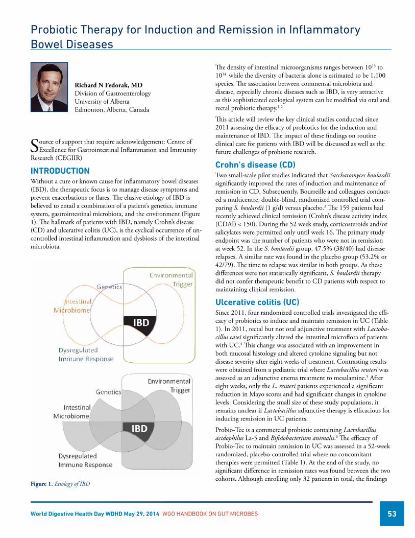
World Digestive Health Day WDHD May 29, 2014 WGO HANDBOOK ON GUT MICROBES 53
Probiotic Therapy for Induction and Remission in Inflammatory Bowel Diseases
Richard N Fedorak, MD Division of Gastroenterology University of Alberta Edmonton, Alberta, Canada
Source of support that require acknowledgement: Centre of Excellence for Gastrointestinal Inflammation and Immunity
Research (CEGIIR)
INTRODUCTIONWithout a cure or known cause for inflammatory bowel diseases (IBD), the therapeutic focus is to manage disease symptoms and prevent exacerbations or flares. The elusive etiology of IBD is believed to entail a combination of a patient’s genetics, immune system, gastrointestinal microbiota, and the environment (Figure 1). The hallmark of patients with IBD, namely Crohn’s disease (CD) and ulcerative colitis (UC), is the cyclical occurrence of un-controlled intestinal inflammation and dysbiosis of the intestinal microbiota.
The density of intestinal microorganisms ranges between 1013 to 1014 while the diversity of bacteria alone is estimated to be 1,100 species. The association between commensal microbiota and disease, especially chronic diseases such as IBD, is very attractive as this sophisticated ecological system can be modified via oral and rectal probiotic therapy.1,2
This article will review the key clinical studies conducted since 2011 assessing the efficacy of probiotics for the induction and maintenance of IBD. The impact of these findings on routine clinical care for patients with IBD will be discussed as well as the future challenges of probiotic research.
Crohn’s disease (CD)Two small-scale pilot studies indicated that Saccharomyces boulardii significantly improved the rates of induction and maintenance of remission in CD. Subsequently, Bourreille and colleagues conduct-ed a multicentre, double-blind, randomized controlled trial com-paring S. boulardii (1 g/d) versus placebo.3 The 159 patients had recently achieved clinical remission (Crohn’s disease activity index (CDAI) < 150). During the 52 week study, corticosteroids and/or salicylates were permitted only until week 16. The primary study endpoint was the number of patients who were not in remission at week 52. In the S. boulardii group, 47.5% (38/40) had disease relapses. A similar rate was found in the placebo group (53.2% or 42/79). The time to relapse was similar in both groups. As these differences were not statistically significant, S. boulardii therapy did not confer therapeutic benefit to CD patients with respect to maintaining clinical remission.
Ulcerative colitis (UC)Since 2011, four randomized controlled trials investigated the effi-cacy of probiotics to induce and maintain remission in UC (Table 1). In 2011, rectal but not oral adjunctive treatment with Lactoba-cillus casei significantly altered the intestinal microflora of patients with UC.4 This change was associated with an improvement in both mucosal histology and altered cytokine signaling but not disease severity after eight weeks of treatment. Contrasting results were obtained from a pediatric trial where Lactobacillus reuteri was assessed as an adjunctive enema treatment to mesalamine.5 After eight weeks, only the L. reuteri patients experienced a significant reduction in Mayo scores and had significant changes in cytokine levels. Considering the small size of these study populations, it remains unclear if Lactobacillus adjunctive therapy is efficacious for inducing remission in UC patients.
Probio-Tec is a commercial probiotic containing Lactobacillus acidophilus La-5 and Bifidobacterium animalis.6 The efficacy of Probio-Tec to maintain remission in UC was assessed in a 52-week randomized, placebo-controlled trial where no concomitant therapies were permitted (Table 1). At the end of the study, no significant difference in remission rates was found between the two cohorts. Although enrolling only 32 patients in total, the findings
Figure 1. Etiology of IBD
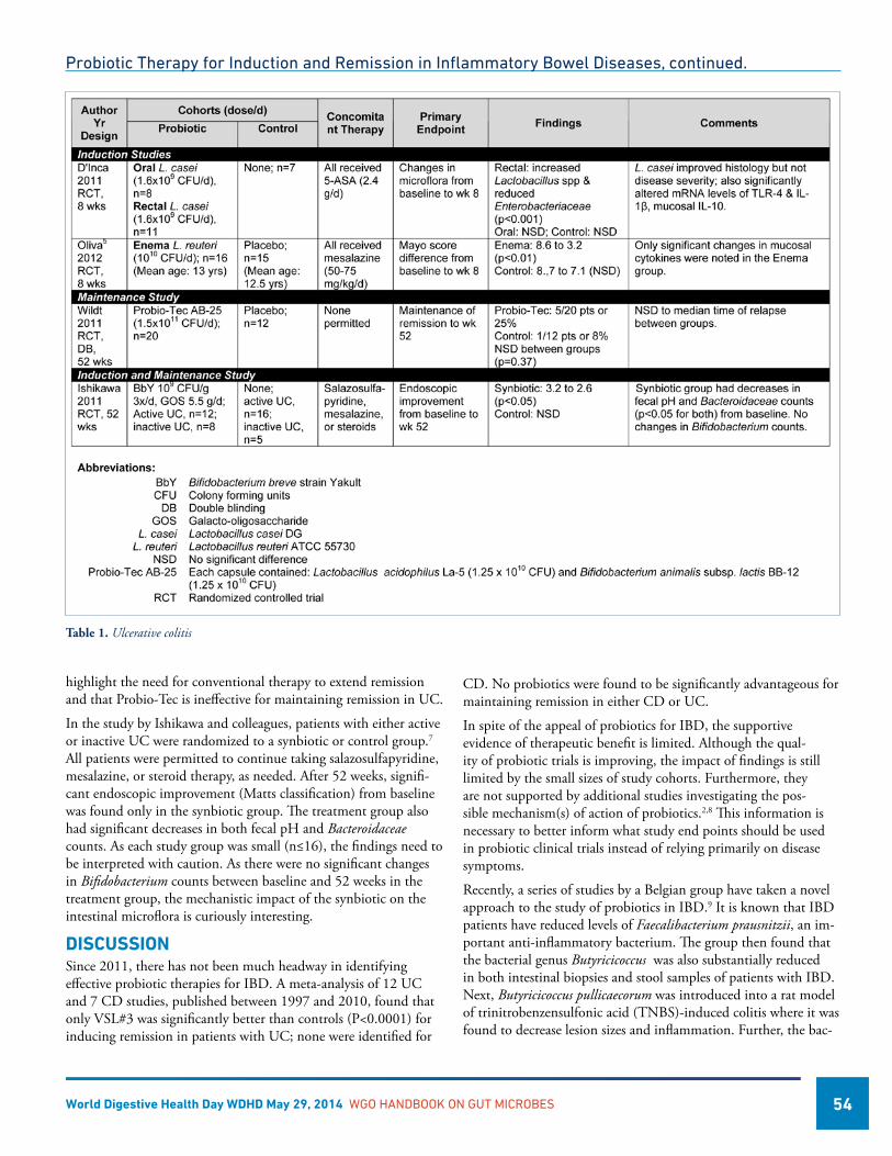
World Digestive Health Day WDHD May 29, 2014 WGO HANDBOOK ON GUT MICROBES 54
highlight the need for conventional therapy to extend remission and that Probio-Tec is ineffective for maintaining remission in UC.
In the study by Ishikawa and colleagues, patients with either active or inactive UC were randomized to a synbiotic or control group.7 All patients were permitted to continue taking salazosulfapyridine, mesalazine, or steroid therapy, as needed. After 52 weeks, signifi-cant endoscopic improvement (Matts classification) from baseline was found only in the synbiotic group. The treatment group also had significant decreases in both fecal pH and Bacteroidaceae counts. As each study group was small (n≤16), the findings need to be interpreted with caution. As there were no significant changes in Bifidobacterium counts between baseline and 52 weeks in the treatment group, the mechanistic impact of the synbiotic on the intestinal microflora is curiously interesting.
DISCUSSIONSince 2011, there has not been much headway in identifying effective probiotic therapies for IBD. A meta-analysis of 12 UC and 7 CD studies, published between 1997 and 2010, found that only VSL#3 was significantly better than controls (P<0.0001) for inducing remission in patients with UC; none were identified for
CD. No probiotics were found to be significantly advantageous for maintaining remission in either CD or UC.
In spite of the appeal of probiotics for IBD, the supportive evidence of therapeutic benefit is limited. Although the qual-ity of probiotic trials is improving, the impact of findings is still limited by the small sizes of study cohorts. Furthermore, they are not supported by additional studies investigating the pos-sible mechanism(s) of action of probiotics.2,8 This information is necessary to better inform what study end points should be used in probiotic clinical trials instead of relying primarily on disease symptoms.
Recently, a series of studies by a Belgian group have taken a novel approach to the study of probiotics in IBD.9 It is known that IBD patients have reduced levels of Faecalibacterium prausnitzii, an im-portant anti-inflammatory bacterium. The group then found that the bacterial genus Butyricicoccus was also substantially reduced in both intestinal biopsies and stool samples of patients with IBD. Next, Butyricicoccus pullicaecorum was introduced into a rat model of trinitrobenzensulfonic acid (TNBS)-induced colitis where it was found to decrease lesion sizes and inflammation. Further, the bac-
Table 1. Ulcerative colitis
Probiotic Therapy for Induction and Remission in Inflammatory Bowel Diseases, continued.
World Digestive Health Day WDHD May 29, 2014 WGO HANDBOOK ON GUT MICROBES 55
terium produced a supernatant that prevented cytokine-induced epithelial integrety impairment in an in vitro cell culture model. Although a clinical trial exploring the therapeutic efficacy of B. pullicaecorum has not yet been conducted, this research approach is mechanistically-focused and promising.
Overall probiotic therapy is well tolerated by IBD patients of all ages and over a range of doses. Further, a cocktail of eight bacterial strains (VSL#3) has been associated with significantly improved remission/response rates in UC patients compared to controls or placebo. To effect major change on the resident dysbiotic intes-tinal microbial community in IBD patients, a relatively massive introduction of many strains may be necessary. Along these lines, fecal microbial transplantation (FMT) is eclipsing the popular-ity of over-the-counter probiotic therapy of late (reviewed in 10). Though the scientific literature is limited to case reports and case series of FMT treatment for IBD, physicians need to be aware that patients are independently performing FMT aided by sites with do-it-yourself instructions (http://thepowerofpoop.com/).
Logically, FMT has the potential to provide a more impressive effect than a single bacterial strain. Yet the probiotic research com-munity needs diagnostic tools to consider a patient’s unique mi-croflora and develop a customized therapeutic probiotic regimen.11 Metabolomics provides an excellent means to monitor metabolic changes in the microflora in response to probiotic treatment. Lon-gitudinal metabolomic studies have the potential to provide much needed insights into the mechanisms of probiotic action, identify beneficial combinations of strains, define treatment frequency, impact on disease progression, and adverse events.
CONCLUSIONFrom a clinical perspective, and in studies before 2011, VSL#3 and E Coli Nissle have both shown to improve the induction of remission/response in UC patients. In contrast, results in CD patients have been disappointing. Over-the-counter probiotics do not improve remission rates or duration for IBD patients, however they are not associated with adverse events. Patients should be advised that probiotics can be used in an adjunctive capacity to conventional treatments.
ACKNOWLEDGEMENTThank you to Kathleen Ismond for assistance with the literature search and manuscript preparation.
References1. Martín R, Miquel S, Ulmer J, et al. Role of commensal and
probiotic bacteria in human health: a focus on inflammatory bowel disease. Microb. Cell Fact. 2013;12:71.
2. Fedorak R, Demeria D. Probiotic bacteria in the prevention and the treatment of inflammatory bowel disease. Gastroen-terol. Clin. North Am. 2012;41(4):821–42.
3. Bourreille A, Cadiot G, Dreau G Le, et al. Saccharomyces boulardii does not prevent relapse of Crohn’s disease. Clin. Gastroenterol. Hepatol. 2013;11(8):982–7.
4. D’Incà R, Barollo M, Scarpa M, et al. Rectal administration of Lactobacillus casei DG modifies flora composition and Toll-like receptor expression in colonic mucosa of patients with mild ulcerative colitis. Dig. Dis. Sci. 2011;56(4):1178–87.
5. Oliva S, Nardo G Di, Ferrari F, et al. Randomised clinical trial: the effectiveness of Lactobacillus reuteri ATCC 55730 rectal enema in children with active distal ulcerative colitis. Aliment. Pharmacol. Ther. 2012;35(3):327–34.
6. Wildt S, Nordgaard I, Hansen U, et al. A randomised double-blind placebo-controlled trial with Lactobacillus acidophilus La-5 and Bifidobacterium animalis subsp. lactis BB-12 for maintenance of remission in ulcerative colitis. J. Crohns. Coli-tis 2011;5(2):115–21.
7. Ishikawa H, Matsumoto S, Ohashi Y, et al. Beneficial effects of probiotic bifidobacterium and galacto-oligosaccharide in patients with ulcerative colitis: a randomized controlled study. Digestion 2011;84(2):128–33.
8. Whelan K, Quigley EMM. Probiotics in the management of irritable bowel syndrome and inflammatory bowel disease. Curr. Opin. Gastroenterol. 2013;29(2):184–9.
9. Eeckhaut V, Machiels K, Perrier C, et al. Butyricicoc-cus pullicaecorum in inflammatory bowel disease. Gut 2013;62(12):1745–52.
10. Allegretti JR, Hamilton MJ. Restoring the gut microbiome for the treatment of inflammatory bowel diseases. World J. Gastroenterol. 2014;20(13):3468–3474.
11. Bellavia M, Tomasello G, Romeo M, et al. Gut microbiota imbalance and chaperoning system malfunction are central to ulcerative colitis pathogenesis and can be counteracted with specifically designed probiotics: a working hypothesis. Med. Microbiol. Immunol. 2013;202(6):393–406.
Probiotic Therapy for Induction and Remission in Inflammatory Bowel Diseases, continued.
World Digestive Health Day WDHD May 29, 2014 WGO HANDBOOK ON GUT MICROBES 56
Probiotics in Pediatrics
For centuries, probiotics have been used in many forms; however, only in the last few decades have probiotics been
systematically assessed for pediatric indications. The potential for probiotics is even more magnified in pediatrics, as there are potential opportunities for prevention of chronic disease, as well as the enhancement of childhood growth and development. There are a number of different indications for probiotics that have been evaluated in children.
The majority of studies have focused on issues related to pediatric diarrhea. Probiotic strains have been shown to be efficacious for the treatment and/or prevention of diarrhea. In addition, the use of probiotics in the treatment and prevention of necrotizing en-terocolitis and colic looks promising. Below is a summary of some of the indications tested in pediatric settings.
• One of the most investigated indications for probiotics in children focuses on the prevention and treatment of infectious diarrhea, including viral and bacterial diarrhea, acute (Allen, 2010), chronic (Bernaola Aponte G, 2013) and antibiotic-associated diarrhea (AAD) (Hempel, 2012). Several meta-analyses have noted that probiotics decrease the duration of infectious diarrheal episodes in in-patient and out-patient settings, particularly, in otherwise healthy children with acute viral gastroenteritis.
• Several studies have shown that probiotics can decrease stool frequency and increase stool consistency during oral antibiotic therapy (Correa, 2005). Probiotic supplementation can be effective in C. difficile diarrhea; however there are few data for studies involving children (Goldenberg, 2013).
• As specific strains of probiotic bacteria may have an immu-nomodulatory effect, probiotics also have been used in the prevention of atopic disorders such as eczema. To date, these studies have demonstrated mixed results for the prevention of allergic disease, eczema and wheezing (Azad, 2013).
• The probiotic strain L. reuteri DSM 17938 has been shown to be effective in the treatment and prevention of colic in several studies. However, further studies are needed in this area (Sung, 2013).
• Probiotics have been used effectively in the prevention of nec-rotizing enterocolitis in premature infants. This effect has been documented in several different trials that have used different probiotic strains. These results suggest that the effect of probi-otic supplementation may not necessarily be strain-specific for the prevention of necrotizing enterocolitis (Alfaleh, 2011).
Michael D. Cabana, MD, MPH Departments of Pediatrics Epidemiology and Biostatistics The Philip R. Lee Institute of Health Policy Studies University of California San Francisco, CA
Dosage and Administration: Issues to ConsiderProbiotics given as supplements can, in theory, provide more consistent and relatively higher doses of probiotics in a much lower ingested volume compared to those incorporated into food products. Food products can, however, offer the additional benefit of other nutritional components and/or prebiotics.
Yogurt products have been marketed to families as a source of pro-biotic supplementation for children. Parents often view yogurt as a convenient and palatable food for infants and children. Although it is widely assumed by consumers that yogurt products have live bacterial cultures, not all ‘yogurt’ has live and active cultures. In addition, the strain or dose or CFU in yogurt may not be suffi-cient, in some cases, for a therapeutic effect.
When reviewing the literature, it is important that the clinician pay close attention to the strain (not just genus and species) being used in a particular study, as the efficacy of one probiotic strain does not im-ply that other related strains will be equally efficacious. Also, multi-strain probiotic products do not necessarily offer more benefit than single-strain products. Rather, it is more important that the product match exactly the probiotic strain and dose that was proven to be ef-fective in randomized controlled trials for the same indication.
There are no uniform dosing recommendations for probiotic sup-plements. Rather, the dose depends on the indication and strain of the probiotic being used. Studies to date have used doses ranging from 107 CFU/day to 1012 CFU/day (Table 1). Some practitioners use half of the adult dose for children of average weight and one-quarter of the adult dose for infants; however, it is not clear if this is necessary.
It is important to check the label of any probiotic product, as many products contain package labels which state either the “through end of shelf life,” which indicates the minimum number of CFUs that should be viable if the product is consumed before the end of its stated shelf life, or “at the time of manufacture,” which indicates the maximum CFU you can expect to obtain from the product but does not guarantee viability up to the end of shelf life.
When used as an adjunct to another treatment, the addition of a probiotic supplement can potentially affect compliance (e.g. adding a probiotic supplement to prevent antibiotic associated diarrhea). As a result, counseling and patient education are es-sential components of probiotic therapy, as the effects of probiotics require compliance to the therapy.
Quality Control of ProductsDifferent studies have noted the variability in quality of over-the-counter probiotic products. For example, Marcobal and colleagues conducted a cross-sectional analysis of 14 commercial probiotic products and noted that many products contained additional, unadvertised Lactobacilli and Bifidobacteria, whereas others were missing species listed on the product label. The label claims on probiotic products may or may not represent the true constituents (Marcobal, 2008).
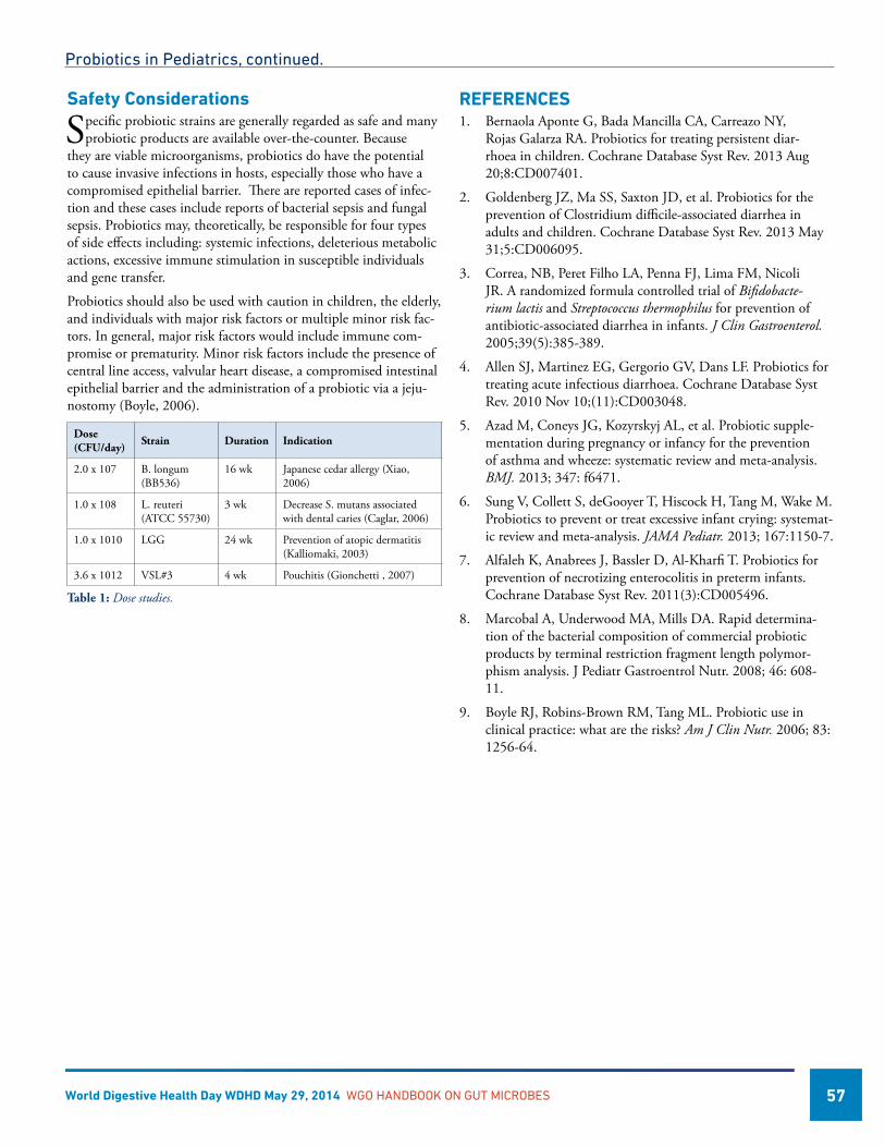
World Digestive Health Day WDHD May 29, 2014 WGO HANDBOOK ON GUT MICROBES 57
Safety Considerations
Specific probiotic strains are generally regarded as safe and many probiotic products are available over-the-counter. Because
they are viable microorganisms, probiotics do have the potential to cause invasive infections in hosts, especially those who have a compromised epithelial barrier. There are reported cases of infec-tion and these cases include reports of bacterial sepsis and fungal sepsis. Probiotics may, theoretically, be responsible for four types of side effects including: systemic infections, deleterious metabolic actions, excessive immune stimulation in susceptible individuals and gene transfer.
Probiotics should also be used with caution in children, the elderly, and individuals with major risk factors or multiple minor risk fac-tors. In general, major risk factors would include immune com-promise or prematurity. Minor risk factors include the presence of central line access, valvular heart disease, a compromised intestinal epithelial barrier and the administration of a probiotic via a jeju-nostomy (Boyle, 2006).
Dose (CFU/day) Strain Duration Indication
2.0 x 10⁷ B. longum (BB536)
16 wk Japanese cedar allergy (Xiao, 2006)
1.0 x 10⁸ L. reuteri (ATCC 55730)
3 wk Decrease S. mutans associated with dental caries (Caglar, 2006)
1.0 x 10¹⁰ LGG 24 wk Prevention of atopic dermatitis (Kalliomaki, 2003)
3.6 x 10¹² VSL#3 4 wk Pouchitis (Gionchetti , 2007)
Table 1: Dose studies.
REFERENCES1. Bernaola Aponte G, Bada Mancilla CA, Carreazo NY,
Rojas Galarza RA. Probiotics for treating persistent diar-rhoea in children. Cochrane Database Syst Rev. 2013 Aug 20;8:CD007401.
2. Goldenberg JZ, Ma SS, Saxton JD, et al. Probiotics for the prevention of Clostridium difficile-associated diarrhea in adults and children. Cochrane Database Syst Rev. 2013 May 31;5:CD006095.
3. Correa, NB, Peret Filho LA, Penna FJ, Lima FM, Nicoli JR. A randomized formula controlled trial of Bifidobacte-rium lactis and Streptococcus thermophilus for prevention of antibiotic-associated diarrhea in infants. J Clin Gastroenterol. 2005;39(5):385-389.
4. Allen SJ, Martinez EG, Gergorio GV, Dans LF. Probiotics for treating acute infectious diarrhoea. Cochrane Database Syst Rev. 2010 Nov 10;(11):CD003048.
5. Azad M, Coneys JG, Kozyrskyj AL, et al. Probiotic supple-mentation during pregnancy or infancy for the prevention of asthma and wheeze: systematic review and meta-analysis. BMJ. 2013; 347: f6471.
6. Sung V, Collett S, deGooyer T, Hiscock H, Tang M, Wake M. Probiotics to prevent or treat excessive infant crying: systemat-ic review and meta-analysis. JAMA Pediatr. 2013; 167:1150-7.
7. Alfaleh K, Anabrees J, Bassler D, Al-Kharfi T. Probiotics for prevention of necrotizing enterocolitis in preterm infants. Cochrane Database Syst Rev. 2011(3):CD005496.
8. Marcobal A, Underwood MA, Mills DA. Rapid determina-tion of the bacterial composition of commercial probiotic products by terminal restriction fragment length polymor-phism analysis. J Pediatr Gastroentrol Nutr. 2008; 46: 608-11.
9. Boyle RJ, Robins-Brown RM, Tang ML. Probiotic use in clinical practice: what are the risks? Am J Clin Nutr. 2006; 83: 1256-64.
Probiotics in Pediatrics, continued.
World Digestive Health Day WDHD May 29, 2014 WGO HANDBOOK ON GUT MICROBES 58
Diet can be used to manage disorders as mediated through the gut microbiome. One popular and long standing approach is
to include live microbial cultures in the diet as probiotics (Sanders et al. 2007). Current probiotic strategies target improved resis-tance to infections, irritable bowel syndrome, chronic gut disorder (inflammatory bowel disease, colon cancer), lactose intolerance, recurrent vaginal thrush, skin problems, food allergy and mineral bioavailability. Many different products exist and new develop-ments are continuing at a rapid pace.
A further concept is that of prebiotics. These are non viable food components (carbohydrates) that have a selective microbial me-tabolism in the human or animal gut. They attempt to induce ben-eficial changes by fortifying levels of certain bacteria indigenous to the gut microbiota.
Prebiotics were first defined as ‘non-digestible food ingredient that beneficially affects the host by selectively stimulating the growth and/or activity of one or a limited number of bacteria already resident in the colon’ (Gibson and Roberfroid. 1995). Thus, the prebiotic approach advocated administration of non viable food ingredients that transfer to the colon and have a selective metabolism therein. The prebiotic concept considers that apparently positive microor-ganisms, such as bifidobacteria and lactobacilli, are already present in the human colon. The original definition was then updated in 2004 to ‘selectively fermented ingredients that allow specific changes, both in the composition and/or activity in the gastrointestinal micro-flora that confers benefits upon host well-being and health’ (Gibson et al., 2004). This extrapolated the concept into other areas of the gut, and not just the colon, that may benefit from a selective targeting of particular micro-organisms.
Currently accepted prebiotics are confined to non-digestible oligo-saccharides, many of which seem to confer the degree of fermenta-tion selectivity that is required (towards bifidobacteria). Inulin type prebiotics occur naturally in several foods such as leek, asparagus, banana, chicory, Jerusalem artichoke, garlic, artichoke, onion and wheat. However, the overall intake from these sources is small. An effective route to achieve a requisite dose (ca. 5g/d in an adult) is the fortification of more frequently eaten foodstuffs with prebiotic ingredients and/or their use as supplements. Prebiotics are there-fore a sub-category of functional foods. They are added to many foods including yogurts, cereals, breads, biscuits, milk desserts, ice-creams, spreads, drinks, as well as animal feeds and supplements. Some prebiotics can be obtained by extraction from crops, e.g. inulin from chicory or agave. They can be commercially produced through hydrolysis (e.g. oligofructose from inulin) or through catabolic enzymatic reactions from lower molecular weight sugars,
e.g. short-chain fructooligosaccharides (scFOS) from sucrose or ga-lactooligosaccharides (GOS)/lactulose from lactose. The review by Crittenden and Playne (1996) gives an overview of various aspects of the production and properties of food grade oligosaccharides. Table 1 lists candidate prebiotics.
Three criteria are required for a prebiotic effect are (Gibson et al., 2004):
• Resistsgastricacidity,hydrolysisbymammalianenzymesandgastrointestinal absorption
• Isfermentedbyintestinalmicroflora
• Selectivelystimulatesthegrowthand/oractivityofintestinalbacteria associated with health and well-being.
As it stands, the prebiotic field is dominated by gastrointestinal events. However, it may be the case that other mixed microbial ecosystems may be modulated by a prebiotic approach, such as the oral cavity, the skin, or urogenital tract. Thus, a dietary prebiotic is: “A selectively fermented ingredient that results in specific changes, in the composition and/or activity of the gastrointestinal microbiota, thus conferring benefit(s) upon host health.” (Gibson et al. 2011). In terms of testing, confirmation of selective metabolism is key. This can be shown using in vitro “gut model” systems (Figure 1) but more definitive results are required from in vivo trials.
Prebiotics
GR Gibson, Ph.D. Food Microbial Sciences Unit Department of Food and Nutritional Sciences The University of Reading UK
• Fructo-oligosaccharides(FOS),includinginulin*• Galacto-oligosaccharides(GOS)*• Lactulose• Isomalto-oligosaccharides(IMO)• Lactosucrose• Polydextrose(PDX)• Xylo-oligosaccharide(XOS)• Mannan-oligosaccharides(MOS)• Soybeanoligosachharide(SOS)• Gluco-oligosaccharide(GlOS)• Genti-oligosaccharides(GiOS)• Arabino-xylo-oligosaccharides(AXOS)• Germinatedbarleyfoodstuffs• Oligodextrans• Gluconicacid• Pectic-oligosaccharides• Lactose• Glutamineandhemicelluloserichsubstrates• Resistantstarchanditsderivatives• Oligosaccharidesfrommelibiose• Lactoferrin-derivedpeptide• N-acetylchitooligosaccharides• Isoflavonicphytoestrogens• Variousfibresandderivatives*Carbohydratesthatcurrentlyhavethestrongestlevelofevidenceasprebiot-ics (as gained from multiple in vitro and in vivo investigations)
Table 1. List of candidate prebiotics as currently reported in the scientific literature.
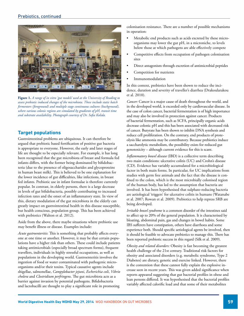
World Digestive Health Day WDHD May 29, 2014 WGO HANDBOOK ON GUT MICROBES 59
Target populationsGastrointestinal problems are ubiquitous. It can therefore be argued that prebiotic based fortification of positive gut bacteria is appropriate to everyone. However, the early and later stages of life are thought to be especially relevant. For example, it has long been recognized that the gut microbiota of breast and formula fed infants differs, with the former being dominated by bifidobac-teria (due to the presence of oligosaccharides and glycoproteins in human beast milk). This is believed to be one explanation for the lower incidence of gut difficulties, like infections, in breast fed infants. Prebiotic use in infant formulae is therefore currently popular. In contrast, in elderly persons, there is a large decrease in levels of gut bifidobacteria, possibly contributing to increased infection rates and the onset of an inflammatory state. In view of this, dietary modulation of the gut microbiota in the elderly can greatly impact on gastrointestinal health in this disease susceptible, but health conscious, population group. This has been achieved with prebiotics (Walton et al. 2012).
Aside from the above, there maybe situations where prebiotic use may benefit illness or disease. Examples include:
Acute gastroenteritis: This is something that probably affects every-one at one time or another. However, it may be that certain popu-lations have a higher risk than others. These could include patients taking antimicrobials (especially broad spectrum forms), frequent travellers, individuals in highly stressful occupations, as well as populations in the developing world. Gastroenteritis involves the ingestion of food or water contaminated with pathogenic micro-organisms and/or their toxins. Typical causative agents include shigellae, salmonellae, Campylobacter jejuni, Escherichia coli, Vibrio cholera and Clostridium perfringens. The gut microbiota acts as a barrier against invasion by potential pathogens. Bifidobacteria and lactobacilli are thought to play a significant role in promoting
colonisation resistance. There are a number of possible mechanisms in operation:
• Metabolicendproductssuchasacidsexcretedbythesemicro-organisms may lower the gut pH, in a microniche, to levels below those at which pathogens are able effectively compete
• Competitiveeffectsfromoccupationofpathogencolonisationsites
• Directantagonismthroughexcretionofantimicrobialpeptides
• Competitionfornutrients
• Immunomodulation
In this context, prebiotics have been shown to reduce the inci-dence, duration and severity of traveller’s diarrhea (Drakoularakou et al. 2010).
Cancer: Cancer is a major cause of death throughout the world, and in the developed world, is exceeded only by cardiovascular disease. In the case of colon cancer, bacterial fermentation is of high importance and may also be involved in protection against cancer. Products of bacterial fermentation, such as SCFA, principally organic acids decrease colonic pH and this has been associated with decreased risk of cancer. Butyrate has been shown to inhibit DNA synthesis and reduce cell proliferation. On the contrary, end products of prote-olysis like ammonia may be contributory. Because prebiotics induce a saccharolytic metabolism, the possibility exists for reduced gut genotoxicity – although current evidence for this is scant.
Inflammatory bowel disease (IBD) is a collective term describing two main conditions: ulcerative colitis (UC) and Crohn’s disease (CD). Evidence has variably accumulated for a microbiological factor in both main forms. In particular, for UC implications from studies with germ free animals and the fact that the disease is con-fined to the colon, which is the most microbially colonised region of the human body, has led to the assumption that bacteria are involved. It has been hypothesised that sulphate-reducing bacteria are aetiological ‘triggers’ for ulcerative colitis in humans (Watanabe et al. 2007; Rowan et al. 2009). Prebiotics to help repress SRB are being developed.
Irritable bowel syndrome is a common disorder of the intestines said to affect up to 20% of the general population. It is characterised by bloating, abdominal pain, gas and changes in bowel habits. Some IBS sufferers have constipation, others have diarrhoea and some experience both. Should specific aetiological agents be involved, then it should be feasible to advocate prebiotics to manage this. There has been reported prebiotic success in this regard (Silk et al. 2009).
Obesity and related disorders: Obesity is fast becoming the greatest health challenge of the 21st century. Traditional risk factors for obesity and associated disorders (e.g. metabolic syndrome, Type 2 Diabetes) are dietary, genetic and exercise linked. However, there is the contention that these cannot fully explain the explosive in-crease seen in recent years. This was given added significance when reports appeared suggesting that gut bacterial profiles in obese and lean persons differed. It was hypothesised that the bacterial profiles variably affected calorific load and that some of their metabolites
Prebiotics, continued
Figure 1. A range of in vitro ‘gut models’ used at the University of Reading to assess prebiotic induced changes of the microbiota. These include static batch fermenters (foreground) and multiple stage continuous cultures (background), where various colonic regions are simulated by gradients of pH, transit time and substrate availability. Photograph courtesy of Dr. Sofia Kolida.
World Digestive Health Day WDHD May 29, 2014 WGO HANDBOOK ON GUT MICROBES 60
could influence satiety. This is still an area of some debate (and it may be that the traditional risk factors themselves affect microflora profiles). However, should gut microbiota differences be a factor in obese related conditions, this then opens up the possibility of alter-ing the situation by using dietary ingredients that have a selective fermentation in situ. Given the recent link between gut microflora and obesity, it makes sense to research whether prebiotics can exert a modulatory role. A recent human study showed some promise in this regard in that prebiotic use positively affected markers of insu-lin resistance, gut inflammation and some blood lipids (Vulevic et al 2013).
ConclusionsThe usual target genera for prebiotics are lactobacilli and bifido-bacteria. However, most success has predominantly been with the latter, probably because they are usually present in higher numbers than lactobacilli. Typical prebiotics are non-digestible oligosaccha-rides like FOS and GOS. As knowledge of gut microbiota diversity has expanded, there may be other target genera for prebiotic approaches such as Roseburia, Eubacterium, Faecalibacterium. In some cases, these may produce desirable metabolites that bifido-bacteria/lactobacilli cannot. Trials that include a functional, as well as compositional, assessment of microbiota changes following prebiotic use are a useful way forward, as are further studies into clinical outcome. What can be said is that the approach is safe and user friendly.
References1. Crittenden, R.G. and Playne, M.J. (1996) Production, prop-
erties and applications of food-grade oligosaccharides Trends in Food Science and Technology 7, 353-361.
2. Drakoularakou, A., Tzortzis, G., Rastall, R.A. and Gibson, G.R. (2010) A double-blind, placebo-controlled, randomized human study assessing the capacity of a novel galacto-oligo-saccharide mixture in reducing travellers’ diarrhoea. European Journal of Clinical Nutrition 64, 146-152.
3. Gibson, G.R., Probert, H.M., van Loo, J.A.E., Rastall, R.A. and Roberfroid, M.B. (2004) Dietary modulation of the hu-man colonic microbiota: Updating the concept of prebiotics. Nutrition Research Reviews 17, 259-275.
Prebiotics, continued
4. Gibson, G.R. and Roberfroid, M.B. (1995) Dietary modu-lation of the human colonic microbiota: introducing the concept of prebiotics. Journal of Nutrition 125, 1401-1412.
5. Gibson, G.R., Scott, K.P., Rastall, R.A., Tuohy, K.M., Hotch-kiss, A., Dubert-Ferrandon, A., Gareau, M., Murphy, E.F., Saulnier, D., Loh, G., Macfarlane, S., Delzenne, N., Ringel, Y., Kozianowski, G., Dickmann, R., 1. 1. Lenoir-Wijnkook, I.,Walker, C. and Buddington, R. (2011) Dietary prebiotics: Current status and new definition. IFIS Functional Foods Bulletin 7, 1–19.
6. Rowan, F., Docherty, N.G., Murphy, M., Murphy, B., Calvin Coffey, J. and O’Connell, P.R. (2010) Desulfovibrio bacte-rial species are increased in ulcerative colitis. Diseases of the Colon and Rectum 53, 1530-1536.
7. Sanders, M.E., Gibson, G.R., Gill, H.S. and Guarner, F. (2007) Probiotics: Their potential to impact human health. CAST Issue Paper 36, 1-20.
8. Silk, D.B., Davis, A., Vulevic, J., Tzortzis, G. and Gibson, G.R. (2009) Clinical trial: the effects of a trans-galactooli-gosaccharide prebiotic on faecal microbiota and symptoms in irritable bowel syndrome. Alimentary Pharmacology and Therapy 29, 508-518.
9. Walton, G.E., van den Heuvel, E.G., Kosters, M.H., Rastall, R.A., Tuohy, K.M. and Gibson G.R. (2012) A randomised crossover study investigating the effects of galacto-oligosac-charides on the faecal microbiota in men and women over 50 years of age. British Journal of Nutrition 107, 1466-1475.
10. Watanabe, K., Mikamo, H., Tanaka, K. (2007) Clinical significance of sulfate-reducing bacteria for ulcerative colitis. Nihon Rinsho 7, 1337-1346.
11. Vulevic, J., Juric, A., Tzortzis, G. and Gibson, G.R. 2013. A mixture of trans-galactooligosaccharides reduces markers of metabolic syndrome and modulates the fecal microbiota and immune function of overweight adults. Journal of Nutrition 143, 324-331.
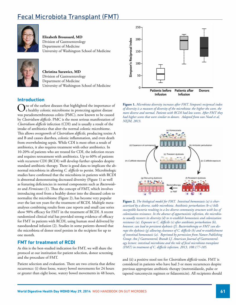
World Digestive Health Day WDHD May 29, 2014 WGO HANDBOOK ON GUT MICROBES 61
Fecal Microbiota Transplant (FMT)
Introduction
One of the earliest diseases that highlighted the importance of a healthy colonic microbiome in protecting against disease
was pseudomembranous colitis (PMC), now known to be caused by Clostridium difficile. PMC is the most serious manifestation of Clostridium difficile infection (CDI) and is usually a result of the intake of antibiotics that alter the normal colonic microbiome. This allows overgrowth of Clostridium difficile, producing toxins A and B and causes diarrhea, colonic inflammation, and even death from overwhelming sepsis. While CDI is most often a result of antibiotics, it also requires treatment with other antibiotics. In 10-20% of patients who are treated for CDI, the infection recurs and requires retreatment with antibiotics. Up to 60% of patients with recurrent CDI (RCDI) will develop further episodes despite standard antibiotic therapy. There is good data to implicate the ab-normal microbiota in allowing C. difficile to persist. Microbiologic studies have confirmed that the microbiota in patients with RCDI is abnormal demonstrating decreased diversity (Figure 1) as well as featuring deficiencies in normal components such as Bacteroide-tes and Firmicutes (1). Thus the concept of FMT, which involves introducing stool from a healthy donor into the diseased colon to normalize the microbiome (Figure 2), has become very popular over the last ten years for the treatment of RCDI. Multiple meta-analyses combining results from case reports and small case series show 90% efficacy for FMT in the treatment of RCDI. A recent randomized clinical trial has provided strong evidence of efficacy for FMT in patients with RCDI, using human stool delivered by nasoduodenal infusion (2). Studies in some patients showed that the microbiota of donor stool persists in the recipient for up to one month.
FMT for treatment of RCDIAs this is the best-studied indication for FMT, we will share the protocol at our institution for patient selection, donor screening and the procedure of FMT.
Patient selection and evaluation. There are two criteria that define recurrence: (i) three loose, watery bowel movements for 24 hours or greater than eight loose, watery bowel movements in 48 hours,
Elizabeth Broussard, MD Division of Gastroenterology Department of Medicine University of Washington School of Medicine
Christina Surawicz, MD Division of Gastroenterology Department of Medicine University of Washington School of Medicine
and (ii) a positive stool test for Clostridium difficile toxin. FMT is considered in patients who have had 3 or more recurrences despite previous appropriate antibiotic therapy (metronidazole, pulse or tapered vancomycin regimen or fidaxomicin). All recipients should
Figure 1. Microbiota diversity increases after FMT. Simpson’s reciprocal index of diversity is a measure of diversity of the microbiota: the higher the score, the more diverse and normal. Patients with RCDI had low scores. After FMT they had higher scores that were similar to donors. Adapted from van Nood et al, NEJM, 2013.
Figure 2. The biological model for FMT. Intestinal homeostasis (a) is char-acterized by a diverse, stable microbiota. Antibiotic perturbation (b–c) kills susceptible bacteria resulting in a less diverse community structure with loss of colonization resistance. In the absence of opportunistic infection, the microbio-ta usually recovers its diversity (d) to re-establish homeostasis and colonization resistance (a). Exposure to C. difficile (e) after antibiotic perturbation (b), however, can lead to persistent dysbiosis (f ). Bacteriotherapy or FMT can dis-rupt the dysbiosis (g) allowing clearance of C. difficile (h) and re-establishment of intestinal homeostasis (a). Reprinted by permission from Nature Publishing Group: Am J Gastroenterol; Brandt LJ. American Journal of Gastroenterol-ogy lecture: intestinal microbiota and the role of fecal microbiota transplant (FMT) in treatment of C. difficile infection. 2013, 108:177-185.
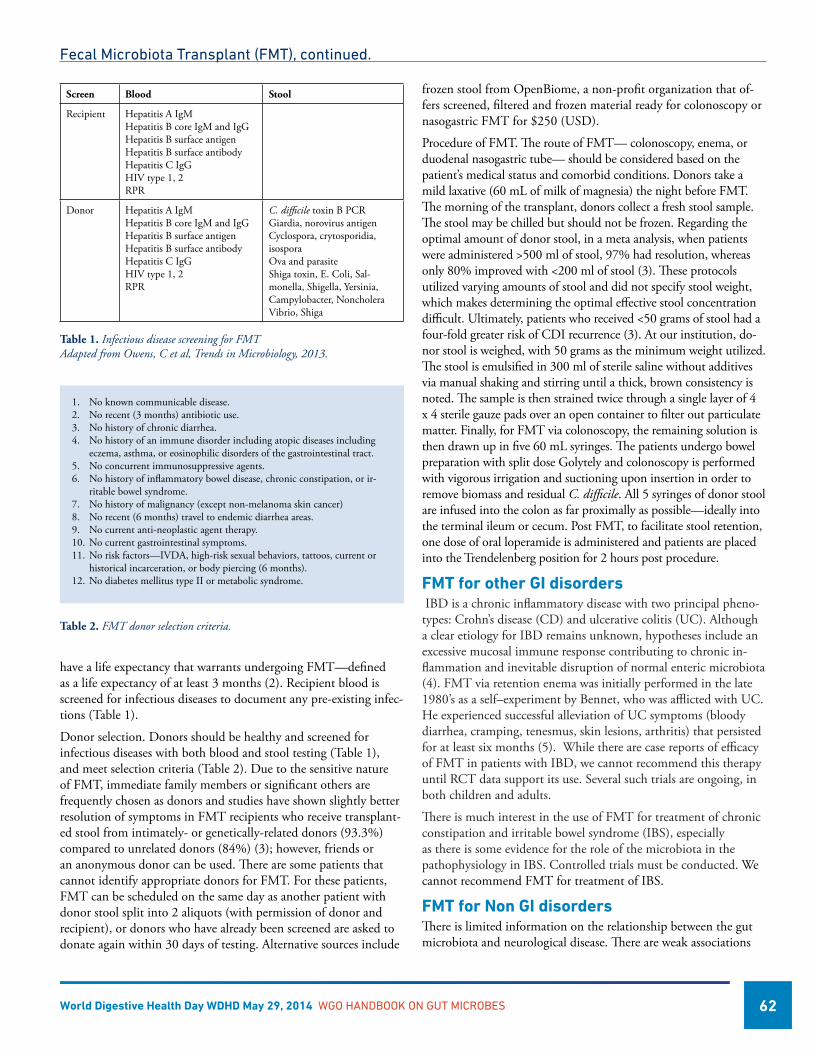
World Digestive Health Day WDHD May 29, 2014 WGO HANDBOOK ON GUT MICROBES 62
have a life expectancy that warrants undergoing FMT—defined as a life expectancy of at least 3 months (2). Recipient blood is screened for infectious diseases to document any pre-existing infec-tions (Table 1).
Donor selection. Donors should be healthy and screened for infectious diseases with both blood and stool testing (Table 1), and meet selection criteria (Table 2). Due to the sensitive nature of FMT, immediate family members or significant others are frequently chosen as donors and studies have shown slightly better resolution of symptoms in FMT recipients who receive transplant-ed stool from intimately- or genetically-related donors (93.3%) compared to unrelated donors (84%) (3); however, friends or an anonymous donor can be used. There are some patients that cannot identify appropriate donors for FMT. For these patients, FMT can be scheduled on the same day as another patient with donor stool split into 2 aliquots (with permission of donor and recipient), or donors who have already been screened are asked to donate again within 30 days of testing. Alternative sources include
frozen stool from OpenBiome, a non-profit organization that of-fers screened, filtered and frozen material ready for colonoscopy or nasogastric FMT for $250 (USD).
Procedure of FMT. The route of FMT— colonoscopy, enema, or duodenal nasogastric tube— should be considered based on the patient’s medical status and comorbid conditions. Donors take a mild laxative (60 mL of milk of magnesia) the night before FMT. The morning of the transplant, donors collect a fresh stool sample. The stool may be chilled but should not be frozen. Regarding the optimal amount of donor stool, in a meta analysis, when patients were administered >500 ml of stool, 97% had resolution, whereas only 80% improved with <200 ml of stool (3). These protocols utilized varying amounts of stool and did not specify stool weight, which makes determining the optimal effective stool concentration difficult. Ultimately, patients who received <50 grams of stool had a four-fold greater risk of CDI recurrence (3). At our institution, do-nor stool is weighed, with 50 grams as the minimum weight utilized. The stool is emulsified in 300 ml of sterile saline without additives via manual shaking and stirring until a thick, brown consistency is noted. The sample is then strained twice through a single layer of 4 x 4 sterile gauze pads over an open container to filter out particulate matter. Finally, for FMT via colonoscopy, the remaining solution is then drawn up in five 60 mL syringes. The patients undergo bowel preparation with split dose Golytely and colonoscopy is performed with vigorous irrigation and suctioning upon insertion in order to remove biomass and residual C. difficile. All 5 syringes of donor stool are infused into the colon as far proximally as possible—ideally into the terminal ileum or cecum. Post FMT, to facilitate stool retention, one dose of oral loperamide is administered and patients are placed into the Trendelenberg position for 2 hours post procedure.
FMT for other GI disorders IBD is a chronic inflammatory disease with two principal pheno-types: Crohn’s disease (CD) and ulcerative colitis (UC). Although a clear etiology for IBD remains unknown, hypotheses include an excessive mucosal immune response contributing to chronic in-flammation and inevitable disruption of normal enteric microbiota (4). FMT via retention enema was initially performed in the late 1980’s as a self–experiment by Bennet, who was afflicted with UC. He experienced successful alleviation of UC symptoms (bloody diarrhea, cramping, tenesmus, skin lesions, arthritis) that persisted for at least six months (5). While there are case reports of efficacy of FMT in patients with IBD, we cannot recommend this therapy until RCT data support its use. Several such trials are ongoing, in both children and adults.
There is much interest in the use of FMT for treatment of chronic constipation and irritable bowel syndrome (IBS), especially as there is some evidence for the role of the microbiota in the pathophysiology in IBS. Controlled trials must be conducted. We cannot recommend FMT for treatment of IBS.
FMT for Non GI disordersThere is limited information on the relationship between the gut microbiota and neurological disease. There are weak associations
1. No known communicable disease.2. No recent (3 months) antibiotic use.3. No history of chronic diarrhea.4. No history of an immune disorder including atopic diseases including
eczema, asthma, or eosinophilic disorders of the gastrointestinal tract.5. No concurrent immunosuppressive agents.6. No history of inflammatory bowel disease, chronic constipation, or ir-
ritable bowel syndrome.7. No history of malignancy (except non-melanoma skin cancer) 8. No recent (6 months) travel to endemic diarrhea areas.9. No current anti-neoplastic agent therapy.10. No current gastrointestinal symptoms.11. No risk factors—IVDA, high-risk sexual behaviors, tattoos, current or
historical incarceration, or body piercing (6 months). 12. No diabetes mellitus type II or metabolic syndrome.
Table 2. FMT donor selection criteria.
Screen Blood Stool
Recipient Hepatitis A IgMHepatitis B core IgM and IgGHepatitis B surface antigenHepatitis B surface antibodyHepatitis C IgGHIV type 1, 2RPR
Donor Hepatitis A IgMHepatitis B core IgM and IgGHepatitis B surface antigenHepatitis B surface antibodyHepatitis C IgGHIV type 1, 2RPR
C. difficile toxin B PCRGiardia, norovirus antigenCyclospora, crytosporidia, isosporaOva and parasiteShiga toxin, E. Coli, Sal-monella, Shigella, Yersinia, Campylobacter, Noncholera Vibrio, Shiga
Table 1. Infectious disease screening for FMT Adapted from Owens, C et al, Trends in Microbiology, 2013.
Fecal Microbiota Transplant (FMT), continued.
World Digestive Health Day WDHD May 29, 2014 WGO HANDBOOK ON GUT MICROBES 63
between depression and carbohydrate malabsorption, levels of Clostridia and autism and alterations in feeding patterns in patients with chronic H. pylori infection (6). For this and other reasons, there is interest in the application of FMT to neurologic diseases including multiple sclerosis and Parkinson’s disease. The role of FMT in these diseases is speculative and positive controlled trials would be needed before this can be incorporated into clinical prac-tice. We cannot recommend FMT for treatment of these disorders.
Risks of FMTIn summary, there has been an explosion of data about the micro-biota and there is intense interest in its role in the pathophysiology and even treatment of various diseases. With all the enthusiasm for FMT, we must not forget that there are risks, both known and unknown. There are risks associated with any procedure, such as colonoscopy, sigmoidoscopy and even nasoduodenal intubation. There have been 2 cases of Norovirus transmission to patients from donor stool (7). There are risks of other infections and the long-term effects of altering the microbiota, even transiently, are not known. For these and other reasons, we recommend establishment of data registries to collect such important clinical information.
ConclusionsThe role of FMT makes sense and appears efficacious and safe in some patients with RCDI. The role of FMT in severe refractory CDI needs further study. FMT in IBD is under study, and its role in IBS, chronic constipation and neurologic disease is speculative at best.
References1. Chang, JY, et al. (2008). Decreased Diversity of the Fecal
Microbiome in Recurrent Clostridium difficile—Associated Diarrhea, J Infect Dis; 197 (3), 435-438.
2. van Nood, E, et al. (2013) Duodenal Infusion of Donor Feces for Recurrent Clostridium difficile, N Engl J Med 2013; 368:407-415.
3. Gough, E., et al. (2011) Systematic review of intestinal mi-crobiota transplantation (fecal bacteriotherapy) for recurrent Clostridium difficile infection. Clin Infect. Dis. 53, 994-1002.
4. Borody TJ, et al. (2004) Bacteriotherapy using fecal flora: toying with human motions. J Clin Gastroenterol. 38(6), 475-483.
5. Bennet JD, et al. (1989) Treatment of ulcerative colitis by implantation of normal colonic flora. Lancet 1(8630), 164.
6. Collins, SM, et al. (2009) The relationship between intestinal microbiota and the central nervous system in normal gas-trointestinal function and disease. Gastroenterology 136(6), 2003-2014.
7. Schwartz M, et al. (2013) Norovirus gastroenteritis after fecal microbiota transplantation for treatment of Clostridium dif-ficile infection despite asymptomatic donors and lack of sick contacts. Am J Gastroenterol. Aug;108(8):1367.
8. Owens, C, et al (2013) Fecal microbiota transplantation and donor standardization. Trends in Microbiology 21(9), 443-445.
9. Brandt LJ. American Journal of Gastroenterology Lecture: Intestinal Microbiota and the Role of Fecal Microbiota Transplant (FMT) in treatment of C difficile infection. Am J Gastroenterol 2013; 108: 177-185.
Fecal Microbiota Transplant (FMT), continued.
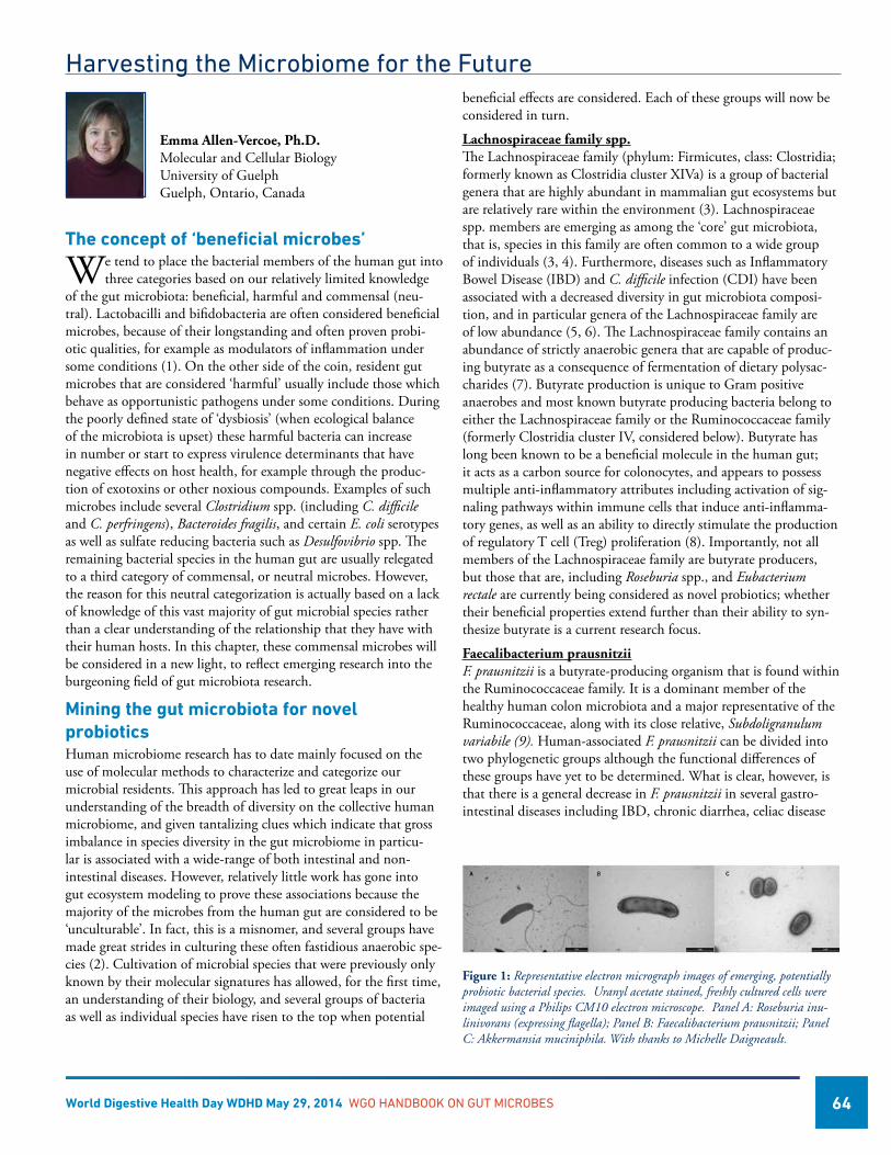
World Digestive Health Day WDHD May 29, 2014 WGO HANDBOOK ON GUT MICROBES 64
Harvesting the Microbiome for the Future
The concept of ‘beneficial microbes’
We tend to place the bacterial members of the human gut into three categories based on our relatively limited knowledge
of the gut microbiota: beneficial, harmful and commensal (neu-tral). Lactobacilli and bifidobacteria are often considered beneficial microbes, because of their longstanding and often proven probi-otic qualities, for example as modulators of inflammation under some conditions (1). On the other side of the coin, resident gut microbes that are considered ‘harmful’ usually include those which behave as opportunistic pathogens under some conditions. During the poorly defined state of ‘dysbiosis’ (when ecological balance of the microbiota is upset) these harmful bacteria can increase in number or start to express virulence determinants that have negative effects on host health, for example through the produc-tion of exotoxins or other noxious compounds. Examples of such microbes include several Clostridium spp. (including C. difficile and C. perfringens), Bacteroides fragilis, and certain E. coli serotypes as well as sulfate reducing bacteria such as Desulfovibrio spp. The remaining bacterial species in the human gut are usually relegated to a third category of commensal, or neutral microbes. However, the reason for this neutral categorization is actually based on a lack of knowledge of this vast majority of gut microbial species rather than a clear understanding of the relationship that they have with their human hosts. In this chapter, these commensal microbes will be considered in a new light, to reflect emerging research into the burgeoning field of gut microbiota research.
Mining the gut microbiota for novel probioticsHuman microbiome research has to date mainly focused on the use of molecular methods to characterize and categorize our microbial residents. This approach has led to great leaps in our understanding of the breadth of diversity on the collective human microbiome, and given tantalizing clues which indicate that gross imbalance in species diversity in the gut microbiome in particu-lar is associated with a wide-range of both intestinal and non-intestinal diseases. However, relatively little work has gone into gut ecosystem modeling to prove these associations because the majority of the microbes from the human gut are considered to be ‘unculturable’. In fact, this is a misnomer, and several groups have made great strides in culturing these often fastidious anaerobic spe-cies (2). Cultivation of microbial species that were previously only known by their molecular signatures has allowed, for the first time, an understanding of their biology, and several groups of bacteria as well as individual species have risen to the top when potential
Emma Allen-Vercoe, Ph.D. Molecular and Cellular Biology University of Guelph Guelph, Ontario, Canada
beneficial effects are considered. Each of these groups will now be considered in turn.
Lachnospiraceae family spp. The Lachnospiraceae family (phylum: Firmicutes, class: Clostridia; formerly known as Clostridia cluster XIVa) is a group of bacterial genera that are highly abundant in mammalian gut ecosystems but are relatively rare within the environment (3). Lachnospiraceae spp. members are emerging as among the ‘core’ gut microbiota, that is, species in this family are often common to a wide group of individuals (3, 4). Furthermore, diseases such as Inflammatory Bowel Disease (IBD) and C. difficile infection (CDI) have been associated with a decreased diversity in gut microbiota composi-tion, and in particular genera of the Lachnospiraceae family are of low abundance (5, 6). The Lachnospiraceae family contains an abundance of strictly anaerobic genera that are capable of produc-ing butyrate as a consequence of fermentation of dietary polysac-charides (7). Butyrate production is unique to Gram positive anaerobes and most known butyrate producing bacteria belong to either the Lachnospiraceae family or the Ruminococcaceae family (formerly Clostridia cluster IV, considered below). Butyrate has long been known to be a beneficial molecule in the human gut; it acts as a carbon source for colonocytes, and appears to possess multiple anti-inflammatory attributes including activation of sig-naling pathways within immune cells that induce anti-inflamma-tory genes, as well as an ability to directly stimulate the production of regulatory T cell (Treg) proliferation (8). Importantly, not all members of the Lachnospiraceae family are butyrate producers, but those that are, including Roseburia spp., and Eubacterium rectale are currently being considered as novel probiotics; whether their beneficial properties extend further than their ability to syn-thesize butyrate is a current research focus.
Faecalibacterium prausnitzii F. prausnitzii is a butyrate-producing organism that is found within the Ruminococcaceae family. It is a dominant member of the healthy human colon microbiota and a major representative of the Ruminococcaceae, along with its close relative, Subdoligranulum variabile (9). Human-associated F. prausnitzii can be divided into two phylogenetic groups although the functional differences of these groups have yet to be determined. What is clear, however, is that there is a general decrease in F. prausnitzii in several gastro-intestinal diseases including IBD, chronic diarrhea, celiac disease
Figure 1: Representative electron micrograph images of emerging, potentially probiotic bacterial species. Uranyl acetate stained, freshly cultured cells were imaged using a Philips CM10 electron microscope. Panel A: Roseburia inu-linivorans (expressing flagella); Panel B: Faecalibacterium prausnitzii; Panel C: Akkermansia muciniphila. With thanks to Michelle Daigneault.
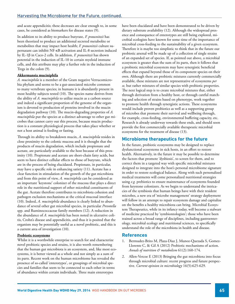
World Digestive Health Day WDHD May 29, 2014 WGO HANDBOOK ON GUT MICROBES 65
and acute appendicitis; these decreases are clear enough to, in some cases, be considered as biomarkers for disease states (9).
In addition to its ability to produce butyrate, F. prausnitzii has been theorized to produce an additional secreted metabolite or metabolites that may impact host health; F. prausnitzii culture su-pernatant can inhibit NF-κB activation and IL-8 secretion induced by IL-1β in Caco-2 cells. In addition, F. prausnitzii has shown potential in the induction of IL-10 in certain myeloid immune cells, and this attribute may play a further role in the induction of Tregs in the colon (9).
Akkermansia muciniphila A. muciniphila is a member of the Gram negative Verrucomicro-bia phylum and seems to be a gut-associated microbe common to many vertebrate species; in humans it is abundantly present in most healthy subjects tested (10). The species name derives from the ability of A. muciniphila to utilize mucin as a carbon source and indeed a significant proportion of the genome of the organ-ism is devoted to production of proteins involved in the mucin degradation pathway (10). The mucin-degrading properties of A. muciniphila put the species at a distinct advantage to other gut mi-crobes that cannot carry out this process, because mucin produc-tion is a constant physiological process that takes place whether or not a host animal is feeding or fasting.
Through its ability to breakdown mucin, A. muciniphila resides in close proximity to the colonic mucosa and it is thought that the products of mucin degradation, which include propionate and acetate, are particularly available to the host because of this prox-imity (10). Propionate and acetate are short-chain fatty acids, but seem to have distinct cellular effects to those of butyrate, which are in the process of being elucidated. Propionate, in particular, may have a specific role in enhancing satiety (11). Acetate has a clear function in stimulation of the growth of the gut microbiota and from this point of view, A. muciniphila can be considered as an important ‘keystone’ colonizer of the mucosa that plays a direct role in the nutritional support of other microbial constituents of the gut. Acetate therefore contributes to microbiota cohesion and pathogen exclusion mechanisms at the critical mucosal interface (10). Indeed, A. muciniphila abundance is clearly linked to abun-dance of several other gut microbial species, in particular Prevotella spp. and Ruminococcaceae family members (12). A reduction in the abundance of A. muciniphila has been noted in ulcerative coli-tis, Crohn’s disease and appendicitis, and thus it is posited that the organism may be potentially useful as a novel probiotic, and this is a current area of investigation (10).
Probiotic ecosystems Whilst it is a worthwhile enterprise to search for and characterize novel probiotic species and strains, it is also worth remembering that the human gut microbiota is an ecosystem, and, like most eco-systems, it is better viewed as a whole and not simply as a sum of its parts. Recent work on the human microbiome has revealed the presence of so-called ‘enterotypes’, or groupings of microbial spe-cies and families that seem to be connected to each other in terms of abundance within certain individuals. Three main enterotypes
have been elucidated and have been demonstrated to be driven by dietary substrate availability (12). Although the widespread pres-ence and consequence of enterotypes are still being explored, mi-crobial ecologists have known for some time of the importance of microbial cross-feeding to the sustainability of a given ecosystem. Therefore it is maybe too simplistic to think that in the future our probiotic arsenal will be made up of a collection of single strains of an expanded set of species. If, as pointed out above, a microbial ecosystem is greater than the sum of its parts, then it follows that a probiotic microbial ecosystem may have synergistic beneficial effects that expand beyond those of its component species on their own. Although there are probiotic mixtures currently commercially available, these mixtures are not representative of ecosystems per se, but rather mixtures of similar species with probiotic properties. The next logical step is to create microbial mixtures that, either through derivation from a healthy host, or through careful match-ing and selection of strains based on phenotype, work together to promote health through synergistic actions. These ecosystems would include proven probiotics as well as a supporting group of microbes that promote their survival and wellbeing through, for example, cross-feeding, environmental buffering capacity, etc. Research is already underway towards these ends, and should soon provide the first commercially available therapeutic microbial ecosystems for the treatment of disease (13).
Microbiome therapeutics for the futureIn the future, probiotic ecosystems may be designed to replace dysfunctional ecosystems in sick hosts, in an effort to restore health. Alternatively, in the future it may be possible to determine the factors that promote ‘dysbiosis’, to screen for them, and to correct them in a targeted way with specific microbial mixtures designed to integrate into the host’s personal microbiome signature in order to restore ecological balance. Along with such personalized medical treatments will come personalized nutritional strategies using e.g. prebiotics to ensure maintenance of ecosystems founded from keystone colonizers. As we begin to understand the intrica-cies of the symbiosis that human beings have with their resident microbes, a new era of ‘microbe management’ strategy deployment will follow in an attempt to repair ecosystem damage and capitalize on the benefits a healthy microbiota can bring. Microbial Ecosys-tem Therapeutics, while in its infancy today, will become a stalwart of medicine practiced by ‘symbiontologists’; those who have been trained across a broad range of disciplines, including gastroenter-ology, microbial ecology and nutritional sciences, to specifically understand the role of the microbiota in health and disease.
References1. Bermudez-Brito M, Plaza-Diaz J, Munoz-Quezada S, Gomez-
Llorente C, & Gil A (2012) Probiotic mechanisms of action. Annals of nutrition & metabolism 61(2):160-174.
2. Allen-Vercoe E (2013) Bringing the gut microbiota into focus through microbial culture: recent progress and future perspec-tive. Current opinion in microbiology 16(5):625-629.
Harvesting the Microbiome for the Future, continued.
World Digestive Health Day WDHD May 29, 2014 WGO HANDBOOK ON GUT MICROBES 66
3. Meehan CJ & Beiko RG (2014) A phylogenomic view of ecological specialization in the Lachnospiraceae, a family of digestive tract-associated bacteria. Genome biology and evolu-tion 6(3):703-713.
4. Sekelja M, Berget I, Naes T, & Rudi K (2011) Unveiling an abundant core microbiota in the human adult colon by a phylogroup-independent searching approach. ISME J 5(3):519-531.
5. Antharam VC, et al. (2013) Intestinal dysbiosis and depletion of butyrogenic bacteria in Clostridium difficile infection and nosocomial diarrhea. J Clin Microbiol 51(9):2884-2892.
6. Berry D & Reinisch W (2013) Intestinal microbiota: a source of novel biomarkers in inflammatory bowel diseases? Best practice & research. Clinical gastroenterology 27(1):47-58.
7. Louis P & Flint HJ (2009) Diversity, metabolism and micro-bial ecology of butyrate-producing bacteria from the human large intestine. FEMS Microbiol Lett 294(1):1-8.
Harvesting the Microbiome for the Future, continued.
8. Vinolo MA, Rodrigues HG, Nachbar RT, & Curi R (2011) Regulation of inflammation by short chain fatty acids. Nutri-ents 3(10):858-876.
9. Miquel S, et al. (2013) Faecalibacterium prausnitzii and human intestinal health. Current opinion in microbiology 16(3):255-261.
10. Belzer C & de Vos WM (2012) Microbes inside--from diver-sity to function: the case of Akkermansia. ISME J 6(8):1449-1458.
11. Havenaar R (2011) Intestinal health functions of colonic mi-crobial metabolites: a review. Beneficial microbes 2(2):103-114.
12. Arumugam M, et al. (2011) Enterotypes of the human gut microbiome. Nature 473(7346):174-180.
13. Petrof EO, Claud EC, Gloor GB, & Allen-Vercoe E (2013) Microbial ecosystems therapeutics: a new paradigm in medi-cine? Beneficial microbes 4(1):53-65.
World Gastroenterology Organisation (WGO)The WGO Foundation (WGO-F)
555 East Wells Street, Suite 1100Milwaukee, WI USA 53202Tel: +1 (414) 918-9798 • Fax: +1 (414) 276-3349Email: [email protected]: www.worldgastroenterology.org • www.wgofoundation.org
WGOF-0616-665