world journal of - bsdwebstorage.blob.core.windows.net · hsin-fu liu, new taipei ... chang-jer wu,...
TRANSCRIPT

Published by Baishideng Publishing Group Inc
World Journal of VirologyWorld J Virol 2017 February 12; 6(1): 1-25
ISSN 2220-3249 (online)

World Journal of VirologyW J V
EDITOR-IN-CHIEFLing Lu, Kansas
ASSOCIATE EDITORChun-Jung Chen, Taichung
GUEST EDITORIAL BOARD MEMBERSChi-Ho Chan, Taichung CityShih-Cheng Chang, TaoyuanHsin-Wei Chen, Miaoli CountyShun-Hua Chen, TainanWei-June Chen, TaoYuanJiann Ruey Hong, TainanReuben Jih-Ru Hwu, HsinchuCheng-Wen Lin, TaichungNa-Sheng Lin, TaipeiTzou-Yien Lin, TaoyuanHsin-Fu Liu, New TaipeiHung-Jen Liu, TaichungMenghsiao Meng, TaichungWen-Ling Shih, PingtungRobert Yung-Liang Wang, TaoyuanChang-Jer Wu, KeelungChi-Chiang Yang, TaichungKung-Chia Young, Tainan
MEMBERS OF THE EDITORIAL BOARD
Argentina
Angela Gentile, Buenos AiresPablo D Ghiringhelli, BernalJorge V Pavan, CórdobaLaura E Valinotto, Buenos Aires
AustraliaShisan Bao, SydneyJiezhong Chen, NswRussell J Diefenbach, NswRussell Diefenbach, WestmeadIan M Mackay, HerstonJohn J Miles, BrisbaneDavid P Wilson, SydneyKong-Nan Zhao, Herston
Austria
Adly MM Abd-Alla, ViennaZoltan Banki, InnsbruckSabine Brandt, ViennaThomas Lion, Vienna
Barbados
Alok Kumar, Bridgetown
Belgium
Jan P Clement, Leuven
Brazil
Luciane P Gaspar, CuritibaJosé P Gagliardi Leite, Rio de JaneiroLuciano K de Souza Luna, Curitiba
Thiago M Lopes e Souza, Rio de JaneiroSonia M Raboni, CuritibaLivia M Villar, Rio De JaneiroClaudia L Vitral, Niterói
Bulgaria
Irena P Kostova, Sofia
Cameroon
Richard Njouom, Yaounde
Canada
Stephen D Barr, LondonEarl G Brown, OttawaIvan Brukner, MontrealJingxin Cao, WinnipegPeter J Krell, GuelphJean F Laliberté, VancouverHonglin Luo, VancouverXianzhou Nie, FrederictonXiaoli L Pang, AlbertaJean-Pierre Routy, MontrealAiming Wang, OntarioDecheng Yang, Vancouver
Chile
Gloria L Arriagada, Vina del MarMarcelo López-Lastra, Santiago
I
Editorial Board2016-2019
The World Journal of Virology Editorial Board consists of 370 members, representing a team of worldwide experts in virology. They are from 59 countries, including Argentina (4), Australia (8), Austria (4), Barbados (1), Belgium (1), Brazil (7), Bulgaria (1), Cameroon (1), Canada (12), Chile (2), China (55), Croatia (2), Cuba (1), Czech Republic (1), Denmark (1), Egypt (3), Ethiopia (1), Finland (5), France (10), Gambia(1), Germany (11), Ghana (1), Greece (2), Hungary (1), India (13), Indonesia (1), Iran (2), Ireland (3), Israel (4), Italy (23), Japan (16), Kazakhstan (1), Kenya (1), Kosovo (1), Mexico (2), Netherlands (5), New Zealand (1), Nigeria (1), Pakistan (1), Palestine (1), Poland (1), Portugal (1), Romania (1), Russia (2), Saudi Arabia (1), Singapore (2), Slovakia (2), Slovenia (2), South Africa (2), South Korea (6), Spain (19), Sweden (4), Thailand (8), Tunisia (1), Turkey (4), United Arab Emirates (1), United Kingdom (8), United States (92), and Uruguay (1).
February 26, 2016WJV|www.wjgnet.com

ChinaKun-Long Ben, KunmingGuang-Wen Cao, ShanghaiPaul KS Chan, HongkongYuan-Ding Chen, KunmingAn-Chun Cheng, Ya’anShang-Jin Cui, HarbinXiao-Ping Dong, BeijingZai-Feng Fan, BeijingJean-Michel Garcia, Hong KongGuan-Zhu Han, NanjingYu-Xian He, BeijingXiu-Guo Hua, ShanghaiWen-Lin Huang, GuangzhouMargaret Ip, Hong KongDao-Hong Jiang, WuhanJian-Qi Lian, Xi’anXiao-Yang Mo, HunanBeatrice Nal, Hong KongCheng-Feng Qin, BeijingHua-Ji Qiu, HarbinXiao-feng Ren, HarbinHong Tang, ChengduJian-Wei Wang, BeijingYou-Chun Wang, BeijingNing Wang, BeijingMary Miu Yee Waye, Hong KongPatrick CY Woo, Hong KongYu-Zhang Wu, ChongqingJian-Qing Wu, NanjingRui Wu, LuoyangXin-Yong Liu, JinanXu-Qing Zhang, ChongqingGuo-Zhong Zhang, BeijingChuang-Xi Zhang, HangzhouPing Zhao, ShanghaiShi-Jun Zheng, Beijing
Croatia
Snjezana Z Lepej, ZagrebPero Lucin, Rijeka
Cuba
Maria G Guzman, Havana
Czech Republic
Daniel Ruzek, Ceske Budejovice
Denmark
Havard Jenssen, Roskilde
Egypt
Mona El SH El-Raziky, CairoSamia A Kamal, CairoAbdel-Rahman N Zekri, Cairo
Ethiopia
Woldaregay E Abegaz, Addis Ababa
FinlandJussi Hepojoki, HelsinkiAnne Jaaskelainen, HelsinkiIrmeli Lautenschlager, HelsinkiPamela Osterlund, HelsinkiAntti Vaheri, Helsinki
France
Christian A Devaux, MontpellierJean Dubuisson, LilleDuverlie Gilles, AmiensBedouelle Hugues, ParisEric J Kremer, MontpellierBelec Laurent, ParisDenis Rasschaert, ToursDominique Salmon-Céron, ParisChristian Trépo, LyonEric Wattel, Lyon
Gambia
Assan Jaye, Banjul
Germany
Claus-Thomas Bock, BerlinElke Bogner, BerlinAndreas Dotzauer, BremenIngo Drexler, DüsseldorfChristoph Eisenbach, HeidelbergThomas Iftner, ErlangenFlorian Lang, TuebingenJochen Mattner, ErlangenMichael Nevels, RegensburgAndreas MH Sauerbrei, JenaFrank Tacke, Aachen
Ghana
Kwamena W Sagoe, Accra
Greece
Apostolos I Beloukas, AthensGeorge V Papatheodoridis, Athens
Hungary
Krisztián Bányai, Budapest
India
Akhil C Banerjea, New DelhiJayanta Bhattacharya, PuneRunu Chakravarty, KolkattaSibnarayan Datta, TezpurKumar Jitendra, PunjabHimansu Kesari Pradhan, New DelhiSachin Kumar, Assam
Sunil K Lal, New DelhiSunil K Mukherjee, New DelhiRamesh S Paranjape, PuneSharma Pradeep, KarnalShamala D Sekaran, New DelhiRasappa Viswanathan, Coimbatore
Indonesia
Andi Utama, Tangerang
Iran
Seyed M Ghiasi, TehranFarzin Roohvand, Tehran
Ireland
Carlo Bidoia, DublinLiam J Fanning, CorkWeifeng Shi, Dublin
Israel
Irit Davidson, Bet DaganYedidya Gafni, Bet DaganMurad Ghanim, Bet DaganIlan Sela, Rehovot
Italy
Alberto Alberti, SassariGiorgio Barbarini, VogheraMassimiliano Berretta, AvianoFranco M Buonaguro, NaplesMaria R Capobianchi, NaplesArnaldo Caruso, BresciaDaniel O Cicero, RomeMarco Ciotti, RomeCristina Costa, TorinoPiergiuseppe De Berardinis, NaplesFederico De Marco, RomeMassimo EA De Paschale, LegnanoMaurizia Debiaggi, PaviaPaolo Fabris, VicenzaDaniele Focosi, PisaSimone Giannecchini, FlorenceFabrizio Maggi, PisaRoberto Manfredi, BolognaVito Martella, ValenzanoGiuseppe Portella, NapoliNicola Principi, MilanGiovanni Rezza, RomaDiego Ripamonti, Bergamo
Japan
Masanori Daibata, NankokuBin Gotoh, OtsuShoji Ikuo, KobeTakashi Irie, HiroshimaHiroki Isomura, MaebashiHideya Kawasaki, Hamamatsu
II February 26, 2016WJV|www.wjgnet.com

III February 26, 2016WJV|www.wjgnet.com
Eiichi N Kodama, SendaiEmoto Masashi, GunmaHiromitsu Moriyama, TokyoKenji Okuda, YokohamaNobuhiro Suzuki, OkayamaTakashi Suzuki, ShizuokaTetsuro Suzuki, HamamatsuYoshiyuki Suzuki, Nagoya-shiAkifumi Takaori-Kondo, KyotoTetsuya Toyoda, Toyohashi
Kazakhstan
Vladimir E Berezin, Almaty
Kenya
George G Maina, Nairobi
Kosovo
Lul Raka, Prishtina
Mexico
Juan E Ludert, Mexico CityJulio Reyes-Leyva, Mexico
Netherlands
Kimberley SM Benschop, AmsterdamBenjamin Berkhout, AmsterdamByron EE Martina, RotterdamWillem JG Melchers, NijmegenMonique Nijhuis, Utrecht
New Zealand
Olga S Garkavenko, Auckland
Nigeria
Olajide A Owolodun, Plateau State
Pakistan
Muhammad I Qadir, Faisalabad
Palestine
Ahmad Y Amro, Jerusalem
Poland
Brygida Knysz, Wroclaw
Portugal
Celso Cunha, Lisbon
RomaniaAnda Baicus, Bucharest
Russia
Anton Buzdin, MoscowElena V Gavrilova, Novosibirsk
Saudi Arabia
Ahmed S Abdel-Moneim, Al-Taif
Singapore
Sophie Bellanger, SingaporeDing X Liu, Singapore
Slovakia
Gabriela Bukovska, BratislavaJulius Rajcani, Bratislava
Slovenia
Uros Krapez, LjubljanaAndrej Steyer, Ljubljana
South Africa
Janusz T Paweska, SandringhamDirk Stephan, Matieland
South Korea
Sang Hoon Ahn, SeoulTae-Jin Choi, BusanYoung-Ki Choi, CheongjuKee-Jong Hong, CheongwonBum-Joon Kim, SeoulJunsoo Park, Wonju
Spain
Alí Alejo, ValdeolmosAlfredo Berzal-Herranz, GranadaRafael Blasco, MadridJulio Collazos, Usánsolo-GaldácanoJuan M Hernández, MadridGómez L Jaime, CórdobaJosep M Llibre, BadalonaCecilio López-Galíndez, MadridF. Xavier López-Labrador, ValenciaJoséA Melero, MadridLuis Menéndez-Arias, MadridAndrés Moya, ValènciaDavid R Pereda, SevillaPilar Perez-Romero, SevillaJosep Quer, BarcelonaDaniel López Rodríguez, Majadahonda
Juan-Carlos Saiz, MadridNoemi Sevilla, MadridNatalia Soriano-Sarabia, Madrid
Sweden
Goran PL Bucht, UmeaAli Mirazimi, SolnaMuhammad Munir, UppdalaBo F Oberg, Huddinge
Thailand
Prasert Auewarakul, BangkokParin Chaivisuthangkura, BangkokWasin Charerntantanakul, Chiang MaiWansika Kiatpathomchai, BangkokSasisopin Kiertiburanakul, BangkokWinyou Mitarnun, Chiang MaiYong Poovorawan, BangkokViroj Wiwanitkit, Bangkok
Tunisia
Olfa Bahri, Tunis
Turkey
Omer Coskun, AnkaraIftihar Koksal, TrabzonAykut Ozdarendeli, KayseriAyca A Sayiner, Izmir
United Arab Emirates
Tahir A Rizvi, Al Ain
United Kingdom
Shiu-Wan Chan, ManchesterSimon R Clegg, PrestonChiriva I Maurizio, NottinghamIain M Morgan, GlasgowMark R Nelson, LondonAdrian W Philbey, GlasgowJames P Stewart, LiverpoolGavin WG Wilkinson, Cardiff
United States
Nafees Ahmad, TucsonAshok Aiyar, Los AngelesHizi Amnon, BethesdaJudith M Ball, TexasIgor M Belyakov, FrederickBradford K Berges, ProvoPreeti Bharaj, OrlandoJay C Brown, CharlottesvilleVictor E Buckwold, WalkersvilleAlexander A Bukreyev, GalvestonJoseph J Carter, SeattleMaria G Castro, Los AngelesYan-Ping Chen, Beltsville

IV February 26, 2016WJV|www.wjgnet.com
Xiaojiang S Chen, Los AngelesChaoping Chen, Fort CollinsPawel S Ciborowski, OmahaHarel Dahari, Los AlamosDavid A Davis, BethesdaDon J Diamond, DuarteDimiter S Dimitrov, FrederickYajarayma JT Feldman, SacramentoVincent N Fondong, DoverPhillip A Furman, PrincetonShou-Jiang Gao, San AntonioKaplan Gerardo, BethesdaDavid R Gretch, SeattleHailong Guo, RochesterHaitao Guo, IndianapolisYoung S Hahn, CharlottesvilleJames M Hill, New OrleansWei Jiang, CharlestonXia Jin, New YorkClinton Jones, LincolnRobert Jordan, CorvallisAdriana E Kajon, AlbuquerqueKrishna MV Ketha, BethesdaPaul R Kinchington, PittsburghPrasad S Koka, San DiegoMajid Laassri, RockvilleFeng Li, BrookingsJin Ling, Corvallis
Yuanan Lu, HonoluluIgor S Lukashevich, LouisvillePaolo Lusso, BethesdaRavi Mahalingam, AuroraBarry J Margulies, TowsonMichael R McConnell, San DiegoGeorge Miller, BostonMohammad Mir, Kansas CityMansour Mohamadzadeh, ChicagoThomas P Monath, Menlo ParkJonathan P Moorman, Johnson CityEgbert Mundt, StillwaterKaruppiah Muthumani, PhiladelphiaEleftherios Mylonakis, BostonHiroyuki Nakai, PittsburghDebiprosad Nayak, Los AngelesOscar A Negrete, LivermoreAnthony V Nicola, RichmondShunbin Ning, MiamiDiana Nurutdinova, St. LouisPhillipe N Nyambi, New YorkSlobodan Paessler, GalvestonKrishan K Pandey, Saint LouisVirendra N Pandey, NewarkEric M Poeschla, RochesterAndrew P Rice, HoustonJacques Robert, RochesterRachel L Roper, Greenville
Paula Saá, RockvilleDeepak Shukla, ChicagoAndrey Staruschenko, MilwaukeeQiyi Tang, PonceSharof M Tugizov, San FranciscoChristophe Vanpouille, BethesdaRobert J Visalli, SavannahAbdul A Waheed, FrederickXiu-Feng Wan, Mississippi StateXiuqing Wang, BrookingsJane H Wang, ChicagoXinzhen Yang, BostonZhiping Ye, BethesdaKyoungjin J Yoon, AmesJianxin You, PhiladelphiaYan Yuan, PhiladelphiaLijuan Yuan, BlacksburgHong Zhang, RockvilleLuwen Zhang, LincolnZhi-Ming Zheng, BethesdaHong Zheng, TampaHeshan S Zhou, Louisville
Uruguay
Matías Victoria, Salto

Contents
IWJV|www.wjgnet.com February 12, 2017|Volume 6|Issue 1|
Quarterly Volume 6 Number 1 February 12, 2017
MINIREVIEWS1 BiologicalandhistoricaloverviewofZikavirus
Armstrong N, Hou W, Tang Q
9 Valueofroutinedenguediagnosisinendemiccountries
Ayukekbong JA, Oyero OG, Nnukwu SE, Mesumbe HN, Fobisong CN
ORIGINAL ARTICLE Basic Study
17 ExpressionofhepatitisBvirussurfaceantigensinducesdefectivegonadphenotypesinCaenorhabditis
elegans
Chen YY, Lee LW, Hong WN, Lo SJ
W J World Journal of VirologyV

ContentsWorld Journal of Virology
Volume 6 Number 1 February 12, 2017
EDITORS FOR THIS ISSUE
Responsible Assistant Editor: Xiang Li Responsible Science Editor: Jin-Xin KongResponsible Electronic Editor: Ya-Jing Lu Proofing Editorial Office Director: Xiu-Xia SongProofing Editor-in-Chief: Lian-Sheng Ma
EDITORIALOFFICEXiu-Xia Song, DirectorWorld Journal of VirologyBaishideng Publishing Group Inc8226 Regency Drive, Pleasanton, CA 94588, USATelephone: +1-925-2238242Fax: +1-925-2238243E-mail: [email protected] Desk: http://www.wjgnet.com/esps/helpdesk.aspxhttp://www.wjgnet.com
PUBLISHERBaishideng Publishing Group Inc8226 Regency Drive, Pleasanton, CA 94588, USATelephone: +1-925-223-8242Fax: +1-925-223-8243E-mail: [email protected] Desk: http://www.wjgnet.com/esps/helpdesk.aspxhttp://www.wjgnet.com
PUBLICATIONDATEFebruary 12, 2017
COPYRIGHT© 2017 Baishideng Publishing Group Inc. Articles published by this Open-Access journal are distributed under the terms of the Creative Commons Attribution Non-commercial License, which permits use, distribution, and reproduction in any medium, provided the original work is properly cited, the use is non-commercial and is otherwise in compliance with the license.
SPECIALSTATEMENTAll articles published in journals owned by the Baishideng Publishing Group (BPG) represent the views and opin-ions of their authors, and not the views, opinions or policies of the BPG, except where otherwise explicitly indicated.
INSTRUCTIONSTOAUTHORShttp://www.wjgnet.com/bpg/gerinfo/204
ONLINESUBMISSIONhttp://www.wjgnet.com/esps/
IIWJV|www.wjgnet.com
ABOUT COVER
AIM AND SCOPE
INDExING/ABSTRACTING
FLYLEAF
February 12, 2017|Volume 6|Issue 1|
NAMEOFJOURNALWorld Journal of Virology
ISSNISSN 2220-3249 (online)
LAUNCHDATEFebruary 12, 2012
FREQUENCYQuarterly
EDITOR-IN-CHIEFLing Lu, MD, PhD, Department of Pathology and Laboratory Medicine, University of Kansas Medical Center, Kansas City, 3901 Rainbow Blvd, WHE 3020, KS 66160, United States
EDITORIALBOARDMEMBERSAll editorial board members resources online at http://www.wjgnet.com/2220-3249/editorialboard.htm
EditorialBoardMemberofWorldJournalofVirology ,SharofMTugizov,DSc,PhD,Professor,DepartmentofMedicine,UniversityofCalifornia,DivisionofInfectiousDiseases,SanFrancisco,CA94143,UnitedStates
World Journal of Virology (World J Virol, WJV, online ISSN 2220-3249, DOI: 10.5501) is a peer-reviewed open access academic journal that aims to guide clinical practice and improve diagnostic and therapeutic skills of clinicians.
WJV covers topics concerning arboviral infections, bronchiolitis, central nervous system viral diseases, DNA virus infections, encephalitis, eye infections, fatigue syndrome, hepatitis, meningitis, opportunistic infections, pneumonia, RNA virus infections, sexually transmitted diseases, skin diseases, slow virus diseases, tumor virus infections, viremia, zoonoses, and virology-related traditional medicine, and integrated Chinese and Western medicine. Priority publication will be given to articles concerning diagnosis and treatment of viral diseases. The following aspects are covered: Clinical diagnosis, laboratory diagnosis, differential diagnosis, imaging tests, pathological diagnosis, molecular biological diagnosis, immunological diagnosis, genetic diagnosis, functional diagnostics, and physical diagnosis; and comprehensive therapy, drug therapy, surgical therapy, interventional treatment, minimally invasive therapy, and robot-assisted therapy.
We encourage authors to submit their manuscripts to WJV. We will give priority to manuscripts that are supported by major national and international foundations and those that are of great basic and clinical significance.
World Journal of Virology is now indexed in PubMed, PubMed Central.
I-IV EditorialBoard

Najealicka Armstrong, Wangheng Hou, Qiyi Tang
MINIREVIEWS
� February �2, 20�7|Volume 6|Issue �|WJV|www.wjgnet.com
Biological and historical overview of Zika virus
Najealicka Armstrong, Wangheng Hou, Qiyi Tang, Deparment of Microbiology, Howard University, College of Medicine, Washington, DC 20059, United States
Author contributions: Armstrong N and Hou W searched most of the references and participated in drafting the manuscript; Armstrong N organized the tables; Tang Q designed the study and drafted the manuscript; all authors read and approved the final manuscript.
Supported by a Charles and Mary Latham Fund (Q.T.), No. NIH/NIAID SC1AI112785 (Q.T.); and National Institute on Minority Health and Health Disparities of the National Institutes of Health, No. G12MD007597.
Conflict-of-interest statement: The authors declare that they have no conflict of interest.
Open-Access: This article is an open-access article which was selected by an in-house editor and fully peer-reviewed by external reviewers. It is distributed in accordance with the Creative Commons Attribution Non Commercial (CC BY-NC 4.0) license, which permits others to distribute, remix, adapt, build upon this work non-commercially, and license their derivative works on different terms, provided the original work is properly cited and the use is non-commercial. See: http://creativecommons.org/licenses/by-nc/4.0/
Manuscript source: Unsolicited manuscript
Correspondence to: Qiyi Tang, PhD, Associate Professor, Department of Microbiology, Howard University, College of Medicine, Seeley Mudd Building, Room 315, 520 W Street, NW, Washington, DC 20059, United States. [email protected]: +1-202-8063915Fax: +1-202-2388518
Received: May 10, 2016Peer-review started: May 12, 2016First decision: June 14, 2016Revised: June 20, 2016Accepted: August 11, 2016Article in press: August 15, 2016Published online: February 12, 2017
Abstract The recent outbreak of the Zika virus attracts worldwide attention probably because the most recently affected country (Brazil) will host the 2016 Olympic Game. Zika virus infected cases are now spreading to many other countries and its infection might be linked to some severe medical sequelae. Since its first isolation from the infected monkey in 1947 in Uganda, only a few studies had been taken until recent outbreak. According to the history of referenced publications, there is a 19-year gap from 1989 to 2007. This might be because only mild diseases were diagnosed from Zika virus infected populations. Obviously, the recent reports that Zika virus infection is probably associated with microcephaly of the neonates makes us reevaluate the medical significance of the viral pathogen. It can be transmitted sexually or by mosquito biting. Sexual transmission of the Zika virus distinguishes it from other members of the Genus Flavivirus. Detailed information of the Zika virus is needed through a thorough investigation covering basic, epidemical, subclinical and clinical studies. Here, we reviewed the published information of Zika virus.
Key words: Zika virus; Flavivirus; Congenital infection; Outbreak; Microcephaly
© The Author(s) 2017. Published by Baishideng Publishing Group Inc. All rights reserved.
Core tip: Zika virus is gaining new ground with the recent outbreaks that are starting to expand worldwide. While normally transmitted by the mosquito, other routes of transmission are being discovered. Also, other medical complications are being detected with Zika virus infections. These recent findings require the scientific community to thoroughly examine Zika virus to better understand it so that better diagnostic options, treatment, and preventative measures can be developed. In order to beat Zika virus, we must understand its history and outbreak patterns as well as gain a full understanding of
Submit a Manuscript: http://www.wjgnet.com/esps/Help Desk: http://www.wjgnet.com/esps/helpdesk.aspxDOI: �0.550�/wjv.v6.i�.�
World J Virol 20�7 February �2; 6(�): �-8ISSN 2220-3249 (online)
© 20�7 Baishideng Publishing Group Inc. All rights reserved.
World Journal of VirologyW J V

2 February �2, 20�7|Volume 6|Issue �|WJV|www.wjgnet.com
Armstrong N et al . Starting a fight with Zika virus
all clinical manifestations associated with this virus.
Armstrong N, Hou W, Tang Q. Biological and historical overview of Zika virus. World J Virol 2017; 6(1): 1-8 Available from: URL: http://www.wjgnet.com/2220-3249/full/v6/i1/1.htm DOI: http://dx.doi.org/10.5501/wjv.v6.i1.1
INTRODUCTIONThe Zika virus, together with the West Nile virus, Yellow fever virus, Japanese encephalitis virus, Dengue fever virus, and many other classified and unclassified viruses, forms the genus Flavivirus that belongs to family Flaviviridae. The family Flaviviridae consists of many other viruses that are summarized in a 2010 review[1]. This family of viruses have enveloped icosahedral capsid that contains a single strand RNA genome (about 11000 nucleotides) with positive sense[2]. Therefore, the infected viral RNA can be directly translated to a large polyprotein precursor, which is co- and post-translationally processed by viral and cellular proteases into structural and non-structural proteins. The three structural proteins are critical for the formation of envelop and capsid, and the seven non-structural (NS) proteins play important roles in virus replication. The three structural proteins are envelope, E; membrane precursor, PrM; and capsid, C. The seven NS proteins include NS1, NS2a, NS2b, NS3, NS4a, NS4b, and NS5 (Figure 1). The names, location in the infected cells, and functions of viral proteins are listed in Table 1. The members of the genus Flavivirus are characterized by similarities in genomic structure, viral protein function, pathogenesis and transmission.
The large polyprotein precursor must be cleaved to generate actively functional proteins. The cleavage of the polyprotein precursor is a sophisticated process and is completed collaboratively by cellular proteases of the PACE (Paired basic Amino acid Cleaving Enzyme)-type or other Golgi-localized proteases and the viral serine protease embedded in the N-terminal domain of non-structural protein 3 (NS3Pro), which requires NS2b for its activity[1]. A distinct feature of genus Flavivirus from other genera of Flaviviridae is that the 5′-end of the (+)ssRNA genome of genus Flavivirus is decorated with an RNA cap structure (N7meGpppA2′Ome-RNA). 5’end capping of the viral RNA is as important as that for eukaryotic mRNAs, not only to initiate the process of translation but also to protect the viral RNA from degradation by endogenous RNA exonucleases. The protein translation happens immediately after the uncoating of viral particle in the cytoplasm. The (+)ssRNA genome is used as a template not only for gene expression but also for viral genome replication. Both viral RNA replication and gene translation occur in the cytoplasm. For RNA replication, viral NS proteins and cellular proteins interact to form a replication compartment (RC). During the period of viral RNA replication in the cytoplasm, the RC consists of morphologically distinct, membrane-bound
compartments that also differ with respect to both function and NS proteins composition[3]. The NS3 and NS5 proteins are central to the viral RC, as together, they harbor most, if not all, of the catalytic activities required to both cap and replicate the viral RNA. Following replication, the protected genomic RNA is packaged by the C protein to form a capsid in a host-derived lipid bilayer in which the E protein is embedded and later integrated into viral envelope. The mature particles subsequently exit from the host cell by exocytosis.
REGIONAL ISOLATION OF ZIKA VIRUSThe Zika virus is phylogenetically close to Spondweni virus and a member of Flaviviridae family[4]. Comparative genomic analysis revealed that coding regions of pre-epidemic and epidemic strains of the Zika virus were similar with the exception of the NS2B. Bootscan analysis and multiple sequence alignment of the Asian lineage suggested that there may be genetic recombination of a fragment (nucleotides 4237-4528) of NS2B with that of the Spondweni virus[5].
African countries In 1947, a group of scientists from United Kingdom led by Haddow et al[4] who were investigating yellow fever isolated Zika virus from a rhesus macaque with fever in the Zika Forest in Uganda[6,7]. The isolated viral strain has been stored in ATCC (ATCC® VR84™, MR 766) and the European Virus Archive (France) and is now still used for studies. The next important step was to find out whether the Zika virus is transmitted by mosquitos. First, Boorman et al[8] demonstrated that Zika virus can infect and replicate in mosquitos, providing experimental evidence that Zika virus may be transmitted by mos-quitos. Later, the United Kingdom Flavivirus research group continued their studies of arboreal mosquitos as virus vectors in Uganda. They isolated 12 strains of Zika virus from Aedes (Stegomyia) africanus in the Zika forest[9]. Zika virus is apparently enzootic in Zika forest, and the evidence collected by Hoddow et al[9] suggested that Aedes africanus is the primary vector and that forest-dwelling monkeys and human are, on occasion, involved. It was not clear whether the mosquito transmitted the virus to other animals because no small mammal trapped in the forest showed serum antibody against the Zika virus. The Zika virus infection in humans was first reported in 1954[10]. It has also been experimentally demonstrated via volunteers that the Zika virus is able to infect humans[11]. In summary, results from these investigations suggest that the Zika virus is an arbovirus, transmitted by mosquitos and infects at least monkeys and humans.
Southern Asian countriesThe first isolation of Zika virus in South-Eastern Asia was reported in 1969 in Malaysia[12]. Some years later, there was another report that the Zika virus was isolated from patients in Indonesia[13]. The event occurred

3 February �2, 20�7|Volume 6|Issue �|WJV|www.wjgnet.com
during the end of the rainy season of 1977 when Aedes aegypti usually flourishes. Seven patients in central Java, Indonesia, appeared in the hospital with high fever, malaise, stomach ache, dizziness and anorexia. Data on these 7 Zika virus cases and several previously reported human infections indicated that clinical characteristics of infection with Zika virus appeared relatively mild, self-limiting, and nonlethal. It was suspected that the virus was transmitted by Aedes aegypti, which had been reported to be a probable vector in Malaysia[12]. A later investigation in Sabah, Malaysia, showed that the Zika virus infected 60 semi-captive and 84 free-ranging orangutans (Pongo pygmaeus pygmaeus)[14]. Another study conducted by the United States Naval Medical Research Unit No. 2 (NAMRU-2) isolated Zika virus in Cambodia in 2010[15]. This case was from a 3-year-old boy who had 4 d of fever, sore throat and cough as well a headache that lasted for 3 d. The studies conducted in
southern Asia further confirmed that mosquitos are the vector and the primates might be the end host of viral infection.
The Zika virus has been also isolated from animals and human in other African countries. For examples, during the years 1964 to 1970, Moore et al[16] isolated 171 arboviruses of 15 different types from humans in Ibadan, Nigeria. Zika virus isolation rates also varied by season, with peaks in rainy seasons (June to August) and lows in dry seasons (January to February). Viruses were isolated from all age groups, with the majority from children one to four years old. The viruses isolated in largest numbers were chikungunya and yellow fever, which caused epidemics in 1969, and dengue types 1 and 2 and Tataguine, which are endemic in Ibadan. The Zika virus was isolated at a low rate. In 1999, three strains of the Zika virus were isolated as part of yellow fever studies in the Ivory Coast[17]. In 2010, it was
Translation and processing of the flavivirus polyprotein
Single stranded (+) sense RNA genome
Single open reading frame
Precursor of the protein
Approximately 3300 Aa
3'NCR5'NCRCAP
AUG
Structural
CprM
E
NS1NS2a
2bNS3
NS4aNS5
4b
Non-structural
Mature proteins
Approximately 11 kbUAG
Figure 1 Genomic structure and gene production of Flavivirus. AUG: Translation start codon; UAG: Translation stop codon; NCR: Non coding RNA sequence; kb: Kilo base; Aa: Amino acid.
Table 1 Roles of viral protein and RNA during viral infection in permissive cells
Name of the vital material Location in cell Function
Viral genome + ssRNA (approximately ��000 nt) Cytoplasm Template for protein translation and for viral genome replication Envelope, E (53 KDa)[52] Cell membrane Viral assembly, budding, attachment to target cells, and viral membrane fusionMembrane precursor, PrM (20 KDa)[53] Cell membrane Facilitating E protein folding and trafficking, and virion maturationCapsid, C (�2 KDa)[54] Cytoplasm Virion maturationNS� (glycoprotein)[55] (46-55 kDa) Endoplasmic reticulum Subverting immune response
vesicular compartments, virus-induced intracellular RNA cell surface replication, neurovirulence
NS2a (25 kDa)[56] Transmembrane Virus assembly, inhibit IFN-responseNS2b (�4 kDa)[�,57] Cytoplasm, nucleus Viral protein cleavageNS3 (69 kDa)[�] Cytoplasm, nucleus Viral protein cleavage, RNA
triphosphatase, mRNA capping,RNA helicase
NS4a (�6 kDa)[58] Transmembrane Viral RNA replicationNS4b (2�.5 kDa)[59] Integral membrane Suppression of (IFN-α/β),
suppression of the host RNAi,negatively regulate the helicase
function, viral replicationNS5 (�03 kDa)[60,6�] Cytoplasm, nucleus The RNA triphosphatase,
RNA-dependent RNA polymerase
Armstrong N et al . Starting a fight with Zika virus

4 February �2, 20�7|Volume 6|Issue �|WJV|www.wjgnet.com
reported that the Zika virus was isolated at a high rate in Cameroon. The research group investigated 102 sera from febrile patients (with negative laboratory findings for malaria and typhoid fever) at clinics in the Fako Division of Cameroon. The Zika virus was isolated at a rate of 11.4%, higher than that of any other members of Genus Flavivirus[18]. Therefore, following the time, the Zika virus has been spread throughout Africa.
More and more Zika virus strains have been isolated from humans worldwide[17]. Studies conducted in Nigeria during 1971-1975 isolated the Zika virus from humans. Serological experiments showed that 40% of the persons tested had neutralizing antibody to the Zika virus[16,19]. The infected populations were detected in other African countries such as Uganda, Tanzania, Egypt, Central African Republic, Sierra Leone, and Gabon, and in parts of Asia, including India, Malaysia, the Philippines, Thai-land, Vietnam, and Indonesia[20]. Table 2 lists the strains that have been sequenced. The data from the viral genomic analysis support the hypothesis that the Zika viruses can be classified by origin into the Southern-Eastern Asian type and African type (Table 2). Other isolates might be derived from these types.
ZIKA VIRUS OUTBREAKS AND CLINICAL COMPLICATIONSThe Zika virus has been considered as a benign pathogen, causing asymptomatic or mild infections. Currently, there is no serological test that can clearly distinguish the Zika virus from other Flaviviruses. Diagnostic tests for Zika include RT-PCR, an IgM ELISA, and a plaque reduction neutralization test (PRNT). Some commercial tests have only become recently available[21]. Even a report from Olson et al[13] in 1981 that a cluster of 7 people with serologic evidence of the Zika virus illness in Indonesia did not attract serious attention and was not considered an outbreak due to the mildness of the associated illness. Later on, the same arbovirus research group performed a serological study that showed that 9/71 (13%) human volunteers in Lombok, Indonesia, had neutralizing antibodies to the Zika virus[22]. However, no serious cases were reported. The first outbreak of Zika virus-caused diseases was reported in 2007 on Yap Island of Micronesia. In April 2007, physicians on Yap Island characterized the disease with rash, conjunctivitis, arthralgia, arthritis, and fever. The disease affected 99 patients in 2 mo. A comprehensive study that combined analysis of patient samples, serological testing and real-time RT-PCR revealed the genetic and serological properties of the Zika virus epidemic[23]. The studies suggested that the 2007 Yap Island Zika virus is distantly related to African subclades and may be spread from Southeast Asia and the Pacific. Duffy et al[24] later conducted an extensive study on the Yap Island Zika virus outbreak. From 185 patients, 49 had been confirmed with the Zika virus illness, only 5 were excluded from Zika virus infection, and all others were suspected of Zika virus infection. They used survey
studies in a large population, and estimated that 73% of the population of the Yap Island was infected with the Zika virus during the epidemic outbreak. Therefore, the outbreak on Yap Island in 2007 suggested that Zika virus infection has been spread outside of Africa and Asia[17]. Of course, whether or not the Zika virus was imported from Africa or Asia or other places remains to be verified.
Another Zika outbreak occurred between Oct. 2013 and Feb. 2014 in French Polynesia - like Yap Island, another island in the Pacific Ocean. In the very beginning of the outbreak, a mild dengue-like illness was observed in the patients within a family (consisting of wife, husband and their son-in-law). The symptoms included low fever (< 38 ℃), asthenia, wrist and fingers arthralgia, headache, rash, and conjunctivitis. The RT-PCR test confirmed that it was a Zika virus infection[25]. The epidemic has been spread to a large population as reported by the syndromic surveillance network (6630 suspected Zika virus infection cases), 333 of which were confirmed by real-time RT-PCR as Zika virus infections. Symptoms of most Zika virus infection cases are mild and self-limited (mean duration of symptoms is 3-6 d)[25-27]. No hospitalizations for acute infection have been reported. In contrast to the outbreak in Yap Island, some severe complications were seen in this outbreak: The first case of Guillain-Barré syndrome (GBS) was found immediately after a Zika virus infection[28], and another case of vertical transmission from an infected pregnant woman to the baby was reported in this out-break[29].
The spread of Zika virus from the outbreak of French Polynesia has been reported. Two Japanese travelers were confirmed to be infected with the Zika virus after they returned from a trip to French Polynesia during the time of the outbreak[30]. In addition, it was found to have spread to other Pacific Islands including New Caledonia, Cook Islands, Easter Island, Vanuatu, and Solomon Islands[31]. The introduction of the Zika virus from French Polynesia into New Caledonia caused an-other outbreak in New Caledonia in 2014[32]; The first cases of Zika virus infection were confirmed in November 2013, and they were imported from French Polynesia. By the end of 2014, a total of 1383 cases were confirmed in a laboratory[32]. Consequently, an outbreak in New Caledonia was declared. Thus far, introduction of the Zika virus from French Polynesia to other countries has been continuously reported.
Between 1947 and 2006, < 20 cases of Zika virus infection have been reported[5]. There have been recent reports of imported cases of Zika virus infections in 18 travelers returning to the Netherlands from Surinam, which is in South America near the northern border of Brazil, and the Dominican Republic[33], 13 infections were imported from Venezuela, Fiji/Samoa, or Suriname to China[34], and 4 infections were imported from Brazil to Portugal[35]. Autochthonous cases were reported in places such as Mexico[36], Colombia[37], and Easter Island, which was the first outbreak (51 cases) reported in a territory of the Americas in early 2014[38].
Armstrong N et al . Starting a fight with Zika virus

5 February �2, 20�7|Volume 6|Issue �|WJV|www.wjgnet.com
The recent outbreak in Brazil has attracted the most attention due to not only its growing infected population but also its likely enhanced severity of the clinical sequelae. In March of 2015, Zanluca et al[39] from the Molecular Virology Laboratory of Carlos Chagas Institute, Oswaldo Cruz Institute, state of Paraná, Brazil, detected the Zika virus genome by RT-PCR from 8 out of 21 acute-phase serum specimens from the patients with dengue-like symptoms. This is the first report of Zika virus outbreak in Brazil. Later, another group reported a similar detection of Zika virus cases (8 out 24 samples) by RT-PCR[40]. The virus has been assumed to have been imported from French Polynesia either by the travelers during the time of the World Cup[39] or by the teams from the Va’a World Sprint Championship canoe race that was held in Rio de Janeiro, Brazil[41]. It has been reported that the virus is carried by the travelers to other countries[42]. Genomic sequencing has been conducted to analyze the similarities between different strains isolated historically. Phylogenetic studies showed that the Brazilian strain is closely related to the one from French Polynesia, and the French Polynesia strain is likely derived from Yap Island. These strains all belong to the Asian lineage[41].
The severe clinical sequelae caused by Zika virus infection include the following. First, during the outbreak of the Zika virus in French Polynesia, the Zika virus was detected from the semen of a patient, which brought out the presumption that the Zika virus might be transmitted sexually[43]. Several cases of Zika virus infected patients
have been reported to be sexually transmitted[44]. This observation implies another transmission route for the Zika virus other than through mosquito. Secondly, the Zika virus was reported to be transmitted vertically (from the infected mother to the fetus). This is a major problem for patients infected by Zika virus because the virus directly results in birth defects. Again, the first cases of congenital Zika virus infection were found during the French Polynesia outbreak[29]. Thirdly, it was reported to be related to some severe syndromes like GBS[28,45]. In addition, Zika virus infection might have been associated with microcephaly[46-51]. However, after more detailed and accurate experimental studies and clinical analysis, the number of Zika-related microcephaly dropped quickly. Therefore, all the linkages to the severe diseases are still informative not conclusive. Systemic research in different aspects for Zika virus is needed to assure that the clinical findings are explained and understood.
FUTURE DIRECTIONSEven though the world has noticed the emergence of Zika virus infection, time is needed to achieve understanding of its pathogenesis, prevention, and treatment. A previously systemic study is lacking, so the Zika virus, from now on, will be another member of Genus Flavivirus to be the center of virological research. The following aspects may be very important in the near future: Animal model for Zika virus infection: It will help researchers understand
Table 2 Origin of the types Zika viruses
Isolation region Isolation year Accession # Strain Ref.
Malaysia �966 HQ234499 P6-740 Haddow et al[4]
Micronesia 2007 EU545988 N/A Lanciotti et al[23]
Cambodia 20�0 JN860885 FSS�3025 Haddow et al[4]
Thailand 20�6 KU68�082 H.sapiens-tc/PHL/20�2/CPC-0740 unpublishedPhilippines 20�6 KU68�08� H.sapiens-tc/THA/20�4/SV0�27 unpublishedChina 20�6 KU744693 VE Ganxian unpublishedChina 20�6 KU740�84 GD0� unpublishedNigeria �968 HQ234500 IBH 30656 Haddow et al[4]
Senegal �984 HQ23450� ArD 4�5�9 Haddow et al[4]
Uganda �947 HQ234498 MR766 Haddow et al[4]
Uganda 2004 NC0�2532 N/A Kuno et al[62]
CAR 20�4 KF268948 ARB�3565 Berthet et al[63]
CAR 20�4 KF268949 ARB�5076 Berthet et al[63]
CAR 20�4 KF268950 ARB770� Berthet et al[63]
Senegal 200� KF383��9 ArD�58084 Faye et al[2]
Senegal 200� KF383��8 ArD�57995 Faye et al[2]
Senegal 200� KF383��7 ArD�28000 Faye et al[2]
Senegal 200� KF383��6 ArD7��7 Faye et al[2]
Brazil 20�6 KU497555 Brazil-ZKV20�5 Calvet et al[64] Brazil 20�6 KU707826 SSABR� Costa et al[65]
Brazil 20�6 KU527608 Natal RGN Mlakar et al[48]
Brazil 20�6 KU50�2�5 PRVABC59 Lanciotti et al[23]
Brazil 20�6 KU32�639 ZikaSPH20�5 Staples et al[66]
Brazil 20�6 KU3�23�2 Z��06033 Enfissi et al[67]
France 20�4 KJ77679� H/PF/20�3 Baronti et al[68]
Martinique 20�6 KU647676 Martinique_PaRi_20�5 Baronti et al[68]
Haiti 20�4 KU509998 Haiti/�225/20�4 Lednicky et al[69]
CAR: Central African Republic; N/A: Not applicable.
Armstrong N et al . Starting a fight with Zika virus

6 February �2, 20�7|Volume 6|Issue �|WJV|www.wjgnet.com
whether and how Zika virus causes neural disorder through interfering with the neural progenitor cell/neural stem cell (NPC/NSC) proliferation and differentiation; vaccine development: Like all other viruses, the best and most effective way to prevent viral infection is by vaccine. Some successful experience in Dengue virus and yellow fever virus may be useful towards developing the Zika vaccine; transmission prevention. Viral transmission needs to be studied, such as whether and how semen components enhance viral infection.
REFERENCES1 Bollati M, Alvarez K, Assenberg R, Baronti C, Canard B, Cook S,
Coutard B, Decroly E, de Lamballerie X, Gould EA, Grard G, Grimes JM, Hilgenfeld R, Jansson AM, Malet H, Mancini EJ, Mastrangelo E, Mattevi A, Milani M, Moureau G, Neyts J, Owens RJ, Ren J, Selisko B, Speroni S, Steuber H, Stuart DI, Unge T, Bolognesi M. Structure and functionality in flavivirus NS-proteins: perspectives for drug design. Antiviral Res 2010; 87: 125-148 [PMID: 19945487 DOI: 10.1016/j.antiviral.2009.11.009]
2 Faye O, Freire CC, Iamarino A, Faye O, de Oliveira JV, Diallo M, Zanotto PM, Sall AA. Molecular evolution of Zika virus during its emergence in the 20(th) century. PLoS Negl Trop Dis 2014; 8: e2636 [PMID: 24421913 DOI: 10.1371/journal.pntd.0002636]
3 Mackenzie J. Wrapping things up about virus RNA replication. Traffic 2005; 6: 967-977 [PMID: 16190978 DOI: 10.1111/j.1600-08 54.2005.00339.x]
4 Haddow AD, Schuh AJ, Yasuda CY, Kasper MR, Heang V, Huy R, Guzman H, Tesh RB, Weaver SC. Genetic characterization of Zika virus strains: geographic expansion of the Asian lineage. PLoS Negl Trop Dis 2012; 6: e1477 [PMID: 22389730 DOI: 10.1371/journal.pntd.0001477]
5 Zhu Z, Chan JF, Tee KM, Choi GK, Lau SK, Woo PC, Tse H, Yuen KY. Comparative genomic analysis of pre-epidemic and epidemic Zika virus strains for virological factors potentially associated with the rapidly expanding epidemic. Emerg Microbes Infect 2016; 5: e22 [PMID: 26980239]
6 Dick GW. Zika virus. II. Pathogenicity and physical properties. Trans R Soc Trop Med Hyg 1952; 46: 521-534 [PMID: 12995441]
7 Dick GW, Kitchen SF, Haddow AJ. Zika virus. I. Isolations and serological specificity. Trans R Soc Trop Med Hyg 1952; 46: 509-520 [PMID: 12995440]
8 Boorman JP, Porterfield JS. A simple technique for infection of mosquitoes with viruses; transmission of Zika virus. Trans R Soc Trop Med Hyg 1956; 50: 238-242 [PMID: 13337908]
9 Haddow AJ, Williams MC, Woodall JP, Simpson DI, Goma LK. Twelve isolations of zika virus from aedes (stegomyia) africanus (theobald) taken in and above a uganda forest. Bull World Health Organ 1964; 31: 57-69 [PMID: 14230895]
10 Macnamara FN. Zika virus: a report on three cases of human infection during an epidemic of jaundice in Nigeria. Trans R Soc Trop Med Hyg 1954; 48: 139-145 [PMID: 13157159]
11 Bearcroft WG. Zika virus infection experimentally induced in a human volunteer. Trans R Soc Trop Med Hyg 1956; 50: 442-448 [PMID: 13380987]
12 Marchette NJ, Garcia R, Rudnick A. Isolation of Zika virus from Aedes aegypti mosquitoes in Malaysia. Am J Trop Med Hyg 1969; 18: 411-415 [PMID: 4976739]
13 Olson JG, Ksiazek TG. Zika virus, a cause of fever in Central Java, Indonesia. Trans R Soc Trop Med Hyg 1981; 75: 389-393 [PMID: 6275 577]
14 Kilbourn AM, Karesh WB, Wolfe ND, Bosi EJ, Cook RA, Andau M. Health evaluation of free-ranging and semi-captive orangutans (Pongo pygmaeus pygmaeus) in Sabah, Malaysia. J Wildl Dis 2003; 39: 73-83 [PMID: 12685070 DOI: 10.7589/0090-3558-39.1.73]
15 Heang V, Yasuda CY, Sovann L, Haddow AD, Travassos da Rosa AP, Tesh RB, Kasper MR. Zika virus infection, Cambodia, 2010.
Emerg Infect Dis 2012; 18: 349-351 [PMID: 22305269 DOI: 10.3201/eid1802.111224]
16 Moore DL, Causey OR, Carey DE, Reddy S, Cooke AR, Akinkugbe FM, David-West TS, Kemp GE. Arthropod-borne viral infections of man in Nigeria, 1964-1970. Ann Trop Med Parasitol 1975; 69: 49-64 [PMID: 1124969]
17 Hayes EB. Zika virus outside Africa. Emerg Infect Dis 2009; 15: 1347-1350 [PMID: 19788800 DOI: 10.3201/eid1509.090442]
18 Fokam EB, Levai LD, Guzman H, Amelia PA, Titanji VP, Tesh RB, Weaver SC. Silent circulation of arboviruses in Cameroon. East Afr Med J 2010; 87: 262-268 [PMID: 23057269]
19 Fagbami AH. Zika virus infections in Nigeria: virological and seroepidemiological investigations in Oyo State. J Hyg (Lond) 1979; 83: 213-219 [PMID: 489960]
20 Saluzzo JF, Ivanoff B, Languillat G, Georges AJ. [Serological survey for arbovirus antibodies in the human and simian populations of the South-East of Gabon (author’s transl)]. Bull Soc Pathol Exot Filiales 1982; 75: 262-266 [PMID: 6809352]
21 Saiz JC, Vázquez-Calvo Á, Blázquez AB, Merino-Ramos T, Escribano-Romero E, Martín-Acebes MA. Zika Virus: the Latest Newcomer. Front Microbiol 2016; 7: 496 [PMID: 27148186 DOI: 10.3389/fmicb.2016.00496]
22 Olson JG, Ksiazek TG, Gubler DJ, Lubis SI, Simanjuntak G, Lee VH, Nalim S, Juslis K, See R. A survey for arboviral antibodies in sera of humans and animals in Lombok, Republic of Indonesia. Ann Trop Med Parasitol 1983; 77: 131-137 [PMID: 6309104]
23 Lanciotti RS, Kosoy OL, Laven JJ, Velez JO, Lambert AJ, Johnson AJ, Stanfield SM, Duffy MR. Genetic and serologic properties of Zika virus associated with an epidemic, Yap State, Micronesia, 2007. Emerg Infect Dis 2008; 14: 1232-1239 [PMID: 18680646 DOI: 10.3201/eid1408.080287]
24 Duffy MR, Chen TH, Hancock WT, Powers AM, Kool JL, Lanciotti RS, Pretrick M, Marfel M, Holzbauer S, Dubray C, Guillaumot L, Griggs A, Bel M, Lambert AJ, Laven J, Kosoy O, Panella A, Biggerstaff BJ, Fischer M, Hayes EB. Zika virus outbreak on Yap Island, Federated States of Micronesia. N Engl J Med 2009; 360: 2536-2543 [PMID: 19516034 DOI: 10.1056/NEJMoa0805715]
25 Cao-Lormeau VM, Roche C, Teissier A, Robin E, Berry AL, Mallet HP, Sall AA, Musso D. Zika virus, French polynesia, South pacific, 2013. Emerg Infect Dis 2014; 20: 1085-1086 [PMID: 24856001 DOI: 10.3201/eid2006.140138]
26 Musso D, Nilles EJ, Cao-Lormeau VM. Rapid spread of emerging Zika virus in the Pacific area. Clin Microbiol Infect 2014; 20: O595-O596 [PMID: 24909208 DOI: 10.1111/1469-0691.12707]
27 Musso D, Nhan T, Robin E, Roche C, Bierlaire D, Zisou K, Shan Yan A, Cao-Lormeau VM, Broult J. Potential for Zika virus transmission through blood transfusion demonstrated during an outbreak in French Polynesia, November 2013 to February 2014. Euro Surveill 2014; 19: [PMID: 24739982]
28 Oehler E, Watrin L, Larre P, Leparc-Goffart I, Lastere S, Valour F, Baudouin L, Mallet H, Musso D, Ghawche F. Zika virus infection complicated by Guillain-Barre syndrome--case report, French Polynesia, December 2013. Euro Surveill 2014; 19: [PMID: 24626205]
29 Besnard M, Lastere S, Teissier A, Cao-Lormeau V, Musso D. Evidence of perinatal transmission of Zika virus, French Polynesia, December 2013 and February 2014. Euro Surveill 2014; 19: pii: 20751 [PMID: 24721538]
30 Kutsuna S, Kato Y, Takasaki T, Moi M, Kotaki A, Uemura H, Matono T, Fujiya Y, Mawatari M, Takeshita N, Hayakawa K, Kanagawa S, Ohmagari N. Two cases of Zika fever imported from French Polynesia to Japan, December 2013 to January 2014 [corrected]. Euro Surveill 2014; 19: pii: 20683 [PMID: 24507466]
31 Musso D, Cao-Lormeau VM, Gubler DJ. Zika virus: following the path of dengue and chikungunya? Lancet 2015; 386: 243-244 [PMID: 26194519 DOI: 10.1016/S0140-6736(15)61273-9]
32 Dupont-Rouzeyrol M, O’Connor O, Calvez E, Daurès M, John M, Grangeon JP, Gourinat AC. Co-infection with Zika and dengue viruses in 2 patients, New Caledonia, 2014. Emerg Infect Dis 2015; 21: 381-382 [PMID: 25625687 DOI: 10.3201/eid2102.141553]
33 Duijster JW, Goorhuis A, van Genderen PJ, Visser LG, Koopmans
Armstrong N et al . Starting a fight with Zika virus

7 February �2, 20�7|Volume 6|Issue �|WJV|www.wjgnet.com
MP, Reimerink JH, Grobusch MP, van der Eijk AA, van den Kerkhof JH, Reusken CB, Hahne SJ. Zika virus infection in 18 travellers returning from Surinam and the Dominican Republic, The Netherlands, November 2015-March 2016. Infection 2016; 44: 797-802 [PMID: 27209175 DOI: 10.1007/s15010-016-0906-y]
34 Zhang Y, Chen W, Wong G, Bi Y, Yan J, Sun Y, Chen E, Yan H, Lou X, Mao H, Xia S, Gao GF, Shi W, Chen Z. Highly diversified Zika viruses imported to China, 2016. Protein Cell 2016; 7: 461-464 [PMID: 27209301 DOI: 10.1007/s13238-016-0274-5]
35 Zé-Zé L, Prata MB, Teixeira T, Marques N, Mondragão A, Fernandes R, Saraiva da Cunha J, Alves MJ. Zika virus infections imported from Brazil to Portugal, 2015. IDCases 2016; 4: 46-49 [PMID: 27134823 DOI: 10.1016/j.idcr.2016.03.004]
36 Jimenez Corona ME, De la Garza Barroso AL, Rodriguez Martínez JC, Luna Guzmán NI, Ruiz Matus C, Díaz Quiñonez JA, Lopez Martinez I, Kuri Morales PA. Clinical and Epidemiological Characterization of Laboratory-Confirmed Autochthonous Cases of Zika Virus Disease in Mexico. PLoS Curr 2016; 8: pii: ecurrents.outbreaks.a2fe1b3d6d71e24ad2b5afe982824053 [PMID: 27158557 DOI: 10.1371/currents.outbreaks.a2fe1b3d6d71e24ad2b5afe982824053]
37 Camacho E, Paternina-Gomez M, Blanco PJ, Osorio JE, Aliota MT. Detection of Autochthonous Zika Virus Transmission in Sincelejo, Colombia. Emerg Infect Dis 2016; 22: 927-929 [PMID: 27089253 DOI: 10.3201/eid2205.160023]
38 Zanluca C, Dos Santos CN. Zika virus - an overview. Microbes Infect 2016; 18: 295-301 [PMID: 26993028]
39 Zanluca C, Melo VC, Mosimann AL, Santos GI, Santos CN, Luz K. First report of autochthonous transmission of Zika virus in Brazil. Mem Inst Oswaldo Cruz 2015; 110: 569-572 [PMID: 26061233 DOI: 10.1590/0074-02760150192]
40 Campos GS, Bandeira AC, Sardi SI. Zika Virus Outbreak, Bahia, Brazil. Emerg Infect Dis 2015; 21: 1885-1886 [PMID: 26401719 DOI: 10.3201/eid2110.150847]
41 Musso D. Zika Virus Transmission from French Polynesia to Brazil. Emerg Infect Dis 2015; 21: 1887 [PMID: 26403318 DOI: 10.3201/eid2110.151125]
42 Zammarchi L, Tappe D, Fortuna C, Remoli ME, Günther S, Venturi G, Bartoloni A, Schmidt-Chanasit J. Zika virus infection in a traveller returning to Europe from Brazil, March 2015. Euro Surveill 2015; 20: [PMID: 26084316]
43 Musso D, Roche C, Robin E, Nhan T, Teissier A, Cao-Lormeau VM. Potential sexual transmission of Zika virus. Emerg Infect Dis 2015; 21: 359-361 [PMID: 25625872 DOI: 10.3201/eid2102.141363]
44 Hills SL, Russell K, Hennessey M, Williams C, Oster AM, Fischer M, Mead P. Transmission of Zika Virus Through Sexual Contact with Travelers to Areas of Ongoing Transmission - Continental United States, 2016. MMWR Morb Mortal Wkly Rep 2016; 65: 215-216 [PMID: 26937739 DOI: 10.15585/mmwr.mm6508e2]
45 Wise J. Study links Zika virus to Guillain-Barré syndrome. BMJ 2016; 352: i1242 [PMID: 26932976 DOI: 10.1136/bmj.i1242]
46 Barreto ML, Barral-Netto M, Stabeli R, Almeida-Filho N, Vasconcelos PF, Teixeira M, Buss P, Gadelha PE. Zika virus and microcephaly in Brazil: a scientific agenda. Lancet 2016; 387: 919-921 [PMID: 26921913 DOI: 10.1016/S0140-6736(16)00545-6]
47 de Paula Freitas B, de Oliveira Dias JR, Prazeres J, Sacramento GA, Ko AI, Maia M, Belfort RJr. Ocular Findings in Infants With Microcephaly Associated With Presumed Zika Virus Congenital Infection in Salvador, Brazil. JAMA Ophthalmol 2016 Feb 9; Epub ahead of print [PMID: 26865554 DOI: 10.1001/jamaophthalmol.20 16.0267]
48 Mlakar J, Korva M, Tul N, Popović M, Poljšak-Prijatelj M, Mraz J, Kolenc M, Resman Rus K, Vesnaver Vipotnik T, Fabjan Vodušek V, Vizjak A, Pižem J, Petrovec M, Avšič Županc T. Zika Virus Associated with Microcephaly. N Engl J Med 2016; 374: 951-958 [PMID: 26862926 DOI: 10.1056/NEJMoa1600651]
49 Stratton SJ. Zika Virus Association with Microcephaly: The Power for Population Statistics to Identify Public Health Emergencies. Prehosp Disaster Med 2016; 31: 119-120 [PMID: 26940218 DOI: 10.1017/S1049023X16000170]
50 Ventura CV, Maia M, Bravo-Filho V, Góis AL, Belfort R. Zika virus in Brazil and macular atrophy in a child with microcephaly. Lancet 2016; 387: 228 [PMID: 26775125 DOI: 10.1016/S0140-6 736(16)00006-4]
51 Werner H, Fazecas T, Guedes B, Lopes Dos Santos J, Daltro P, Tonni G, Campbell S, Araujo Júnior E. Intrauterine Zika virus infection and microcephaly: correlation of perinatal imaging and three-dimensional virtual physical models. Ultrasound Obstet Gynecol 2016; 47: 657-660 [PMID: 26923098 DOI: 10.1002/uog.15901]
52 Heinz FX, Mandl CW, Holzmann H, Kunz C, Harris BA, Rey F, Harrison SC. The flavivirus envelope protein E: isolation of a soluble form from tick-borne encephalitis virus and its crystallization. J Virol 1991; 65: 5579-5583 [PMID: 1716695]
53 Li L, Lok SM, Yu IM, Zhang Y, Kuhn RJ, Chen J, Rossmann MG. The flavivirus precursor membrane-envelope protein complex: structure and maturation. Science 2008; 319: 1830-1834 [PMID: 18369147 DOI: 10.1126/science.1153263]
54 Jones CT, Ma L, Burgner JW, Groesch TD, Post CB, Kuhn RJ. Flavivirus capsid is a dimeric alpha-helical protein. J Virol 2003; 77: 7143-7149 [PMID: 12768036]
55 Muller DA, Young PR. The flavivirus NS1 protein: molecular and structural biology, immunology, role in pathogenesis and application as a diagnostic biomarker. Antiviral Res 2013; 98: 192-208 [PMID: 23523765 DOI: 10.1016/j.antiviral.2013.03.008]
56 Leung JY, Pijlman GP, Kondratieva N, Hyde J, Mackenzie JM, Khromykh AA. Role of nonstructural protein NS2A in flavivirus assembly. J Virol 2008; 82: 4731-4741 [PMID: 18337583 DOI: 10.1128/JVI.00002-08]
57 Pastorino BA, Peyrefitte CN, Grandadam M, Thill MC, Tolou HJ, Bessaud M. Mutagenesis analysis of the NS2B determinants of the Alkhurma virus NS2B-NS3 protease activation. J Gen Virol 2006; 87: 3279-3283 [PMID: 17030861 DOI: 10.1099/vir.0.82088-0]
58 McLean JE, Wudzinska A, Datan E, Quaglino D, Zakeri Z. Flavivirus NS4A-induced autophagy protects cells against death and enhances virus replication. J Biol Chem 2011; 286: 22147-22159 [PMID: 21511946 DOI: 10.1074/jbc.M110.192500]
59 Zou J, Xie X, Lee le T, Chandrasekaran R, Reynaud A, Yap L, Wang QY, Dong H, Kang C, Yuan Z, Lescar J, Shi PY. Dimerization of flavivirus NS4B protein. J Virol 2014; 88: 3379-3391 [PMID: 24390334 DOI: 10.1128/JVI.02782-13]
60 Grun JB, Brinton MA. Dissociation of NS5 from cell fractions containing West Nile virus-specific polymerase activity. J Virol 1987; 61: 3641-3644 [PMID: 2959795]
61 Laurent-Rolle M, Morrison J, Rajsbaum R, Macleod JM, Pisanelli G, Pham A, Ayllon J, Miorin L, Martínez-Romero C, tenOever BR, García-Sastre A. The interferon signaling antagonist function of yellow fever virus NS5 protein is activated by type I interferon. Cell Host Microbe 2014; 16: 314-327 [PMID: 25211074 DOI: 10.1016/j.cho m.2014.07.015]
62 Kuno G, Chang GJ. Full-length sequencing and genomic chara-cterization of Bagaza, Kedougou, and Zika viruses. Arch Virol 2007; 152: 687-696 [PMID: 17195954 DOI: 10.1007/s00705-006-0903-z]
63 Berthet N, Nakouné E, Kamgang B, Selekon B, Descorps-Declère S, Gessain A, Manuguerra JC, Kazanji M. Molecular characterization of three Zika flaviviruses obtained from sylvatic mosquitoes in the Central African Republic. Vector Borne Zoonotic Dis 2014; 14: 862-865 [PMID: 25514122 DOI: 10.1089/vbz.2014.1607]
64 Calvet G, Aguiar RS, Melo AS, Sampaio SA, de Filippis I, Fabri A, Araujo ES, de Sequeira PC, de Mendonça MC, de Oliveira L, Tschoeke DA, Schrago CG, Thompson FL, Brasil P, Dos Santos FB, Nogueira RM, Tanuri A, de Filippis AM. Detection and sequencing of Zika virus from amniotic fluid of fetuses with microcephaly in Brazil: a case study. Lancet Infect Dis 2016; 16: 653-660 [PMID: 26897108 DOI: 10.1016/S1473-3099(16)00095-5]
65 Costa F, Sarno M, Khouri R, de Paula Freitas B, Siqueira I, Ribeiro GS, Ribeiro HC, Campos GS, Alcântara LC, Reis MG, Weaver SC, Vasilakis N, Ko AI, Almeida AR. Emergence of Congenital Zika Syndrome: Viewpoint From the Front Lines. Ann Intern Med 2016; 164: 689-691 [PMID: 26914810 DOI: 10.7326/M16-0332]
66 Staples JE, Dziuban EJ, Fischer M, Cragan JD, Rasmussen SA, Cannon MJ, Frey MT, Renquist CM, Lanciotti RS, Muñoz JL, Powers AM, Honein MA, Moore CA. Interim Guidelines for the Evaluation
Armstrong N et al . Starting a fight with Zika virus

8 February �2, 20�7|Volume 6|Issue �|WJV|www.wjgnet.com
and Testing of Infants with Possible Congenital Zika Virus Infection - United States, 2016. MMWR Morb Mortal Wkly Rep 2016; 65: 63-67 [PMID: 26820387 DOI: 10.15585/mmwr.mm6503e3]
67 Enfissi A, Codrington J, Roosblad J, Kazanji M, Rousset D. Zika virus genome from the Americas. Lancet 2016; 387: 227-228 [PMID: 26775124 DOI: 10.1016/S0140-6736(16)00003-9]
68 Baronti C, Piorkowski G, Charrel RN, Boubis L, Leparc-Goffart I, de
Lamballerie X. Complete coding sequence of zika virus from a French polynesia outbreak in 2013. Genome Announc 2014 Jun 5; 2: [PMID: 24903869 DOI: 10.1128/genomeA.00500-14]
69 Lednicky JA, Butel JS, Luetke MC, Loeb JC. Complete genomic sequence of a new Human polyomavirus 9 strain with an altered noncoding control region. Virus Genes 2014; 49: 490-492 [PMID: 25260554 DOI: 10.1007/s11262-014-1119-z]
P- Reviewer: Arriagada GL, Cunha C, De Berardinis P, Ghiringhelli PD, Giannecchini S S- Editor: Qiu S L- Editor: A
E- Editor: Lu YJ
Armstrong N et al . Starting a fight with Zika virus

James Ayukepi Ayukekbong, Olufunmilayo G Oyero, Samuel Ekpesu Nnukwu, Henry Nzike Mesumbe, Cajetang Nkong Fobisong
MINIREVIEWS
� February 12, 2017|Volume 6|Issue 1|WJV|www.wjgnet.com
Value of routine dengue diagnosis in endemic countries
James Ayukepi Ayukekbong, Centre for Continuing and Online Learning, Algonquin College, Ottawa, ON K2G 1V8, Canada
Olufunmilayo G Oyero, Institute for Advanced Medical Research and Training, College of Medicine, University of Ibadan, Ibadan PB200005, Nigeria
Samuel Ekpesu Nnukwu, Department of Medical Laboratory Science, Faculty of Allied Medical Sciences, University of Calabar, Calabar PB3651, Nigeria
Henry Nzike Mesumbe, Cajetang Nkong Fobisong, Section for Clinical Virology, Redeem Biomedical Buea, Buea SWR MILE 16, Cameroon
Author contributions: Ayukekbong JA performed the majority of the writing, prepared the figures and tables; Oyero OG performed data accusation and writing; Nnukwu SE and Mesumbe HN provided the relevant input in writing the paper; Fobisong CN designed the outline and coordinated the writing of the paper.
Conflict-of-interest statement: The authors whose names are listed below certify that they have NO affiliations with or involvement in any organization or entity with any financial interest (such as honoraria; educational grants; participation in speaker’ bureaus; membership, employment, consultancies, stock ownership, or other equity interest; and expert testimony or patent-licensing arrangements), or non-financial interest (such as personal or professional relationships, affiliations, knowledge or beliefs) in the subject matter or materials discussed in this manuscript.
Open-Access: This article is an open-access article which was selected by an in-house editor and fully peer-reviewed by external reviewers. It is distributed in accordance with the Creative Commons Attribution Non Commercial (CC BY-NC 4.0) license, which permits others to distribute, remix, adapt, build upon this work non-commercially, and license their derivative works on different terms, provided the original work is properly cited and the use is non-commercial. See: http://creativecommons.org/licenses/by-nc/4.0/
Manuscript source: Invited manuscript
Correspondence to: Dr. James Ayukepi Ayukekbong, Centre for Continuing and Online Learning, Algonquin College, 1385
Woodroffe Avenue, Ottawa, ON K2G 1V8, Canada. [email protected]: +1-819-6391160
Received: October 10, 2016 Peer-review started: October 11, 2016 First decision: November 14, 2016Revised: November 24, 2016 Accepted: December 7, 2016Article in press: December 9, 2016Published online: February 12, 2017
AbstractDengue is one of the most common arthropod-borne viral diseases in humans and it is a leading cause of illness and death in the tropical and subtropical regions of the world. It is thought to account for 400 million cases annually among approximately 3.97 billion people at risk of infection in 128 endemic countries. Despite the global prevalence of the disease, the availability of a vaccine is limited in most countries in the endemic areas. Most endemic countries in South America, South East Asia and Africa serve as attractive touristic sites for people from non-endemic countries who become infected and export the virus to dengue-free regions. Dengue fever typically resembles malaria and in endemic countries most cases of dengue are treated as presumptive malaria. Consequently, routine dengue diagnosis among persons with fever will offer early treatment and reduce the burden of the disease. Also, routine testing among travellers from endemic countries will reduce importation and prevent the geographical expansion of dengue. In this essay, we seek to highlight the usefulness of routine dengue testing in endemic countries.
Key words: Dengue virus; Endemic; Mosquito; Vector-borne
© The Author(s) 2017. Published by Baishideng Publishing Group Inc. All rights reserved.
Submit a Manuscript: http://www.wjgnet.com/esps/Help Desk: http://www.wjgnet.com/esps/helpdesk.aspxDOI: 10.5501/wjv.v6.i1.�
World J Virol 2017 February 12; 6(1): �-16ISSN 2220-324� (online)
© 2017 Baishideng Publishing Group Inc. All rights reserved.
World Journal of VirologyW J V

10 February 12, 2017|Volume 6|Issue 1|WJV|www.wjgnet.com
Ayukekbong JA et al . Value of routine dengue diagnosis
increased[13]. In the absence of routine vaccination and specific antivirals, the main method to reduce the burden of dengue is to reduce the vector population, educate people on protective measures such as spraying of insecticides and wearing protective clothing[7,8,14]. DF typically resembles malaria and in endemic countries most cases of dengue are treated as presumptive malaria. Therefore, routine and differential diagnosis of dengue will provide a basis for evidence-based treatment and reduce the irrational use of antimalarial or antibiotics to treat febrile diseases. Routine screening in endemic countries will provide a better estimate on the burden of dengue disease for public health action.
DENGUE EPIDEMIOLOGYDengue is currently regarded as the most important arboviral disease internationally as over half of the world’s population lives in dengue endemic countries[7]. A global estimate suggests about 50-200 million cases of dengue with 500000 episodes of DHF/DSS occur annually culminating in about 20000 dengue related deaths[15,16]. The determining factors of dengue epidemiology trends include, but not limited to: (1) rapid urban population growth and density due to rural to urban migration; (2) poor sewage disposal system and land use pattern; (3) global warming; and (4) trade necessitating movement of people[17]. It is now known that every WHO region has evidence of dengue transmission[18]. Almost 75% of the world’s population at risk of dengue, live in South East Asia (SEA) and the Western Pacific region and the disease is the leading cause of hospitalization and death in children from these regions (Figure 1)[18]. Dengue is also recognized as an emerging infection in the Eastern Mediterranean region with multiple outbreaks occurring in Pakistan, Yemen, and Saudi Arabia[19]. Almost all countries in the Americas are now hyperendemic for dengue with epidemics occurring every three-to-five years especially in Latin America[16,18]. Due to the sig-nificant endemicity of malaria throughout Africa, the majority of “febrile illnesses” including dengue is likely to be mistreated as malaria. This negatively affects our understanding of the epidemiology of dengue in the region. Dengue is indeed underreported in Africa and is not a notifiable disease to WHO by most countries from the continent. A review of the subject by Amarasinghe et al[20] suggested that dengue is endemic in 34 countries in Africa and that the four main dengue serotypes circulate in Africa with serotype 2 responsible for most epidemics. Although the threat of dengue is rare in Europe, imported cases by European travellers to and from endemic countries continue to rise (Table 1). A report suggests the importation of dengue to 13 European countries by returning travellers[8].
CLINICAL ASPECTS OF DENGUE INFECTION Dengue infection may present as a mild asymptomatic
Core tip: Dengue is an emerging arborvirus infection currently endemic in 128 countries in the world. In the absence of routine vaccination and specific antivirals, the main method to reduce the burden of dengue is to reduce the vector population, educate people on protective measures and timely laboratory identification. Unfortunately this routine laboratory investigation is currently neglected in most endemic countries and most cases of fevers are often misconstrued as malaria. This review provides a comprehensive summary of dengue infection and highlights the fact that routine dengue diagnosis will reduce the burden and global expansion of dengue.
Ayukekbong JA, Oyero OG, Nnukwu SE, Mesumbe HN, Fobisong CN. Value of routine dengue diagnosis in endemic countries. World J Virol 2017; 6(1): 9-16 Available from: URL: http://www.wjgnet.com/2220-3249/full/v6/i1/9.htm DOI: http://dx.doi.org/10.5501/wjv.v6.i1.9
INTRODUCTIONDengue virus (DENV) is the most common arthropod-borne viral disease in humans and it is endemic in most tropical and sub-tropical countries[1]. It has been designated a major international public health concern by the World health Organization (WHO) as it accounts for 400 million cases annually among 3.97 billion people at risk of infection[1,2]. Previous phylogenetic analysis suggests that there are four distinct DENV serotypes (DENV 1 to 4)[3,4]. However, a 5th serotype associated with milder disease was isolated in 2013 in Malaysia[5]. Over the years, DENV has spread from less than 9 endemic countries to presently about 128 endemic countries[6,7]. Factors such as unrestricted large-scale international travel and trade, urbanization, global warming, virus and vector evolution contributed to its rapid spread to other regions of the World[8].
The main arthropod vectors for the transmission of DENVs are Aedes aegypti and Aedes albopictus mosquitoes are predominant in both tropical and sub-tropical regions of the world[9,10]. Infected individuals may be asymptomatic or may present with dengue fever (DF) - a mild febrile illness, dengue hemorrhagic fever (DHF) - a life-threatening complication, or dengue shock syndrome (DSS). The incubation period is between 3-15 d following an infected blood meal. Rare cases of human - human transmission via needle stick injuries, contaminated blood products, donor organs and vertical transmission from infected mother to an unborn child have been documented[11]. Dengue endemicity in 128 countries makes it a year round occurrence with peak prevalence during the rainy season when environmental conditions are optimal for the Aedes vector breeding[12]. As a result, epidemics are common during the rainy season when the vector population is high and the chances for human exposure to mosquito bites is

11 February 12, 2017|Volume 6|Issue 1|WJV|www.wjgnet.com
infection to severe illness that may lead to death in some cases. The disease may start as an undifferentiated febrile illness (UF), which may culminate to a diverse and complicated clinical condition such as: DF, DHF, DSS[21]. Clinically, UF illness mimics malaria and other tropical fevers and in the absence of specific serological testing, UF illness could easily be misdiagnosed or labeled as fever of unknown origin. DF is considered to be a mild disease because death is rarely reported, but may be associated with high fever, severe headache, pain behind the eyes, muscle and bone or joint pains, nausea, vomiting, rash and skin haemorrhages. Leukopenia and thrombocytopenia may also occur. The clinical pre-sentation of DHF is similar to DF but the latter is char-acterized by plasma leakage resulting from alteration in microvascular permeability. The plasma leakage occurs into the pleural and peritoneal cavities that may result in pleural effusion and ascites. Typical presentation of DHF includes high fever, haemorrhagic phenomena, thrombocytopenia, hepatomegaly and circulatory failure. On the other hand, the clinical features of DSS are also similar to those of DHF but the plasma leakage is so severe that the patient develops shock[21,22]. Other signs of circulatory failure such as the skin becoming cool, blotchy, and congested; circumoral cyanosis may be observed. Also, the patients may initially be lethargic, then become restless and then rapidly enter a critical stage of shock. DSS is usually characterized by weak pulse with narrowing of the pulse, hypotension with cold, clammy skin and restlessness. Death may occur in the absence of appropriate treatment.
DENGUE AMONG TRAVELLERS TO ENDEMIC COUNTRIES The contribution of dengue expansion through inter-national travel and intercontinental movement of goods is on the rise[23]. As the global community trades and travel more and more, so too do communicable and vector-borne diseases. Most dengue-endemic countries are popular touristic destinations and the frequency of international travel to these regions plays a role in the infection and transmission of the disease. With increasing growing markets and international trade in Africa, Asia and Latin America, the risk of dengue infection by travelers is high. It was observed in 2011 that air travel frequency was 40-times higher compared to the frequency during mid 20th century[24]. Human travel to endemic areas as well as travel of infected persons to non-endemic areas is the main driver in the global transmission and expansion of the disease. Overcrowded airports located in most tropical countries serve as ideal breeding ground and distribution source of dengue viruses and travelers contribute in the importation of the disease (Table 1)[25]. Other globalization factors such as international transport of cargo and goods, especially via commercial sea shipment also contribute in the importation or exportation of the dengue’s primary and secondary vectors, Aedes aegypti and Aedes albopictus, respectively[26]. The transatlantic transport of used cars and tires has been linked with the introduction of exotic mosquitoes from America to Europe, which contributed to other vector-borne disease epidemics[27,28].
Dengue, countries of areas at risk, 2011
Countries or areas where dengue has been reported The contour lines of the January and July isotherms indicate areas at risk, defined by the geographical limits of the northern
and Southern hemispheres for year-round survival of Aedes aegypti , the principal mosquito vector of dengue viruses.
Figure 1 Countries or areas of the world where dengue was reported in 2011, as per data collected by the World Health Organization. Reprinted with permission from Murray et al[8]. The boundaries and names shown and the designations used on this map do not imply the expression of any opinion whatsoever on the part of the World Health Organization concerning the legal status of any country, territory, city or area or of its authorities, or concerning the delimitation of its frontiers or boundaries. Dotted and dashed lines on maps represent approximate borderlines for which there may not yet be full agreement.
Ayukekbong JA et al . Value of routine dengue diagnosis
January isotherm
10.C
July isotherm
10.C

12 February 12, 2017|Volume 6|Issue 1|WJV|www.wjgnet.com
ASSESSING DENGUE DISEASE BURDEN IN ENDEMIC COUNTRIESWhile geographical expansion of dengue and its vector are evident, the true burden of the disease is under-estimated due to lack of an efficient public health surveillance system for dengue. Dengue diagnosis is not routinely performed in endemic countries and most febrile illnesses are treated as presumptive malaria or fever of unknown origin. Also, most dengue cases are asymptomatic and go undetected and infected persons do not seek medical attention. Consequently, the number of dengue cases is underreported and the disease burden is grossly underestimated. However, DF, DHF and DSS cause significant humanitarian and economic hardship and it is suggested that about 3.97 billion people living in 128 endemic countries globally are at risk of dengue[7,29]. The disability adjusted life year (DALY) lost due to dengue infection globally was 700000 per year in 2009 while an estimate of aggregate annual cost of dengue was USD 2.1 billion in the Americas in 2000-2007[15,30-32]. Prior to 1990, dengue was endemic in only 9 countries but the disease is currently endemic in 128 countries across Africa, the Americas, the Eastern Mediterranean, SEA and the Western Pacific regions. A study involving twelve countries in the SEA region from 2001 to 2010 suggest an annual economic burden of US $950 million amongst the studied nation[8]. Overall, due to inadequate disease surveillance, low level of reporting, low case fatality rate, lack of routine diagnosis, the true incidence and burden of the disease is unclear.
DENGUE AND MALARIA ENDEMICITYMosquitoes are widespread in most tropical and sub-
tropical regions of the world. Dengue vectors as well as those responsible for the transmission of yellow fever, chikungunya (Aedes spp) and those responsible for malaria (Plasmodium spp,) are known to be well established in these regions. Dengue - malaria co-infection has been recognized as an important clinical problem in endemic regions[33]. Vector expansion is driven partly by population growth, unplanned urbanization, crowded humans settlements and inadequate water, sewage and waste management[24]. These factors with the lack of effective vector control programs increase the exposure of humans to the disease vectors. Concurrent dengue and malaria co-infection has been reported in many areas of the world with predominance in the Americas, Asian tropical and sub-Saharan Africa regions[34-36]. The profound endemicity of both diseases and similar and overlapping clinical presentations often lead to misdiagnosis or misinterpretation as mono infections[37]. We previously reported that 10% of malaria patients in Ibadan, Nigeria had active dengue infection. Also, all malaria patients were positive for dengue IgG antibodies which is suggestive of a previous infection[33]. This concomitant dengue/malaria co-infection is consistent with the endemicity of both infections in the region. Despite this endemicity, routine diagnosis of dengue is often neglected and more focus is on malaria. Dengue misdiagnosis or under-diagnosis poses a great risk of increased morbidity and mortality in endemic countries[38]. Therefore, routine dengue diagnosis is very essential in endemic countries as misdiagnosis or lack of diagnosis is likely to have tremendous public health consequences in the general management of febrile conditions in these regions.
DENGUE DIAGNOSTIC METHODS Dengue diagnosis is relevant in epidemiological sur-
Table 1 Randomly selected articles revealing dengue importation by travelers from endemic countries
Year Import country
Source country No. of cases
Age group Serotype Assay Ref.
2010 France Benin 1 40s Unknown IgG/IgM seology Gautret et al[54]
2001-200� Denmark Southeast Asia, South Asia, Central America, Africa, Caribbean, South America
114 6-7� DENV 1, 2, 3 and 4
IgG/IgM serology, PCR Vinner et al[55]
2010 Italy Caribbean, India, Indonesia, Brazil, Thailand, Venezuela, Nicaragua-Honduras
17 16-63 DENV 1, 3 IgG/IgM immunofluorescence, PCR
Pierro et al[56]
2013 France Guadeloupe 1 50s DENV 2 PCR Marchand et al[57]
2010 France, Sweden
Tanzania 5 41-6� DENV 3 PCR Gautret et al[58]
2012 Germany, United
Kingdom
Madeira 42 20-73 Unknown Unknown Frank et al[5�]
2007-200� Sweden Thailand 100 Unknown DENV 2 Unknown Heddini et al[60]
200� Italy Senegal 1 40s DENV 3 PCR Nisii et al[61]
2012 Finland Madeira 5 50-60 DENV 3 IgG/IgM, NS1 and PCR Huhtamo et al[62]
2013 Germany Japan 1 50s Unknown IgG/IgM, NS1 and PCR Schmidt-Chanasit et al[63]
2010 Germany Croatia 1 72 Unknown IgG/IgM, NS1 and PCR Schmidt-Chanasit et al[64]
DENV: Dengue virus; PCR: Polymerase chain reaction.
Ayukekbong JA et al . Value of routine dengue diagnosis

13 February 12, 2017|Volume 6|Issue 1|WJV|www.wjgnet.com
veillance, outbreak control, routine diagnosis in endemic countries among people with febrile diseases as well as diagnosis among travellers visiting or returning from endemic countries. There are several diagnostic assays such as virus culture, RNA detection, antigen detection and serology. These assays are associated with many advantages and disadvantages as well as different level of specificity and sensitivity.
Virus cultureThis is usually done by inoculation of samples (serum, plasma or buffy coat) into mosquitoes cell lines such as C6/36 and AP61 or mammalian cell lines such as Vero and LLC-MK2 cells[39]. Sucking mice inoculated intra-cerebrally have also been used for the isolation of DENV[40]. Autopsy tissues from spleen, liver, thymus and lymph nodes have been used to isolate the virus from fatal cases[3]. After virus isolation, serotype identification is achieved by immunofluorescence using serotype-specific monoclonal antibodies. Despite the sensitivity of this method, the routine use is limited in endemic countries due to the fact that it requires improved laboratory safety capacity. The assay is also labor intensive and time consuming requiring adequate professional training. Also, virus detection is effective mostly during the early stages of the infection. It is therefore thought that, for better results, cultures should be performed using patient sample collected during the acute phase of the infection that may contain high viral copies. Acute infection is associated with rapid viral replication with high viral load that peaks before the onset of symptoms[3]. Therefore, timing of sample collection is very important for reliable test results.
Virus RNA detectionDengue RNA can be detected by polymerase chain reaction (PCR) from tissues, blood or sera collected during the acute phase of the infection using primers directed to serotype-specific regions of the genome[41,42]. Common genomic regions for PCR include; E, NS1, E/NS1, prM/E, NS5 and NS5/3. Viral load may be quantified by RT-PCR while strain typing could be per-formed by nucleotide sequencing and phylogenetic analysis[43]. However, it is essential that laboratories performing nested PCR take every precaution to prevent false-positive results that can occur as a result of contamination[40]. A recent development of real-time PCR enables a simpler and faster assay with less exposure to contamination during concurrent dengue detection and typing. The assay utilizes oligonucleotide primers and dual-labeled hydrolysis probes for in vitro qualitative detection of DENV serotypes in a singleplex or multiplex reactions. However, this method is hampered by late sample collection (> 5 d after onset of symptoms). Therefore a negative result does not preclude dengue diagnosis and samples should be subjected to an anti-IgM ELISA for laboratory confirmation of infection. Although highly sensitive, the method is financially prohibitive as most dengue endemic countries lack the capacity to perform such nucleic acid amplification test.
Antigen detectionThe detection of viral antigen has emerged as a potential alternative to PCR and virus culture. The main antigen target is the non-structural protein 1 (NS1). The NS1 antigen is produced during viral replication and can be detected in patients with primary and secondary dengue infections up to 9 d after the onset of disease[44]. NS1 is secreted in all infected cells during the acute phase of the infection and the presence in blood stimulates a strong humoral response. Quantification of the NS1 is a prognostic maker for dengue disease and higher levels have been linked to progression to DHF[45]. A recent study that evaluates a rapid NS1 assay in both Vietnam and Malaysia revealed the following; in Vietnam the sensitivity and specificity of the test was 69.2% (95%CI: 62.8% to 75.6%) and 96% (95%CI: 92.2% to 99.8%) respectively. In Malaysia the performance was similar with 68.9% sensitivity (95%CI: 61.8% to 76.1%) and 96.7% specificity (95%CI: 82.8% to 99.9%) compared to RT-PCR[46].
Serological methodsSerology is based on screening for dengue IgG/IgM antibodies. It is suggested that IgM production occurs 4-8 d after the onset of fever and last for a couple of weeks. On the other IgG production is low after primary infection but matures slowly within weeks and months and may last for several years[47]. ELISA-based IgM assays have become an important tool for the surveillance of dengue. Although these IgM-based assays are a useful diagnostic tool, results from these tests should be interpreted with caution. In addition, there is cross reactivity with other flaviviruses including West Nile virus (WNV), St. Louis encephalitis virus (SLE), Japanese encephalitis virus (JEV) and yellow fever virus (YFV). Therefore, during interpretation of results, patient’s past medical history, recent travel history, and vaccination record (especially yellow fever vaccination) should be reviewed in order to determine the likelihood that the current acute febrile illness is due to an infection with DENV. There may also be false-negative results due to an extended sero-conversion period[48]. The presence of anti-dengue IgM suggests recent infection while IgG antibody detection may be used for the classification of both primary and secondary infection[49]. That is a ratio of IgM/IgG greater than 1.78 represents primary infection and lesser than 1.78 represents secondary infections[48]. Also, the diagnostic value of IgA has been suggested and it has been shown that significantly higher levels of IgA antibodies occur in DHF/DSS than in DF cases[50]. The sensitivity and specificity of IgM-based assays is influenced by the quality of the antigen used and can vary greatly between commercially available products.
ROUTINE DENGUE DIAGNOSIS IN ENDEMIC COUNTRIES In spite of the methods listed above, their routine use
Ayukekbong JA et al . Value of routine dengue diagnosis

14 February 12, 2017|Volume 6|Issue 1|WJV|www.wjgnet.com
in dengue endemic countries is limited due to either lack of laboratory capacity or skilled personnel. An ideal routine diagnostic test in endemic countries would fulfill the ASSURED criteria: (1) Affordable by those at risk of infection; (2) Sensitive; (3) Specific; (4) User-friendly; (5) Rapid and robust; (6) Equipment-free; and (7) Delivered to those who need it. The recent developments in rapid point-of-care (POC) immunochromatograhic tests (ICT) offer hopes for improved diagnosis of early dengue infection[51]. ICT for the detection of DENV NS1 antigen, IgG, IgM, and IgA antibodies promises to offer tremendous opportunities for the rapid detection of DENV in clinical samples. These ICT are manufactured in lateral flow cassettes and strips and allows the flow of sample by capillary action[52]. Considering that the majority of patients in developing countries are treated in primary health centers without the availability of a laboratory, POC testing may offer a unique advantage in the routine diagnosis of DENV among febrile patients. The tests can be performed in approximately 10-15 min and requires no specialized equipment or training.
PREVENTION AND CONTROL OF DENGUE In the absence of specific antivirals and a vaccine, the main method of dengue control is to reduce the vector population and to educate people in endemic countries as well as travellers to these regions on basic protection measures such as wearing protective clothing and the use of anti-insecticide sprays.
Effective dengue control in endemic countries also requires more governmental, public and other stakeholder commitment and intervention at all levels. We recommend the following areas for action: Institution of policies for dengue to be a notifiable disease and the provision of POC diagnosis of dengue in febrile patients in endemic regions. Vector reduction activities by sustained environment and space spraying with larvicide as complementary measure. Unfortunately, there is lack of key indicator measurements for vector control programs at national surveillance systems in most tropical countries. Mobilization for public awareness on dengue control and the need to establish a sustained and integrated disease surveillance-response information and knowledge generation programs in vulnerable countries[53]. Effective waste disposal and water supply system management to reduce vector breeding grounds. Operational research is needed to generate evidence-based and cost-effective knowledge for innovative policies to outwit dengue from the region. Innovative approach on genetically modified mosquitoes to reduce vector population and interrupt transmission. Availability of the dengue vaccine, Dengvaxia (Sanofi Pasteur) for use in 9 to 45 years old persons in hotspots areas as well as accelerating dengue drug discovery and the availability of treatment.
CONCLUSIONThis review outlined some of the basic public health issues associated with dengue control in endemic countries. Despite the increasing contribution of DENV as a major cause of febrile disease in developing countries, there is a high rate of misdiagnosis and underreporting from endemic countries, as well as lack of routine surveillance and public health prioritization. In summary, we suggest the need for public health commitment to include dengue as a notifiable disease, implement routine laboratory diagnosis and personnel training in endemic countries. Also, dengue NS1 ICTs or IgM antibody tests should be available at all primary health care centers to enable early detection of cases. Travellers visiting dengue endemic countries should be fully informed on symptoms of dengue and strongly urged to do a dengue test prior to departure or immediately after entering their own country, if they suspect infection by the virus, to reduce the risk of importation of the disease.
REFERENCES1 Bhatt S, Gething PW, Brady OJ, Messina JP, Farlow AW, Moyes CL,
Drake JM, Brownstein JS, Hoen AG, Sankoh O, Myers MF, George DB, Jaenisch T, Wint GR, Simmons CP, Scott TW, Farrar JJ, Hay SI. The global distribution and burden of dengue. Nature 2013; 496: 504-507 [PMID: 23563266 DOI: 10.1038/nature12060]
2 Gubler DJ. The global emergence/resurgence of arboviral diseases as public health problems. Arch Med Res 2002; 33: 330-342 [PMID: 12234522 DOI: 10.1016/S0188-4409(02)00378-8]
3 Bäck AT, Lundkvist A. Dengue viruses - an overview. Infect Ecol Epidemiol 2013; 3 [PMID: 24003364 DOI: 10.3402/iee.v3i0.19839]
4 Mustafa MS, Agrawal VK. Dengue Vaccine: The Current Status. Med J Armed Forces India 2008; 64: 161-164 [PMID: 27408122 DOI: 10.1016/S0377-1237(08)80065-2]
5 Mustafa MS, Rasotgi V, Jain S, Gupta V. Discovery of fifth serotype of dengue virus (DENV-5): A new public health dilemma in dengue control. Med J Armed Forces India 2015; 71: 67-70 [PMID: 25609867 DOI: 10.1016/j.mjafi.2014.09.011]
6 Ebi KL, Nealon J. Dengue in a changing climate. Environ Res 2016; 151: 115-123 [PMID: 27475051 DOI: 10.1016/j.envres.2016.07.026]
7 Brady OJ, Gething PW, Bhatt S, Messina JP, Brownstein JS, Hoen AG, Moyes CL, Farlow AW, Scott TW, Hay SI. Refining the global spatial limits of dengue virus transmission by evidence-based consensus. PLoS Negl Trop Dis 2012; 6: e1760 [PMID: 22880140 DOI: 10.1371/journal.pntd.0001760]
8 Murray NE, Quam MB, Wilder-Smith A. Epidemiology of dengue: past, present and future prospects. Clin Epidemiol 2013; 5: 299-309 [PMID: 23990732 DOI: 10.2147/CLEP.S34440]
9 McCall PJ, Lenhart A. Dengue control. Lancet Infect Dis 2008; 8: 7-9 [PMID: 18156083 DOI: 10.1016/S1473-3099(07)70298-0]
10 Murray JV, Jansen CC, De Barro P. Risk Associated with the Release of Wolbachia-Infected Aedes aegypti Mosquitoes into the Environment in an Effort to Control Dengue. Front Public Health 2016; 4: 43 [PMID: 27047911 DOI: 10.3389/fpubh.2016.00043]
11 Wagner D, de With K, Huzly D, Hufert F, Weidmann M, Breisinger S, Eppinger S, Kern WV, Bauer TM. Nosocomial acquisition of dengue. Emerg Infect Dis 2004; 10: 1872-1873 [PMID: 15504282 DOI: 10.3201/eid1010.031037]
12 de Wet N, Ye W, Hales S, Warrick R, Woodward A, Weinstein P. Use of a computer model to identify potential hotspots for dengue fever in New Zealand. N Z Med J 2001; 114: 420-422 [PMID: 11700749]
Ayukekbong JA et al . Value of routine dengue diagnosis

15 February 12, 2017|Volume 6|Issue 1|WJV|www.wjgnet.com
13 Hales S, de Wet N, Maindonald J, Woodward A. Potential effect of population and climate changes on global distribution of dengue fever: an empirical model. Lancet 2002; 360: 830-834 [PMID: 12243917 DOI: 10.1016/S0140-6736(02)09964-6]
14 Ooi EE, Goh KT, Gubler DJ. Dengue prevention and 35 years of vector control in Singapore. Emerg Infect Dis 2006; 12: 887-893 [PMID: 16707042 DOI: 10.3201/eid1206.051210]
15 Shepard DS, Coudeville L, Halasa YA, Zambrano B, Dayan GH. Economic impact of dengue illness in the Americas. Am J Trop Med Hyg 2011; 84: 200-207 [PMID: 21292885 DOI: 10.4269/ajtmh.2011.10-0503]
16 Shepard DS, Undurraga EA, Halasa YA, Stanaway JD. The global economic burden of dengue: a systematic analysis. Lancet Infect Dis 2016; 16: 935-941 [PMID: 27091092 DOI: 10.1016/S1473-3099(16)00146-8]
17 Brady OJ, Smith DL, Scott TW, Hay SI. Dengue disease outbreak definitions are implicitly variable. Epidemics 2015; 11: 92-102 [PMID: 25979287 DOI: 10.1016/j.epidem.2015.03.002]
18 Ferreira GL. Global dengue epidemiology trends. Rev Inst Med Trop Sao Paulo 2012; 54 Suppl 18: S5-S6 [PMID: 23011450 DOI: 10.1590/S0036-46652012000700003]
19 Rasheed SB, Butlin RK, Boots M. A review of dengue as an emerging disease in Pakistan. Public Health 2013; 127: 11-17 [PMID: 23219263 DOI: 10.1016/j.puhe.2012.09.006]
20 Amarasinghe A, Kuritsk JN, Letson GW, Margolis HS. Dengue virus infection in Africa. Emerg Infect Dis 2011; 17: 1349-1354 [PMID: 21801609 DOI: 10.3201/eid1708.101515]
21 Kalayanarooj S. Dengue classification: current WHO vs. the newly suggested classification for better clinical application? J Med Assoc Thai 2011; 94 Suppl 3: S74-S84 [PMID: 22043757]
22 Kalayanarooj S. Clinical Manifestations and Management of Dengue/DHF/DSS. Trop Med Health 2011; 39: 83-87 [PMID: 22500140 DOI: 10.2149/tmh.2011-S10]
23 Wilder-Smith A, Gubler DJ. Geographic expansion of dengue: the impact of international travel. Med Clin North Am 2008; 92: 1377-1390, x [PMID: 19061757 DOI: 10.1016/j.mcna.2008.07]
24 Gubler DJ. Dengue, Urbanization and Globalization: The Unholy Trinity of the 21(st) Century. Trop Med Health 2011; 39: 3-11 [PMID: 22500131 DOI: 10.2149/tmh.2011-S05]
25 Gardner LM, Fajardo D, Waller ST, Wang O, Sarkar S. A predictive spatial model to quantify the risk of air-travel-associated dengue importation into the United States and europe. J Trop Med 2012; 2012: 103679 [PMID: 22523497 DOI: 10.1155/2012/103679]
26 Banu S, Hu W, Hurst C, Tong S. Dengue transmission in the Asia-Pacific region: impact of climate change and socio-environmental factors. Trop Med Int Health 2011; 16: 598-607 [PMID: 21320241 DOI: 10.1111/j.1365-3156.2011.02734.x]
27 Napoli C, Salcuni P, Pompa MG, Declich S, Rizzo C. Estimated imported infections of Chikungunya and Dengue in Italy, 2008 to 2011. J Travel Med 2012; 19: 294-297 [PMID: 22943269 DOI: 10.1111/j.1708-8305.2012.00640.x]
28 Sutherst RW. Global change and human vulnerability to vector-borne diseases. Clin Microbiol Rev 2004; 17: 136-173 [PMID: 14726459 DOI: 10.1128/CMR.17.1.136-173.2004]
29 Senn N, Luang-Suarkia D, Manong D, Siba PM, McBride WJ. Contribution of dengue fever to the burden of acute febrile illnesses in Papua New Guinea: an age-specific prospective study. Am J Trop Med Hyg 2011; 85: 132-137 [PMID: 21734138 DOI: 10.4269/ajtm h.2011.10-0482]
30 Cattand P, Desjeux P, Guzman MG, Jannin J, Kroeger A, Medici A, Musgrove P, Nathan MB, Shaw A, Schofield CJ. Tropical Diseases Lacking Adequate Control Measures: Dengue, Leishmaniasis, and African Trypanosomiasis. In: Jamison DT, Breman JG, Measham AR, Alleyne G, Claeson M, Evans DB, Jha P, Mills A, Musgrove P, editors. Disease Control Priorities in Developing Countries. 2nd edition. Washington (DC): World Bank, 2006: Chapter 23 [PMID: 21250331]
31 Hotez PJ, Fenwick A, Savioli L, Molyneux DH. Rescuing the bottom billion through control of neglected tropical diseases. Lancet 2009; 373: 1570-1575 [PMID: 19410718 DOI: 10.1016/S0140-6 736(09)60233-6]
32 Hotez PJ. The neglected tropical diseases and their devastating health and economic impact on the member nations of the Organisation of the Islamic Conference. PLoS Negl Trop Dis 2009; 3: e539 [PMID: 19859530 DOI: 10.1371/journal.pntd.0000539]
33 Oyero OG, Ayukekbong JA. High dengue NS1 antigenemia in febrile patients in Ibadan, Nigeria. Virus Res 2014; 191: 59-61 [PMID: 25087878 DOI: 10.1016/j.virusres.2014.07.023]
34 Deresinski S. Concurrent plasmodium vivax malaria and dengue. Emerg Infect Dis 2006; 12: 1802 [PMID: 17283647 DOI: 10.3201/eid1211.060341]
35 Kaushik RM, Varma A, Kaushik R, Gaur KJ. Concurrent dengue and malaria due to Plasmodium falciparum and P. vivax. Trans R Soc Trop Med Hyg 2007; 101: 1048-1050 [PMID: 17568646 DOI: 10.1016/j.trstmh.2007.04.017]
36 Charrel RN, Brouqui P, Foucault C, de Lamballerie X. Concurrent dengue and malaria. Emerg Infect Dis 2005; 11: 1153-1154 [PMID: 16032797 DOI: 10.3201/eid1107.041352]
37 Yong LS, Koh KC. A case of mixed infections in a patient presenting with acute febrile illness in the tropics. Case Rep Infect Dis 2013; 2013: 562175 [PMID: 23533853 DOI: 10.1155/2013/562175]
38 Zaki SA. Malaria and dengue co-infection. Ann Indian Acad Neurol 2011; 14: 141-142 [PMID: 21808485 DOI: 10.4103/0972-2327.82821]
39 Rosen L, Gubler D. The use of mosquitoes to detect and propagate dengue viruses. Am J Trop Med Hyg 1974; 23: 1153-1160 [PMID: 4429185]
40 Peeling RW, Artsob H, Pelegrino JL, Buchy P, Cardosa MJ, Devi S, Enria DA, Farrar J, Gubler DJ, Guzman MG, Halstead SB, Hunsperger E, Kliks S, Margolis HS, Nathanson CM, Nguyen VC, Rizzo N, Vázquez S, Yoksan S. Evaluation of diagnostic tests: dengue. Nat Rev Microbiol 2010; 8: S30-S38 [PMID: 21548185 DOI: 10.1038/nrmicro2459]
41 Johnson BW, Russell BJ, Lanciotti RS. Serotype-specific detection of dengue viruses in a fourplex real-time reverse transcriptase PCR assay. J Clin Microbiol 2005; 43: 4977-4983 [PMID: 16207951 DOI: 10.1128/JCM.43.10.4977-4983.2005]
42 Wu SJ, Lee EM, Putvatana R, Shurtliff RN, Porter KR, Suharyono W, Watts DM, King CC, Murphy GS, Hayes CG, Romano JW. Detection of dengue viral RNA using a nucleic acid sequence-based amplification assay. J Clin Microbiol 2001; 39: 2794-2798 [PMID: 11473994 DOI: 10.1128/JCM.39.8.2794-2798.2001]
43 Chow VT, Chan YC, Yong R, Lee KM, Lim LK, Chung YK, Lam-Phua SG, Tan BT. Monitoring of dengue viruses in field-caught Aedes aegypti and Aedes albopictus mosquitoes by a type-specific polymerase chain reaction and cycle sequencing. Am J Trop Med Hyg 1998; 58: 578-586 [PMID: 9598444]
44 Dussart P, Labeau B, Lagathu G, Louis P, Nunes MR, Rodrigues SG, Storck-Herrmann C, Cesaire R, Morvan J, Flamand M, Baril L. Evaluation of an enzyme immunoassay for detection of dengue virus NS1 antigen in human serum. Clin Vaccine Immunol 2006; 13: 1185-1189 [PMID: 16988003 DOI: 10.1128/CVI.00229-06]
45 Libraty DH, Young PR, Pickering D, Endy TP, Kalayanarooj S, Green S, Vaughn DW, Nisalak A, Ennis FA, Rothman AL. High circulating levels of the dengue virus nonstructural protein NS1 early in dengue illness correlate with the development of dengue hemorrhagic fever. J Infect Dis 2002; 186: 1165-1168 [PMID: 12355369 DOI: 10.1086/343813]
46 Fry SR, Meyer M, Semple MG, Simmons CP, Sekaran SD, Huang JX, McElnea C, Huang CY, Valks A, Young PR, Cooper MA. The diagnostic sensitivity of dengue rapid test assays is significantly enhanced by using a combined antigen and antibody testing approach. PLoS Negl Trop Dis 2011; 5: e1199 [PMID: 21713023 DOI: 10.1371/journal.pntd.0001199]
47 Rubens Costa Lima J, Rouquayrol MZ, Monteiro Callado MR, Florindo Guedes MI, Pessoa C. Interpretation of the presence of IgM and IgG antibodies in a rapid test for dengue: analysis of dengue antibody prevalence in Fortaleza City in the 20th year of the epidemic. Rev Soc Bras Med Trop 2012; 45: 163-167 [PMID: 22534985 DOI: 10.1590/S0037-86822012000200005]
48 Schwartz E, Mileguir F, Grossman Z, Mendelson E. Evaluation of ELISA-based sero-diagnosis of dengue fever in travelers. J Clin
Ayukekbong JA et al . Value of routine dengue diagnosis
16 February 12, 2017|Volume 6|Issue 1|WJV|www.wjgnet.com
Virol 2000; 19: 169-173 [PMID: 11090753 DOI: 10.1016/S1386-65 32(00)00114-1]
49 Blacksell SD, Jarman RG, Gibbons RV, Tanganuchitcharnchai A, Mammen MP, Nisalak A, Kalayanarooj S, Bailey MS, Premaratna R, de Silva HJ, Day NP, Lalloo DG. Comparison of seven commercial antigen and antibody enzyme-linked immunosorbent assays for detection of acute dengue infection. Clin Vaccine Immunol 2012; 19: 804-810 [PMID: 22441389 DOI: 10.1128/CVI.05717-11]
50 Koraka P, Murgue B, Deparis X, Setiati TE, Suharti C, van Gorp EC, Hack CE, Osterhaus AD, Groen J. Elevated levels of total and dengue virus-specific immunoglobulin E in patients with varying disease severity. J Med Virol 2003; 70: 91-98 [PMID: 12629649 DOI: 10.1002/jmv.10358]
51 Peeling RW, Mabey D. Point-of-care tests for diagnosing infections in the developing world. Clin Microbiol Infect 2010; 16: 1062-1069 [PMID: 20670288 DOI: 10.1111/j.1469-0691.2010.03279.x]
52 Blacksell SD. Commercial dengue rapid diagnostic tests for point-of-care application: recent evaluations and future needs? J Biomed Biotechnol 2012; 2012: 151967 [PMID: 22654479 DOI: 10.1155/2012/151967]
53 Badurdeen S, Valladares DB, Farrar J, Gozzer E, Kroeger A, Kuswara N, Ranzinger SR, Tinh HT, Leite P, Mahendradhata Y, Skewes R, Verrall A. Sharing experiences: towards an evidence based model of dengue surveillance and outbreak response in Latin America and Asia. BMC Public Health 2013; 13: 607 [PMID: 23800243 DOI: 10.1186/1471-2458-13-607]
54 Gautret P, Simon F, Hervius Askling H, Bouchaud O, Leparc-Goffart I, Ninove L, Parola P. Dengue type 3 virus infections in European travellers returning from the Comoros and Zanzibar, February-April 2010. Euro Surveill 2010; 15: 19541 [PMID: 20429996]
55 Vinner L, Domingo C, Ostby AC, Rosenberg K, Fomsgaard A. Cases of travel-acquired dengue fever in Denmark 2001-2009. Clin Microbiol Infect 2012; 18: 171-176 [PMID: 21745259 DOI: 10.1111/j.1469-0691.2011.03543.x]
56 Pierro A, Varani S, Rossini G, Gaibani P, Cavrini F, Finarelli AC, Macini P, Cagarelli R, Mattivi A, Angelini P, Landini MP, Sambri
V. Imported cases of dengue virus infection: Emilia-Romagna, Italy, 2010. Clin Microbiol Infect 2011; 17: 1349-1352 [PMID: 21745260 DOI: 10.1111/j.1469-0691.2011.03544.x]
57 Marchand E, Prat C, Jeannin C, Lafont E, Bergmann T, Flusin O, Rizzi J, Roux N, Busso V, Deniau J, Noel H, Vaillant V, Leparc-Goffart I, Six C, Paty MC. Autochthonous case of dengue in France, October 2013. Euro Surveill 2013; 18: 20661 [PMID: 24342514 DOI: 10.2807/1560-7917.ES2013.18.50.20661]
58 Gautret P, Botelho-Nevers E, Charrel RN, Parola P. Dengue virus infections in travellers returning from Benin to France, July-August 2010. Euro Surveill 2010; 15: pii: 19657 [PMID: 20843471]
59 Frank C, Höhle M, Stark K, Lawrence J. More reasons to dread rain on vacation? Dengue fever in 42 German and United Kingdom Madeira tourists during autumn 2012. Euro Surveill 2013; 18: 20446 [PMID: 23594519 DOI: 10.2807/1560-7917.ES2013.18.14.20446]
60 Heddini A, Janzon R, Linde A. Increased number of dengue cases in Swedish travellers to Thailand. Euro Surveill 2009; 14: pii: 19111 [PMID: 19215716]
61 Nisii C, Carletti F, Castilletti C, Bordi L, Meschi S, Selleri M, Chiappini R, Travaglini D, Antonini M, Castorina S, Lauria FN, Narciso P, Gentile M, Martini L, Di Perri G, Audagnotto S, Biselli R, Lastilla M, Di Caro A, Capobianchi M, Ippolito G. A case of dengue type 3 virus infection imported from Africa to Italy, October 2009. Euro Surveill 2010; 15: pii: 19487 [PMID: 20184855]
62 Huhtamo E, Korhonen E, Vapalahti O. Imported dengue virus serotype 1 from Madeira to Finland 2012. Euro Surveill 2013; 18: pii: 20405 [PMID: 23449230]
63 Schmidt-Chanasit J, Emmerich P, Tappe D, Gunther S, Schmidt S, Wolff D, Hentschel K, Sagebiel D, Schoneberg I, Stark K, Frank C. Autochthonous dengue virus infection in Japan imported into Germany, September 2013. Euro Surveill 2014; 19: pii: 20681 [PMID: 24480059 DOI: 10.2807/1560-7917.ES2014.19.3.20681]
64 Schmidt-Chanasit J, Haditsch M, Schoneberg I, Gunther S, Stark K, Frank C. Dengue virus infection in a traveller returning from Croatia to Germany. Euro Surveill 2010; 15: pii: 19677 [PMID: 20946759]
P- Reviewer: Bonilauri P, Garg RK, Kim ST, Santos-Lopez G S- Editor: Ji FF L- Editor: A E- Editor: Lu YJ
Ayukekbong JA et al . Value of routine dengue diagnosis

Yi-Yin Chen, Li-Wei Lee, Wei-Ning Hong, Szecheng J Lo
ORIGINAL ARTICLE
17 February 12, 2017|Volume 6|Issue 1|WJV|www.wjgnet.com
Expression of hepatitis B virus surface antigens induces defective gonad phenotypes in Caenorhabditis elegans
Yi-Yin Chen, Li-Wei Lee, Szecheng J Lo, Department of Biomedical Science, College of Medicine, Chang Gung University, Tao Yuan 333, Taiwan
Yi-Yin Chen, Wei-Ning Hong, Szecheng J Lo, Graduate Institute of Biomedical Science, College of Medicine, Chang Gung University, Tao Yuan 333, Taiwan
Author contributions: Chen YY, Lee LW and Lo SJ conceived and designed experiments; Chen YY, Lee LW and Hong WN performed the experiments; Chen YY and Lo SJ analyzed data; Lee LW and Lo SJ wrote the paper.
Supported by Chang Gung Memorial Hospital, grants Nos. CMRPD1C0812, CMRPD1C0813 and BMRP742 (to Lo SJ).
Institutional review board statement: No.
Informed consent statement: No.
Conflict-of-interest statement: The authors declare no conflict of interest.
Data sharing statement: No.
Open-Access: This article is an open-access article which was selected by an in-house editor and fully peer-reviewed by external reviewers. It is distributed in accordance with the Creative Commons Attribution Non Commercial (CC BY-NC 4.0) license, which permits others to distribute, remix, adapt, build upon this work non-commercially, and license their derivative works on different terms, provided the original work is properly cited and the use is non-commercial. See: http://creativecommons.org/licenses/by-nc/4.0/
Manuscript source: Invited manuscript
Correspondence to: Szecheng J Lo, PhD, Professor, Depart-ment of Biomedical Science, College of Medicine, Chang Gung University, #259 Wen-Hwa 1st Road, Tao Yuan 333, Taiwan. [email protected] Telephone: +886-3-2118800-3259Fax: +886-3-2118392
Received: June 24, 2016 Peer-review started: June 27, 2016 First decision: September 5, 2016 Revised: October 9, 2016 Accepted: November 27, 2016Article in press: November 29, 2016Published online: February 12, 2017
AbstractAIMTo test whether a simple animal, Caenorhabditis elegans (C. elegans), can be used as an alternative model to study the interaction between hepatitis B virus antigens (HBsAg) and host factors.
METHODSThree plasmids that were able to express the large, middle and small forms of HBsAgs (LHBsAg, MHBsAg, and SHBsAg, respectively) driven by a ubiquitous promoter (fib-1) and three that were able to express SHBsAg driven by different tissue-specific promoters were constructed and microinjected into worms. The brood size, egg-laying rate, and gonad development of transgenic worms were analyzed using microscopy. Levels of mRNA related to endoplasmic reticulum stress, enpl-1 , hsp-4 , pdi-3 and xbp-1 , were determined using reverse transcription polymerase reaction (RT-PCRs) in three lines of transgenic worms and dithiothreitol (DTT)-treated wild-type worms.
RESULTSSevere defects in egg-laying, decreases in brood size, and gonad retardation were observed in transgenic worms expressing SHBsAg whereas moderate defects were observed in transgenic worms expressing LHBsAg and MHBsAg. RT-PCR analysis revealed that enpl-1 , hsp-4 and pdi-3 transcripts were significantly elevated in worms expressing LHBsAg and MHBsAg and in wild-
Submit a Manuscript: http://www.wjgnet.com/esps/Help Desk: http://www.wjgnet.com/esps/helpdesk.aspxDOI: 10.5501/wjv.v6.i1.17
World J Virol 2017 February 12; 6(1): 17-25ISSN 2220-3249 (online)
© 2017 Baishideng Publishing Group Inc. All rights reserved.
World Journal of VirologyW J V
Basic Study
18 February 12, 2017|Volume 6|Issue 1|WJV|www.wjgnet.com
Chen YY et al . HBsAg expression in C. elegans
type worms pretreated with DTT. By contrast, only pdi-3 was increased in worms expressing SHBsAg. To further determine which tissue expressing SHBsAg could induce gonad retardation, we substituted the fib-1 promoter with three tissue-specific promoters (myo-2 for the pharynx, est-1 for the intestines and mec-7 for the neurons) and generated corresponding transgenic animals. Moderate defective phenotypes were observed in worms expressing SHBsAg in the pharynx and intestines but not in worms expressing SHBsAg in the neurons, suggesting that the secreted SHBsAg may trigger a cross-talk signal between the digestive track and the gonad resulting in defective phenotypes.
CONCLUSIONEctopic expression of three forms of HBsAg that causes recognizable phenotypes in transgenic worms suggests that C. elegans can be used as an alternative model for studying virus-host interactions because the resulting phenotype is easily detected through microscopy.
Key words: Hepatitis B virus; Caenorhabditis elegans ; Green fluorescence proteins; Endoplasmic reticulum stress; Gonad retardation; Surface antigens
© The Author(s) 2017. Published by Baishideng Publishing Group Inc. All rights reserved.
Core tip: In the past, mouse and cell culture models have been used for studying the effects of hepatitis B virus antigens (HBsAg) on hosts. Both models have advantages and disadvantages in terms of economic and time concerns. In this study, we provide an alternative animal model, Caenorhabditis elegans (C. elegans) , to demonstrate that SHBsAg can induce observable phenotypes which has never been reported in mouse and cell culture models. We suggest that C. elegans can serve as a new plateform for studying various viral pathogenesis.
Chen YY, Lee LW, Hong WN, Lo SJ. Expression of hepatitis B virus surface antigens induces defective gonad phenotypes in Caenorhabditis elegans. World J Virol 2017; 6(1): 17-25 Available from: URL: http://www.wjgnet.com/2220-3249/full/v6/i1/17.htm DOI: http://dx.doi.org/10.5501/wjv.v6.i1.17
INTRODUCTION Human hepatitis B virus (HBV), a member of the family Hepadnaviride, is a partially double-stranded DNA virus. The genome of HBV contains approximately 3200 nucleotides that encodes four open reading frames, namely surface (S), core (C), polymerase (P) and X in an overlapping but frame-shifted manner[1-3]. Infection with HBV inducers a broad range of clinical outcomes, from asymptomatic hepatitis to fulminant hepatitis. Chronic hepatitis B carriers are highly associated with the development of liver cirrhosis and hepatocellular carcinoma[4,5]. Molecular biology analyses from tumor
samples have revealed that HBV DNA integration could activate genes associated with the cell cycles, leading to abnormal cell proliferation[6,7]. Pathogenesis and etiology studies have found that X and truncated preS proteins play oncogenic roles[8-10].
Woodchucks were the first animal model for studying liver carcinogenesis caused by natural woodchuck hepatitis virus (WHV) infection[11,12]. A high incidence of liver tumor formation occurs in newborn woodchucks infected with WHV. Molecular dissection has revealed that c-myc oncogene level are highly elevated in liver tumors[13,14]. Later, the transgenic mouse model was applied to express an individual viral protein, such as the large hepatitis B surface antigen (LHBsAg) and X protein, which are driven by the albumin promoter for specific expression in the liver, to study the mechanisms of liver carcinogenesis induced by viral proteins[15,16]. In combining molecular biology analyses of the HBV X (HBx) gene in transfected cells, numerous studies have elucidated that the X protein is multifunctional and induces transactivation activity, signal transduction and cell death[9,17,18]. Recently, Geng et al[19] employed Caenorhabditis elegans (C. elegans), a soil nematode, to express HBx under a heat shock control and found that HBx induced cell apoptosis and necrosis through the interaction of HBx and CED-9, a human homolog of Bcl-2.
C. elegans was first used as a model organism for studying development and the nervous system because the species is transparent throughout its life span and in its adult form, possesses approximately 300 neurons out of 1000 somatic cells[20,21]. Because of its short-life cycle, simplicity, numerous available mutated forms, and ease of handling for knockingdown specific gene, C. elegans is now a model for studying various biological topics, such as aging, human diseases, host-pathogen interaction and viral pathogenesis[22-25]. Because of a high percentage of genes in numerous cellular pathways is conserved across nematodes to vertebrates, a study of PEG-mediated Poxviridae infection in C. elegans revealed that the core genes of apoptosis (ced-3 and ced-4) control vaccinia virus replication in worms[26,27]. Therefore, C. elegans could serve as a new platform for virologists to study virus-host interaction and pathogenesis in addition to the currently used cell culture and mammalian models. In this study, we expressed three forms of HBsAg in C. elegans to determine different degrees of defects in gonad development.
MATERIALS AND METHODSPlasmid constructionsPfib-1::gfp::icr::SHBsAg: A 1.5 kb fragment excised from the Pfib1::gfp::LD plasmid[28] by cutting with HindⅢ and AgeⅠ and was isolated and then inserted into the HindⅢ and AgeⅠ sites of pPD95.75 to generate Pfib1::
gfp. Pfib1::gfp was then cut with EcoRⅠ and ligated with a 0.8 kb of icr::SHBsAg fragment which was isolated from Pfib1::LD::icr::SHBsAg to generate a 6.8 kb of Pfib1::
19 February 12, 2017|Volume 6|Issue 1|WJV|www.wjgnet.com
gfp::icr::SHBsAg. When microinjection of Pfib1::gfp::icr::SHBsAg into N2 strain, worms expressed green fluorescence proteins (GFP) and HBV small surface antigens (SHBsAg).
Pfib-1::gfp::icr::linker: A linker was designed to contain EcoRⅠ, NotⅠ , BglⅡ, SalⅠ , NsiⅠ and SacⅠ cutting sites. This linker was ligated to Pfib1::gfp and generated plasmid Pfib1::gfp::linker. Pfib1::gfp::linker was cut with EcoRⅠ and NotⅠ and ligated with an icr fragment which was isolated from Pfib1::gfp::icr::SHBsAg to generate a 6.2 kb of Pfib1::gfp::icr::linker plasmid.
Pfib-1::gfp::icr::MHBsAg: A 680 bp of MHBsAg DNA fragment was amplified from pMH3/3097[29] using primers HBVs(M)-NotⅠ-F and HBVs-SalⅠ-R. This fragment was ligated with Pfib1::gfp::icr::linker to generate a 7.0 kb of Pfib1::gfp::icr::MHBsAg. Transgenic worm carrying Pfib1::
gfp::icr::MHBsAg expressed GFP and MHBsAg.
Pfib-1::gfp::icr::LHBsAg: A 1.1 kb of LHBsAg DNA fragment was amplified from pMH3/3097[29] using primers HBVs(L)-NotI-F and HBVs-SalⅠ-R. This fragment was ligated with Pfib1::gfp::icr::linker to generate a 7.3 kb of Pfib1::gfp::icr::LHBsAg. When microinjection of Pfib1::
gfp::icr::LHBsAg into N2 strain, worms expressed GFP and LHBsAg.
Pmyo-2::gfp::icr::SHBsAg: The fragment of fib-1 promoter was cut from Pfib1GFP-icr-SHBsAg by digestion with HindⅢ and AgeI, and replaced with the myo-2 promoter which was isolated from Pmyo2::gfp::icr::DsRed::LD[28] to create a 6.6 kb of Pmyo2::gfp::icr::SHBsAg. Transgenic worms carrying this plasmid expressed both GFP and SHBsAg in pharynx.
Pmec-7::gfp::icr::SHBsAg: The plasmid was generated by substitution of the fib-1 promoter of Pfib1::gfp::icr::SHBsAg with the mec-7 promoter which was isolated from Pmec7::gfp::icr::DsRed::LD[28] to create a 6.3 kb of Pmec7::GFP::icr::SHBsAg. Transgenic worms carrying this plasmid expressed GFP and SHBsAg in neurons.
Pges-1::gfp::icr::SHBsAg: The plasmid was generated by substitution of the fib-1 promoter of Pfib1::gfp::icr::SHBsAg with the ges-1 promoter which was isolated from Pges1::gfp::icr::DsRed::LD[28] to create a 6.6 kb of Pges1::gfp::icr::SHBsAg. Transgenic worms carrying this plasmid expressed GFP and SHBsAg in intestinal cells.
Primers used in this studyFor plasmid constructions and RT-PCR analyses, the following paired primers were used: linkerF: 5’aat tcaaaaagcggccgcagatctgtcgacatgcatgagctc3’; linkerR: 5’gtttttcgccggcgtctagacagctgtacg tactcgagttaa3’; HBVs(L)+NotⅠF: 5’gggaacaagag cggccgcatggggcag3’; HBVs(M)+NotⅠF: 5’acactc atcgcggccgcatgcagtgg3’; HBVs+SalⅠR: 5’gttttgtgtcgacttaaatgtataccc3’;
eft2F: 5’ggtggtcaaatcatcccaac3’; eft2R: 5’tcctcgaaaacgtgtcctct3’; endoplasminF: 5’t gaaaa cctccaacagcaca3’; endoplasminR: 5’gcagtttccttg agccagtc3’; hsp4F: 5’ttttcgaggttcttgccact3’; hsp4R: 5’tctccggtattttcgacacc3’; PDIF: 5’gccgtttcca aca aagaaga3’; PDIR: 5’cccttgagc ccatcagtaga3’; xbp1F: 5’cgtcgtcta cgaagaagaagtcgtc3’; xbp1R: 5’gatg ata gttagatacatatccacactg3’.
Worm strains and cultureN2 (wild-type) worm was obtained from the Caenorhabditis Genetics Center (CGC, University of Minnesota) and cultured on Nematode Growth Medium (NGM) following standard methods[20]. Images of transgenic worms were acquired using Leica DM2500 equipped with CoolSNAP K4 (photometrics) and processed with a MetaMorph (version 6.1).
MicroinjectionPlasmid DNA was prepared by using QIAprep spin miniprep kit and the concentration was adjusted at 100 ng/μL in injection buffer (20 mmol/L potassium phosphate, pH 7.5, 3 mmol/L potassium citrate, pH 7.5, 2% polyethylene glycol, M.W. 6000). The injection mixture also contained pRF-4 which was included as a screening marker. Worm was placed onto 2% agarose pads and injected by capillary needle loaded with DNA mixture using a FemtoJet system (Eppendorf AG, Hamburg, Germany). The glass capillaries were purchased from World Precision Instruments (Kwik-Fil™, borosilicate 16 glass capillaries, item number 1B100F-6, United States) and pull by Flaming/Brown micropipette puller (MODEL P97, Sutter Instrument Co., United States).
Measurement of egg-laying activity and brood sizeWorms were first synchronized and placed one worm per a single plate. The offspring in each plate were counted every day.
Microscopy For visualization of GFP expression in transgenic worms, an upright fluorescence microscope (Leica DM2500) was used. For visualization of gonad structure and development a differential interference contrast (DIC) microscope was used and images were captured using a cool CCD (CoolSNAP K4).
Reverse transcriptionThe total RNA was extracted from transgenic worms expressed both GFP and HBsAgs with TRIzol reagent. The reverse transcription reaction was first carried out with 4 μg of RNA, 2 μL of dNTP (10 mmol/L), 2 μL of OligodT (10 mmol/L), and added DEPC H2O to 12 μL. After incubated at 68 ℃ for 5 min, the mixture was then added 4 μL of 5 × firststrand buffer (invitrogen), 2 μL of DTT (0.1 mmol/L, invitrogen), 1 μL of RNase inhibitor (invitrogen) and 1 μL of Reverse Transcriptase (invitrogen) and incubated at 42 ℃ for 50 min, and then 70 ℃ for
Chen YY et al . HBsAg expression in C. elegans

20 February 12, 2017|Volume 6|Issue 1|WJV|www.wjgnet.com
15 min. The primers used in PCR analyses were listed as above.
RESULTS Expression of three forms of HBsAg reduces egg-laying capabilityTo determine whether C. elegans can be a new platform for studying virus-host interaction we ectopically ex-pressed three lengths of HBsAg (SHBsAg, MHBsAg, and LHBsAg) in worms under the control of the ubiquitous promoter fibrillarin (fib-1). Three HBsAg gene sequences were individually placed in a bicistronic vector behind a reporter gene, green fluorescence protein (GFP), which was used as a selection marker[30]. Transgenic worms were selected for the expression of GFP (Figure 1AF) and maintained to characterize phenotypes. After
synchronization, transgenic worms were singled out and placed on single plates, and the numbers of eggs produced by individual worms were counted every day. The results showed different egg-laying averages in a total of 60 transgenic animals in three groups expressing SHBsAg, MHBsAg, and LHBsAg (Figure 1G). On the fourth day after hatching, the wild-type worms (N2) displayed an egg-laying rate of 100% whereas those worms expressing SHBsAg, MHBsAg, and LHBsAg demonstrated laying-egg rates of approximately 8.6%, 10%, and 53.3%, respectively. Although the egg-laying rates of the three lines of transgenic worms increased in the following days, the maximum egg-laying rate was 41.1% for worms expressing SHBsAg, 70% for worms expressing MHBsAg and 81.7% for worms expressing LHBsAg at 7 d post-hatching (Figure 1B). The reduced rate of egg-laying in the three lines of HBsAg-expressing
200 μm
4.2
0.0
0.0
8.6 10.0
86.7
53.3
100.
0
76.7
35.0
30.3
100.
0
100.
0
100.
0
37.8
65.0
81.7
81.7
70.0
41.1
100
75
50
25
0
Egg
layi
ng (
%)
Egg laying ability
Days post hatch3 4 5 6 7
Pfib-1::gfp::icr::LHBsAg (n = 60)
Pfib-1::gfp::icr::MHBsAg (n = 60)
Pfib-1::gfp::icr::SHBsAg (n = 60)
N2 (n = 17)
Figure 1 Expression of various lengths of hepatitis B virus antigens in whole worms induced defects in the rate of egg-laying. A-F: Micrographs of transgenic worms expressing LHBsAg (A and B), MHBsAg (C and D), and SHBsAg (E and F) were captured under a bright-field microscope (A, C, and E) and a fluorescence microscope (B, D, and F). The heads of the worms are shown toward the left. The scale bar indicates 200 μm. G: Egg-laying capability of three lines of transgenic worms and wild-type worms (N2) shown using various color bars. The rate of egg-laying in 3 to 7 d post-hatching is shown above the bar. HBsAgs: Hepatitis B virus antigens.
A B
C D
E F
G
Chen YY et al . HBsAg expression in C. elegans

21 February 12, 2017|Volume 6|Issue 1|WJV|www.wjgnet.com
worms was unlikely to have caused by the ectopic expression of GFP because worms carrying a plasmid with the sole function of expressing GFP throughout the body exhibited egg-laying capability of 100% (Table 1).
Expression of SHBsAg causes the most severe gonad retardationTo understand why the expression of various lengths of HBsAg in transgenic worms caused a reduction in egg-
laying capability, we examined the gonad development of the three types of transgenic worms under a DIC microscope. As shown in Figure 2 (upper two rows), at 52 h after hatching, wild-type worms had nearly completed the gonad development; by contrast, worms expressing SHBsAg exhibited a dramatic retardation of gonad development in larval stage 3 (L3). The process of oogenesis was observed in N2 worms 68 h post hatching whereas the gonads of worms expressing SHBsAg were
Table 1 Comparison of egg-laying ability and brood size among various transgenic worms
Construct Strain Ecotopic proteins Protein expression site Egg-laying ability (%) Brood size
N2 100 (n = 17) 290 ± 15 (n = 17)Pfib-1::gfp::icr N2 GFP Whole worm 100 (n = 10) 268 ± 29 (n = 18)Pfib-1::gfp::icr::SHBsAg N2 GFP, SHBsAg Whole worm 9 (n = 60) 66 ± 15 (n = 14)Pfib-1::gfp::icr::MHBsAg N2 GFP, MHBsAg Whole worm 10 (n = 60) 175 ± 50 (n = 15)Pfib-1::gfp::icr::LHBsAg N2 GFP, LHBsAg Whole worm 54 (n = 60) 239 ± 14 (n = 15)Pmyo-2::gfp::icr::SHBsAg N2 GFP, SHBsAg Pharynx 83 (n = 60) 163 ± 20 (n = 26)Pges-1::gfp::icr::SHBsAg N2 GFP, SHBsAg Intestine 97 (n = 60) 203 ± 50 (n = 32)Pmec-7::gfp::icr::SHBsAg N2 GFP, SHBsAg Neuron 100 (n = 60) 270 ± 42 (n = 26)
HBsAgs: Hepatitis B virus antigens; GFP: Green fluorescence proteins; SHBsAg: Human hepatitis B virus small surface antigens; MHBsAg: Human hepatitis B virus middle surface antigens; LHBsAg: Human hepatitis B virus large surface antigens.
N252 h post-hatching 68 h post-hatching
Pfib-1::gfp::icr::SHBsAg52 h post-hatching 68 h post-hatching
Pfib-1::gfp::icr::MHBsAg52 h post-hatching 68 h post-hatching
Pfib-1::gfp::icr::LHBsAg52 h post-hatching 68 h post-hatching
100 μm
100 μm
100 μm
100 μm 100 μm
100 μm
100 μm
100 μm
*
**
*
*
*
*
*
*
Figure 2 Differential interference contrast micrographs of gonad development in transgenic worms at various post-hatching times. The upper row shows a wild-type worm (N2); the second row shows a transgenic worm expressing SHBsAg; the third row shows a transgenic worm expressing MHBsAg; and the bottom row shows a transgenic worm expressing LHBsAg. The right column shows the gonads at 52 h post-hatching and the left column at shows gonads at 68 h post-hatching expect the worm expressing SHBsAg is at 72 h post-hatching. The gonad contour is indicated with dotted lines and the tip (distal end) is marked by asterisk.
Chen YY et al . HBsAg expression in C. elegans

22 February 12, 2017|Volume 6|Issue 1|WJV|www.wjgnet.com
only just beginning to turn as mid-stage of larva 4 as 72 h posthatching. Oogenesis was observed in some worms expressing SHBsAg until 96 h post-hatching (data not shown). Worms expressing MHBsAg and LHBsAg at 52 h and 68 h post-hatching showed a retardation of gonad development that was less severe than that observed in worms expressing SHBsAg at a similar stage (Figure 2, lower two rows). The severity of gonad retardation clearly reflected the reduced percentage of egg-laying (9%, 10%, and 54%, respectively) and average brood size (66, 175, and 239, respectively) in the three lines of transgenic worms, as shown in Table 1.
Gonad retardation caused by SHBsAg may operate through unknown pathwaysA previous study reported that endoplasmic reticulum (ER) stress could cause retardation of gonad development[31]. To determine whether the defective phenotypes of the three lines of transgenic worms resulted from ER stress, we performed RT-PCR analysis of ER stress markers. Total RNA from the three lines of transgenic worms and N2 worms with or without DTT treatment was isolated and analyzed for the expression levels of enpl-1, hsp-4, pdi-3 and xbp-1 through RT-PCR. The results of the gel-electrophoresis of RT-PCR products indicated that the levels of enpl-1, hsp-4 and pdi-3 were substantially elevated in worms expressing LHBsAg and MHBsAg, being similar to
those N2 worms pretreated with the ER stress-inducer DTT whereas the xbp-1 level had increased only slightly (Figure 3). Only a slight increase in the level of pdi-3 was observed in worms expressing SHBsAg compared with that of wild-type worms, and no obvious elevation of other ER stress-related transcripts was detected. We concluded that the defective phenotypes caused by the expression of LHBsAg and MHBsAg were likely attributable to ER stress signals. By contrast, the defect induced by the expression of SHBsAg might have been caused by other unknown pathways.
Expression of SHBsAg in the pharynx and intestines induces defective phenotypesTo determine which tissues expressing SHBsAg were responsible for gonad retardation, we substituted the fib-1 promoter with myo-2, ges-1, and mec-7, to express SHBsAg in the pharynx, intestines, and neurons, respectively. Transgenic worms expressing GFP in the pharynx, intestines, and neurons were selected (Figure 4A) and analyzed for egglaying capability and gonad development. As shown in Table 1, 100% and 97% egg-laying capability were found in worms expressing SHBsAg in the neurons and intestines, respectively, compared with 83% egg-laying capability in worms expressing SHBsAg through myo-2. When examining the gonad development of the three lines of transgenic worms using DIC, we found no obvious retardation in worms expressing SHBsAg in neuron at 52 h and 68 h post-hatching and a moderate level of gonad retardation in worms expressing SHBsAg in the pharynx and intestines (Figure 4B). This suggested that cross-talk between the digestive and reproductive system may be triggered by secreted SHBsAg, causing defects in a small portion of the population.
DISCUSSION In this study, we demonstrated that transgenic worms expressing three forms of HBsAgs throughout the body exhibited lower rates of egg-laying, reduced brood sizes and retardation of gonad development to various degrees. Unexpectedly, worms expressing SHBsAg displayed the most severe defects (Table 1). No study has yet reported that the expression of SHBsAg can induce detectable phenotypes in cultured cells or animals; however, ERstress and tumor formation have been observed in cells and animals expressing LHBsAg and MHBsAg[16,32]. Consistent with previous studies, worms expressing LHBsAg and MHBsAg were found to possess higher levels of enpl-1, hsp-4, pdi-3 and xbp-1 transcripts as did N2 worms pretreated with DTT (Figure 3). Because the gonad is the organ most sensitive to environmental changes[33], we suggest that ER-stress signal occurring autonomously or non-autonomously in the gonad can lead to gonad retardation, a reduced rate of egglaying, and a smaller brood sizes in transgenic worms expressing LHBsAg and MHBsAg.
The unexpected results of the most severe pheno-
enpl-1
hsp-4
pdi-3
xbp-1
eft-1
N2
DTT
Pfib-
1 ::gf
p::ic
r::L
HBs
Ag
Pfib-
1 ::gf
p::ic
r::M
HBs
Ag
Pfib-
1 ::gf
p::ic
r::S
HBs
Ag
Figure 3 Reverse transcription polymerase reaction analyses of mRNA levels in various transgenic worms. The four transcripts (enpl-1, hsp-4, pdi-3, and xbp-1), ER-stress markers, from wild-type worms (N2) with or without DTT pretreatment, an ER stress inducer, and three lines of transgenic worms were analyzed using RT-PCR and gel-electrophoresis. The transcript of the translation factor (eft-2) served as a loading control. N2 worms treated with and without DTT and transgenic worms are indicated above the gel. DTT-treated worms severed as positive controls of ER-stress responses. DTT: Dithiothreitol; RT-PCR: Reverse transcription polymerase reaction.
Chen YY et al . HBsAg expression in C. elegans
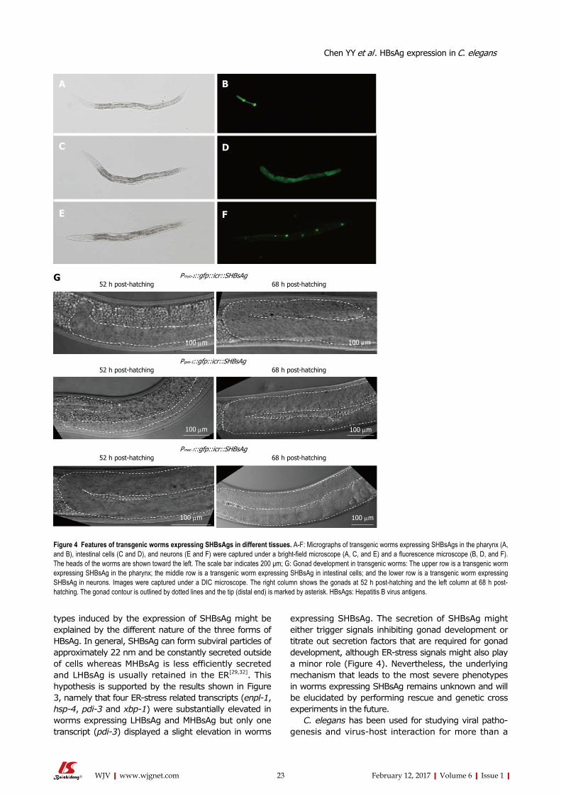
23 February 12, 2017|Volume 6|Issue 1|WJV|www.wjgnet.com
types induced by the expression of SHBsAg might be explained by the different nature of the three forms of HBsAg. In general, SHBsAg can form subviral particles of approximately 22 nm and be constantly secreted outside of cells whereas MHBsAg is less efficiently secreted and LHBsAg is usually retained in the ER[29,32]. This hypothesis is supported by the results shown in Figure 3, namely that four ER-stress related transcripts (enpl-1, hsp-4, pdi-3 and xbp-1) were substantially elevated in worms expressing LHBsAg and MHBsAg but only one transcript (pdi-3) displayed a slight elevation in worms
expressing SHBsAg. The secretion of SHBsAg might either trigger signals inhibiting gonad development or titrate out secretion factors that are required for gonad development, although ER-stress signals might also play a minor role (Figure 4). Nevertheless, the underlying mechanism that leads to the most severe phenotypes in worms expressing SHBsAg remains unknown and will be elucidated by performing rescue and genetic cross experiments in the future.
C. elegans has been used for studying viral patho-genesis and virus-host interaction for more than a
Pmyo-2::gfp::icr::SHBsAg52 h post-hatching 68 h post-hatching
Pges-1::gfp::icr::SHBsAg52 h post-hatching 68 h post-hatching
Pmec-7::gfp::icr::SHBsAg52 h post-hatching 68 h post-hatching
100 μm
100 μm
100 μm 100 μm
100 μm
100 μm
**
*
Figure 4 Features of transgenic worms expressing SHBsAgs in different tissues. A-F: Micrographs of transgenic worms expressing SHBsAgs in the pharynx (A, and B), intestinal cells (C and D), and neurons (E and F) were captured under a bright-field microscope (A, C, and E) and a fluorescence microscope (B, D, and F). The heads of the worms are shown toward the left. The scale bar indicates 200 μm; G: Gonad development in transgenic worms: The upper row is a transgenic worm expressing SHBsAg in the pharynx; the middle row is a transgenic worm expressing SHBsAg in intestinal cells; and the lower row is a transgenic worm expressing SHBsAg in neurons. Images were captured under a DIC microscope. The right column shows the gonads at 52 h post-hatching and the left column at 68 h post-hatching. The gonad contour is outlined by dotted lines and the tip (distal end) is marked by asterisk. HBsAgs: Hepatitis B virus antigens.
A B
C D
E F
G
Chen YY et al . HBsAg expression in C. elegans

24 February 12, 2017|Volume 6|Issue 1|WJV|www.wjgnet.com
decade[27,34]. In comparison with the number of pub-lications using C. elegans study viral, bacterial and fungal pathogenesis, relatively few papers have focused on viral pathogenesis and virus-host interaction in the past 10 years. The bottleneck could be due to the difficulty of creating transgenic worms expressing viral antigens. Currently, two methods for delivering ectopic genes into C. elegans are microinjection and gene bombardment, neither of which are easily achievable in general biology laboratories. To use C. elegans as a platform for studying virus-host interaction, virologists must collaborate with worm scientists. Alternatively, virologists could engineer the three viruses (Orsay, Santeui, and Le Blanc virus[35,36]), that naturally infect C. elegans to become versatile vectors for the easy deliver of different viral genes into worms through infection.
ACKNOWLEDGMENTSWe would like to thank Wallace Academic Editing for editing the manuscript, the Caenorhabditis Genetics Center for providing worm strains, and Kuei-Ching Hsiung for preparing the figures. This work is supported by grants from the Chang Gung Memorial Hospital (CMRPD1C0812, CMRPD1C0813 and BMRP742) to to Lo SJ.
COMMENTSBackgroundThe purpose of this research was to develop a model for human hepatitis B viral antigen interaction with host factors in the standard model animal Caenorhabditis elegans (C. elegans).
Research frontiersThe hepatitis B viral surface proteins (large, small and middle sized) (LHBsAg, MHBsAg, SHBsAg) are essential in viral assembly and infection. Here the three viral surface proteins were synthesized in C. elegans after microinjection of bacterial plasmids containing the genes for the proteins expressed under the control of a ubiquitous animal promoter. Severe reduction in egg laying and brood size as well as gonad retardation occurred with expression of the small hepatitis B virus (HBV) surface antigen in the worm. Smaller effects were found with the middle and larger sized surface antigens.
Innovations and breakthroughsThe specific effects of human HBV surface antigen expression of the C. elegans worm demonstrates the worm as a useful model for understanding viral infection and its effects on animal tissues.
ApplicationsSimple and rapid animal cell and model animal systems are essential for understanding of the infection process and disruptions caused by important human disease agents such as HBV. Here a new and useful model is established and its characteristics presented.
TerminologyHBV is the familiar abbreviation for human hepatitis B virus, although with HAV and HCV, a major cause of liver damage and morbidity. LHBsAg, MHBsAg and SHBsAg are the abbreviations for the large, middle-sized and small versions of the viral surface antigen, the proteins involved in the initial phase of virus surface attachment and infection.
Peer-reviewThe paper is well written.
REFERENCES1 Tiollais P, Pourcel C, Dejean A. The hepatitis B virus. Nature 1985;
317: 489-495 [PMID: 2995835 DOI: 10.1038/317489a0]2 Ganem D, Varmus HE. The molecular biology of the hepatitis B
viruses. Annu Rev Biochem 1987; 56: 651-693 [PMID: 3039907 DOI: 10.1146/annurev.bi.56.070187.003251]
3 Locarnini S, Littlejohn M, Aziz MN, Yuen L. Possible origins and evolution of the hepatitis B virus (HBV). Semin Cancer Biol 2013; 23: 561-575 [PMID: 24013024 DOI: 10.1016/j.semcancer.2013.08.006]
4 Brechot C, Kremsdorf D, Soussan P, Pineau P, Dejean A, Paterlini-Brechot P, Tiollais P. Hepatitis B virus (HBV)-related hepatocellular carcinoma (HCC): molecular mechanisms and novel paradigms. Pathol Biol (Paris) 2010; 58: 278-287 [PMID: 20667665 DOI: 10.1016/j.patbio.2010.05.001]
5 Beasley RP, Hwang LY, Lin CC, Chien CS. Hepatocellular carcinoma and hepatitis B virus. A prospective study of 22 707 men in Taiwan. Lancet 1981; 2: 1129-1133 [PMID: 6118576 DOI: 10.1016/S0140-67 36(81)90585-7]
6 Wang J, Chenivesse X, Henglein B, Bréchot C. Hepatitis B virus integration in a cyclin A gene in a hepatocellular carcinoma. Nature 1990; 343: 555-557 [PMID: 1967822 DOI: 10.1038/343555a0]
7 Wang J, Zindy F, Chenivesse X, Lamas E, Henglein B, Bréchot C. Modification of cyclin A expression by hepatitis B virus DNA integration in a hepatocellular carcinoma. Oncogene 1992; 7: 1653-1656 [PMID: 1321406]
8 Wang HC, Chang WT, Chang WW, Wu HC, Huang W, Lei HY, Lai MD, Fausto N, Su IJ. Hepatitis B virus pre-S2 mutant upregulates cyclin A expression and induces nodular proliferation of hepatocytes. Hepatology 2005; 41: 761-770 [PMID: 15726643 DOI: 10.1002/hep.20615]
9 Bouchard MJ, Schneider RJ. The enigmatic X gene of hepatitis B virus. J Virol 2004; 78: 12725-12734 [PMID: 15542625 DOI: 10.1128/JVI.78.23.12725-12734.2004]
10 Yen TT, Yang A, Chiu WT, Li TN, Wang LH, Wu YH, Wang HC, Chen L, Wang WC, Huang W, Chang CW, Chang MD, Shen MR, Su IJ, Wang LH. Hepatitis B virus PreS2-mutant large surface antigen activates store-operated calcium entry and promotes chromosome instability. Oncotarget 2016; 7: 23346-23360 [PMID: 26992221 DOI: 10.18632/oncotarget.8109]
11 Tennant BC, Toshkov IA, Peek SF, Jacob JR, Menne S, Hornbuckle WE, Schinazi RD, Korba BE, Cote PJ, Gerin JL. Hepatocellular carcinoma in the woodchuck model of hepatitis B virus infection. Gastroenterology 2004; 127: S283-S293 [PMID: 15508096 DOI: 10.1053/j.gastro.2004.09.043]
12 Gerin JL, Cote PJ, Korba BE, Tennant BC. Hepadnavirus-induced liver cancer in woodchucks. Cancer Detect Prev 1989; 14: 227-229 [PMID: 2695243]
13 Transy C, Fourel G, Robinson WS, Tiollais P, Marion PL, Buendia MA. Frequent amplification of c-myc in ground squirrel liver tumors associated with past or ongoing infection with a hepadnavirus. Proc Natl Acad Sci USA 1992; 89: 3874-3878 [PMID: 1570307 DOI: 10.1073/pnas.89.9.3874]
14 Hsu T, Möröy T, Etiemble J, Louise A, Trépo C, Tiollais P, Buendia MA. Activation of c-myc by woodchuck hepatitis virus insertion in hepatocellular carcinoma. Cell 1988; 55: 627-635 [PMID: 3180223 DOI: 10.1016/0092-8674(88)90221-8]
15 Wu BK, Li CC, Chen HJ, Chang JL, Jeng KS, Chou CK, Hsu MT, Tsai TF. Blocking of G1/S transition and cell death in the regenerating liver of Hepatitis B virus X protein transgenic mice. Biochem Biophys Res Commun 2006; 340: 916-928 [PMID: 16403455 DOI: 10.1016/j.bbrc.2005.12.089]
16 Chisari FV, Klopchin K, Moriyama T, Pasquinelli C, Dunsford HA, Sell S, Pinkert CA, Brinster RL, Palmiter RD. Molecular pathogenesis of hepatocellular carcinoma in hepatitis B virus transgenic mice. Cell 1989; 59: 1145-1156 [PMID: 2598264 DOI: 10.1016/0092-8674(89)90770-8]
17 Kim A, Kwon OS, Kim SO, He L, Bae EY, Lee MS, Jeong SJ, Shim JH, Yoon DY, Kim CH, Moon A, Kim KE, Ahn JS, Kim BY. Caspase-3 activation as a key factor for HBx-transformed cell death.
COMMENTS
Chen YY et al . HBsAg expression in C. elegans

25 February 12, 2017|Volume 6|Issue 1|WJV|www.wjgnet.com
Cell Prolif 2008; 41: 755-774 [PMID: 18700866 DOI: 10.1111/j.1365-2184.2008.00550.x]
18 Zhang XD, Wang Y, Ye LH. Hepatitis B virus X protein accelerates the development of hepatoma. Cancer Biol Med 2014; 11: 182-190 [PMID: 25364579]
19 Geng X, Harry BL, Zhou Q, Skeen-Gaar RR, Ge X, Lee ES, Mitani S, Xue D. Hepatitis B virus X protein targets the Bcl-2 protein CED-9 to induce intracellular Ca2+ increase and cell death in Caenorhabditis elegans. Proc Natl Acad Sci USA 2012; 109: 18465-18470 [PMID: 23091037 DOI: 10.1073/pnas.1204668109]
20 Brenner S. The genetics of Caenorhabditis elegans. Genetics 1974; 77: 71-94 [PMID: 4366476]
21 Ankeny RA. The natural history of Caenorhabditis elegans research. Nat Rev Genet 2001; 2: 474-479 [PMID: 11389464 DOI: 10.1038/35076538]
22 Diogo J, Bratanich A. The nematode Caenorhabditis elegans as a model to study viruses. Arch Virol 2014; 159: 2843-2851 [PMID: 25000902 DOI: 10.1007/s00705-014-2168-2]
23 Kurz CL, Ewbank JJ. Caenorhabditis elegans: an emerging genetic model for the study of innate immunity. Nat Rev Genet 2003; 4: 380-390 [PMID: 12728280 DOI: 10.1038/nrg1067]
24 Culetto E, Sattelle DB. A role for Caenorhabditis elegans in understanding the function and interactions of human disease genes. Hum Mol Genet 2000; 9: 869-877 [PMID: 10767309 DOI: 10.1093/hmg/9.6.869]
25 Olsen A, Vantipalli MC, Lithgow GJ. Using Caenorhabditis elegans as a model for aging and age-related diseases. Ann N Y Acad Sci 2006; 1067: 120-128 [PMID: 16803977 DOI: 10.1196/annals.1354.015]
26 Zarski JP, Kuhns M, Berck L, Degos F, Schalm SW, Tiollais P, Bréchot C. Comparison of a quantitative standardized HBV-DNA assay and a classical spot hybridization test in chronic active hepatitis B patients undergoing antiviral therapy. Res Virol 1989; 140: 283-291 [PMID: 2772413 DOI: 10.1016/S0923-2516(89)80108-6]
27 Liu WH, Lin YL, Wang JP, Liou W, Hou RF, Wu YC, Liao CL. Restriction of vaccinia virus replication by a ced-3 and ced-4-dependent pathway in Caenorhabditis elegans. Proc Natl Acad
Sci USA 2006; 103: 4174-4179 [PMID: 16537504 DOI: 10.1073/pnas.0506442103]
28 Lee LW, Chang TY, Lo HW, Lo SJ. Hepatitis D antigens cause growth retardation and brood-size reduction in C. elegans. Front Biosci (Elite Ed) 2011; 3: 380-390 [PMID: 21196318]
29 Sheu SY, Lo SJ. Biogenesis of the hepatitis B viral middle (M) surface protein in a human hepatoma cell line: demonstration of an alternative secretion pathway. J Gen Virol 1994; 75 (Pt 11): 3031-3039 [PMID: 7964612 DOI: 10.1099/0022-1317-75-11-3031]
30 Lee LW, Lo HW, Lo SJ. Vectors for co-expression of two genes in Caenorhabditis elegans. Gene 2010; 455: 16-21 [PMID: 20149852 DOI: 10.1016/j.gene.2010.06.007]
31 Safra M, Ben-Hamo S, Kenyon C, Henis-Korenblit S. The ire-1 ER stress-response pathway is required for normal secretory-protein metabolism in C. elegans. J Cell Sci 2013; 126: 4136-4146 [PMID: 23843615 DOI: 10.1242/jcs.123000]
32 Hung JH, Su IJ, Lei HY, Wang HC, Lin WC, Chang WT, Huang W, Chang WC, Chang YS, Chen CC, Lai MD. Endoplasmic reticulum stress stimulates the expression of cyclooxygenase-2 through activation of NF-kappaB and pp38 mitogen-activated protein kinase. J Biol Chem 2004; 279: 46384-46392 [PMID: 15319438 DOI: 10.1074/jbc.M403568200]
33 Hubbard EJ, Greenstein D. The Caenorhabditis elegans gonad: a test tube for cell and developmental biology. Dev Dyn 2000; 218: 2-22 [PMID: 10822256]
34 Lu R, Maduro M, Li F, Li HW, Broitman-Maduro G, Li WX, Ding SW. Animal virus replication and RNAi-mediated antiviral silencing in Caenorhabditis elegans. Nature 2005; 436: 1040-1043 [PMID: 16107851 DOI: 10.1038/nature03870]
35 Jiang H, Franz CJ, Wang D. Engineering recombinant Orsay virus directly in the metazoan host Caenorhabditis elegans. J Virol 2014; 88: 11774-11781 [PMID: 25078701 DOI: 10.1128/JVI.01630-14]
36 Franz CJ, Renshaw H, Frezal L, Jiang Y, Félix MA, Wang D. Orsay, Santeuil and Le Blanc viruses primarily infect intestinal cells in Caenorhabditis nematodes. Virology 2014; 448: 255-264 [PMID: 24314656 DOI: 10.1016/j.virol.2013.09.024]
P- Reviewer: Ghiringhelli PD, Liu ZW, Striker R S- Editor: Ji FF L- Editor: A E- Editor: Lu YJ
Chen YY et al . HBsAg expression in C. elegans

© 2017 Baishideng Publishing Group Inc. All rights reserved.
Published by Baishideng Publishing Group Inc8226 Regency Drive, Pleasanton, CA 94588, USA
Telephone: +1-925-223-8242Fax: +1-925-223-8243
E-mail: [email protected] Desk: http://www.wjgnet.com/esps/helpdesk.aspx
http://www.wjgnet.com