world journal of surgical oncology - home - springer · biomed central page 1 of 4 (page number not...
TRANSCRIPT

BioMed Central
World Journal of Surgical Oncology
ss
Open AcceCase reportMesenteric desmoid tumor of the interposed jejunal pouch after total gastrectomyKoichi Tamura, Masaji Tani*, Hiroyuki Kinoshita and Hiroki YamaueAddress: Second Department of Surgery, Wakayama Medical University, School of Medicine, Wakayama, Japan
Email: Koichi Tamura - [email protected]; Masaji Tani* - [email protected]; Hiroyuki Kinoshita - [email protected]; Hiroki Yamaue - [email protected]
* Corresponding author
AbstractBackground: Desmoid tumor is a rare entity, and most desmoid tumors are located in abdominalwall or extra-abdominal tissues. Occurrence of desmoid tumor in mesentry is extremely rare.
Case presentation: we report a mesenteric desmoid tumor in a 73-years-old woman who hadundergone total gastrectomy reconstructed with jejunal pouch interposition for gastric carcinoma.After 1 year, a tumor was originating from mesentery of the interposed jejunal pouch wasidentified, and the patient underwent resection of the large mass which was found to invadepancreas. Histological examination revealed desmoid tumor.
Conclusion: Desmoid tumor is rare, and it was difficult for the differential diagnosis of desmoidtumor or recurrent tumor.
BackgroundDesmoid is derived from the Greek word "desmos",meaning band-like. The tumors are defined as benignfibrous tissue tumors arising in the musculoaponeuroticstructures throughout the body. There is no report ofmetastasis for desmoid tumor, however, desmoid tumorsometimes shows locally invasive growth [1]. Histologicalexamination usually shows uniformed mature fibroblastsin both size and shape without karyomitosis [2]. Anannual incidence of desmoid is rare, and it is reportedonly in 2–4 cases per 1 million populations [3]. Moreover,desmoid tumors commonly occur as an extracolonicmanifestation of familial adenomatous polyposis (FAP),especially Gardner syndrome [4]. There is no report ofintra-abdominal desmoid tumor after total gastrectomy,originating from the interposed jejunal pouch.
Case presentationA 73-years-old woman had undergone total gastrectomyand D2 lymphadenectomy for gastric carcinoma, whichwas pathologically diagnosed as stage I (T1, N1, H0, P0,M0, CY0) by Japanese Classification of Gastric Carcinoma2nd English edition[5]. The patient was followed-up bycomputed tomography (CT), and there was no tumor at 1year after the total gastrectomy. However, another yearlater, she was admitted to Wakayama Medical Universityhospital because of asymptomatic intra-abdominaltumor. Laboratory investigations revealed a hemoglobin(Hb) of 11.7 g/dl, while the levels of serum carcinoembry-onic antigen (CEA), carbohydrate antigen 125 (CA125)and carbohydrate antigen 19-9 (CA19-9) were within nor-mal range. CT showed a 50 × 65 mm round-shaped solidmass at the right side of the reconstructed jejunal pouch,which showed weak enhancement at arterial phase bycontrast medium and local invasion into pancreas body
Published: 01 June 2006
World Journal of Surgical Oncology 2006, 4:27 doi:10.1186/1477-7819-4-27
Received: 13 December 2005Accepted: 01 June 2006
This article is available from: http://www.wjso.com/content/4/1/27
© 2006 Tamura et al; licensee BioMed Central Ltd.This is an Open Access article distributed under the terms of the Creative Commons Attribution License (http://creativecommons.org/licenses/by/2.0), which permits unrestricted use, distribution, and reproduction in any medium, provided the original work is properly cited.
Page 1 of 4(page number not for citation purposes)

World Journal of Surgical Oncology 2006, 4:27 http://www.wjso.com/content/4/1/27
(Figure. 1). Magnetic resonance imaging (MRI) showed alow signal intensity of the mass in both T1-weighted andT2-weighted images (Figure. 2). Abdominal ultrasoundrevealed a hypoechoic mass without blood-flow.
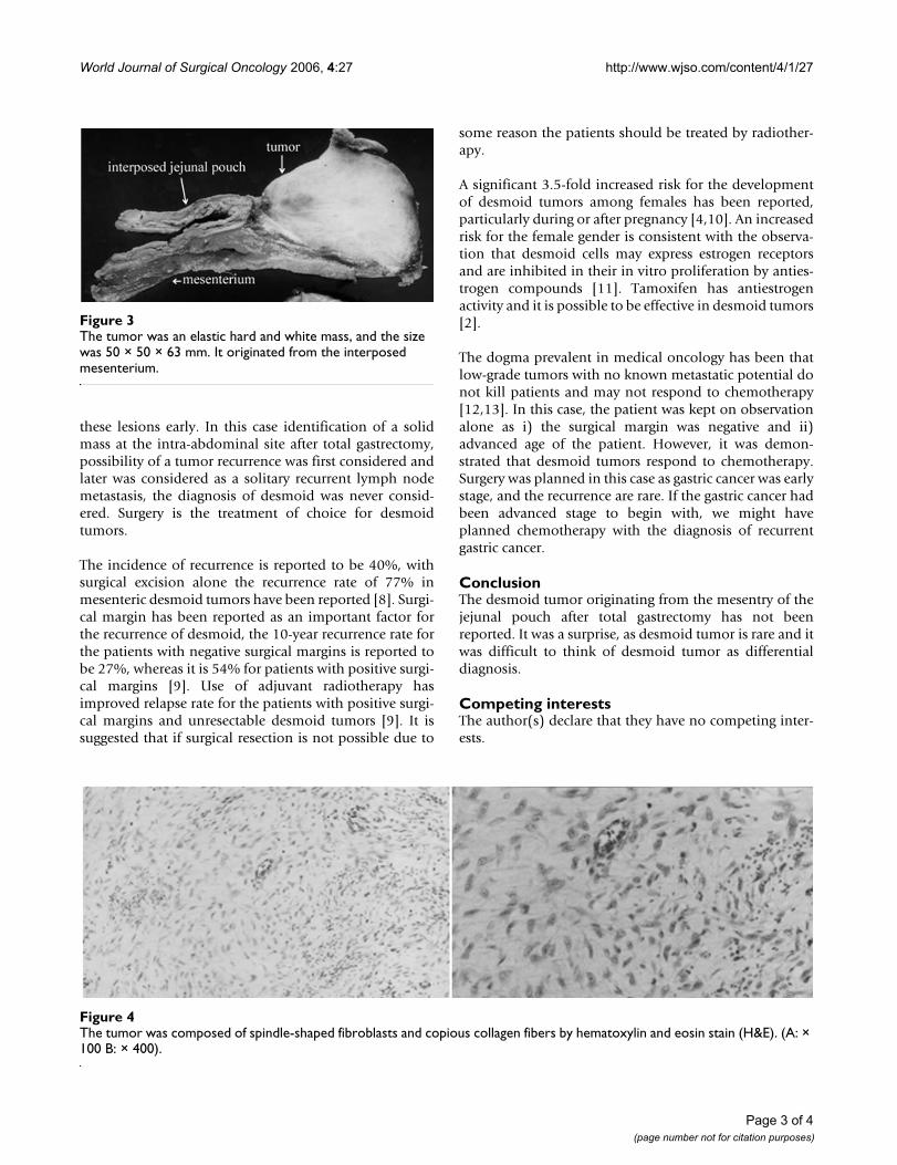
At laparotomy, a 50 × 50 × 63 mm tumor was found to belocated in the mesentery of the jejunal pouch which wasused for reconstruction after total gastrectomy the mass
was adhered to the pancreas and it was possible to sepa-rate it from pancreas. The tumor was excised and thereconstruction was performed by double tract methodwith esphagojejunostomy and jejunoduodenostomy. Theresected specimen was shown in Figure. 3. Microscopi-cally, the tumor was composed of spindle-shaped fibrob-lasts and copious collagen fibers by hematoxylin andeosin stain. Collagen fibers were positive for immunolog-ical staining of α-smooth muscle actin, confirming adesmoid tumor (Figure. 4).
She is presently doing well and has no sign of any recur-rent tumor 4 years after the operation.
DiscussionSurgical trauma, which is one of the most important etio-logic factors for desmoid, can induce desmoid growth [6].However, desmoid tumor originating from the jejunalpouch, which had been interposed for reconstructionafter total gastrectomy for improvement of early postoper-ative eating capacity, body weight and quality of life hasnot been reported before. The desmoid tumor originatedfrom mesentery of reconstructed jejunal pouch, which hasnot been affected surgical manipulation. Although anas-tomotic leakage, abscess, wound infection, and fistula arethe known complications of total gastrectomy, occurrenceof desmoid tumor is rare [7]. In our case, there were nopostoperative complications, and there was no intra-abdominal inflammatory lesion that could have led toformation of desmoid tumor.
Mesenteric desmoid tumor is rare and has few symptomsassociated with this tumor, thus it is difficult to diagnose
Computed tomography (CT) shows round-shaped solid mass at the right side of the reconstructed jejunal pouch, and invaded into pancreas (arrow)Figure 1Computed tomography (CT) shows round-shaped solid mass at the right side of the reconstructed jejunal pouch, and invaded into pancreas (arrow).
Magnetic resonance imaging (MRI) showed a low signal intensity of the mass in both T1-weighted (left) and T2-weighted images (right)Figure 2Magnetic resonance imaging (MRI) showed a low signal intensity of the mass in both T1-weighted (left) and T2-weighted images (right).
Page 2 of 4(page number not for citation purposes)
World Journal of Surgical Oncology 2006, 4:27 http://www.wjso.com/content/4/1/27
these lesions early. In this case identification of a solidmass at the intra-abdominal site after total gastrectomy,possibility of a tumor recurrence was first considered andlater was considered as a solitary recurrent lymph nodemetastasis, the diagnosis of desmoid was never consid-ered. Surgery is the treatment of choice for desmoidtumors.
The incidence of recurrence is reported to be 40%, withsurgical excision alone the recurrence rate of 77% inmesenteric desmoid tumors have been reported [8]. Surgi-cal margin has been reported as an important factor forthe recurrence of desmoid, the 10-year recurrence rate forthe patients with negative surgical margins is reported tobe 27%, whereas it is 54% for patients with positive surgi-cal margins [9]. Use of adjuvant radiotherapy hasimproved relapse rate for the patients with positive surgi-cal margins and unresectable desmoid tumors [9]. It issuggested that if surgical resection is not possible due to
some reason the patients should be treated by radiother-apy.
A significant 3.5-fold increased risk for the developmentof desmoid tumors among females has been reported,particularly during or after pregnancy [4,10]. An increasedrisk for the female gender is consistent with the observa-tion that desmoid cells may express estrogen receptorsand are inhibited in their in vitro proliferation by anties-trogen compounds [11]. Tamoxifen has antiestrogenactivity and it is possible to be effective in desmoid tumors[2].
The dogma prevalent in medical oncology has been thatlow-grade tumors with no known metastatic potential donot kill patients and may not respond to chemotherapy[12,13]. In this case, the patient was kept on observationalone as i) the surgical margin was negative and ii)advanced age of the patient. However, it was demon-strated that desmoid tumors respond to chemotherapy.Surgery was planned in this case as gastric cancer was earlystage, and the recurrence are rare. If the gastric cancer hadbeen advanced stage to begin with, we might haveplanned chemotherapy with the diagnosis of recurrentgastric cancer.
ConclusionThe desmoid tumor originating from the mesentry of thejejunal pouch after total gastrectomy has not beenreported. It was a surprise, as desmoid tumor is rare and itwas difficult to think of desmoid tumor as differentialdiagnosis.
Competing interestsThe author(s) declare that they have no competing inter-ests.
The tumor was composed of spindle-shaped fibroblasts and copious collagen fibers by hematoxylin and eosin stain (H&E)Figure 4The tumor was composed of spindle-shaped fibroblasts and copious collagen fibers by hematoxylin and eosin stain (H&E). (A: × 100 B: × 400).
The tumor was an elastic hard and white mass, and the size was 50 × 50 × 63 mmFigure 3The tumor was an elastic hard and white mass, and the size was 50 × 50 × 63 mm. It originated from the interposed mesenterium.
Page 3 of 4(page number not for citation purposes)

World Journal of Surgical Oncology 2006, 4:27 http://www.wjso.com/content/4/1/27
Publish with BioMed Central and every scientist can read your work free of charge
"BioMed Central will be the most significant development for disseminating the results of biomedical research in our lifetime."
Sir Paul Nurse, Cancer Research UK
Your research papers will be:
available free of charge to the entire biomedical community
peer reviewed and published immediately upon acceptance
cited in PubMed and archived on PubMed Central
yours — you keep the copyright
Submit your manuscript here:http://www.biomedcentral.com/info/publishing_adv.asp
BioMedcentral
Authors' contributionsTamura, K: Preparation of manuscript, Collection of clin-ical data, Operation
Tani, M: Proofreading of manuscript, Operation, Collec-tion of clinical data
Kinoshita H: Collection of data, Operation
Yamaue, H: Proofreading of manuscript
AcknowledgementsWritten consent of the patient was obtained for publication of his case report.
References1. Naylor EW, Gardner EJ, Richards RC: Desmoid tumors and
mesenteric fibromatosis in Gardner's syndrome:report ofkidred 109. Arch Surg 1979, 114:1181-1185.
2. Anika H, Claudia A, Tilmann V, Andreas U, Gabriela M: High-dosetamoxifen and sulindac as fiest-line treatment for desmoidtumors. Cancer 2004, 100:612-620.
3. Reitamo JJ, Hayry P, Nykyri E, Saxen E: The desmoid tumor. Inci-dence, sex, age and anatomical distribution in the Finnishpopulation. Am J Clin Pathol 1982, 77:665-673.
4. Rodriguez-Bigas MA, Mahoney MC, Karakousis CP, Petrelli NJ:Desmoid tumors in patients with familial adenomatous poly-posis. Cancer 1994, 74:1270-1274.
5. Japanese Gastric Cancer Association: Japanese Classification of GastricCarcinoma 2nd edition. Tokyo: Kanehara & Co., Ltd.; 1998.
6. Friedl W, Caspari R, Sengteller R, Uhlhaas S, Lamberti C, Jungck M,Kadmon M, Wolf M, Fahnenstich J, Gebert J, Moslein G, Mangold E,Propping P: Can APC mutation analysis contribute to thera-peutic decisions in familial adenomatous polyposis ? Experi-ence from 680 FAP families. Gut 2001, 48:515-521.
7. Smith JW, Shiu MH, Kelsey L: Morbidity of radical lymphadenec-tomy in the curative resection of gastric carcinoma. Arch Surg1981, 126:1469-1473.
8. Easter DW, Halasz NA: Recent trends in the management ofdesmoid tumors. Summary of 19 cases and review of the lit-erature. Ann Surg 1989, 210:765-769.
9. Ballo MT, Zagars GK, Pollack A: Desmoid tumor: prognostic fac-tors and outcome after surgery, radiation therapy, or com-bined surgery and radiation therapy. J Clin Oncol 1999,17:158-167.
10. Clark SK, Phillips RK: Desmoids in familial adenomatous poly-posis. Br J Surg 1996, 83:1494-1504.
11. Lim CL, Walker MJ, Mehta RR, Das Gupta TK: Estrogen and anti-estrogen binding sites in desmoid tumors. Eur J Cancer ClinOncol 1986, 22:583-587.
12. Patel SR, Benjamin RS: Desmoid tumors respond to chemother-apy: deflying the dogma in oncology. J Clin Oncol 2006, 24:11-12.
13. Papagelopoulos PJ, Mavrogenis AF, Mitsiokapa EA, PapaparaskevaKTH, Galanis EC, Soucacos PN: Current trends in the manage-ment of extra-abdominal desmoid tumours. World J Surg Oncol2006 in press.
Page 4 of 4(page number not for citation purposes)