worthy regarding - microbiology and molecular biology …mmbr.asm.org/content/4/4/227.full.pdf ·...
TRANSCRIPT
NON-SPOREFORMING ANAEROBIC BACTERIA OFMEDICAL IMPORTANCE
G. M. DACKFrom the Department of Bacteriology and Parasitology, University of Chicago
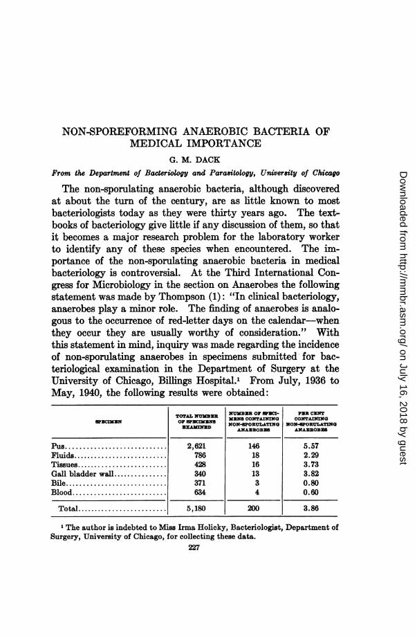
The non-sporulating anaerobic bacteria, although discoveredat about the turn of the century, are as little known to mostbacteriologists today as they were thirty years ago. The text-books of bacteriology give little if any discussion of them, so thatit becomes a major research problem for the laboratory workerto identify any of these species when encountered. The im-portance of the non-sporulating anaerobic bacteria in medicalbacteriology is controversial. At the Third International Con-gress for Microbiology in the section on Anaerobes the followingstatement was made by Thompson (1): "In clinical bacteriology,anaerobes play a minor role. The finding of anaerobes is analo-gous to the occurrence of red-letter days on the calendar-whenthey occur they are usually worthy of consideration." Withthis statement in mind, inquiry was made regarding the incidenceof non-sporulating anaerobes in specimens submitted for bac-teriological examination in the Department of Surgery at theUniversity of Chicago, Billings Hospital., From July, 1936 toMay, 1940, the following results were obtained:
TOTAL NMBDt NMBEIR OF SPECK- PER CETQzcimm opgtcnTOTAOF MENS CONTAINING CONTAINING
NAOSIMENSD NON-SPORULATING NON-PORULATINGANAUROBES ANAUROBES
Pus....................... 2,621 146 5.57Fluids....................... 786 18 2.29Tissues....................... 428 16 3.73Gall bladder wall............... 340 13 3.82Bile....................... 371 3 0.80Blood....................... 634 4 0.60
Total....................... 5,180 200 3.86
1 The author is indebted to Miss Irma Holicky, Bacteriologist, Department ofSurgery, University of Chicago, for collecting these data.
227
on July 16, 2018 by guesthttp://m
mbr.asm
.org/D
ownloaded from
G. M. DACK
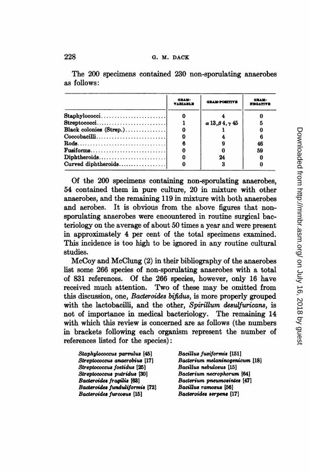
The 200 specimens contained 230 non-sporulating anaerobesas follows:
Gx-GRAM- PO N GRAM-VARM-PAZT!VD NUGATTVN
Staphylococci........................ 0 4 0Streptococci.......................... 1 a13,0 4, y45 5Black colonies (Strep.)............... 0 1 0Coccobacilli .......................... 4 6Rods............................ 6 9 46Fusiforms............................ 0 0 59Diphtheroids......................... 0 24 0Curved diphtheroids.................. 0 3 0
Of the 200 specimens containing non-sporulating anaerobes,54 contained them in pure culture, 20 in mixture with otheranaerobes, and the remaining 119 in mixture with both anaerobesand aerobes. It is obvious from the above figures that non-sporulating anaerobes were encountered in routine surgical bac-teriology on the average of about 50 times a year and were presentin approximately 4 per cent of the total specimens examined.This incidence is too high to be ignored in any routine culturalstudies.McCoy and McClung (2) in their bibliography of the anaerobes
list some 266 species of non-sporulating anaerobes with a totalof 831 references. Of the 266 species, however, only 16 havereceived much attention. Two of these may be omitted fromthis discussion, one, Bacteroides bifidus, is more properly groupedwith the lactobacilli, and the other, Spirillum desulfuricars, isnot of importance in medical bacteriology. The remaining 14with which this review is concerned are as follows (the numbersin brackets following each organism represent the number ofreferences listed for the species):
Staphylococcus parvulus (45] Bacillus fusiformis [151]Streptococcus anaerobius [17] Baderium melaninogenicum [18]Streptococcus foetidus [25] Bacillus nebulosus (15]Streptococcus putridus [30] Bacterium necrophorum [64]Bacteroidee fragilis [63] Bacterium pneumosintes [47]Baderoide funduliformis (72] BaciUus ramosus [56]Bacteroides furcosus (15] Bacteroides serpen [17]
228
on July 16, 2018 by guesthttp://m
mbr.asm
.org/D
ownloaded from
NON-SPOREFORMING ANAEROBIC BACTERIA
Several generic names are cited for a single species, which isin keeping with the confusion now existing in bacteriologicalnomenclature. Recently Pr6vot (3) has proposed new genericnames for the anaerobes. Bergey's Manual (3a) classifies thegram-negative members of this group under the generic name ofBacteroides. Since these organisms are so different in morphologyand physiology, it seems absurd to group them under one genus.In this review mention will be made of some of the generic andspecific names applied to these micro6rganisms, but no attemptwill be made to name them. As will be pointed out subsequently,we do not know enough about many of them to assign them tospecial genera.Most of the work on the non-sporeforming anaerobes has been
done by clinicians, who have isolated and described new species.The descriptions of many species have been so inadequate thatthe same organism may be again discovered by another investi-gator and given a new name. Another difficulty is that no twoworkers will use the same technique in isolation and identification,thus affording no comparative basis for the examination of newlyisolated strains. In many hospitals no effort is made to look forthis group of organisms, hence the laboratory diagnosis of "sterilepus" is made on specimens from abscesses, if no growth appearson aerobic blood-agar plates.
Nearly all the non-sporulating anaerobes of medical importanceare normal inhabitants of the mucous membranes of the body,inhabiting the upper respiratory tract, the colon and the genitaltract. Under conditions which give rise to necrosis of the mucousmembranes one or more of these species may become establishedand invade the tissue. It is when they produce abscesses orenter the blood stream that they are usually detected. Theyare not found at their original portal of entry because of poormethods for their isolation. Most workers in bacteriology seemto think that a search for anaerobes is too arduous a task toattempt, although with modern apparatus it is possible to isolateand study anaerobes in relatively convenient routine fashion.In the author's experience the early method proposed by Veillonfor the isolation of anaerobes is entirely inadequate when the
229
on July 16, 2018 by guesthttp://m
mbr.asm
.org/D
ownloaded from
G. M. DACK
anaerobe sought is greatly outnumbered by other bacteria. Nooriginality is claimed for the method used successfully in thislaboratory for many years. The method is as follows: A blood-agar plate containing 10 per cent of sheep blood is streaked withspecimens to be examined. When the specimen is heavily con-taminated a second plate is streaked from the first. Anaerobicconditions are provided by using ordinary pyrex desiccatorswithground glass stoppers. The ground joints of the jar are sealedwith a preparation made of equal parts of rubber, paraffin, andvaseline. This sealing preparation is effective at incubator tem-peratures. The plates after streaking are placed in the jar overa solution of pyrogallic acid and sodium carbonate. The jar isclosed and evacuated with an oil pump to approximately 10 cm.mercury pressure. Carbon dioxide is allowed to flow into thejar until atmospheric pressure is reached, after which the jaris again exhausted to 25 cm. mercury pressure and is then sealedand placed in the incubator. Oxygen dissolved in the blood agaris absorbed by the sodium pyrogallate. The blood-agar assumesa cyanotic color when good anaerobic conditions are attained,thus serving as an indicator of the relative absence of oxygen inthe jar. Non-sporulating anaerobes of diverse types have beensuccessfully isolated by this simple procedure.
ANAEROBIC COCCI OF MEDICAL IMPORTANCE
Numerous species of anaerobic cocci have been described, butonly four have been sufficiently studied to warrant space in thisreview.
Staphylococcus parvulus. This organism has also been listedunder the genus Micrococcus, although Weinberg et al. (4) haveplaced it in a genus Veillonella, because it is gram-negative anddiffers from the members of the genus Neisseria in having aninterstitial substance (ectoplasm) which may be demonstratedby Giemsa's stain. This organism was first described by Veillonand Zuber (5), who claimed to have found it in pus in appendicitis,either in the abscesses about the cecum or in the peritonealcavity in a generalized peritonitis. Subcutaneous abscesses havebeen produced in rabbits and guinea pigs with these strains.
230
on July 16, 2018 by guesthttp://m
mbr.asm
.org/D
ownloaded from
NON-SPOREFORMING ANAEROBIC BACTERIA
These workers noted the predominance of this organism overEscherichia coli. All cultures produce gas and a very foul odor.Gelatin is not liquefied and milk is not affected. Glucose isfermented and certain strains apparently attack other carbo-hydrates (3, 4), although there seems to be considerable irregu-larity in this respect.
Streptococcus anaerobius and Streptococcus foetidus. These twospecies are listed together, since the description of their activityin different media is identical. The only difference ascribed tothem is in morphology, and with the recognition of variationamong other species of bacteria, it does not seem at the presenttime that this difference is in itself adequate to separate theseas two distinct species. Streptococcus anaerobius is describedas having long regular chains, whereas Streptococcus foetidusappears in short chains and irregular arrangement with occa-sional tetrads. S. anaerobius was first described by Kronig andMenge (6) and S. foetidus by Veillon (7).Both organisms are gram-positive and are generally found in
the oral cavity, intestine and vagina in abundance. They pro-duce gas and foul odors in all media. Gelatin is not liquefiedand neither milk nor heat-coagulated proteins are affected, al-though good growth occurs in serum and a very foul odor results.In man, the organisms have been found in purulent gangrenousprocesses involving the genital tract, lungs and viscera, and insepticemias. They do not produce strong toxins, neither do theycause hemolysis. There seems to be a difference of opinion amongworkers regarding their pathogenicity for animals. Pr6vot (3)claims that local edematous lesions with gangrenous suppurationhave been produced in guinea pigs with S. foetidus.
Streptococcus putridus. Schottmiiller (8) first described Strep-tococcus putridus and reported 25 cases in which it was found inthe lesions. These cases comprised meningitis, cystopyelitis,gangrene of the lung, salpingitis with pelvic abscesses, and septicabortion (ten cases, of which eight had thrombophlebitis withother complications). S. putridus was isolated in pure culturein 12 of the cases, and found along with other organisms inthe rest.
231
on July 16, 2018 by guesthttp://m
mbr.asm
.org/D
ownloaded from
G. M. DACK
Schottmiiller observed that on artificial media long and shortchains were present and the individual organisms were usuallyflattened and appeared attached as diplococci. In old cultureshe noticed different shapes, with some cells appearing as rods.The strains were all gram-positive. S. putridus was often iso-lated from blood by placing the blood immediately in bouillonand incubating it without mixing or further handling. Onblood-agar plates the colonies were porcelain white in color, andof the size of the head of a needle.The main characteristic by which S. putridus may be differ-
entiated from other anaerobic streptococci is the reaction in blood-broth cultures. The blood takes on a characteristic poppy-redcolor; and spectroscopically H2S may be demonstrated in bloodcultures. In about ten days blood-broth cultures are black incolor. Schottmuiller believed S. putridus was not a simplesaprophyte, since it was able to invade and produce lesions else-where in the body. He called attention to the ability of thisorganism to dissolve fibrin, since in the pleural exudates onenever finds the slightest trace of fibrin clots.
S. putridus is strictly anaerobic. Heat-coagulated proteinmedia are not attacked, whereas sterile protein solutions such asserum are attacked and give rise to a foul odor. Glucose,levulose and maltose are fermented (3).
Since SchottmUller's original description of S. putridus in1910 (8) there has been little attention given this organism. Afew reports have appeared, some of which will be mentioned.Schwarz and Dieckmann (9, 10) in St. Louis made a search forthis organism in puerperal infections. Of 165 uterine culturesand blood cultures from suspected cases, they found that 46contained S. putridus (9). More recently Stone (11) has studied26 strains of anaerobic streptococci, which he isolated fromparturient and post-abortal women by means of a sterile pipetteinserted through the cervix into the uterus. The organismswere found to be gram-variable. Stone attempted to applysome of the tests used in differentiating aerobic streptococciof the beta-hemolytic type, such as growth in 10 and 40 percent bile, final pH in glucose medium, hydrolysis of sodium
232
on July 16, 2018 by guesthttp://m
mbr.asm
.org/D
ownloaded from
NON-SPOREFORMING ANAEROBIC BACTERIA
hippurate, fermentation of trehalose and sorbitol, and finallythe precipitin reactions. He found that it was impossible toset up definite groups of these strains by means of their precipitinreactions. Stone made no attempt to differentiate S. putridusfrom other anaerobic streptococci by its reaction in blood-broth,as recommended by Schottmiiller (8).
ANAEROBIC NON-SPORULATING GRAM-NEGATIVE RODS
Organisms of this group have been placed in many genera.Castellani and Chalmers (12) have listed a Tribe Bacteroideaein the Family BACILLACEAE with the following description:"Bacillaceae with good growth on ordinary laboratory media,without endospores, fluorescence, or pigment formation, andobligatory anaerobes." They named a genus Bacteroides withthese tribal characters and Bacteroides fragilis was given as thetype species. This generic name, Bacteroides, includes bothgram-negative and gram-positive species, although it is pro-posed (13) that it be restricted to the gram-negative species.From the author's experience with the gram-negative non-
sporulating organisms, it seems unwise to place all of these in asingle genus, since they represent greatly different morpholog-ical types. In this review, however, these organisms will belisted under the names appearing in the McCoy and McClungbibliography (2).
Bacteroides fragilisPr6vot (3) has listed this organism in a genus which he calls
Ristella, and defines as containing asporulating simple rods,non-ciliated, non-motile, straight or slightly curved, non-capsu-lated and gram-negative. Under this genus he has listed 25species. Topley and Wilson (14) list this organism in a genusFusiformis, which they characterize as "obligate parasites,anaerobic or microaerophilic. Cells frequently elongated andfusiform, staining somewhat unevenly. Filaments sometimesformed; non-branching. Non-motile. No spores. Reaction toGram variable. Growth in laboratory media feeble." Veillonand Zuber (5) called this organism Bacillus fragilis. In addition
233
on July 16, 2018 by guesthttp://m
mbr.asm
.org/D
ownloaded from
G. M. DACK
to having four different generic names, Bacillu8, Bacteroides,Fusiformis and Ristella, it has another species name: sassmanns-hausen, given it by Heyde in 1911 (4).The description given this organism by Veillon and Zuber
(5), who found it in 22 cases of appendicitis, may be translatedas follows:
"This bacillus appears to us to be the most abundant and the mostconstant in the pus from appendicitis. It is a fine rod, a little smallerthan that of diphtheria, rounded at the ends and regular. It presentsitself in the form of rods, isolated or united two by two by one of theirextremities. Sometimes certain bacilli are slightly curved. In culturethey have the same appearance, although they appear a little largerand certain rods are longer. It is gram-negative and non-motile. Al-though this bacillus is mi great abundance in the pus from appendicitis,it is difficult to isolate. At incubator temperature the colonies do notappear in the depth of the agar until the third or fourth day. Theyform little round or slightly irregular colonies, ovoid, brownish yellow,rather opaque with smooth borders. These colonies remain discreteand although they are not far separate, one from the other, they remainpunctiform. The most isolated colonies are less than one millimeter indiameter, and it is necessary to tramnsplant them as soon as they areevident because they die quickly. A culture left 7 or 8 days in theincubator is no longer viable. On agar at the surface this organismforms extremely fine colonies, very transparent, grayish, scarcely moremarked than those of pneumococcus and, like those, at the end of severaldays they become less visible and seem to be reabsorbed.
"Cultures can be obtained on gelatin at room temperature. Thecolonies which appear at the end of 8-10 days are punctiform, yellowishgranules with wet edges. The medium is not liquefied. The culture isviable for 20-30 days. In broth growth occurs easily and in relativeabundance. The medium is uniformly cloudy and there is a fine whitishdeposit at the bottom of the receptacle. The cultures do not give offenough gas to break up the agar, but do give off a fetid odor. We havenot established the production of spores and, as we have said, this bacil-lus is very fragile and non-resistant. It is pathogenic for guinea pigsand forms abscesses when injected subcutaneously. If the animal doesnot die of its abscess, the pus is eliminated and the guinea pig becomescachectic and dies in about a month. This bacillus is much morevirulent for rabbits. By subcutaneous inoculation there is produced a
234
on July 16, 2018 by guesthttp://m
mbr.asm
.org/D
ownloaded from
NON-SPOREFORMING ANAEROBIC BACTERIA
large phlegmon with separation of the skin and death in 6 to 7 days.An inoculation in the veins produces death by cachexia; but one isunable to find the bacilli in the body. It is probable that toxins arethe agents in this case, because one obtains the same results with deadcultures."
This description given by Veillon and Zuber is useful in theisolation and identification of the orgaiism. Cohen (15) listedthis organism in 4 out of 16 cases of abscess of the lung, althoughhis summary stated that he found it five times. His strainsfermented maltose, glucose, sucrose and lactose, but not man-nitol and inulin. Litmus milk was not acidified and gelatinnot liquefied. Henthorne, Thompson and Beaver (16) isolatedstrains from the following: pelvic abscess in a patient with carci-noma of rectum, hepatic abscess and appendix in a patient withgangrenous appendicitis, abscess over sacrum in a patient withpilonidal cyst, and from the appendix in gangrenous appendicitis.Three of these cases were fatal. Their strains fermented glu-cose, maltose, lactose, sucrose, levulose, inulin, dextrin, xylose,raffinose, galactose and glycogen. Only one of the 4 strains fer-mented rhamnose, arabinose and trehalose, whereas none of themfermented mannitol, inositol, dulcitol, glycerol, salicin or sorbitol.They did not produce H2S, hemolysis on blood-agar plates, re-duction of nitrates or liquefaction of gelatin. Acid coagulationwas produced in milk and gas was produced in the carbohydratesthat were fermented.
Bacteroides fragilis is not limited to lesions about the appendixor the intestinal tract. It has been found, as previously men-tioned, in lung abscesses as well as in many other conditions,such as periurethral and other infections of the urinary tract.It has also been found in septicemias with metastatic abscesses.No toxins have been demonstrated, in spite of the suggestion oftheir presence by Veillon and Zuber.Not all workers are in agreement with Veillon and Zuber re-
garding the pathogenicity of these strains for rabbits and guineapigs. The problem of differences of opinion concerning patho-genicity will be discussed in the review under Bacterium necro-phorum.
235
on July 16, 2018 by guesthttp://m
mbr.asm
.org/D
ownloaded from
G. M. DACK
Bacteroides funduliformis and Bacterium necrophorumThese two organisms are grouped together, since they have
common properties and there seems to be no good reason forclassifying them separately. Damman (17) in 1884 probablysaw Bacterium necrophorum in the lesions of calf diphtheria.Loeffler (18) in the same year observed the organisms in calfdiphtheria and succeeded in producing necrotic lesions in miceby subcutaneous inoculation of the necrotic membrane. Heobtained a primary isolation of the organism from mice on calfserum but failed to subculture it. In 1891 Schmorl (19) reportedan epidemic among rabbits in his laboratory, which was charac-terized by caseous necrotic lesions of the mucosa. He isolatedan organism from the lesions which he named Streptothrix cuni-culi. Much work by veterinary bacteriologists followed theseearly studies and consequently infections in animals caused byBacterium necrophorum are commonly recognized.
Bacteroides funduliformis may have been first studied by Veil-lon and Zuber in 1894 (5) and described by them as species C.The first clear-cut recognition of this! organism was by Hall6(20), who found it in the vagina in the healthy state, in exudatesfrom retained placentas, and in pus in bartholinitis. He de-scribed the organism in pus as a rod, generally slightly curved.He observed that when it is the only organism in pus it is neververy abundant, that it does not stain well and that sometimesits ends are better colored than its center.
Bacterium necrophorum has received numerous names, whichfollow (3, 4):
Bacillus of Schmorl (Weinberg et al.)Bacillus necrophorus (Flitgge)Actinomyces necrophorus (Bergey, 1930)Bacillus necrosus (Jensen)Bacillus diphtheriae vitulorum (FlUgge)Bacillus Jfliformis (Schultz)Nekrosebacillus (Bang)Streptothrix cuniculi (Schmorl)Actinomyces cuniculi (Gasperini)Bacillus necroseos (Salomonsen)Bacillus des Kilbernoma (Ritter)Streptothrix necrophora (Kitt)
23Q006
on July 16, 2018 by guesthttp://m
mbr.asm
.org/D
ownloaded from
NON-SPOREFORMING ANAEROBIC BACTERIA
Actinomyces necrophorus (Neukirch)Corynebacterium necrophorum (Lehmann and Neumann)Fusiformis necrophorus (Topley and Wilson)Corynebacterium de la necrose (Hornach)Spherophorus necrophorus
The names given Bacteroides funduliformis (4) are:Espkce C (Veillon and Zuber, 1894)Bacterium funduliformeBacillus funduliformisBacillus thetoides (Rist and Guillemot, 1898)Spherophorus funduliformis
This multiplicity of names is sufficient evidence for the con-fusion in the isolation and identification of these organisms.Pr6vot (3) has recently given the family name SPHEROPHORACEAto the gram-negative organisms in the Class ACTINOMY-CETALES. He has given the generic name Spherophorus anddefined it as follows: Rods, straight or slightly curved, verypolymorphic, occurring in exudates, ovoid with bipolar staining,in cultures forms are variable: filamentous, swollen, in form ofsausage, ramified with constant presence of spheroids of variableshape, sometimes very large, metachromatism in elongated andfilamentous forms, non-motile, non-ciliated, non-sporeformingand gram-negative.
This generic description by Pr6vot is sufficient excuse for themany names which these organisms have received. Animaand human strains of Bacterium necrophorum grow well on thesurface of anaerobic blood-agar plates, prepared according tothe method previously described. When the plates are removedfrom the anaerobic environment and exposed to air, a greenishzone appears about the colonies, which upon prolonged exposureto the air may change to a clear hemolysis. In the anaerobicstate when the hemoglobin is reduced no hemolysis may beseen about the colonies. Colonies vary in size on differentanaerobic blood-agar plates; sometimes they appear very smalland at other times large. No reason can be ascribed for thiscondition. There is no significant difference in the colonies fromhuman and animal origin (21). A foul odor is produced in allcultures.
237
on July 16, 2018 by guesthttp://m
mbr.asm
.org/D
ownloaded from
G. M. DACK
The morphology of the cells of Bact. necrophorum is variable,as Pr6vot (3) has stated in the definition of the genus in whichhe places these organisms. The morphology varies with thetype of medium used, so that it may even be questioned whethercultures are pure (Hall6, 20). In general, animal strains pro-duce long, filamentous forms in broth and in anaerobic blood-agar slant cultures, whereas human strains have more "ghostforms" and short forms. Irregular staining and granules arecommonly found in the cells. The morphological difference,however, is not absolute or clear-cut enough to warrant makingspecies differentiation. All strains are gram-negative.
Biochemical reactions. Many difficulties are encountered indetermining the biochemical properties of this group of bacteria.Some strains fail to grow in a basic medium of veal infusion brothor they grow with great irregularity, unless a fermentable carbo-hydrate is present. The addition of 10 per cent serum, 0.05per cent cystine or 0.1 per cent cysteine has been found effective(21) in supporting growth in the basic medium. Glucose, mal-tose and levulose are fermented and, in general, more acid isproduced from glucose and levulose than from maltose. Lactose,sucrose, mannitol and glycerol are not fermented and litmusmilk is unchanged. Indole is produced in tryptophane veal in-fusion broth containing 0.05 per cent cysteine. Gelatin is notliquefied and none of the strains digests coagulated egg white.Many strains cause a drop in pH of only about 0.1 in the basicmedium of veal infusion broth containing 0.05 per cent cysteineand frequently produce a small amount of gas in solid agarmedium. The irregularity of growth due to the sensitivity ofthese organisms to oxygen, together with the property whichsome of them possess of producing slight amounts of acids andgas in basic medium, probably accounts for the lack of uniform-ity in the reported biochemical reactions.
Pathogenicity for animals. When strains isolated from manyanimal lesions are injected subcutaneously into the rabbit, aspreading necrotic lesion develops which kills the animal in 6days or longer. Not all strains are lethal in this way. Orcutt(22) found that one of 10 cultures isolated from a bovine liver
238
on July 16, 2018 by guesthttp://m
mbr.asm
.org/D
ownloaded from
NON-SPOREFORMING ANAEROBIC BACTERIA
abscess produced only a local abscess upon subcutaneous injec-tion into a rabbit. Some of the human strains produce spread-ing lesions and death, but usually only local abscesses (23, 24)which are slow in healing. Organisms may be found in the pusfor long periods of time (23). Intravenous injection of humanstrains sometimes gives rise to joint lesions.
In experimental infections in rabbits sulfanilamide has givengood therapeutic results (25), although in certain infections inman the results have been discouraging (26).The guinea pig appears to be quite resistant to human strains
of Bacterium necrophorum (27). This is in contrast to the resultsof Hall6 (20), who reported abscesses from subcutaneous injec-tions. Guinea pigs on a vitamin C deficient diet, however, read-ily develop lesions when injected with human strains (27). Itmay well be that the success which early investigators had inproducing lesions in laboratory animals was due to the deficientdiets of their experimental animals.
Immunological reactions. The problem as to whether Bac-terium necrophorum produces toxins is one that has been muchdebated. Beveridge (28) in a study of 12 animal strains con-cluded that these organisms produce a soluble toxin and an endo-toxin, the latter being resistant to heat and chemical agents.He claimed to demonstrate exotoxins by filtering a broth cultureand injecting rabbits intradermally with 0.1 ml. of filtrate. Sub-cutaneous inoculations of rabbits with 1 to 3 ml. of filtrate pro-duced no obvious local reaction, but a slight hemorrhagic ap-pearance to underlying muscles was observed through the skin.Four milliliters of a Berkefeld N filtrate given intravenously toa 2000-gram rabbit killed the animal in one hour, whereas 3ml. of a "Seitz EK special" filtrate similarly given to a 1500-gram rabbit caused collapse in one hour, but the rabbit survived.In guinea pigs, 1.5 ml. of a fresh Chamberland L3 filtrate inocu-lated intravenously or subcutaneously had no effect, whereas0.1 ml. intradermally produced only very slight swellings 0.5cm. in diameter. One to 2 ml. of whole culture intravenouslykilled 3 of 4 guinea pigs in from 4 to 20 hours. A sheep inocu-lated intravenously with 20 ml. of fresh Berkefeld N filtrate
239
on July 16, 2018 by guesthttp://m
mbr.asm
.org/D
ownloaded from
G. M. DACK
developed diarrhea, labored breathing and anorexia for 3 days,then recovered.
Beveridge (28) demonstrated endotoxins by treating suspen-sions of the organisms in different ways, such as (a) formalinizing(0.5 per cent) and incubating for one and for 8 weeks at 37°C.,(b) heating at 10000. for one hour, and (c) heating at 60°C. for15 minutes. All of these preparations, when inoculated intra-dermally into rabbits in doses of 0.1 ml., produced well-markednodular swellings 0.5 to 1 cm. in diameter and necrosis of thedeep layers of the skin. The swellings persisted for severalweeks.The soluble toxins which Beveridge demonstrated were cer-
tainly not very strong. Scrivner and Lee (29), on the other hand,prepared filtrates from pure cultures of Bacterium necrophorumstrains of animal origin and found that they did not containsufficient toxin to affect rabbits injected subcutaneously or in-traperitoneally. Furthermore, they failed to find a toxin suffi-ciently strong to affect calves when the filtrates were injectedsubcutaneously. They found that immunization with a filtratewas of questionable value in protecting rabbits and calves fromartificial infection with the organism.
Filtrates from human strains of Bacterium necrophorum wheninjected intravenously into rabbits may give rise to some lossin weight, but the toxicity of such filtrates is not very marked.No satisfactory immunity is built up against infections withthis organism. Rabbits immunized with human strains developabscesses when injected with living cultures just as readily asnon-immunized animals. With strains of animal origin, vaccina-tion has been unsuccessful (28).The various strains of Bacterium necrophorum do not form a
homogeneous agglutinating group, such as occurs in the case ofEberthella typhosa. Many of the strains have agglutinogenswhich are unrelated, so that a single agglutination test is insuffi-cient for identification of these organisms (24, 28, 30, 31).
Bacterium necrophorum infections in man. Numerous reportshave been made of infections of man with this organism.Schmorl (19) and one of his assistants each developed a small
240
on July 16, 2018 by guesthttp://m
mbr.asm
.org/D
ownloaded from
NON-SPOREFORMING ANAEROBIC BACTERIA
abscess on one finger while working with their Streptothrix cuni-culi. Harris (32) described an anaerobic organism, Bacillusmortiferus, that he isolated from a liver abscess in man. B.mortiferus has many of the features of Bacterium necrophorum.Norris (33) found an organism resembling B. necrophorum in aliver abscess of a man. This organism was associated withanaerobic cocci, the colon bacillus and Proteus vulgaris. Nopathologic condition of the intestine was reported in either of theliver abscess cases. This does not exclude the possibility thatlesions were present in the colon at the time of the entrance ofthe emboli into the blood stream.
In 1910 Stemen and Shaw (34) described an acute infection ofthe skin in a patient who was a government meat inspector.While dissecting an ulceration on the lip of a sheep, the patienthad scratched his hand on one of the sheep's teeth and subse-quently developed an infection of the hand from which B.necrophorum was isolated. Shaw (34) isolated B. necrophorumin apparently pure culture in pus from a patient with a lungabscess. Cunningham (35) studied two cases which came toautopsy. In one case, B. necrophorum was isolated from ab-scesses and necrotic tissue of the hip joint, lung infarcts, andblood. There was a 15 cm. bluish hemorrhagic ulceration inthe lower part of the ileum which was thought to be the portal ofentry of the organism. In the other case, it was found in a retro-pharyngeal abscess with gangrene and extension to the peri-tracheal and subcutaneous tissue and mediastinum. There weresubmucous hemorrhages into the ileum. Harris and Brown(36) isolated an organism which they named Actinomyces pseudo-necrophorus from the uteri of women with puerperal infection.Their strains did not produce spreading necrosis when injectedsubcutaneously into rabbits.
In 1934 Shaw and Bigger (37) described a case of necrobacil-losis of the lung following an upper respiratory infection, inwhich the organism was found in a surgical specimen in pureculture. Henthorne, Thompson and Beaver (16) found B.necrophorum (Bacteroides funduliformis) in pure culture fromfour liver abscesses, three of them from patients with carcinoma
241
on July 16, 2018 by guesthttp://m
mbr.asm
.org/D
ownloaded from
G. M. DACK
of the rectum. They also isolated it in pure culture from afecal (?) fistula in a patient with carcinoma of the sigmoid flexureand in mixture with other organisms from a patient with apulmonary abscess.No attempt has been made to review all of the reported cases,
but rather to point out the various types of lesions in which theseorganisms are found. In France there has been considerableactivity among clinicians and bacteriologists in recognizing theseinfections, as evidenced by the reviews of Teissier (38), PhamHuu Chi (39), and Lemierre (40). In Germany, Brunner (41)has reviewed the literature, and described three cases of his ownin which "Bacilu funduliformis" had caused pleural empyemas.The author has studied strains of Bacterium necrophorum
from lesions in many parts of the body, such as in chronic ulcera-tive colitis and cancer of the rectum, iliopsoas abscess, sub-acromial abscess, chronic fistula draining from the breast, andosteomyelitis of femur following middle ear infection. It wasalso isolated from the blood stream of a child who had a severeangina and developed lung abscesses.The r6le which B. necrophorum plays in chronic ulcerative
colitis is not clearly understood. From a group study madeover the past eight years the following information has been ob-tained (26). When the seriously diseased colon is isolated fromthe fecal stream, as by end ileostomy, aerobic organisms aregreatly reduced in number from the colon discharges, the florabecomes almost entirely anaerobic and B. necrophorum predom-inates (23, 24, 43). Such an isolated colon often remains dis-eased for years with intermittent periods of quiescence andexacerbation. During periods of quiescence B. necrophorumusually disappears, only to become plentiful again with each newexacerbation. This organism has been found in the great ma-jority of cases of typical ulcerative colitis when appropriatemethods for its detection have been used, but it is not found inthe normal colon. It is pathogenic for rabbits, producing inthem local abscesses and systemic infection, and also for man asindicated by its isolation in pure culture from liver abscesses,from persistent purulent sinuses, from empyema thoracis, and
242
on July 16, 2018 by guesthttp://m
mbr.asm
.org/D
ownloaded from
NON-SPOREFORMING ANAEROBIC BACRIA
from a portal thrombus in a patient who died of ulcerative coli-tis. Recently a strain was isolated in mixed culture with ananaerobic coccus from a lymph node in the mesocolon in a sur-gical specimen from a patient upon whom a colectomy was per-formed. Specific antibodies for this organism have been foundin the blood in cases of chronic ulcerative colitis and not in theblood of normal individuals, indicating (43, 30) that the or-ganism is implicated in some way in the mechanism of the dis-ease, either as a cause or as a secondary invader.From this summary of Bacterium necrophorum it is evident
that the organism is probably a normal inhabitant of the mucousmembranes of man and animals. This is further suggested bythe fact that necrotic lesions have been experimentally producedin the colon of monkeys, following which the organism has beenisolated (21, 23, 24), whereas they were not found in the normalcolon. The fact that B. necrophorum has not been found inthe normal colon does not indicate that it is not present there,but probably that it is present in insufficient numbers to be de-tected. The strains in general are not highly pathogenic, al-though once metastatic abscesses or blood stream invasion occursthe mortality is high. It is important that this bacterium beconsidered when dealing with pus which is foul smelling or whenstudying cases of a septic nature where aerobes are not found.More study needs to be given this group of bacteria, since
little is known regarding their metabolism (44). It is desirablethat technics be worked out and put into general use so that theresults of various investigators may be adequately compared.
Bacteroides furcosusAlthough McCoy and McClung (2) list 15 references for this
organism, a review of these references reveals that some authorswere merely repeating Veillon and Zuber's (5) description andshowing wherein their strains agree or disagree with the originaldescription. According to Veillon and Zuber, this bacillus israre and is distinguished principally by its morphology. Itappears in pus as a very small rod, terminating in two littlebranches which give it a Y shape. In culture it forms rods, but
243
on July 16, 2018 by guesthttp://m
mbr.asm
.org/D
ownloaded from
G. 'M. DACK
many elemenits are elongated and divide at one extremity intotwo branches that end in a swelling or knob; others bear brancheswhich in turn subdivide. The bodies of the bacilli and the ramifi-cations are never very long. The round, or more often pear-shaped swellings are numerous. This bacillus is scarcely largerthan Mycobacterium tuberculosis, is not motile and is gram-negative. Colonies appear only after 3 or 4 days at 37°C., andnot at all at room temperature.On the surface of agar the colonies are fine, form little gray
dots hardly raised above the medium, and remain separate andvery small. When magnified they appear as little yellowishmasses, transparent at the edges, and very finely granular.Within the agar the colonies are so fine and so transparent thatone can scarcely see them; under the microscope they are round,yellowish, with thin, regular edges. They never become large,even when they are well isolated.
In broth the culture forms a fine precipitate. This bacillusdoes not give off enough gas to make any appreciable bubbles,but it yields a sour, slightly fetid odor. Development is slowbut the cultures remain alive 15 to 20 days.
Guinea pigs inoculated under the skin develop abscesses fromwhich they generally recover; some die of cachexia after severalweeks.The foregoing description by Veillon and Zuber (5) does not
clearly distinguish these organisms from the Bacterium necro-phorum group. However, the pear-like swellings in cultures aretypical of Bacteroides furcosus, and the Y-shaped forms in pus arenot characteristic of B. necrophorum; but in view of its greatpleomorphism it would not be unusual to expect to find suchforms in pus. Pr6vot (3) has named this organism Ristellafurcosa. Cohen (15) claims to have found it in 2 of 16 specimensfrom lung abscess. He reports that it produced gas and a fetidodor in Smith-Noguchi medium. His statements are somewhatcontradictory regarding dextrose fermentation, since he states:"Gas was not produced in broth or in dextrose broth. Gas wasproduced in dextrose, maltose, saccharose and mannite." Lac-tose and inulin were not fermented. Milk was not coagulated
244
on July 16, 2018 by guesthttp://m
mbr.asm
.org/D
ownloaded from
NON-SPOREFORMING ANAEROBIC BACTERIA
and gelatin was not liquefied. Aside from the appearance of theorganisms in pus, the other properties are not sufficient to dis-tinguish this species from Bacterium necrophorum.
Bacillus fusiformis (Fusiformis fusiformis)This species is referred to by Pr6vot under the generic name
Fusiformis given by Topley and Wilson (14). The literatureconcerning this organism is well reviewed by Weinberget al. (4).
Isolation of Bacillus fusiformis is rather easily accomplished.It is usually found in mixture with many other bacteria, so thatit is desirable to streak out adequately on a suitable medium thespecimens to be examined. Dilution in fluid media and pourplates involves too much exposure to oxygen for successful isola-tion. In our experience these organisms may be isolated on10 per cent sheep-blood agar plates incubated under anaerobicconditions as previously described. Slanetz and Rettger (45)have found that gentian violet at 1: 10,000 dilution in 5 per centblood-agar or 1: 20,000 dilution in potato-extract agar permittedgood growth of the fusiform bacteria and inhibited heterogeneoustypes. These workers found that carbon dioxide in an anaerobicenvironment was satisfactory for good growth of these organisms.This has also been the experience of the author. The coloniesare small, and on blood-agar plates a greenish zone of hemolysismay be seen about them.
Morphologically this group of bacteria varies considerably.In lesions, the fusiform bacilli are associated with spirilla. Tun-nicliff (46) grew cultures on agar containing ascitic fluid, and inthe old cultures found spiral forms. Subsequently (47), sheconsidered the spirilla and fusiform organisms as two phases inthe developmental cycle of the same organism. Later (48),she studied the smooth and rough colonies and found the straightforms associated with the smooth colonies, whereas the spiralforms were much more numerous in the rough colonies.Varney (49) studied 18 cultures from various sources and
separated them into four different types based on morphologicaland serological differences. His types 3 and 4 could be identified
245
on July 16, 2018 by guesthttp://m
mbr.asm
.org/D
ownloaded from
G. M. DACK
by morphology, whereas types 1 and 2 varied greatly in size andshape and could be differentiated from each other only byserological tests. There seems to be no correlation betweenVarney's four types and the following.
Slanetz and Rettger (45) have divided these organisms intofour groups based on their morphological, cultural and biochem-ical characteristics. Morphologically they may be distin-guished as follows:
"Type I occurs as single cells and in pairs. The ends are definitelypointed. In young cultures they vary in length from 3-6ju, and inwidth from 0.4-0.6,A. They remain fairly uniform in size, even in oldcultures, differing from the other types in this respect. They oftencontain one or two granules. The cells are shorter than those of anyof the other types, and they can often be identified by their morphologi-cal appearance."
"Type II is long and slender, often growing in long filaments. Theends are definitely pointed, as a rule. The shorter forms vary in lengthfrom 6 to 20,u and in width from 0.3 to 0.6,. Numerous granules arepresent in old cultures."
"Types III is thicker and often longer than type II. Chains canfrequently be observed. They measure from 6-25ju in length and from0.6 to 0.8,M in width. The ends are only slightly pointed. In old cul-tures the cell outline fades away and granules develop."
"Type IV cells are usually larger than those of the other three types.They occur in characteristic chain formation, and it is often difficult todistinguish between individual cells and a chain of cells. They varyfrom 8 to 25,u in length, and from 0.7 to 1.0,u in thickness. Granulesappear in old cultures. On agar medium containing 25 per cent carrotextract the cells increase in size, and numerous granules develop after48 hours incubation. They present an entirely different morphologicalappearance on this medium than when growing on potato extract agar."
These authors found that types I and IV could usually be iden-tified by morphology, whereas II and III were difficult to sepa-rate in this way but could be separated by other tests. Spiralforms were not found in types I, II and III, but a few wereformed in type IV which appeared to develop from filaments orfrom deep staining bodies within the cells, as described by Tun-
246
on July 16, 2018 by guesthttp://m
mbr.asm
.org/D
ownloaded from
NON-SPOREFORMING ANAEROBIC BACTERIA2
nicliff. Colony types could not be distinguished on blood-agarbut could on potato-extract agar. They detected no differencein the types from their growth in broth media.
Gelatin is not liquefied by strains of B. fusiformis. Slanetzand Rettger (45) studied the fermentation of glucose, sucrose,lactose and mannitol. Their type I and II strains fermentglucose only, type III glucose and sucrose and type IV glucose,sucrose, and lactose; but none ferment mannitol. None producegas, which is in accord with the results of most investigators,although Pr6vot (3) reports that very little gas is produced inglucose-agar with serum. The latter also states that mannitolis fermented by most strains. Milk is coagulated but notdigested.
Although Rosenow's brain medium is used for the cultivationof many of the non-sporulating anaerobes, in our experience thefusiform bacilli fail to grow in this medium without the additionof blood or serum. A fetid odor is produced when growth occurs.Although Varney (49) has been successful in separating mem-
bers of this group by their agglutination reactions, most otherworkers have failed. In this connection, Slanetz and Rettger(45) make the following statement: "It was frequently difficultto distinguish between specific and spontaneous agglutination.Furthermore, no definite correlation between the agglutinationreaction and the type of organism could be established. Mostof the strains were either agglutinated by all of the antisera, ordid not react definitely with any of them."
Pathogenicity for man. Weinberg et al. (4) credit Miller (50),rather than Plaut (51) or Vincent (52), as the first to observefusiform bacilli in ulcerative stomatitis. Since then (1890),many investigators have found this organism in a variety ofulcerative processes. The name of Vincent is frequently appliedto one type of infection, namely Vincent's angina. Vincent (52)published a number of papers on this subject. In the first, whichappeared in 1896 and was concerned with hospital gangrene, hedescribed B. fusiformis as rectilinear or incurving, frequentlyfilamentous, with the extremities pointed and gram-negative.He noticed the formation of granules or vacuoles, the frequency
247
on July 16, 2018 by guesthttp://m
mbr.asm
.org/D
ownloaded from
G. M. DACK
of involution forms, immotility and absence of spores. In 40of 47 cases spirilla were associated with the bacilli.The fusiform bacilli have been found in normal throats and
in ulcerative processes involving the mucous membranes of thethroat, colon and vagina, also in noma and lung abscesses. Inour laboratory we have frequently encountered them in theulcerated colon.The interesting question arises as to why stomatitis (trench
mouth) and Vincent's angina are not more prevalent. Thefactors which contribute to natural resistance against these or-ganisms are not understood. Wallace, Wallace and Robertson(53) have shown that daily intravenous injection of 0.25 lethaldose of scillaren B, a squill glucoside, induces a typical clinicalpicture of Plaut-Vincent's angina in the dog. Typical fusiformbacilli and spirilla were found in smears from the lesions. Whenthe injections were discontinued, some of the dogs recovered.
Lichtenberg, Werner and Lueck (54) found fusospirochetalorganisms in about 45 per cent of tonsils removed from 108 chil-dren. They observed these organisms in 91 per cent of themembranes that formed over the tonsillar beds after tonsillec-tomy, and usually in greater numbers than in the tonsilsthemselves. Sixteen consecutive cases of severe ulcerativestomatitis in children healed in some 4 to 7 days without treat-ment, which they found to compare favorably with the reportsof cases treated with various drugs and other forms of therapy.Recently (55) nicotinic acid has been reported to be a specifictherapeutic agent in stomatitis, which would suggest a metabolicdisturbance in the host as responsible for infection with theseorganisms. King (55) inoculated his own mouth with infectedmaterial from a severe case of Vincent's disease and failed toinduce inflammation or ulceration. The organisms grew inabundance for a short time but disappeared three days afterinoculation.
Bacterium melaninogenicumThis organism is particularly interesting due to the coal-black
appearance of the colonies which develop on anaerobic blood-
248
on July 16, 2018 by guesthttp://m
mbr.asm
.org/D
ownloaded from
NON-SPOREFORMING ANAEROBIC BACTERIA
agar plates. Pr6vot (3) has placed this organism in the genusRistella. Bacterium melaninogenicum, Bacteroides melanino-genicus and Ristella melaninogenica are synonyms.
Oliver and Wherry (56) described and named this orgamam.They cultured it from the mouth, tonsils, infected abdominalwounds, focal infection of the kidneys, the feces, and from thestools of patients with chronic amebic dysentery. Considerablestudy has been given this organism by Burdon (57), who describesit as a very small, non-spore-bearing, gram-negative anaerobicdiplococcobacillus. Its growth in pure culture is feeble, but itgrows readily in mixture with other bacteria with which it isfound. Mixed anaerobic blood-agar cultures are characterizedby a very extensive destruction of hemoglobin, the formation oflarge amounts of a brownish-black melanin-like pigment, andthe production of a foul odor. The pigment develops slowly sothat the characteristic black colonies on blood-agar may not beobvious until after 4 or 5 days. The pigment is similar to butnot identical with melanin. In culture the organism has markedproteolytic powers, causing rapid digestion of coagulated serumand other native proteins. Burdon (57) questions the purityof the cultures of Oliver and Wherry (56), since the reactionsreported by them were similar to those which he obtained inmixed cultures. B. melaninogenicum grows in the same colonieswith other organisms and is difficult to isolate and maintain inpure culture. Oliver and Wherry (56) did not use plating meth-ods for the isolation of their cultures.Burdon (57) examined the anaerobic blood-agar slant cultures
from 5 cases of uterine infection studied by Schwarz and Dieck-mann (9, 10) and identified B. melaninogenicum as the pigment-producing organism in them. Schwarz and Dieckmann (9) hadfound that puerperal fever, (of the type doubtless due to auto-infection), frequently involving the pigment-producing organism,is extremely rare in patients observing good personal hygiene andoccurs most commonly in the less cleanly colored ward patients.Whether Bacterium melaninogenicum is a true pathogen or asecondary invader is still unknown.
249
on July 16, 2018 by guesthttp://m
mbr.asm
.org/D
ownloaded from
G. M. DACK
Bacillus nebulosusThis organism was described by Hall6 (20). It is briefly dis-
cussed by Weinberg et al. (4) in a chapter entitled "Insufficientlydescribed gram-negative bacilli." It is a small bacillus re-sembling the bacillus of mouse septicemia (32). Usually straight,it sometimes is curved, appearing as a rod swollen at the centerand tapering at the extremities. It is gram-negative, non-sporulating and shows no involution forms. Growth at 37°C.is slow, and no growth is obtained at room temperature. Nogas is formed in sugar media. It is inconstant in its pathogenicproperties and occasionally produces abscesses in rabbits andguinea pigs.
Bacterium pneumosintesThis bacterium has been placed in the Family RISTELLACEAE
by Pr6vot (3) and in a genus Dialister, which he defines as in-cluding very small, non-motile, gram-negative, non-sporeformingorganisms which pass through Berkefeld V and Chamberland L2filters. Olitsky and Gates (58 to 62) first isolated these organismsfrom the nasopharyngeal washings of patients in the early stageof influenza. The cultural characteristics are well summarizedby Topley and Wilson. This species may be cultured in Smith-Noguchi medium (human ascitic fluid containing a piece ofsterile rabbit kidney and covered with a vaseline seal). After3 to 4 subcultures in this medium, it will grow anaerobically onblood-agar, chocolate agar and Bordet's medium. Morphologi-cally the organisms are minute bodies, arranged singly, in pairs,or short chains; the length varies from 0.15-0.3 ,u and the breadthfrom one-half to one-third of the length. The center stainsmore deeply than the ends.On horse blood agar, the colonies after 7 days' incubation at
37°C. are round, convex, milky-white, opaque, and measureabout 0.5 mm. in diameter. They are amorphous with a smoothglistening surface and an entire edge; there is no hemolysis.Following incubation at 370C. for 5 to 7 days in Smith-Noguchimedium, they remain viable at room temperature for two anda half years. The organisms withstand freezing and drying in
'250
on July 16, 2018 by guesthttp://m
mbr.asm
.org/D
ownloaded from
NON-SPOREFORMING ANAEROBIC BACTERIA
vacuo, and remain viable for a long time when dried. Organismsin infected rabbit lungs kept in 50 per cent glycerol at 4°C. survivefor 9 months, during which time the virulence is maintained.At 56°C. for 30 minutes the organisms in the moist state aredestroyed. They pass through Berkefeld N and V filters.Acid is produced from dextrose; indole, nitrite and catalase
are not produced, and methylene blue is not reduced. Ag-glutinins are formed following injection of cultures into rabbits.Injection of mass cultures intratracheally into rabbits producesa rise of temperature in 24 hours and sometimes a conjunctivitisand a mononuclear leucopenia. Recovery occurs in 2 to 3 days.If the rabbit is killed during the acute stage of the illness, edemaand emphysema are found in the lungs. Numerous hemorrhages,discrete or diffuse, are seen on the surface of the lungs; but thepleura is not involved. On section of the lung a frothy blood-stained fluid escapes and hemorrhages are found scattered throughthe parenchyma. A muco-purulent exudate is present in thetrachea and bronchi. These organisms are non-pathogenic tomonkeys when injected intratracheally.
Mills, Shibley and Dochez (63) found these gram-negativefilter-passing anaerobes in individuals throughout the year, andfor that reason consider that no causative r6le can be assigned tothem in the etiology of influenza or the common cold.
Bacillus ramosusThis organism is known under several other names, such as
Bacteroides ramosus, Fusiformis ramosus and Ramibacteriumramosum. Pr6vot (3) has placed it in the Family BACTERIACEAEand has given the generic name Ramibacterium to rods straightor curved, non-sporulating, non-motile, not ciliated, not en-capsulated, gram-positive and presenting pseudo-branching.B. ramosus was first described by Veillon and Zuber. A transla-tion of their description follows.
This bacillus is as constant as B. fragilis in pus of appendicitis, butit seems less abundant. It appears identical with B. ramosus encoun-tered in pulmonary gangrene. In pus it occurs as a small fine rod, notas long as the tubercle bacillus, and is either isolated or grouped in
251
on July 16, 2018 by guesthttp://m
mbr.asm
.org/D
ownloaded from
G. M. DACK
cluters. In culture, the rods have the same form as in pus, but a largenumber of them are somewhat larger and especially longer; some assumeirregular forms and are straight or concave; the ends are sharp and thethickness is variable; some contain swellings. Some cells are unitedtwo by two in a V shape; others are isolated. Certain rods are branched,and a quite long bacillary form may be seen which is divided at one endinto two little branches like a V; others bear several of these littlebranches throughout their length. In some several branches seem toshoot off from one swelling. The bacillus is non-motile, gram-positive,grows only at 370C., and requires 3 to 4 days' incubation for goodgrowth. On gelatin no growth is observed. In deep agar the coloniesare round or oval, granular, brownish-yellow, at first with smooth edgeswhich later appear bristling with very fine short filaments. On thesurface of agar the colonies are very small, gray-white and transparent.
Broth is uniformly clouded in 3 or 4 days and forms a muddy, grayishmass. A little gas and a sour fetid odor are given off. Cultures remainviable for about a month; these organisms are non-sporeforming.When B. ramosus is injected into guinea pigs subcutaneous abscesses
are formed; in rabbits abscesses are formed and the animals die in 8 to10 days. Intravenous injection into rabbits causes the death of theanimals in several days and gives rise to intoxication and cachexia.
This description by Veilon and Zuber, although incomplete,is very useful in the isolation and identification of B. ramous.Other characteristics of this species are added in the review ofthe literature by Weinberg et al. (4). In peptone-water growthis meagre and indole is not produced, 'gelatin is not liquefied,and milk is coagulated. Acid is formed in glucose, maltose,galactose, sucrose, mannitol and lactose. No hemolysis occurson blood-agar.Weinberg and Prevot grew B. ramosus in glucose brothculture
for 24 to 48 hours, and then centrifuged the culture. When 3 ml.of the supernatant fluid was injected intramuscularly into guineapigs, pain was produced in the part injected; dyspnea and respira-tory paralysis followed, after which the heart continued to beatfor a short time. Upon injection of a sub-lethal dose (1 to 2 ml.)the muscles went into spasm at the site of injection, dyspneadeveloped and then gradual recovery followed. The toxin isnot hemolytic either in vitro or in vtivo and is not precipitated
252
on July 16, 2018 by guesthttp://m
mbr.asm
.org/D
ownloaded from
NON-SPOREFORMING ANAEROBIC BACTERIA
with ammonium sulfate. Antigenicity was not determined be-cause of the transient nature of the toxin which, together withvirulence, was lost after 6 weeks to 2 months of cultivation.Agglutinins were obtained for homologous strains.
Pathogenicity for man. B. ramomw has been found in numerousinfections in man, such as mastoiditis, chronic otitis, pulmonarygangrene, putrid pleurisy, cavernous tuberculosis and gangrenousappendicitis. It is sometimes found in infections of the urinarytract, intestinal ulceration, liver abscess and osteomyelitis.
Lemierre, Reilly and Bloch-Michel (64) have reported fivecases of B. ramosu infection observed at the Claude-BernardHospital, Paris. The first was one of gas gangrene due to B.ramou and aerobic hemolytic streptococci following a hypo-dermic injection. The patient recovered after incision and drain-age of the wound. In the four other patients B. ramosu wasisolated from the blood. In one of the latter patients the factthat the blood-culture was positive only once out of five timesraises the question as to whether the bacillus might have beenonly a transitory invader. In the three others, whose blood-cultures were positive, the authors were inclined to believe thatthe organism was a secondary invader. Compared to the gravityof infections due to Bacterium necrophorum, those due to B.ramosus are relatively benign.
Bacteroides serpensThis organism has been classed in the Family RISTELLACEAB
and in a genus Zuberellz by Pr6vot (3), who defines the genus ascontaining: Straight rods, non-sporulating, gram-negative,motile, ciliated and not encapsulated.
This organism is known under the names Bacillus serpens,BaciUus radiiformis and Zuberella serpens. The species was firstdescribed by Veillon and Zuber (5) as a small rod, quite bulky,regular, with rounded ends. In cultures, the cells are oftenunited two by two or form pseudo-filaments. It is slightlymotile and progresses especially by undulation. It developsbetween 200 and 37°C. Gelatin is liquefied. In agar at 37°C.at the end of 24 hours little colonies appear. When magnified,
253
on July 16, 2018 by guesthttp://m
mbr.asm
.org/D
ownloaded from
G. M. DACK
they look like little round masses, clear, grayish, granular andshaded, and sometimes a bunch of threads appears at one ofthe poles. Later the colony, growing larger, becomes moreopaque and the edges more clearly defined.On the surface of anaerobic plates little dots appear which
are scarcely visible at the end of 48 hours; later the colonies formlittle cloudy masses which are transparent. Broth becomes veryturbid during growth and then clears, leaving a white sedimentin the bottom of the tube. The cultures give off a fetid odor butdeep agar is not broken. The cultures remain viable for 20 to25 days. Bacteroides serpenrs is gram-negative and strictlyanaerobic.The cultures are pathogenic to the mouse, guinea pig, and
especially the rabbit. Inoculated under the skin, they produceabscesses, and the animals die of cachexia at the end of 7 to 8days. Pus containing this organism in mixture with others ismore virulent than the pure cultures alone. Veillon and Zuber(5) obtained their culture of Bacteroides serpens in mixture withB. ramosus from a child with a mastoiditis, who was operatedupon and died 24 hours later. At autopsy an otitis media,gangrenous abscess in the sphenoidal lobe and gangrenous fociin two lobes of the lungs were found. The abscesses in the lungsand the one in the brain contained foul-smelling pus.
Pr6vot (3) lists the following additional characteristics ofBacteroides serpens. Clouding occurs in peptone-water. No in-dole is produced. Milk is acidified, then coagulated and gas isgiven off. Brain medium is blackened. Acid and gas are pro-duced in glucose, levulose, maltose, galactose and lactose broths.No toxin or hemolysin has been demonstrated.
DISCUSSION
In this review only the non-sporulating anaerobic bacteria ofmedical importance have been considered. Those of non-medicalimportance are little known, even though in the intestinal tractthey may outnumber Escherichia coli (14). There are manyreasons why our knowledge of this group of anaerobes is someager, although the organisms have been encountered in a wide
254
on July 16, 2018 by guesthttp://m
mbr.asm
.org/D
ownloaded from
NON-SPOREFORMING ANAEROBIC BACTERIA
variety of lesions. In the first place, they have been assignednames and characteristics on the basis of inadequate study. Fur-thermore, medical bacteriologists know practically nothing aboutthe group, and their attempts to inform themselves have led moreto confusion than to systematic knowledge. In this review onlythose organisms which have been most studied are considered,and of 266 species only 4 members of the coccus group (Strepto-coccus anaerobius, S. foetidus, S. putridus and Staphylococcusparvulus) and ten of the rod-shaped organisms were selectedfrom the bibliography of McCoy and McClung (2). Of the 10members of the bacterium group two may be the same species(Bacterium necrophorum and Bacillus funduliformis) and of theremaining eight only four have been studied to any extent (Bac-teroidesfragilis, B. fusiformis, Bacterium pneumosintes and Bacillusramos). Not more than 18 references have been listed (2)for any of the remainder (Bacteroides furcosus, Bacteriummelaninogenicum, Bacillus nebulosus and Bacteroides serpens).One characteristic of all of these organisms is that they are
associated with ulcerative processes involving the mucous mem-branes and that under certain circumstances they may invadethe tissues and produce abscesses from which foul-smelling pusis obtained. They are frequently found in the blood streamin septicemias. An effort should be made on the part of teachersin medical schools to inform students of this group of organismsand to teach those in laboratories of medical bacteriology to beaware of and to recognize them. The author apologizes for usingthe assortment of generic names commonly applied to thesebacteria and listed in the subject bibliography of McCoy andMcClung. It would seem better to use the generic nameBacterium for the non-sporeforming rod-shaped species untilsuch a time as a suitable classification may be given. Pr6votis to be complimented on his attempt to classify them. Thedifficulty with his classification is that he has accepted inadequatedescriptions of organisms and thus increased the number of species.A careful study of this group of bacteria, using uniform methods,would eliminate the species which were created on the basis ofinadequate study.
255
on July 16, 2018 by guesthttp://m
mbr.asm
.org/D
ownloaded from
G. M. DACK
REFERENCES
(1) THOMPSON, L. 1939 Proc., Third Intern. Congr. Microbiology, New York,p. 674.
(2) MCCOY, E. AND MCCLUNG, L. S. 1939 The anaerobic bacteria and theiractivities in nature and disease: A subject bibliography. 2 Vol.University of California Press, Berkeley.
(3) PRtVOT, A. R. 1940 Manuel de classification et de d6termination desbacteries ana6robies. Monographie de l'Institut Pasteur. Massonet Cie., Paris.
(3a) Bergey's Manual of Determinative Bacteriology 1939 Fifth Edition.The Williams & Wilkins Co., Baltimore.
(4) WEINBERG, M., NATIVELLE, R. AND PRAvoTr, A. R. 1937 Les microbesana6robies. Masson et Cie., Paris,
(5) VEILLON, A. AND ZUBER, A. 1898 Recherches surl quelques microbesstrictement ana6robies et leur r6le en pathologie. Arch. m6d. exptl.,10, 517-545.
(6) KR6NIG AND MENGE 1897 Bakteriologie des weiblichen Genital-KanalsLeipzig.
(7) VEILLON, A. 1893 Sur un microcoque anabrobie trouv6 dans des sup-purations f6tides. Compt. rend. soc. biol., 45, 807-09.
(8) SCHOTTMtLLER, H. 1910 Zur Bedeutung einiger Anaeroben in der Pathol-ogie, insbesondere bei pueperalen Erkrankungen., Mitt.-Grenzg. Med.Chir., 21, 450-490.
(9) SCHWARZ, 0. AND DIECKMANN, W. J. 1926 Anaerobic streptococci: Theirr6le in puerperal infection. Southern Med. J., 19, 470-479.
(10) SCHWARZ, 0. H. AND DIECKMANN, W. J. 1927 Puerperal infection due toanaerobic streptococci. Am. J. Obstet. Gynecol., 18, 467-485.
(11) STONE, M. L. 1940 Studies on the anaerobic streptococcus. I. Certainbiochemical and immunological properties of anaerobic streptococci.J. Bact., 89, 559-582.
(12) CASTELLANI, A. AND CHALMERs, A. J. 1920 Manual of tropical medicine.Third edition. Wm. Wood and Company, 959.
(13) Personal communication with Dr. Robert Breed.(14) TOPLEY, W. W. C. AND WILSON, G. 5. 1938 The principles of bacteriology
and immunity. Wm. Wood and Company, 2d ed. 354 to 359.(15) COHEN, J. 1932 The bacteriology of abscess of the lung and methods for
its study. Arch. Surg., 24, 171-188.(16) HENTHORNE, J. C., THOMPSON, L. AND BEAVER, D. C. 1936 Gram-negative
bacilli of the genus Bacteroides. J. Bact., 31, 255-274.(17) DAMMAN. 1877 Die Diphterie der Kalber eine neue auf den Menschen
tlbertragbare Zoonose. Deut. Z. Tiermed.(18) LOEFIrLER, F. 1884 Untersuchungen tiber die Bedeutung der Mikroorgan-
ismen fur die Entstehung der Diphtherie beim Menschen, bei derTaube und beim Kalbe. I. Die Diphtherie beim Menschen. Mitt.kaiserl. Gesundh., 2, 421-499.
(19) SCHMORL, G. 1891 Ueber ein pathogenes Fadenbakterium (Streptothrixcuniculi). Deut. Z. Thiermed., 17, 375-6.
256
on July 16, 2018 by guesthttp://m
mbr.asm
.org/D
ownloaded from
NON-SPOREFORMING ANAEROBIC BACTERIA 257
(20) HALLt, J. 1898 Recherches sur la bact6riologie du canal g6nital de lafemme (6tat normal et pathologique). These de Paris.
(21) DACK, G. M., DRAGSTEDT, L. R., JOHNSON, R. AND MCCULLOUGH, N. B.1938 Comparison of Bacterium necrophorum from ulcerative colitisin man with strains isolated from animals. J. Infectious Diseases,62, 169-180.
(22) ORCUTT, M. L. 1930 A study of Bacillus necrophorus obtained from cows.J. Bact., 20, 343-360.
(23) DACK, G. M., HEINZ, T. E. AND DRAGSTEDT, L. R. 1935 Ulcerative colitis.Study of bacteria in the isolated colons of three patients by culturesand by inoculation of monkeys. Arch. Surg., 31, 225-240.
(24) DACK, G. M., DRAGsTEDT, L. R. AND HEINZ, T. E. 1937 Further studieson Bacterium necrophorum isolated from cases of chronic ulcerativecolitis. J. Infectious Diseases, 40, 335-355.
(25) HMmNs, E. S. AND DAcK, G. M. 1939 The effect of sulfanilamide onexperimental infections with Bacterium necrophorum in rabbits. J.Infectious Diseases, 64, 43-48.
(26) DAcK, G. M., KIRsNER, J. B., DRAGSTEDT, L. R. ANi JOHNSON, R. 1939A study of Bacterium necrophorum in chronic ulcerative colitis and ofthe effect of sulfanilamide in treatment. Am. J. Digestive DiseasesNutrition 6, 305-308.
(27) MCCULLOUGH, N. B. 1938 Vitamin C and resistance of the guinea pig toinfection with Bacterium necrophorum. J. Infectious Diseases, 63,34-53.
(28) BEVERIDGE, W. I. B. 1934 A study of twelve strains of Bacillus necro-phorus, with observations on the oxygen intolerance of the organism.J. Path. Bact., 38, 467-491.
(29) &S RvNR, L. H. AND LEE, A. M. 1934 The morphology, culture, isolationand immunity studies of Actinomyces necrophorous in calf diphtheria.J. Am. Vet. Med. Assoc., 85, 360-378.
(30) DAcK, G. M., KiRsNER, J. B., DRAGSTEDT, L. R. AND JOHNSON, R. 1939Agglutinins for Bacterium necrophorum in the serum of patients withchronic ulcerative colitis. J. Infectious Diseases, 65, 200-205.
(31) WALKER, P. H, AND DACK, G. M. 1939 Antigenic relationships of strainsof Bacterium necrophorum. J. Infectious Diseases, 65, 285-290.
(32) HARRIs, N. MAcL. 1901-1905 Bacillus mortiferus (Nov. Spec.). J.Exptl. Med., 6, 519-547.
(33) NORRIS, C. 1901 Suppurative pylephlebitis associated with anaerobiemicro-organisms. J. Med. Research, 6, 97-104.
(34) SAw, F. W. 1933 Human necrobacillosis. Zentr. Bakt. Parasitenk.,1. Abt. 129, 132-138.
(35) CUNNINGHAM, J. S. 1930 Human infection with Actinomyces necrophorus.Bacteriologic and pathologic report of two cases terminating fatally.Arch. Path., 9, 843-868.
(36) HARRIS, J. W. AND BROWN, J. H. 1927 Description of a new organism thatmay be a factor in the causation of puerperal infection. Bull. JohnsHopkins Hosp., 40, 203-215.
on July 16, 2018 by guesthttp://m
mbr.asm
.org/D
ownloaded from
258 G. M. DACK
(37) SHAw, F. W. AND BIGGER, I. A. 1934 Necrobacillosis of the lung. J. Am.Med. Assoc., 102, 688-689.
(38) TEISSIER, P., REILLY, J., RIVALIER, E. AND STEFANESCO, V. 1931 Lesseptic6mies primitives dues au Bacillusfunduliformis. 1tude clinique,bact6riologique et exp6rimentale. Ann. m6d., 30, 97-144.
(39) PHAM Huu Cm. 1935 Les septic6mies dues au Bacillus funduliformis.These pour le Doctorat en M6dicine. Paris.
(40) LENIERRE, A. 1936 On certain septicaemias due to anaerobic organisms.Lancet, 1, 701-703.
(41) BRUNNER, W. 1937 Ueber Bacillus funduliformie-Infektionen unterbesonderer Berucksichtigung der pleuralen Erkrankungsformen.Munch. med. Wochschr., 84, 2032-2036.
(42) ORTMAYER, M. 1938 Bilateral non-tuberculous iliopsoas abscess. Surg.,Gynecol. Obstet., 66, 778-784.
(43) DACK, G. M., DRAGSTEDT, L. R. ANDHEINZ, T. E. 1936 Bacerium necro-phorum in chronic ulcerative colitis. J. Am. Med. Assoc., 106, 7-10.
(44) DACK, G. M. AND BURRows, W. 1934-35. Oxidation-reduction potentialsof some non-sporulating obligate anaerobes. Proc. Soc. Exptl. Biol.Med., 32, 1441-144.
(45) SLANETZ, L. W. AND RETTGER, L. F. 1933 A systematic study of the fusi-form bacteria. J. Bact., 26, 599-622.
(46) TUNNICLIFF, R. 1906 The identity of fusiform bacilli and spirilla. J.Infectious Diseases, 3, 148-155.
(47) TUNNICLIFF, R. 1923 The life cycle of Bacillus fusiformis. J. InfectiousDiseases, 33, 147-154.
(48) TUNNICLIFF, R. 1933 Relation of spiral organisms to the rough colony ofBacterium fusiformis. J. Infectious Diseases, 53, 280-286.
(49) VARNEY, P. L. 1927 The serological classification of fusiform bacilli. J.Bact., 13, 275-314.
(50) MILLER. 1890 Die Bedeutung der Mikroorganismen der Mundhohle fuirden menschlichen Organismus. Prager med. Wochschr., 34, 475.
(51) PLAuT, H. C. 1894 Studien zur bacteriellen Diagnostik der Diphterie undder Anginen. Deut. med. Wochschr., 29, 920-923.
(52) VINCENT, H. 1896 Sur l'6tiologie et sur les l6sions anatomo-pathologiquesde la pourriture d'hopital. Ann. inst. Pasteur, 10, 488-510.
(53) WALLACE, H., WALLACE, E. W. AND ROBERTSON, O. H. 1933 The produc-tion of experimental Plaut-Vincent's angina in the dog. J. Clin.Investigations, 12, 909-923.
(54) LICHTENBERG, H. H., WERNER, M. AND LUCK, E. V. 1933 The pathogeni-city of the fusiform bacillus and spirillum of Plaut-Vincent. J. Am.Med. Assoc., 100, 707-711.
(55) KING, J. D. 1940 Vincent's disease treated with nicotinic acid. Lancet,239, 32-35.
(56) OLIVER, W. W. AND WHERRY, W. B. 1921 Notes on some bacterial para-sites of the human mucous membranes. J. Infectious Diseases,28, 341-344.
(57) BURDON, K. L. 1928 Bacterium melaninogenicum from normal and patho-logic tissues. J. Infectious Diseases, 42, 161-171.
on July 16, 2018 by guesthttp://m
mbr.asm
.org/D
ownloaded from
NON-SPOREFORMING ANAEROBIC BACTERIA 259
(58) OLUTSKY, P. K. AND GATEs, F. L. 1921 Experimental studies of the naso-pharyngeal secretions from influenza patients. I. Transmission ex-periments with nasopharyngeal washings. J. Exptl. Med., 38, 125-145.
(59) OLIT5KY, P. K. AND GATEr, F. L. 1921 Experimental studies of the naso-pharyngeal secretions from influenza patients. II. Filterability andresistance to glycerol. J. Exptl. Med., 88, 361-372.
(60) OLTSKY, P. K. AND GATES, F. L. 1921 Experimental studies of the naso-pharyngeal secretions from influenza patients. IV. Anaerobic culti-vation. J. Exptl. Med., 88, 713-729.
(61) OLITSKY, P. K. AND GATEs, F. L. 1922 Experimental studies of the naso-pharyngeal secretions from influenza patients. VIII. Furtherobser-vations on the culture and morphological characters of Bacteriumpneumosintes. J. Exptl. Med., 36, 813-821.
(62) OLITSKY, P. K. AND GATES, F. L. 1922 Experimental studies of the naso-pharyngeal secretions from influenza patients. IX. The recurrenceof 1922. J. Exptl. Med., 36, 501-519.
(63) MILLS, K. C., SHIBLEY, G. S. AND DOCHEz, A. R. 1928 Studies in thecommon cold. II. A study of certain Gram-negative ifiter-passinganaerobes of the upper respiratory tract. J. Exptl. Med., 47, 193-206.
(64) LEzIERRE, A., REILLY, J., AND BLOCH-MICHEL, H. 1937 Sur les infectionshumaines A Bacillus ramo8u8. Bull. acad. m6d. (Paris), 3, s6r., 117,322-326.
(65) EGGERTH, A. H. AND GAGNON, B. H. 1933 The bacteroides of humanfeces. J. Bact., 25, 389-413.
on July 16, 2018 by guesthttp://m
mbr.asm
.org/D
ownloaded from