wound clinic referrals, venous, arterial, dfu’s
TRANSCRIPT
10/29/2018
1
Wound Clinic Referrals, Venous,
Arterial, DFU’sGenevieve Tatco-Villamayor, APRN, MSN, FNP-C,
CWON, PHN
Objectives• Discuss wound healing goal and wound care tips
• Discuss criteria for wound clinic referrals
• Differentiate between Venous Disease, Cellulitis
• Know how to treat Venous Stasis Ulcers, Cellulitis
• Discuss Arterial Wounds
• Discuss Neuropathic/Diabetic Foot Ulcers
Wound Healing Goals:
• Prevent infection
• Pain Free Dressings
• Absorb excess drainage and keep wound bed moist
• Keep intact skin dry
10/29/2018
2
Basic Tips
• Never Dry out an open Wound unless it is stable heel eschars
• Do not clean with alcohol, hydrogen peroxide- plain soap and water
• Whirlpools are no longer used
• Gentian Violet not recommended
• Provide adequate nutrition
• No need to culture every wound
• Wet to Dry – nonspecific debridement – out of date
• Pain management to be addressed
Wound Clinic Referral Guidelines:
• > 1 mo. Non-healing open wound under PCP care
• Non healing surgical wounds
• Pressure Injury related wounds
• Venous Stasis ulcers
• Lymphedema wounds
• Venous HTN/edema wounds
• Trauma related (may need collaboration with Ortho PRN)
• Arterial (collaboration with Vascular)
Do not refer to Wound Clinic for:
• Intact skin
• Foot or ankle wounds – Podiatry consult first
• Footwear, Diabetic shoes- Podiatry consult
• Lymphedema or BLE edema WITHOUT wounds; need compression stockings- Physical
Therapy consult or PCP to order compression stockings (may/may not have coverage)
• Cellulitis without open wounds- PT , possible ID involvement
• Lumps and Bumps (e.g cysts) excision- I&D- Refer to General Surgery or Surgical Urgent
Care, Dermatology or ENT (refer to KPHC referral guidelines)
• Pain management for the wound
• Rash management – Derm
• Suture removal- Surgical urgent care nurse visit
10/29/2018
3
FRIENDLY REMINDERS:
• We do not manage patient’s pain
• Done by PCP or Pain Clinic referral through PCP
LOWER EXTREMITY VENOUS DISEASE: VENOUS STASIS ULCERS (VSU’s)
Prevalence
▶Cost of venous leg ulcer treatment = $1.9-3.5 billion/yr US
▶Venous ulcers/wounds= 80-90% of ALL leg ulcers
▶Recurrence rate – 57%-97% with 26-28% recurring in 12 mo.
WOCN LEVD pg. 1
10/29/2018
4
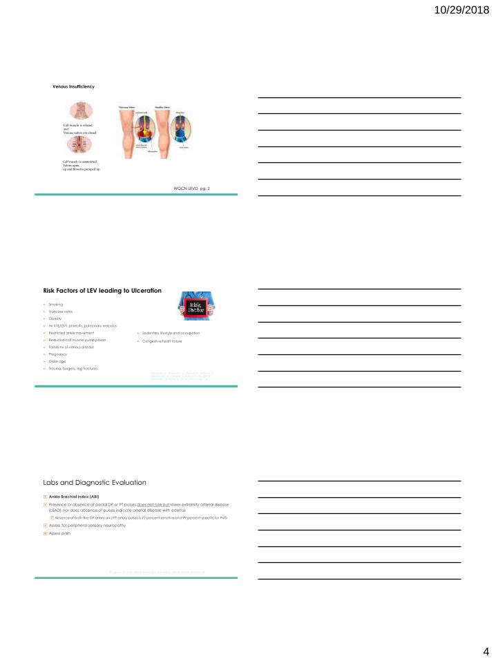
Low Extremity Venous Disease
WOCN LEVD pg. 2
Calf muscle is relaxed and Venous valves are closed
Calf muscle is contracted. Valves openup and blood is pumped up.
Venous Insufficiency
Risk Factors of LEV leading to Ulceration
➢ Smoking
➢ Varicose veins
➢ Obesity
➢ Hx VTE/DVT, phlebitis, pulmonary embolus
➢ Restricted ankle movement
➢ Reduced calf muscle pump power
➢ Family hx of venous disease
➢ Pregnancy
➢ Older age
➢ Trauma, Surgery, leg fractures
Giugliano, D., Di Serafino, L., Perrino, C., Schiano, V., Laurenzano, E., Cassese, S., & Esposito, G. (2013);
Kaminski, J. & Thank, D. (2015). WOCN pg. 1, 4
➢ Sedentary lifestyle and occupation
➢ Congestive heart failure
Labs and Diagnostic Evaluation
▶ Ankle Brachial Index (ABI)
▶ Presence or absence of pedal DP or PT pulses does not rule out lower extremity arterial disease
(LEAD) nor does absence of pulses indicate arterial disease with edema
▶ Absence of both the DP artery and PT artery pulses is 72 percent sensitive and 99 percent specific for PVD
▶ Assess for peripheral sensory neuropathy
▶ Assess pain
Giugliano, G. et al., (2013); Kaminski, J. & Thank, D. (2015); WOCN LEVD 5-6, 31
10/29/2018
5
Physical Exam for Venous Ulcers
▶ Physical assessment- Look for:
▶ Edema
▶ Hemosiderosis (hemosiderin staining, hyperpigmentation)
▶ Venous dermatitis (erythematous, scaly pruritic, weepy wounds)
▶ Atrophie blanche
▶ Varicose veins
▶ Ankle flaring (cluster of reticular/spider veins)
▶ Scarring from previous wounds
▶Warm to hot skin/elevated temp vs. Cool skin
▶ Lipodermatosclerosis
WOCN pg. 5-6, 31
Characteristics of Venous Leg Ulcers
▶ Location: superior to medial malleolus, can
be present anywhere on lower leg including
posterior calf
▶ Wound edges: irregular
▶ Wound bed: ruddy red, yellow
adherent/loose slough, granulation tissue,
shallow and No undermining or tunneling
▶ Exudate: varies – mild, mod, large
▶ Periwound: macerated, crusty, scaling,
hyperpigmented
▶ Odor: +/-Giugliano, G., Di Serafino, L., Perrino, C., Schiano, V.,
Laurenzano, E., Cassese, S., & Esposito, G. (2013).
Kaminski, J. & Thank, D. (2015). WOCN pg. 5-6
▶ Bleeding: +/-▶ Brownish/black discoloration of the
lower extremity▶ Non pitting (brawny) edema▶ Stasis dermatitis- “Tree bark" skin
appearance
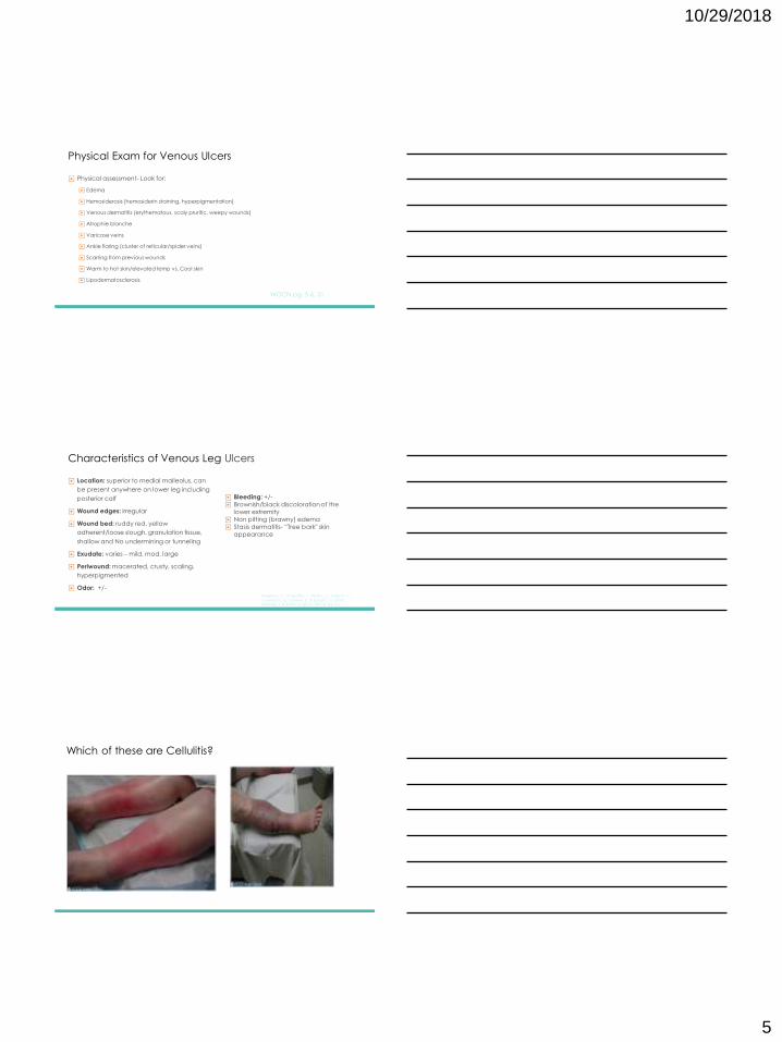
Which of these are Cellulitis?

10/29/2018
6
Venous Stasis vs. CellulitisSymptoms -Afebrile
-Itching-Varicose veins/VTE
-May have fever
-Painful-No relevant history
Signs -Normal body temp
-Erythema, inflamed-May be tender
-Vesicles crusting
-Eczematic lesions may be on
other parts other leg
-Unilateral or bilateral
-Feverish
-Erythema, inflamed-Tender
-One or few bullae/no crusting
-No lesions elsewhere
-Unilateral
Portal of Entry N/A Unknown, breaks in skin, ulcers,
trauma, tinea pedis, intertrigo
Labs -WBC normal
-Skin swabs S. aureus common
-WBC high
-Usually negative except for necrotic tissue
Stasis Dermatitis
Location: medial aspect of lower leg and ankle, superior to medial malleolus
10/29/2018
7
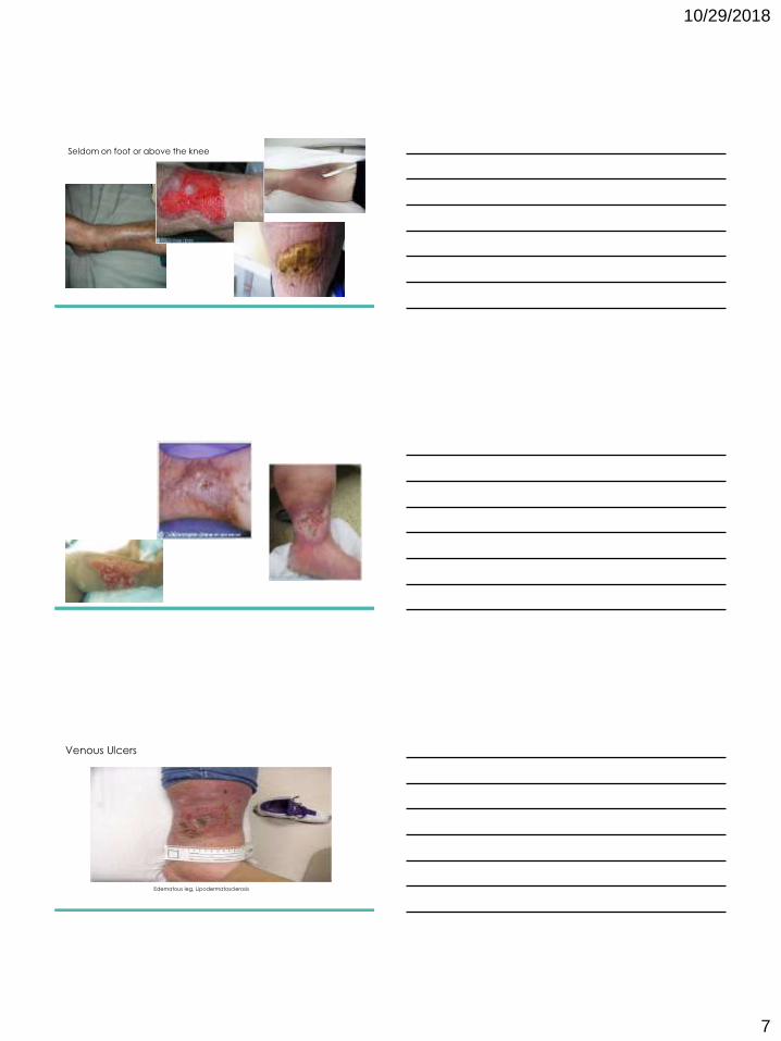
Seldom on foot or above the knee
Venous Ulcers
Edematous leg, Lipodermatosclerosis
10/29/2018
8
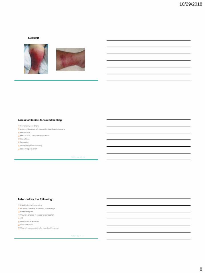
Cellulitis
Assess for Barriers to wound healing:
▶ Comorbidity conditions
▶ Lack of adherence with prevention/treatment programs
▶ Medications
▶ BMI < or = 20 – related to malnutrition
▶ Malnutrition
▶ Depression
▶ Decreased physical activity
▶ Lack of leg elevation
WOCN pg. 8-9, 18
Refer out for the following:
▶ Cellulitis that isn’t improving
▶ Increased swelling, tenderness, skin changes
▶ Intractable pain
▶ Wound is atypical in appearance/location
▶ VTE
▶ Unresponsive Dermatitis
▶ Variceal bleeds
▶ Wound is unresponsive after 4 weeks of treatment
WOCN pg. 9, 13

10/29/2018
9
Prevention of Venous Ulcers is key!
▶ Compression - stockings, garments, wraps, bandages DAILY
▶ Light compression 20-30mmHg for LEVD and/or those who can’t wear tolerate stronger
garments/higher compression
▶ Compression of 40-50mmHg for those with normal arterial blood flow or
what can be tolerated
▶ Tx of varicosities: weight management, physical activity- walking to improve calf muscle
strength, and ROM, avoid wearing constricting garments and crossing legs
▶ Leg elevations
WOCN pg. 31
Treatment and Patient Education▶ Smoking cessation
▶ Wear compression stockings daily
▶ Healthy nutrition and Weight management
▶ Avoid trauma to lower leg
▶ Avoid crossing legs and prolonged standing, avoid high heels
▶ Elevate legs above heart x 30 mins (3-4x a day) when possible
▶ Physical Activity/Exercise– Brisk walks BID, treadmill on incline
▶ Resistance calf muscle exercises
▶ Chair bound patients/non ambulatory- rocking exercise for calf muscles
▶ Discourage self treatment with OTC meds
▶ Topical corticosteroid for no more than 2 weeks - Dermatitis
WOCN pg. 11-12, 14, 27
Compression – Gold Standard
▶ Done throughout lifetime
▶ ABI:
▶ ABI < or = 0.9 = significant arterial disease
▶ ABI > 0.5 but < 0.8mmHg = mixed venous and arterial etiology
▶ Stockings need to be fitted by trained personnel
▶ Apply stockings in AM before getting OOB.
▶ Remove stockings at night prior to sleeping
▶ Replace stockings Q 3-6 mo.
WOCN pg. 11-12,33
10/29/2018
10
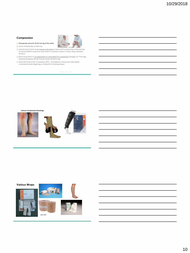
Compression
▶ Therapeutic amount= 30-42 mm Hg at the ankle
▶ Avoid: Antiembolism or TED hose
▶ Inelastic/short stretch wraps require ambulation to force calf muscle pump contraction (Ex.
Unna boot/UBZ kit, Duke boot, Short stretch Comprilan, Coban 2, Farrow Wrap, Rosidal K,
CircPlus)
▶ Elastic/long stretch is not dependent on ambulation for compression (Tubigrip (14-17mm Hg),
Setopress/Surepress (25-35), Profore 4 layer (35-40mm Hg)
▶ Intermittent Pneumatic Compression (IPC) – immobile pts or those who need higher
compression levels (larger legs or intolerant of stockings/wraps)
WOCN pg. 21, 34
Various Compression Stockings
Various Wraps
UBZ/UBC
10/29/2018
11
Stocking clinic guide for compression
• Needs to be measured by certified compression consultant
• Start low compression 20-30mmhg- RX not needed unless want DME coverage
• Activelife- contracted vendor with Kaiser
• If not covered- Compression stocking clinic guide may be available at your wound or
vascular clinic
• Covered if lymphedema, burn or venous stasis with ulcer- Medicare age 65+
PLAN
▶ Refer to Vascular or Outpatient Wound Clinic
▶ Treating prophylactically is not warranted.
▶ Culture guided antibiotic therapy- do not culture slough, Needs topical not systemic
antibiotics
▶ Deep tissue infection and cellulitis- Warrants Systemic treatment
▶ Superficial infection – topical antimicrobial/antibiotics or trial antimicrobial dressings (silver,
PHMB, or cadoxemer iodine)
WOCN pg. 14-18, 33
Cellulitis
TREATMENT:
• If already on PO ABX but cellulitis is not improved:
1. Get aerobic culture- Not on slough/necrotic areas
2. Call Infectious Disease for antibiotic guidance- Doxy or Clinda, Bactrim DS
3. Send pt to ER/Urgent Care for IV Antibiotics
* We do not start IV Antibiotics at the Wound clinic - done in Urgent Care/ER
10/29/2018
12
Other wound care treatments available
▶ Skin grafting
▶ Negative Pressure Wound Therapy (NPWT)
▶ Biologic skin substitutes
▶ Small intestine submucosa wound matrix (Oasis)
▶ Whirlpool- no longer used
▶ Venous- Superficial venous surgery- to prevent recurrence
▶ Venous- Subendoscopic perforator surgery (SEPS)- improve VLU healing and reduce
recurrence since it is for tx of chronic venous insufficiency
WOCN pg. 32
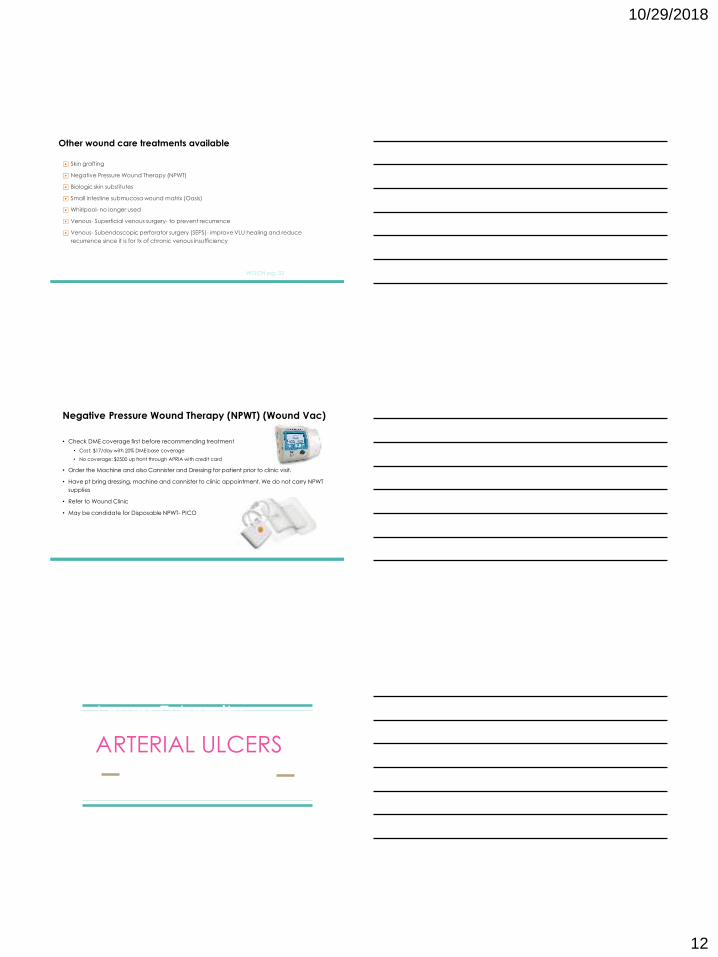
Negative Pressure Wound Therapy (NPWT) (Wound Vac)
• Check DME coverage first before recommending treatment
• Cost: $17/day with 20% DME base coverage
• No coverage: $2500 up front through APRIA with credit card
• Order the Machine and also Cannister and Dressing for patient prior to clinic visit.
• Have pt bring dressing, machine and cannister to clinic appointment. We do not carry NPWT
supplies
• Refer to Wound Clinic
• May be candidate for Disposable NPWT- PICO
Lower Extremity Arterial Disease (LEAD) ARTERIAL ULCERS
10/29/2018
13
Lower Extremity Arterial Disease
▶ US Hospitalization costs LEAD treatment= $4.37-21 billion Medicare pts
▶ Marker of systemic atherosclerosis
▶ Risk of death inc over time. > 5 years, similar to patients with acute MI or
ischemic stroke
▶ US - LEAD affects 8-12 million adults > 40 y/o
▶ 80 years +, Prevalence = 40%
▶ Prevalence lower in women vs. men but severity of disease is higher in
women
WOCN, LEAD pg. 10, 13
Risk Factors
▶ Tobacco use
▶ DM
▶ HTN
▶ Dyslipidemia
▶ Chronic Renal insufficiency
▶ Prevalence increases with age
▶ Hyperhomocysteinemia
▶ Family hx of cardiovascular disease
▶ Sedentary lifestyleWOCN, LEAD pg 1, 14, 20, 21, Mayo Clinic. Peripheral Artery Disease (2012).
Key Points of the Physical Exam
▶ Check for ischemic skin and nail changes
▶ Perform vascular assessment
▶ Check perfusion status
▶ Auscultate fem/popliteal arteries for bruits
▶ Check for neuropathy signs
▶ Screen feet for loss of protective sensation (monofilament, tuning fork,
percussion hammer)- Podiatry
▶ ABI: (Vascular) ; Normal = > or = 1.00
WOCN, LEAD pg 2
10/29/2018
14
Peripheral Artery Disease Assessment ▶ Parathesia
▶ Intermittent claudication
▶ Pain with activity that is relieved by rest
▶ Leg numbness or weakness
▶ Coldness in lower leg or foot
▶ Diminished pulses
▶ Cyanosis or pallor
▶ Hair loss
▶ Dependent rubor
▶ Thin, shiny skin
▶ Ulcer/gangrene
▶ Leg hurts when elevated
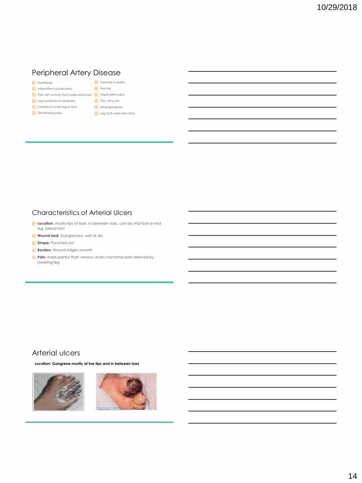
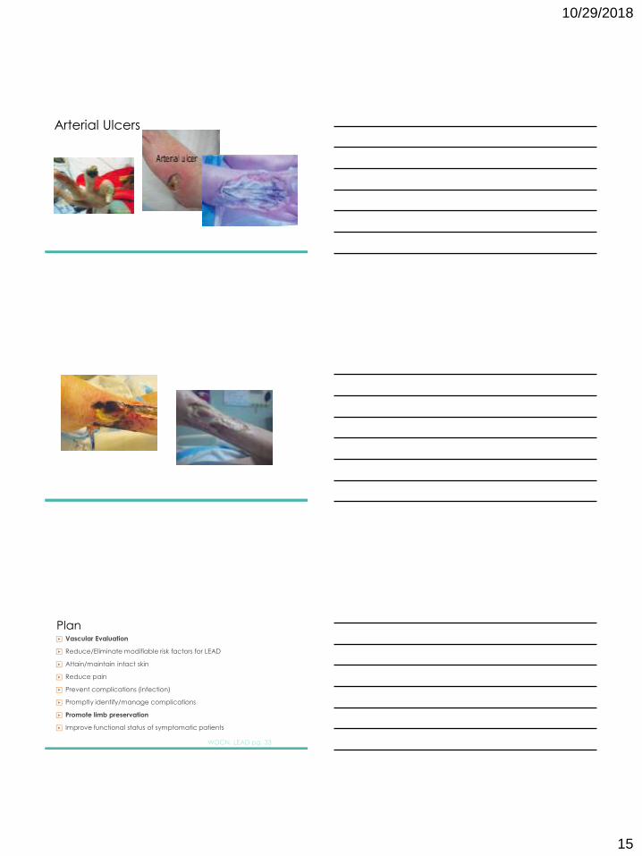
Characteristics of Arterial Ulcers
▶ Location: mostly tips of toes, in between toes, can be mid foot or mid
leg, lateral foot
▶ Wound bed: Gangrenous- wet or dry
▶ Shape: Punched out
▶ Borders: Wound edges smooth
▶ Pain: more painful than venous ulcers; nocturnal pain relieved by
lowering leg
Arterial ulcers
Location: Gangrene mostly at toe tips and in between toes
10/29/2018
15
Arterial Ulcers
Plan▶ Vascular Evaluation
▶ Reduce/Eliminate modifiable risk factors for LEAD
▶ Attain/maintain intact skin
▶ Reduce pain
▶ Prevent complications (infection)
▶ Promptly identify/manage complications
▶ Promote limb preservation
▶ Improve functional status of symptomatic patients
WOCN, LEAD pg. 33
10/29/2018
16
Treatment
▶ Treatment of choice for limb salvage: Revascularization and surgical removal of necrotic
tissue from infected wound on ischemic leg
▶ Offload heels
▶ Maintain dry stable eschar/blisters in noninfected ischemic wounds
▶ Debridement is contraindicated especially with stable eschar until perfusion is determined
▶ LEAD/critical limb ischemia with infection or cellulitis- Culture guided systemic ABX therapy
▶ Encourage regular exercise if stable with intermittent claudication
▶ Analgesia for persistent pain – Consider Pain Clinic referral
WOCN, LEAD pg. 4
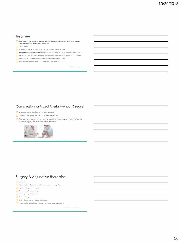
Compression for Mixed Arterial/Venous Disease
▶ Manage edema due to venous disease
▶ Monitor compression for pt with neuropathy
▶ Compression stockings to manage postop edema post lower extremity
bypass surgery- TEDS are contraindicated
WOCN, LEAD pg. 6, 186
Surgery & Adjunctive therapies
▶ Amputation
▶ Prophylactic ABX post amputation, revascularization; grafts
▶ Bypass vs. angioplasty surgery
▶ Conservative topical therapy
▶ Low frequency ultrasound
▶ Electrotherapy
▶ HBOT – Goal: resolve periwound hypoxia
▶ Intermittent pneumatic compression- for non surgical candidates
WOCN, LEAD pg. 7-8, 109

10/29/2018
17
Patient Education
▶ Manage chronic diseases- DM, HTN, cholesterol, weight, medication adherence
▶ Smoking cessation
▶ Promote blood flow, maintain intact skin, prevent trauma
▶ Avoid leg elevation, use dependent position for legs
▶ Routine Daily leg/foot exam for wounds/blisters, nail and foot care
▶ Protect feet, toes, heels- proper fitting shoes with socks
▶ Compression therapy precautions
▶ Increase regular exercise and activity
WOCN, LEAD pg. 8-9
Lower Extremity Neuropathic DiseaseNEUROPATHIC & DIABETIC FOOT ULCERS (DFU’s)
Prevalence
▶ 3x as many pts are admitted to the hospital for neuropathic foot ulcers
than with ischemic ulceration
▶ DM with complications peripheral neuropathy = 50-70% of all non-
traumatic amputations
▶ Diabetic neuropathy wound relapse rate = 66% over 5 years and 12%
progress to amputation
▶ Hospital AKA mortality = 5%, 50-84% subsequent amputation of other
limb in 2-3 years. 5 year survival rate bilateral amputations < 50%
WOCN, LEND pg. 1
10/29/2018
18
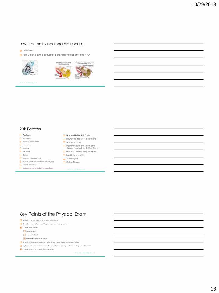
Lower Extremity Neuropathic Disease
▶ Diabetes
▶ Foot ulcers occur because of peripheral neuropathy and PVD
WOCN, LEND pg. 2-3
Risk Factors
▶ Modifiable:
▶ Prediabetes
▶ Hypo/hyperthyroidism
▶ Alcoholism
▶ Smoking
▶ HTN, COPD
▶ Obesity
▶ Exposure to heavy metals
▶ Malabsorption syndrome (bariatric surgery)
▶ Vitamin deficiency
▶ Abdominal, pelvic, and ortho proceduresWOCN, LEND pg. 4-5
▶ Non modifiable Risk Factors:
▶ Raynaud’s disease/ Scleroderma
▶ Advanced age
▶ Neuromuscular and spinal cord
diseases/injuries (MS, Guillain-Barre)
▶ HIV, AIDS, related drug therapies
▶ Familial neuropathy
▶ Acromegaly
▶ Celiac Disease
Key Points of the Physical Exam▶ DM pts- Annual comprehensive foot exam
▶ Check temperature, foot hygiene, shoe wear practices
▶ Check for calluses:
▶ Focal Callus
▶ Insensate feet
▶Hemorrhage into a callus
▶ Check for fissures, moisture, nails, tinea pedis, edema, inflammation
▶ Erythema + edema indicate inflammation= early sign of impending foot ulceration
▶ Check for loss of protective sensation
WOCN, LEND pg. 8-9,15
10/29/2018
19
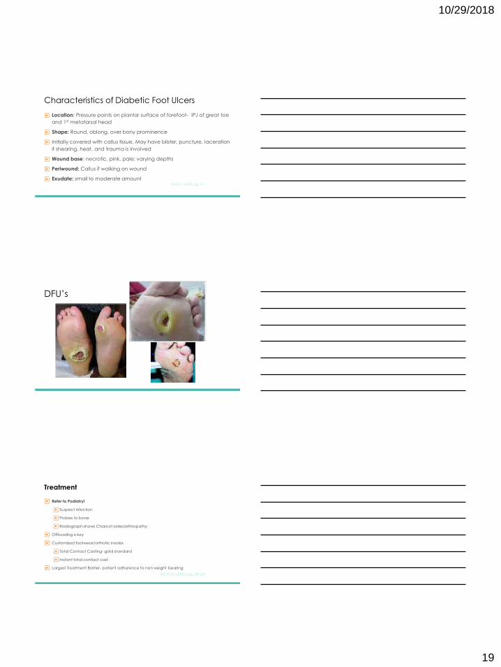
Characteristics of Diabetic Foot Ulcers
▶ Location: Pressure points on plantar surface of forefoot- IPJ of great toe
and 1st metatarsal head
▶ Shape: Round, oblong, over bony prominence
▶ Initially covered with callus tissue. May have blister, puncture, laceration
if shearing, heat, and trauma is involved
▶ Wound base: necrotic, pink, pale; varying depths
▶ Periwound: Callus if walking on wound
▶ Exudate: small to moderate amountWOCN, LEND pg. 10
DFU’s
Treatment
▶ Refer to Podiatry!
▶ Suspect infection
▶ Probes to bone
▶ Radiograph shows Charcot osteoarthropathy
▶ Offloading is key
▶ Customized footwear/orthotic insoles
▶ Total Contact Casting- gold standard
▶ Instant total contact cast
▶ Largest Treatment Barrier- patient adherence to non weight bearing
WOCN, LEND pg. 28-29
10/29/2018
20
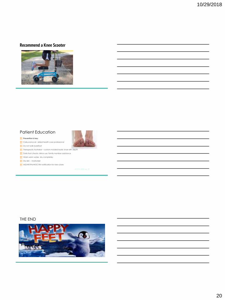
Recommend a Knee Scooter
Patient Education
▶ Prevention is key:
▶ Callus removal - skilled health care professional
▶ Do not walk barefoot
▶ Therapeutic footwear – custom molded insole; shoe with depth
▶ Daily foot checks- Mirror use; family member assistance
▶ Wash warm water, dry completely
▶ Dry skin – moisturizer
▶ MD/NP/PA/WOC RN notification for new ulcers
WOCN, LEND pg. 29
THE END
10/29/2018
21
References
▶ Giugliano, G., Di Serafino, L., Perrino, C., Schiano, V., Laurenzano, E., Cassese, S., & Esposito, G. (2013).
Effects of successful percutaneous lower extremity revascularization on cardiovascular outcomes in patients
with peripheral arterial disease. International Journal of Cardiology, 16(6), 25666-2571. Retrieved from
http:///www.researchgate.net/publication/229081240
▶ Kaminski, J. & Thank, D. (2015). Key To Assessing Peripheral Vascular Disease. Podiatry Today, 28(4). Retrieved
from http://www.podiatrytoday.com/keys-assessing-peripheral-vascular-disease
▶ Margolis, D.J., Malay, D.S., Hoffstad, O., Leonard, C.E., MaCurdy, T., de Nava, K.L., & Seigel, K.L. (2011).
Incidence of diabetic foot ulcer and lower extremity amputation among Medicare beneficiaries, 2006 to
2008. Retrieved from http:///www.nih.gov,
▶ Mayo Clinic. Peripheral Artery Disease (2012). Retrieved from http://www.mayoclinic.org/disease-
condition/peripheral-artery.
References
▶ Wound Ostomy and Continence Nurses Society. (2011). Guideline for management of wounds in patients
with lower-extremity venous disease. WOCN practice guideline series 4. Mount Laurel: New Jersey. Kelechi, T.
& Johnson, J. J.
▶ Wound Ostomy and Continence Nurses Society. (2014). Guideline for management of wounds in patients
with lower-extremity arterial disease. WOCN practice guideline series 1. Mount Laurel: New Jersey. Bonham,
P.A. & Flemister, B.G.
▶ Wound Ostomy and Continence Nurses Society. (2012). Guideline for management of wounds in patients
with lower-extremity neuropathic disease. WOCN practice guideline series 3. Mount Laurel: New Jersey.
Crawford, P. E. & Fields-Varnado, M.