wound healing in ckd management &...
TRANSCRIPT
Wound Healing in CKDManagement & Prevention
Cora Espina, MN, ARNP, CWCNTeaching Associate
University of Washington, School of [email protected]
Objectives
• Understand physiology of wound healing
• Apply appropriate wound management therapies
• Discuss strategies to prevent wound development in vulnerable patients

Classification of Tissue
Based on involved Tissue
• Partial Thickness
– Epidermis
– Dermis (2mm)
• Full Thickness
– Subcutaneous
– Muscle
– Bone

Partial Thickness Wounds
• Shallow, moist, painful
• Exposure of nerve endings
• Partial Dermis loss
– Wound Bed appears pale pink with red islets
• Epidermal Loss
– Wound bed appears bright pink or
red when basement membrane
exposed

Partial Thickness Wound Repair
• Inflammatory Response (<24h)– Erythema, serous exudate containing leukocytes
• Epithelial Proliferation & Migration (24-72h)– Basal cells at the wound edge and throughout
wound bed elongate and migrate laterally.
– Moist wound healing
– Fragile, at risk from shear/friction
• Dermal Repair– Collagen synthesis.
– Dermal cells contribute to repair – scalp heals faster.

Full Thickness Wounds
1. Deeper2. Granulation Tissue3. Slough4. Formation of Scar Tissue5. Stage 3 and 4 Pressure Ulcers

Full Thickness Wound RepairPrimary Intention
1. Hemostasis– Bleeding offers temporary barrier to bacterial invasion. – Platelet activation and aggregation
2. Inflammation (D3)– Breakdown of damaged tissue & bacteria – WBC, Neutrophils, Macrophages– Tensile strength to incision is 0%
3. Proliferation (D5)– Granulation tissue is formed by Extracellular Matrix– Angiogenesis, new capillaries restore O2 delivery– Collagen Synthesis– Epithelium covering restored as stiff scar lacks elastin
4. Maturation/Remodeling (D21-1year)– New collagen is stronger – 80% tensile strength

Categorize by Age
• Acute
– Traumatic or surgical
– Occur suddenly, progress rapidly toward healing
• Chronic
– Fail to proceed normally through repair process
– Etiology: vascular compromise, chronic inflammation, repetitive injury, failure to close

Principles of Wound Healing
• Identify and address underlying factors
• Reduce edema
• Optimize nutrition status
• Control blood sugar
• Protect from trauma
• Treat infection

Dressing SelectionCreate an optimal environment for healing
• Manage drainage • Provide thermal insulation• Be impermeable to water and bacteria• Not traumatic to tissue when removed• Cost effective• Minimal pain on removal
• Dowsett, C. (2008). Exudate management: a patient-centered approach. Journal of Wound Care, 17(6): 249-252.• Armstrong, AH, & Price, P. (2004). Wet-to-dry gauze dressings: fact and fiction. Wounds, 16(2): 56-62.• Modern wound management dressings. Prescribing Nurse Bulletin (1999), 1(2): 5-8.• Hutchinson, J, & McGuckin, M (1990). Occlusive dressings: a microbiologic and clinical review. American Journal of Infection control, 18(4): 257-268.

TREATMENTFactors Influencing Dressing Choice
• Anatomical site
• Surrounding skin
• Available Dressings
• Caregiver ability
• Aggressive therapy vs. palliative care

1. Cleanse the Wound
• Normal Saline
• Antiseptic Agents
– May be cytotoxic to fibroblasts (collagen & granulation tissue )
• Povidone Iodine
• Sodium Hypochlorite (Dakin’s)
• Hydrogen Peroxide
• Iodoform

2. Protect peri-woundSkin barriers
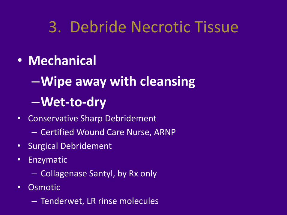
• Mechanical
–Wipe away with cleansing
–Wet-to-dry• Conservative Sharp Debridement
– Certified Wound Care Nurse, ARNP
• Surgical Debridement
• Enzymatic
– Collagenase Santyl, by Rx only
• Osmotic
– Tenderwet, LR rinse molecules
3. Debride Necrotic Tissue

4. Manage Drainage
• Gauze (Kerlix, string/nu-gauze, Iodoform)
• Hydrocolloid (Duo-derm)
• Calcium Alginate (Maxorb)
– 20x weight, moist gel
• Hydrofiber (Hydrogel)
– Contain without expansion
• Foam (Alevyn, Mepilex)
– Non-adherent

Contain High OutputDonate Moisture when dry

5. Fill Dead Space
• Gauze• Calcium Alginates• Foam (Wound VAC)
Decrease bio-burden– Silver
• Amorphous gel (Silvasorb)• Sheet (Silvasorb)• Powder (Arglaes)

6. Cover UP
•Provide thermal insulation
•Keeps bacteria out
•Protects from injury
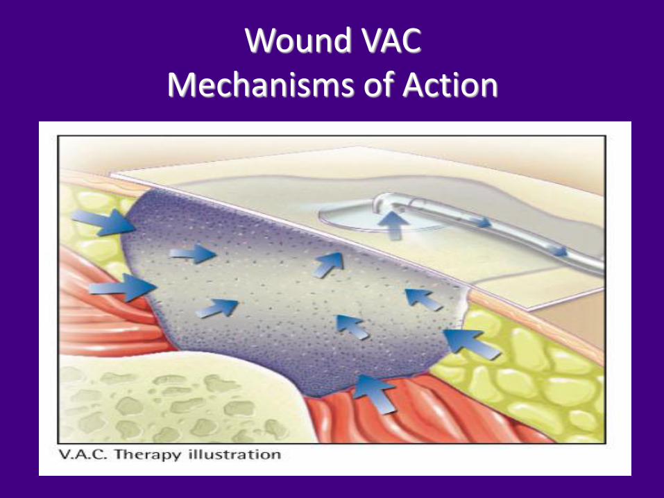
Wound VACMechanisms of Action

Isolation of Fistula

Lower Extremity WoundsTypical Assessment Findings
Poor Circulation, Poor Nerve Function
• Skin - warm
• Pulses - present
• Pain
• Neuropathy
21

Ischemic Ulcers
• Associated with Lower Extremity Arterial Disease
• Painful• Punched Out
22

Neuropathic Ulcers
• Associated with Neuropathic Disease
– Diabetes
• Loss of Protective Sensation
• Area of repeated injury

Venous Insufficiency
• Prominent vein (varicosities)• Telangiectasia (spider veins)• Ankle flare
– Collection of visible dilated capillaries at medial malleolus
• Exudate– Moderate to heavy drainage typical
• Typical location– “Gaiter Area” area of lower leg/ankle
at or above medial malleolus
24

Venous Ulcer Characteristics
• Hemosiderin stain: Brownish, rust color in the skin from leakage of fluid & breakdown of hemoglobin in the tissue
• Areas of avascular or poorly vascularized skin
• Smooth white plaques of white tissue or lacking in normal color
25

Management
• Control edema
• Control exudate & protect skin
• Promote venous return
• Eliminate infection
• Stimulate granulation tissue
• Avoid sensitizers/irritants that cause dermatitis
26
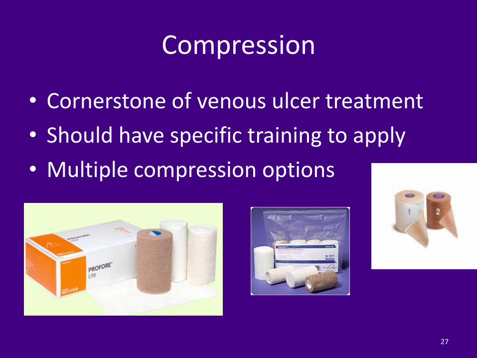
Compression
• Cornerstone of venous ulcer treatment
• Should have specific training to apply
• Multiple compression options
27

Prevention
• High Risk Patients
– Poor circulation, poor sensation, risk for infection
– Steroids
• Promote Circulation and Nerve Function
– Exercise, BP control
• Protect from Injury
– Shoes, wheelchair
– Fragile skin, skin tears

Compression stockings
Minimize Edema
• Light support 20-30 mmHg
• Medium support 30-40 mmHg
• Strong support 40-50 mmHg
• Very strong support 50-60 mm Hg*
• Cost varies from $30-$150/pair
29*WOCN guide for management of LEVD 2005
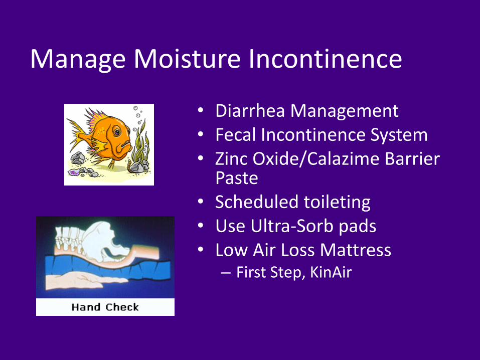
Manage Moisture Incontinence
• Diarrhea Management• Fecal Incontinence System• Zinc Oxide/Calazime Barrier
Paste• Scheduled toileting• Use Ultra-Sorb pads• Low Air Loss Mattress
– First Step, KinAir
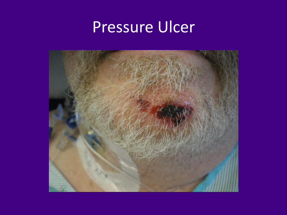
Pressure Ulcer

32
Incontinence Related Dermatitis
Partial Thickness
Interventions:
• Barrier cream with zinc
• Absorptive wicking pads
• Frequent turning to avoid further breakdown
Increases risk:
Pressure Ulcer