wound healing - rcsi dublin · wound healing, preventing overproliferation in response to injury 47...
TRANSCRIPT

Copyright © 2016 American Society of Plastic Surgeons. Unauthorized reproduction of this article is prohibited.
www.PRSJournal.com82S
THE IMPORTANCE OF DEBRIDEMENT: ERADICATING INFECTION,
COMBATING BIOFILM, AND REDUCING SENESCENT CELLS
Debridement is derived from the French débridement, which means to remove a constraint. In its original description by Henri François Le Dran (1685–1770), debridement referred to the use of an incision to promote drainage and to reduce tension under the skin.1 The clinical definition of debridement has subsequently evolved to include the removal of nonviable or contaminated tissue that impedes normal tissue growth, as previously
reviewed.2 Debridement renews the wound and surrounding tissue to promote normal healing by removing infection, biofilm, and senescent cells.
Although each of these factors has the capac-ity to impede healing, they often present simul-taneously in chronic wounds, and their negative effects on the healing cascade tend to be synergis-tic. The common denominator in this equation is chronic inflammation, which is abnormally pro-longed in response to each of these factors. Other issues include physical barriers formed by these ele-ments that decrease vascular supply as well as the delivery of progenitor cells to the wound. Through
Disclosure: Dr. Attinger is a consultant for KCI, an Acelity company, Smith & Nephew, and Integ-ra. Dr. Janis is a consultant for LifeCell, an Acelity company; has received prior honoraria from Bard, Pacira, and KCI, an Acelity company; and receives royalties from CRC Press. None of the other authors has any financial disclosures.Copyright © 2016 by the American Society of Plastic Surgeons
DOI: 10.1097/PRS.0000000000002651
Ersilia L. Anghel, BS, BAMichael V. DeFazio, MD
Jenny C. Barker, MD, PhDJeffrey E. Janis, MD
Christopher E. Attinger, MD
Washington, DC; and Columbus, Ohio
Background: The establishment of a healthy wound bed through adequate debridement of infected, senescent, and/or devitalized tissue is central to the progression of normal wound healing. Although a variety of surgical and non-surgical strategies have been proposed, none have proven completely effective in all settings. This review focuses on the principles and techniques of modern debridement practices employed in the management of complex wounds.Methods: A comprehensive review of the PubMed/Medline and Ovid data-bases was performed to identify basic science and clinical studies using key words most relevant to biofilm, debridement, and wound healing. English lan-guage articles that were peer reviewed and that met the standard of evidence-based medicine were included. Level of evidence for various debridement approaches was rated utilizing the American Society of Plastic Surgeons Rating Levels of Evidence and Grading Recommendations.Results: The value of both operative and nonoperative debridement tech-niques, their indications, and limitations are described. With an emphasis placed on surgical debridement, this review highlights technical adjuncts that can be used to optimize wound bed preparation, including preoperative topi-cal staining of the wound, as well as the use of color-guided endpoints to pre-vent removal of excess healthy tissue. The indications for using temporizing measures for wound control such as negative pressure wound therapy with and without installation are also discussed.Conclusion: Optimal management requires a multimodal approach that centers around operative debridement and incorporates the use of adjunctive measures to facilitate the removal of infected tissue, biofilm, and/or senescent cells that impede the progression of normal wound healing. (Plast. Reconstr. Surg. 138: 82S, 2016.)
From the Department of Plastic Surgery, MedStar George-town University Hospital; and Department of Plastic Sur-gery, Ohio State University Medical Center.Received for publication March 14, 2016; accepted June 10, 2016.
Current Concepts in Debridement: Science and Strategies
WOUND HEALING

Copyright © 2016 American Society of Plastic Surgeons. Unauthorized reproduction of this article is prohibited.
Volume 138, Number 3S • Current Concepts in Debridement
83S
reduction of these impediments, debridement con-verts a chronic wound to an acute and dynamic state where the stages of healing can progress normally.
It should be noted, however, that debride-ment is no substitute for perfusion, as tissues still require adequate perfusion for healing, allow-ing oxygen, nutrients, and cells to be delivered to wound.3–5 Hypoxia in wounds (5–20 mm Hg) can cause cell death, tissue necrosis, and promote growth of microorganisms.6
Ultimately, durable restoration of soft-tissue coverage is dependent on the satisfaction of a number of requisite objectives, including eradica-tion of infection and/or reduction of bioburden, improvement of local blood flow, revitalization of the wound bed, and correction of biomechanical abnormalities. Debridement, when performed correctly, serves to optimize wound healing by meeting these objectives.
Acute InfectionBacteria exist in multiple phenotypic states
and may present as rapidly dividing planktonic, single cells or as an aggregate of bacterial spe-cies embedded within a polymeric matrix (i.e., biofilm). These phenotypic changes are driven by adaptive shifts in gene expression that occur in response to environmental cues. Acute infec-tion is characterized by rapidly dividing organisms within a wound, resulting in the characteristic host response of erythema, warmth, swelling, pain, malodor, and drainage. If not recognized and treated early, acute infection can spread systemi-cally, leading to overt sepsis.7 The line between chronic colonization and acute infection may be minute, so prompt identification is critical to heal-ing wounds and minimizing complications. For most microorganisms, a bacterial count (biobur-den) of 105 bacteria per gram of tissue or greater has been defined as the quantitative threshold for acute infection and wound inhibition. If left alone, wounds with bacterial counts of less than 105 bac-teria per gram of tissue can often heal secondarily in healthy hosts. Skin grafts, on the other hand, are more sensitive to bacterial burden and, thus, may represent an exception to this rule.7–9 With a higher bacterial load, the potential for healing is restricted due to chronic local inflammation, which can inhibit the migration of fibroblasts and keratinocytes.10 Development of a protease-rich environment further exacerbates bacterial prolif-eration and may contribute to progressive tissue necrosis. Although surgical debridement remains the mainstay treatment in the setting of infected and/or nonviable tissue, topical antibiotics such as
silver sulfadiazine, mafenide acetate, cadexomer iodine, or silver nitrate can be used as adjuncts to help lower the bacterial burden within a wound (Fig. 1).7,9
Biofilm InfectionThe Centers for Disease Control and NIH
estimate that 65% to 80% of all human infec-tious diseases are caused by bacteria with a bio-film phenotype.11 It is now well established that infection in problematic wounds is not always the result of opportunistic growth of microorganisms in the planktonic or single-cell state, but rather as polymicrobial aggregates within acellular matri-ces such as biofilm.12 In fact, the perception that infection always results from pure, single-organ-ism culture is an artifact of medicine’s historical diagnostic capabilities, and does not represent the way most microbial species naturally exist. Occult biofilm infection likely contributes to a wide spec-trum of wound therapy failures.
Biofilm, itself, is a protective film composed of protein, polysaccharides, and extracellular DNA termed “extracellular polymeric substance.” The stages of formation and components of the bio-film matrix termed “EPS” are reviewed in detail in other publications and are beyond the scope of this article.13 Briefly, biofilm represents a pro-tected mode of growth for bacteria that allows them to evade eradication by conventional thera-pies and avoid detection by standard diagnostic techniques.14 Biofilm alters wound healing by a number of mechanisms.
First, biofilm is capable of subverting the host immune response, not only by shielding key antigens but also by hijacking the host immune response and augmenting the prolonged inflam-mation observed in nonhealing wounds. Biofilm chronically releases exotoxins, bacteria, and bacterial DNA to stimulate the innate immune system, directly inducing the expression of proin-flammatory cytokines15 and also preventing both appropriate function and appropriate clearance of neutrophils from the wound bed.16,17
Biofilms are recalcitrant to antimicrobial therapies and they both exhibit antimicrobial tolerance and contribute to antimicrobial resis-tance.18,19 Antimicrobial tolerance, or the ability of bacteria to avoid death owing to the physical state of the bacterium despite known susceptibil-ity to the antibiotic in the planktonic state, is the predominant mechanism of antimicrobial recalci-trance in the biofilm state. This is accomplished through slow growth rates, physical barriers of the biofilm matrix that resist antibiotic penetration,

Copyright © 2016 American Society of Plastic Surgeons. Unauthorized reproduction of this article is prohibited.
84S
Plastic and Reconstructive Surgery • September Supplement 2016
Fig.
1. R
ole
of d
ebrid
emen
t and
adj
uvan
t the
rapy
in c
ompl
ex w
ound
man
agem
ent.

Copyright © 2016 American Society of Plastic Surgeons. Unauthorized reproduction of this article is prohibited.
Volume 138, Number 3S • Current Concepts in Debridement
85S
and the binding of antibiotics by EPS macromole-cules rendering them inactive.20–23 Of note, antimi-crobial tolerance is reversible when bacteria revert back to the planktonic state,24,25 which is an impor-tant consideration for biofilm disruption through debridement. Biofilm also contributes to antimi-crobial resistance, which is the ability to not only avoid death, but also grow in the presence of anti-biotic owing to inherent bacterial characteristics. This is accomplished because the biofilm matrix structurally facilitates horizontal gene transfer of resistant plasmids. Antibiotic recalcitrance in the biofilm state is up to 1000 times greater than that of planktonic bacteria alone.25–32
Aside from antibiotic recalcitrance, bio-film is also difficult to adequately debride.11,33–35 Moreover, debridement can translocate micro-colonies deeper into tissues raising the con-cern that debridement may increase the risk of inoculation for deeper, more persistent biofilm infection.33 Evidence suggests that pathogenic biofilms in vivo (as opposed to the in vitro sur-face biofilm model) more likely exist as semisolid microcolonies within deeper tissue rather than being strictly adherent to a wound surface.18,36,37 Multiple alternative dressings have been inves-tigated to determine their role in disrupting biofilm. Among these, lactoferrin, xylitol, sali-cylic acid, erythritol, cadexomer iodine, silver- and iodine-impregnated dressings, and honey have demonstrated limited efficacy (Fig. 1).38–44 Thus, debridement is currently the mainstay of therapy for biofilm eradication. When biofilm is disrupted, bacteria revert to a planktonic state and demonstrate increased susceptibility to antimicrobials.24,25 These observations highlight the importance of a multimodal approach to biofilm—centered around surgical debridement and including the use of antibiotics—to reduce biofilm burden and stimulate healing of these challenging wounds.
Senescent CellsThe presence of senescent cells (cells that
have transitioned to a nondividing pheno-type) in problematic wounds has been well documented. These cells are not dead or bio-logically inert, but they participate in potent cell-signaling interactions45 and are present in higher proportions in nonhealing wounds.46 It has been more recently described, however, that induction of replicative senescence is both a physiologic and beneficial occurrence in normal wound healing, preventing overproliferation in response to injury47 and reducing tissue fibrosis
by promoting balanced extracellular matrix metabolism.48 Animal models that are incapable of inducing a senescent phenotype have abnor-mal and delayed wound healing.49,50
However, in a nonhealing wound, a lack of clearance and persistence of senescent cells may preclude normal wound healing and allow these cells to serve as a factory for the abundance of enzymes, such as matrix metalloproteases,50 that ultimately become pathologic for wound healing. The extent to which senescent cells contribute to pathologic wound healing, rather than as a con-current finding, has not been well defined. It is possible that debridement serves to restore tissue homeostasis by removing excess senescent cells.
DEBRIDEMENT: TECHNIQUES, INDICATIONS, AND LIMITATIONSMultiple techniques are used to debride.
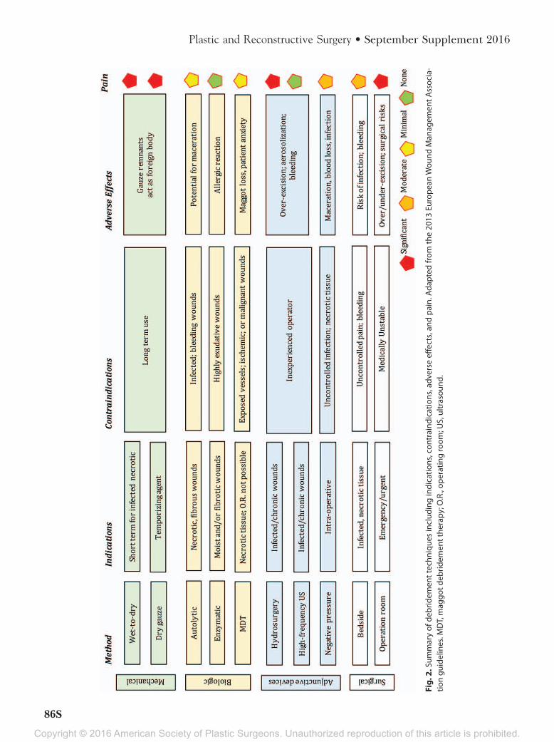
The different options available can be separated into mechanical, biologic, technical, and surgi-cal methods. Determination of the most appro-priate technique mandates the consideration of both host-specific (i.e., comorbidities, compli-ance, social support, etc.) and wound-related (i.e., infection/contamination, perfusion, viability, etc.) factors, as well as the resources available at the treatment facility. The European Wound Man-agement Association guidelines for debridement provide specific information regarding the indi-cations, contraindications, and potential adverse effects associated with each technique.51 These parameters have been summarized in Figure 2.
Mechanical DebridementMechanical debridement includes the use
of both wet-to-dry dressings and dry gauze, to facilitate the removal of infected and/or nonvi-able tissue. The level of evidence for mechanical debridement is rated III and IV. These dressings may act as temporizing measure to facilitate inter-val debridement until definitive reconstruction can be achieved. Alternatively, they serve to pro-mote healing by secondary intention in nonop-erative candidates. Wet-to-dry dressing involves applying moist gauze to a wound, then remov-ing it once dry and adherent to underlying tis-sue. Complete removal is critical, as small gauze remnants can act as foreign bodies and potenti-ate infection within a wound.52 Dry gauze has a lower debridement potential and should be used only as a temporary barrier to minimize environ-mental contamination.53 Both wet-to-dry dressings and dry gauze erratically tear necrotic tissue from
Copyright © 2016 American Society of Plastic Surgeons. Unauthorized reproduction of this article is prohibited.
86S
Plastic and Reconstructive Surgery • September Supplement 2016
Fig.
2. S
umm
ary
of d
ebrid
emen
t tec
hniq
ues
incl
udin
g in
dica
tions
, con
trai
ndic
atio
ns, a
dver
se e
ffect
s, an
d pa
in. A
dapt
ed fr
om th
e 20
13 E
urop
ean
Wou
nd M
anag
emen
t Ass
ocia
-tio
n gu
idel
ines
. MD
T, m
aggo
t deb
ridem
ent t
hera
py; O
.R.,
oper
atin
g ro
om; U
S, u
ltras
ound
.

Copyright © 2016 American Society of Plastic Surgeons. Unauthorized reproduction of this article is prohibited.
Volume 138, Number 3S • Current Concepts in Debridement
87S
the underlying wound and are often painful and insufficient for adequate wound bed preparation for several reasons. These dressings may also cause evaporative fluid loss, surfacing cooling, vasocon-striction, impaired immune response, and local tissue hypoxia, thereby increasing the wound’s susceptibility to infection. They provide no physi-cal obstacle to the penetration of bacteria and/or other contaminants. Lawrence54 showed that bacteria were capable of infiltrating through up to 64 layers of gauze.
Biologic DebridementBiologic debridement is an umbrella term that
encompasses autolytic, enzymatic, as well as honey and maggot therapies.51 The level of evidence for biologic debridement is rated III and IV.
Autolytic dressings (i.e. hydrogels, hydrocol-loids, polymeric membrane formulations) are indicated for wounds with necrotic tissue and/or fibrin coats and act to soften fibrotic wound mar-gins as they stimulate the release of endogenous proteolytic enzymes.55 They essentially exploit the body’s inherent ability to digest/rid itself of necrotic tissue and enhance the formation of granulation tissue. These dressings are relatively painless in nature, which represents a major advantage for patients, particularly those who are sensate. They most often benefit patients who have minimal necrotic loads and cannot tolerate more aggressive forms of debridement.56
Enzymatic ointments, which rely on directly hydrolyzing peptide bonds, are recommended for moist and/or fibrotic wounds, particularly in patients who are poor surgical candidates. Enzymes selectively digest devitalized tissue. This causes less trauma to healthy tissue than surgical debride-ment. Potential side effects of both autolytic and enzymatic agents include allergic reaction and maceration of the wound bed with prolonged use. Furthermore, they are generally labor- and time-intensive and can be painful as well.
Medicinal honey has been employed in the management of wounds for centuries. It uses osmotic properties to treat a wide range of wound types by absorbing exudation. Both honey’s low pH value (i.e., 3.0–4.5) and its stimulated release of hydrogen peroxide have been cited to con-tribute to its antimicrobial effect.57 However, this claim is controversial. Currently, the use of medic-inal honey is only FDA approved as a wound dress-ing and is not considered an active debriding or antimicrobial agent. Caution should be used when selecting honey for patients with history of allergic reaction to bee venom.
Maggot debridement therapy (MDT), using the radiated larvae of the blowfly Phaenicia seri-cata, is a proven, cost-effective alternative for treat-ing drug-resistant, chronically infected wounds in patients who are poor operative candidates.58 Maggots secrete an enzyme that selectively dis-solves necrotic tissue and biofilm into a nutrient-rich food source, while sparing healthy tissue. This reduces the bacterial burden that often plagues gangrenous, recalcitrant wounds. A recent meta-analysis evaluating the safety and effectiveness of MDT revealed significant improvements in both the rate and efficiency of chronic wound heal-ing, longer antibiotic-free intervals, and a lower amputation risk among patients with diabetic foot ulcers.59 MDT is contraindicated near exposed blood vessels, the eyes, the upper gastrointestinal tract, and the upper respiratory tract. It has been associated with pain in some patients likely due to agitation of the wound bed by larvae.
Adjunctive ModalitiesAdjunctive modalities for debridement
include hydrosurgery, ultrasound, and negative-pressure wound therapy (NPWT) with and with-out instillation. The level of evidence for these adjunctive measures is rated III and IV.
Hydrosurgery uses a powerful jet stream of water traveling high speeds, which aspirates surrounding tissue and pulverizes it. The flow carries the waterjet, necrotic tissue, and debris into the evacuator port without the need for separate suction, aside from that provided in the hand piece. It affords minimal splash, vaporization, and aerosol effect (as in high-pressure pulsed lavage and electrocautery) and allows good visibility. Studies have demonstrated effective decrease in quantitative bacterial counts and greater reduction of foreign bodies than high-pressure pulsatile lavage.60,61 Mosti et al. showed that use of hydrosurgical debridement decreases time to reach desired endpoint (i.e. a graftable wound bed) by nearly 5 days when compared with traditional moist dressings (6.1 vs 1.4).62 In this study, most pro-cedures were performed at the bedside, although, in sensate patients, pain intolerance may preclude its use in this fashion. Seventy-six percent required just one procedure with a mean time of 5.9 min-utes. Other studies have shown reductions in cost, time, and number of required to reach clinical end-points.62–65 Care must be taken, however, to avoid over-resection of healthy underlying tissue, which can be aided by the use of preoperative topical wound staining (see below). Hydrosurgery, when done correctly, can significantly reduce bacterial count compared with sharp debridement.66

Copyright © 2016 American Society of Plastic Surgeons. Unauthorized reproduction of this article is prohibited.
88S
Plastic and Reconstructive Surgery • September Supplement 2016
Ultrasound can be used for wound debride-ment. Conventional/high-frequency ultrasound operates between 1 and 3 MHz, requiring con-tact with tissue. It acts by disrupting cellular bod-ies and protein material as well as dislocation of debris. Similar to hydrosurgery, this technology is operator dependent and can result in some aero-solization. Its efficacy is hampered by a lack of evi-dence supporting its use.
Negative-Pressure Wound Therapy ± InstillationNPWT can be used in the interval between
debridements to sterilely occlude the wound and provide intermittent irrigation when combined with instillation (NPWTi). NPWT potentiates healing through increased local blood flow and granulation, reduced tissue edema, and control of bacterial proliferation. This adjunct is most effec-tive in wounds that cannot be immediately closed due to persistent infection or in planned delayed closure. Wound temporization with NPWT mini-mizes the effects of edema and elastic recoil that promote induration and progressive tissue retrac-tion, and therefore can reduce soft-tissue deficits.67 NPWTi is a novel therapy that combines negative
pressure (125– 150 mm Hg) with automated, intermittent instillation of a topical wound solu-tion (antiseptic, antimicrobial, or saline) of set volume (until sponge is visibly saturated), dwell time (30 s to 20 min), frequency (every 1–6 h), and duration (2–10 d). Both systems are contra-indicated in the setting of exposed blood vessels, viscera, and/or malignancy. The level of evidence for NPWT with and without installation is rated II and I, respectively.
Studies in both clinical and animal models have shown that NPWTi with a variety of solutions can significantly reduce the wound bioburden,68,69 whereas NPWT alone can lead to an increase in infection.70,71 In the setting of complex infected wound (those with underlying osteomyelitis, and wounds with exposed orthopedic hardware), NPWTi can be used to routinely wash tissue and prevent the progression of infection between operative debridements. In a retrospective, histor-ical, cohort-controlled trial examining the impact of NPWT with (n = 68) and without (n = 74) instil-lation, NPWTi was shown to reduce the total num-ber of operative procedures, expedite the time to wound closure, and shorten the length of hospital
Fig. 3. Wound stained with methylene blue and surgically debrided. (Above, left) Gross wound before intervention. (Above, right) Methylene blue is liberally applied to wound base using a cot-ton tip applicator, with demarcation of margins to be excised. (Below, left) Following 3 mm exci-sion of senescent wound margin. (Below, right) Debridement of entire wound base with removal of all visibly stained tissues. Healthy bleeding wound base indicating intact vascular supply.

Copyright © 2016 American Society of Plastic Surgeons. Unauthorized reproduction of this article is prohibited.
Volume 138, Number 3S • Current Concepts in Debridement
89S
stay when compared with NPWT alone for patients with infected wounds requiring hospitalization.72 In a separate prospective randomized controlled trial, saline was shown to be equally effective as an antiseptic (0.1% polyhexanide + 0.1% betaine) in the management of infected wounds requiring hospitalization.73
Surgical DebridementExcisional debridement can be accomplished as
a minor procedure at the bedside or under general anesthesia in the operating room. This approach involves the direct excision of all infected and necrotic tissue within a wound using a combination
of scalpel blades, mayo scissors, curettes, rongeurs, power burrs, sagittal saws, and hydrosurgical instru-ments. Surgical debridement should be utilized in situations where urgent/emergent wound decom-pression is required and/or deeper structures (i.e., bone, joints, tendon, etc.) are involved. Bedside debridement can be an effective means of wound temporization or definitive treatment in some cases and can easily be performed under local anesthetic in patients with retained sensibility. Bleeding is more common with this approach, and, thus, exci-sional debridement of large surface area wounds and those with exposed vascular structures should be reserved for the operating room setting. The
Fig. 4. Wound stained with methylene blue and surgically debrided. (Above, left) Chronic wound of lateral left foot. (Above, right) Wound liberally stained with methy-lene blue. (Below, left) Incomplete attempt at debridement using curette. (Below, right) Acute wound after sharp scalpel debridement. Healthy bleeding wound base indicating intact vascular supply.
Copyright © 2016 American Society of Plastic Surgeons. Unauthorized reproduction of this article is prohibited.
90S
Plastic and Reconstructive Surgery • September Supplement 2016
principles and techniques guiding adequate surgi-cal debridement are based mainly on expert opin-ion (Level V) and are discussed below.
SURGICAL DEBRIDEMENT TECHNIQUE: STAINING, EXCISION,
AND COLOR-GUIDED ENDPOINTThe most effective approach to surgical debride-
ment involves tangential excision of all grossly con-taminated and devitalized tissue only until normal tissue is present. This minimizes the amount of via-ble tissue sacrificed, while ensuring that only healthy tissue remains. Gentle tissue handling, sharp dissec-tion, skin hook retraction, and pinpoint monopolar or bipolar cauterization of bleeding vessels serve to minimize trauma and promote tissue viability. One should avoid harmful maneuvers such as crushing skin edges with forceps or clamps, burning tissues with electrocautery, and/or suture-ligating healthy perivascular tissues.74 Three technical adjuncts can be used in combination to ensure adequate debride-ment of the entire wound: (1) topical staining of the wound surface with methylene blue, (2) utiliz-ing color to guide debridement, and (3) tangential excision of senescent peripheral wound margins. Before debridement, methylene blue is liberally applied to the wound base using a cotton tip appli-cator. This allows the surgeon to address all por-tions of the wound by ensuring complete removal of all visibly stained tissue from the wound base. In addition, the surgeon must be familiar with normal tissue colors (i.e., red, white, and yellow). Using these colors as a guide provides an endpoint to debridement and helps prevent removal of healthy
tissue.75 Finally, excision of 3 to 5 mm of peripheral marginal tissue during initial debridement removes senescent cells from chronic wound edges and per-mits healthy underlying cells to progress through the stages of normal wound healing.76
Staining with Methylene BlueTopical staining of chronic wound beds with
methylene blue dye enhances the accuracy of debridement and increases the likelihood that sur-face biofilm and senescent cells will be removed. The approach to preoperative staining is dependent on wound morphology: flat versus tunneling (i.e., containing sinus tracts). For flat wounds, the stain can be “painted” on using cotton-tipped applicators. It is important that the blue dye is not diluted before its application as this will dampen the intensity of the stain. Care should be taken to ensure that the wound bed and peripheral margins have been coated entirely. For tunneling wounds or those containing sinus tracts, the flat portions of the wound should be stained as previously described. A 5-mL syringe (without a needle/catheter) containing methylene blue dye can be used to identify the underlying tun-nels/tracts. The syringe tip is inserted directly into surface openings, creating a seal with the surround-ing tissue, and the dye is injected into the tract. The dye should be immediately aspirated without break-ing the seal to prevent inadvertent staining of unaf-fected tissue (Figs. 3 and 4).
Tangential Excision of the Wound Base and Peripheral Wound Margins
After staining of all tissue with methylene blue, the surgeon can begin debridement of all visibly
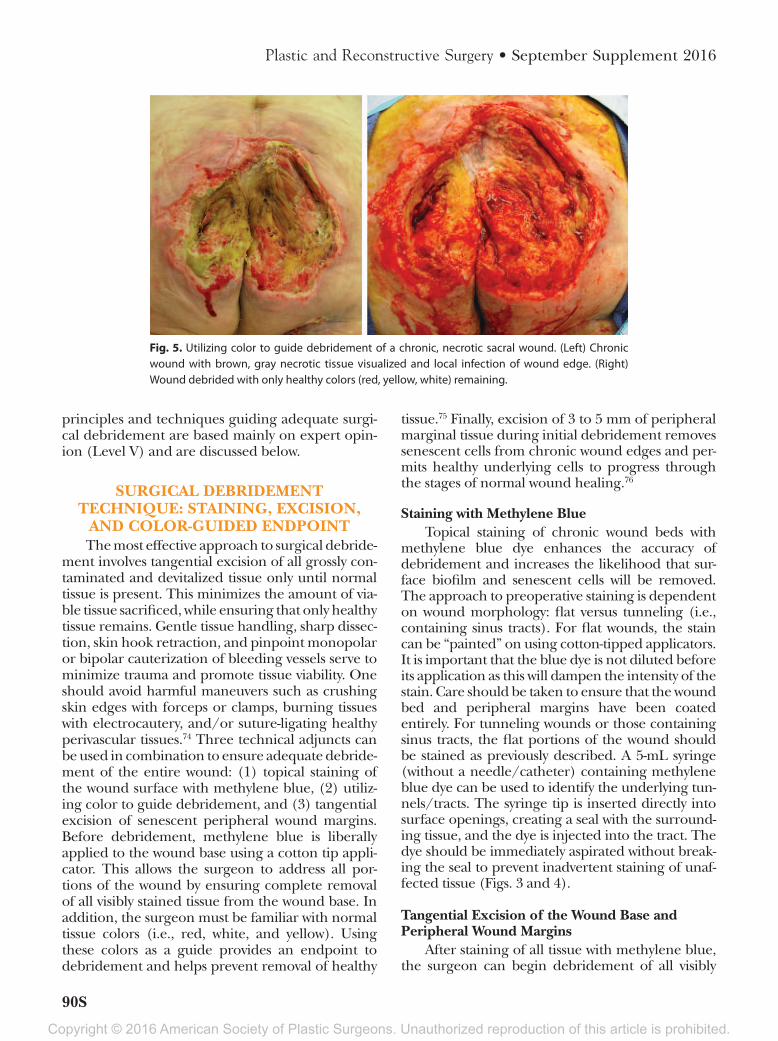
Fig. 5. Utilizing color to guide debridement of a chronic, necrotic sacral wound. (Left) Chronic wound with brown, gray necrotic tissue visualized and local infection of wound edge. (Right) Wound debrided with only healthy colors (red, yellow, white) remaining.
Copyright © 2016 American Society of Plastic Surgeons. Unauthorized reproduction of this article is prohibited.
Volume 138, Number 3S • Current Concepts in Debridement
91S
stained surfaces. First, tangential excision of the wound margin, using a scalpel, is performed to remove senescent/indurated soft tissue. Excision of approximately 3 to 5 mm of peripheral tissue is often necessary to reach soft, healthy subcutane-ous fat. Next, removal of all topically stained tissue from the wound bed, itself, is accomplished using sharp or a combination of sharp and hydrosurgi-cal debridement techniques. Instruments that are useful for the excision of soft tissue include surgi-cal blade, rongeur, electrocautery, and curette. For bone, saw, burr, rasp, and rongeur are all useful. Viable underlying tissue will not be stained and should be preserved to minimize the soft-tissue deficit and facilitate reconstruction at a later date. For tunneling wounds and sinus tracts, the overly-ing tissue should be opened to allow complete visu-alization, exploration, and debridement. Typically, at least part of the resulting incision can be closed primarily.
Color-Guided EndpointAfter the removal of all topically stained tis-
sue, hemostasis should be achieved. The wound must be reevaluated by the surgeon to ensure that all contaminated, devitalized, and/or ques-tionable materials have been removed. Utilizing color as an endpoint helps to ensure that only healthy tissue remains and prevents overaggres-sive resection of otherwise viable tissue. In gen-eral, normal tissue colors include red, white, and yellow—representing healthy, vascularized tissue such as muscle, paratenon/fascia, and fat/bone, respectively. On the other hand, gray, green, purple, brown, and black are not colors normally found in healthy wound environments and tissues of this nature should be removed (Fig. 5). Specifically, dark purple spots signify the presence of coagulated veins, which indi-cates that the local circulation is compromised or absent. Further debridement should be per-formed in this setting until pinpoint bleeding is identified throughout. The removal of indu-rated tissue at the wound base and edges can help ensure that normal tissue has been reached. Often the induration is only 2 to 3 mm thick. If more extensive, it is wiser to limit the resection of wound edges, in a single stage, by 2 to 4 mm and monitor for a subsequent reduction in sur-rounding induration.
Negative Deep Tissue Cultures as an EndpointIn our practices, aerobic and anaerobic cul-
tures of the wound are obtained before and at
the completion of each debridement by taking samples deep to the wound surface. Superficial swabs and tissue cultures are of limited use, as they usually reflect surface flora rather than the underlying bacteria that are responsible for infec-tion. Although still controversial, deep negative tissue cultures can be considered in determining the endpoint of debridement. In a recent series of 60 local flaps performed at Georgetown, the presence of positive postdebridement cultures at the time of definitive closure was a signifi-cant predictor of 90-day wound complications, including infection, dehiscence, and necrosis (OR: 17.3, CI 95%: 4.7–80.1, P < 0.001).77 The persistence of positive cultures despite serial debridement may warrant extended antibiotic therapy and may delay closure. The appropriate antibiotic choice and duration are best deter-mined and monitored by an infectious disease specialist, who can also manage the complica-tions of prolonged antibiotic therapy. Further studies are needed to determine the association between culture status and outcomes in the set-ting of different reconstructive modalities (i.e. primary closure, skin graft, local flap, free flap).
SUMMARYDebridement is an essential component in the
management of both acute and chronic wounds, as it removes infected tissue and contaminants, biofilm, and senescent cells that impede the nor-mal progression of wound healing. This article provides a broad overview of the most common mechanical, biologic, technical, and surgical tech-niques currently employed. Appropriate use of technical adjuncts such as topical tissue staining, tangential excision, color-guided debridement, and NPWT/NPWTi can enhance the efficiency of wound bed preparation and expedite time to closure. Although our debridement methodolo-gies have improved over time, there is still poten-tial for further refinement, especially as plastic surgeons are more often presented with compli-cated wounds in compromised hosts. Future pro-spective, randomized controlled trials will aide in the establishment of evidence-based guidelines to standardize debridement practices for complex wound management.
Jeffrey E. Janis, MD, FACSDepartment of Plastic Surgery
University HospitalsOhio State University Wexner Medical Center
915 Olentangy River Road, Suite 2100, Room 2114Columbus, OH 43212
Copyright © 2016 American Society of Plastic Surgeons. Unauthorized reproduction of this article is prohibited.
92S
Plastic and Reconstructive Surgery • September Supplement 2016
REFERENCES 1. O’Brien M. Exploring methods of wound debridement. Br J
Comm Nurs. 2002:10–18. 2. Attinger CE, Janis JE, Steinberg J, et al. Clinical approach to
wounds: débridement and wound bed preparation includ-ing the use of dressings and wound-healing adjuvants. Plast Reconstr Surg. 2006;117:72S–109S.
3. Faris I, Duncan H. Skin perfusion pressure in the prediction of healing in diabetic patients with ulcers or gangrene of the foot. J Vasc Surg. 1985;2:536–540.
4. Bunte MC, Shishehbor MH. Treatment of infrapopliteal crit-ical limb ischemia in 2013: the wound perfusion approach. Curr Cardiol Rep. 2013;15:363.
5. DeFazio MV, Han KD, Akbari CM, et al. Free tissue transfer after targeted endovascular reperfusion for complex lower extremity reconstruction: setting the stage for success in the presence of multivessel disease. Ann Vasc Surg. 2015;29:1316.e7–1316.e15.
6. Sheffield PJ. Tissue oxygen measurements. In: Davis JC, Hunt TK, eds. Problem Wounds: The Role of Oxygen. New York: Elsevier; 1988:17–52.
7. Steed DL, Donohoe D, Webster MW, et al. Effect of exten-sive debridement and treatment on the healing of diabetic foot ulcers. Diabetic Ulcer Study Group. J Am Coll Surg. 1996;183:61–64.
8. Robson MC, Heggers JP. Delayed wound closure based on bacterial counts. J Surg Oncol. 1970;2:379–383.
9. Krizek TJ, Robson MC. Evolution of quantitative bacteriol-ogy in wound management. Am J Surg. 1975;130:579–584.
10. Brem H, Stojadinovic O, Diegelmann RF, et al. Molecular markers in patients with chronic wounds to guide surgical debridement. Mol Med. 2007;13:30–39.
11. Wolcott R, Dowd S. The role of biofilms: are we hitting the right target? Plast Reconstr Surg. 2011;127(Suppl 1):28S–35S.
12. Gunn JS, Bakaletz LO, Wozniak DJ. What’s on the out-side matters: the role of the extracellular polymeric sub-stance of Gram-negative biofilms in evading host immunity and as a target for therapeutic intervention. J Biol Chem. 2016;291:12538–12546.
13. Costerton JW, Stewart PS, Greenberg EP. Bacterial bio-films: a common cause of persistent infections. Science. 1999;284:1318–1322.
14. Hall-Stoodley L, Costerton JW, Stoodley P. Bacterial biofilms: from the natural environment to infectious diseases. Nat Rev Microbiol. 2004;2:95–108.
15. Jahoor A, Patel R, Bryan A, et al. Peroxisome prolifera-tor-activated receptors mediate host cell proinflamma-tory responses to Pseudomonas aeruginosa autoinducer. J Bacteriol. 2008;190:4408–4415.
16. Wolcott RD, Rhoads DD, Dowd SE. Biofilms and chronic wound inflammation. J Wound Care. 2008;17:333–341.
17. Prince LR, Bianchi SM, Vaughan KM, et al. Subversion of a lysosomal pathway regulating neutrophil apopto-sis by a major bacterial toxin, pyocyanin. J Immunol. 2008;180:3502–3511.
18. Cooper RA, Bjarnsholt T, Alhede M. Biofilms in wounds: a review of present knowledge. J Wound Care. 2014;23:570, 572–574, 576.
19. Lebeaux D, Ghigo JM, Beloin C. Biofilm-related infections: bridging the gap between clinical management and funda-mental aspects of recalcitrance toward antibiotics. Microbiol Mol Biol Rev. 2014;78:510–543.
20. Chiang WC, Nilsson M, Jensen PØ, et al. Extracellular DNA shields against aminoglycosides in Pseudomonas
aeruginosa biofilms. Antimicrob Agents Chemother. 2013;57:2352–2361.
21. Mah TF, Pitts B, Pellock B, et al. A genetic basis for Pseudomonas aeruginosa biofilm antibiotic resistance. Nature. 2003;426:306–310.
22. Mulcahy H, Charron-Mazenod L, Lewenza S. Extracellular DNA chelates cations and induces antibiotic resis-tance in Pseudomonas aeruginosa biofilms. PLoS Pathog. 2008;4:e1000213.
23. Billings N, Millan M, Caldara M, et al. The extracellu-lar matrix Component Psl provides fast-acting antibiotic defense in Pseudomonas aeruginosa biofilms. PLoS Pathog. 2013;9:e1003526.
24. Alhede M, Kragh KN, Qvortrup K, et al. Phenotypes of non-attached Pseudomonas aeruginosa aggregates resem-ble surface attached biofilm. PLoS One. 2011;6:e27943.
25. Stewart PS, Costerton JW. Antibiotic resistance of bacteria in biofilms. Lancet. 2001;358:135–138.
26. James GA, Swogger E, Wolcott R, et al. Biofilms in chronic wounds. Wound Repair Regen. 2008;16:37–44.
27. Brady RA, Leid JG, Calhoun JH, et al. Osteomyelitis and the role of biofilms in chronic infection. FEMS Immunol Med Microbiol. 2008;52:13–22.
28. Wolcott RD, Rhoads DD, Bennett ME, et al. Chronic wounds and the medical biofilm paradigm. J Wound Care. 2010;19: 45–46, 48–50, 52–53.
29. Costerton W, Veeh R, Shirtliff M, et al. The application of biofilm science to the study and control of chronic bacterial infections. J Clin Invest. 2003;112:1466–1477.
30. Davis SC, Martinez L, Kirsner R. The diabetic foot: the importance of biofilms and wound bed preparation. Curr Diab Rep. 2006;6:439–445.
31. Martin JM, Zenilman JM, Lazarus GS. Molecular microbiol-ogy: new dimensions for cutaneous biology and wound heal-ing. J Invest Dermatol. 2010;130:38–48.
32. Schierle CF, De la Garza M, Mustoe TA, et al. Staphylococcal biofilms impair wound healing by delaying reepithelializa-tion in a murine cutaneous wound model. Wound Repair Regen. 2009;17:354–359.
33. Roy S, Elgharably H, Sinha M, et al. Mixed-species biofilm compromises wound healing by disrupting epidermal bar-rier function. J Pathol. 2014;233:331–343.
34. Bradley BH, Cunningham M. Biofilms in chronic wounds and the potential role of negative pressure wound ther-apy: an integrative review. J Wound Ostomy Continence Nurs. 2013;40:143–149.
35. Scali C, Kunimoto B. An update on chronic wounds and the role of biofilms. J Cutan Med Surg. 2013;17:371–376.
36. Bjarnsholt T, Alhede M, Alhede M, et al. The in vivo biofilm. Trends Microbiol. 2013;21:466–474.
37. Kirketerp-Møller K, Jensen PØ, Fazli M, et al. Distribution, organization, and ecology of bacteria in chronic wounds. J Clin Microbiol. 2008;46:2717–2722.
38. Akiyama H, Oono T, Saito M, et al. Assessment of cadexomer iodine against Staphylococcus aureus biofilm in vivo and in vitro using confocal laser scanning microscopy. J Dermatol. 2004;31:529–534.
39. Alandejani T, Marsan J, Ferris W, et al. Effectiveness of honey on Staphylococcus aureus and Pseudomonas aeruginosa bio-films. Otolaryngol Head Neck Surg. 2009;141:114–118.
40. Ammons MC, Ward LS, Fisher ST, et al. In vitro susceptibility of established biofilms composed of a clinical wound isolate of Pseudomonas aeruginosa treated with lactoferrin and xyli-tol. Int J Antimicrob Agents. 2009;33:230–236.

Copyright © 2016 American Society of Plastic Surgeons. Unauthorized reproduction of this article is prohibited.
Volume 138, Number 3S • Current Concepts in Debridement
93S
41. Dowd SE, Sun Y, Smith E, et al. Effects of biofilm treatments on the multi-species Lubbock chronic wound biofilm model. J Wound Care. 2009;18:508, 510–508, 512.
42. Leitch EC, Willcox MD. Lactoferrin increases the suscepti-bility of S. epidermidis biofilms to lysozyme and vancomy-cin. Curr Eye Res. 1999;19:12–19.
43. Percival SL, Bowler P, Woods EJ. Assessing the effect of an antimicrobial wound dressing on biofilms. Wound Repair Regen. 2008;16:52–57.
44. Thorn RM, Austin AJ, Greenman J, et al. In vitro comparison of antimicrobial activity of iodine and silver dressings against biofilms. J Wound Care. 2009;18:343–346.
45. Demaria M, Desprez PY, Campisi J, et al. Cell autonomous and non-autonomous effects of senescent cells in the skin. J Invest Dermatol. 2015;135:1722–1726.
46. Mendez MV, Stanley A, Park HY, et al. Fibroblasts cultured from venous ulcers display cellular characteristics of senes-cence. J Vasc Surg. 1998;28:876–883.
47. Takeuchi S, Takahashi A, Motoi N, et al. Intrinsic coopera-tion between p16INK4a and p21Waf1/Cip1 in the onset of cellular senescence and tumor suppression in vivo. Cancer Res. 2010;70:9381–9390.
48. Jun JI, Lau LF. The matricellular protein CCN1 induces fibroblast senescence and restricts fibrosis in cutaneous wound healing. Nat Cell Biol. 2010;12:676–685.
49. Demaria M, Ohtani N, Youssef SA, et al. An essential role for senescent cells in optimal wound healing through secretion of PDGF-AA. Dev Cell. 2014;31:722–733.
50. Coppé JP, Patil CK, Rodier F, et al. A human-like senescence-associated secretory phenotype is conserved in mouse cells dependent on physiological oxygen. PLoS One. 2010;5:e9188.
51. Strohal R. The EWMA document: debridement. J Wound Care. 2013;22:5.
52. Eneroth M, van Houtum WH. The value of debridement and Vacuum-Assisted Closure (V.A.C.) Therapy in dia-betic foot ulcers. Diabetes Metab Res Rev. 2008;24(Suppl 1):S76–S80.
53. Vermeulen H, Ubbink D, Goossens A, et al. Dressings and topical agents for surgical wounds healing by secondary intention. Cochrane Database Syst Rev. 2004;(2):CD003554.
54. Lawrence JC. Dressings and wound infection. Am J Surg. 1994;167(1A):21S–24S.
55. Gray D, Acton C, Chadwick P. Consensus guidance for the use of debridement techniques in the UK. Wounds UK. 2011;7:77–84.
56. Lewis R, Whiting P, ter Riet G, et al. A rapid and systematic review of the clinical effectiveness and cost-effectiveness of debriding agents in treating surgical wounds healing by sec-ondary intention. Health Technol Assess. 2001;5:1–131.
57. Schneider LA, Korber A, Grabbe S, et al. Influence of pH on wound-healing: a new perspective for wound-therapy? Arch Dermatol Res. 2007;298:413–420.
58. Sherman RA. Maggot therapy takes us back to the future of wound care: new and improved maggot therapy for the 21st century. J Diabetes Sci Technol. 2009;3:336–344.
59. Sun X, Jiang K, Chen J, et al. A systematic review of maggot debridement therapy for chronically infected wounds and ulcers. Int J Infect Dis. 2014;25:32–37.
60. Klein MB, Moore ML, Costa B, et al. Primer on the manage-ment of face burns at the University of Washington. J Burn Care Rehabil. 2005;26:2–6.
61. Granick MS, Posnett J, Jacoby M, et al. Efficacy and cost-effectiveness of a high-powered parallel waterjet for wound debridement. Wound Repair Regen. 2006;14:394–397.
62. Mosti G, Iabichella ML, Picerni P, et al. The debridement of hard to heal leg ulcers by means of a new device based on Fluidjet technology. Int Wound J. 2005;2:307–314.
63. Granick M, Boykin J, Gamelli R, et al. Toward a common language: surgical wound bed preparation and debride-ment. Wound Repair Regen. 2006;14(Suppl 1):S1–10.
64. Caputo WJ, Beggs DJ, DeFede JL, et al. A prospective ran-domised controlled clinical trial comparing hydrosurgery debridement with conventional surgical debridement in lower extremity ulcers. Int Wound J. 2008;5:288–294.
65. Gurunluoglu R. Experiences with waterjet hydrosur-gery system in wound debridement. World J Emerg Surg. 2007;2:10.
66. Skärlina EM, Wilmink JM, Fall N, et al. Effectiveness of con-ventional and hydrosurgical debridement methods in reduc-ing Staphylococcus aureus inoculation of equine muscle in vitro. Equine Vet J. 2015;47:218–222.
67. Huang C, Leavitt T, Bayer LR, et al. Effect of negative pres-sure wound therapy on wound healing. Curr Probl Surg. 2014;51:301–331.
68. Davis K, Bills K, Barker J, et al. Simultaneous irrigation and negative pressure wound therapy enhances wound healing and reduces wound bioburden in a porcine model. Wound Repair Regen. 2013;21:869–875.
69. Phillips PL, Yang Q, Schultz GS. The effect of negative pressure wound therapy with periodic instillation using antimicrobial solutions on Pseudomonas aeruginosa bio-film on porcine skin explants. Int Wound J. 2013;10(Suppl 1):48–55.
70. Armstrong DG, Lavery LA, Diabetic Foot Study Consortium. Negative pressure wound therapy after partial diabetic foot amputation: a multicentre, randomised controlled trial. Lancet. 2005;366:1704–1710.
71. Goss SG, Schwartz JA, Facchin F, et al. Negative pressure wound therapy with instillation (NPWTi) better reduces post-debridement bioburden in chronically infected lower extremity wounds than NPWT alone. J Am Coll Clin Wound Spec. 2014;4:74–80.
72. Kim PJ, Attinger CE, Steinberg JS, et al. The impact of neg-ative-pressure wound therapy with instillation compared with standard negative-pressure wound therapy: a retrospec-tive, historical, cohort, controlled study. Plast Reconstr Surg. 2014;133:709–716.
73. Kim PJ, Attinger CE, Oliver N, et al. Comparison of out-comes for normal saline and an antiseptic solution for neg-ative-pressure wound therapy with instillation. Plast Reconstr Surg. 2015;136:657e–664e.
74. Edgerton M. The Art of Surgical Technique. Baltimore, MD: Williams and Wilkins; 1988.
75. Endara M, Attinger C. Using color to guide debridement. Adv Skin Wound Care. 2012;25:549–555.
76. Harding KG, Moore K, Phillips TJ. Wound chronicity and fibroblast senescence–implications for treatment. Int Wound J. 2005;2:364–368.
77. Shuck J, Nolan J, Kanuri A, et al. The effect of positive post-debridement cultures on local muscle flap reconstruc-tion of the lower extremity. Plast Reconstr Surg. 2015;136 (Suppl 4):9–10.