wound management in palliative patients · injury (dti) birmingham community healthcare graphic...
TRANSCRIPT

Wound Management in Palliative Patients
Louise Morris
Prevention of Harms Practitioner for
Pressure Ulcers

Agenda
• Defining palliative wound care
• Common types of wounds associated with patients at end of life
• Symptoms
• Goals with management
• Wound management products commonly used to treat palliative patients with wounds

Palliative Care & Wounds
• ‘Palliative care’ is used to describe care given to patients with advanced, life-limiting illness of any aetiology. It is a philosophy of care that is patient and family-centred, designed to meet the needs of the patient and family’
• ‘A holistic & integrated approach that encompasses 1) symptom management, 2) the improvement of psychosocial well-being, 3) a multidisciplinary team approach, and 4) patient/family driven goals
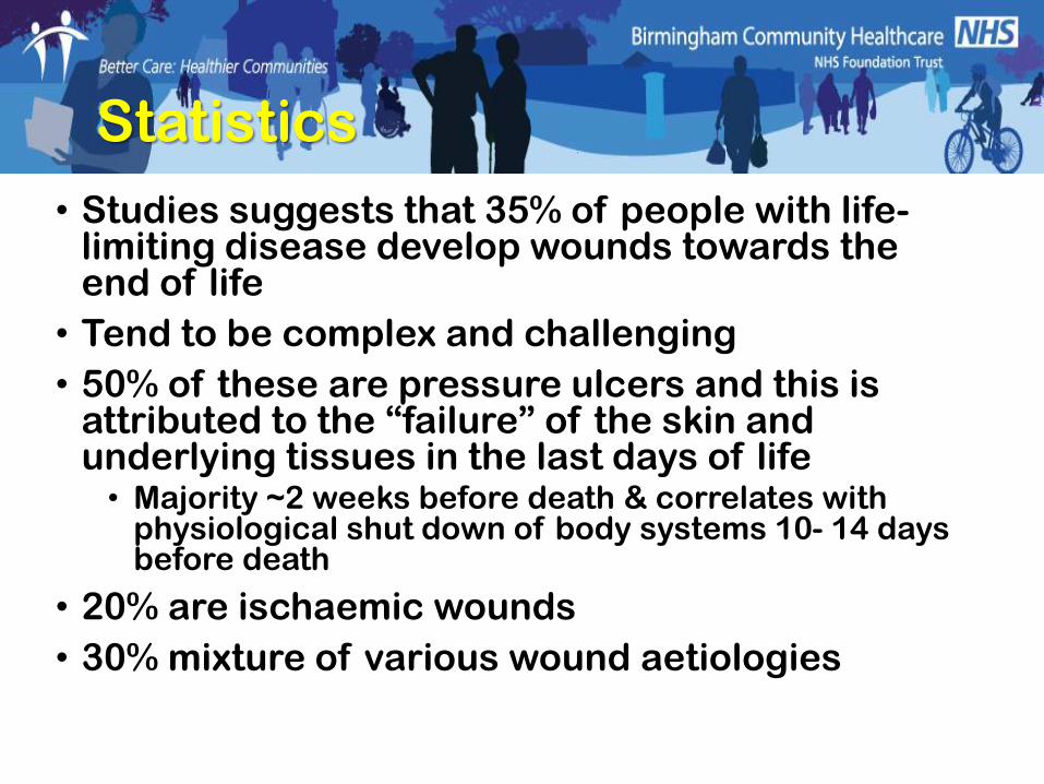
Statistics
• Studies suggests that 35% of people with life-limiting disease develop wounds towards the end of life
• Tend to be complex and challenging
• 50% of these are pressure ulcers and this is attributed to the “failure” of the skin and underlying tissues in the last days of life• Majority ~2 weeks before death & correlates with
physiological shut down of body systems 10- 14 days before death
• 20% are ischaemic wounds
• 30% mixture of various wound aetiologies

Types of Wound
Pressure Ulcers Malignant or
Fungating
WoundsSkin Tears
Leg &
Foot Ulcers
Radiotherapy
Lesions
Fistula
IschaemicLymphoedema
VenousNeuropathic
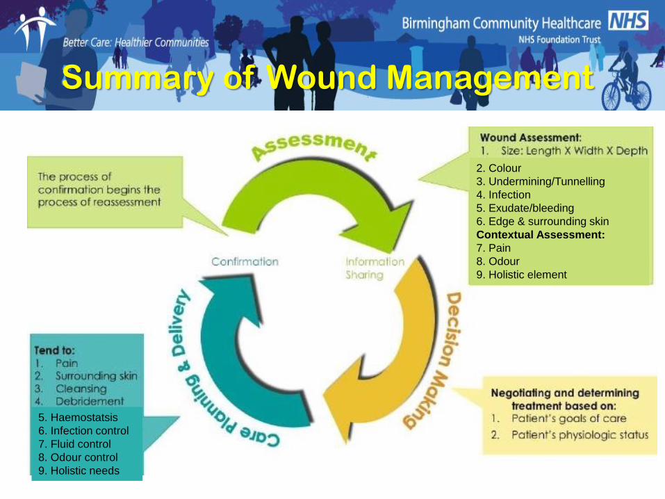
2. Colour
3. Undermining/Tunnelling
4. Infection
5. Exudate/bleeding
6. Edge & surrounding skin
Contextual Assessment:
7. Pain
8. Odour
9. Holistic element
5. Haemostatsis
6. Infection control
7. Fluid control
8. Odour control
9. Holistic needs
Summary of Wound Management

Goals in Palliative Wound Care:
• Prevent if possible
• Comfort for the individual
• Limit the impact of the wound on quality of life
• May not be wound healing, but control of symptoms

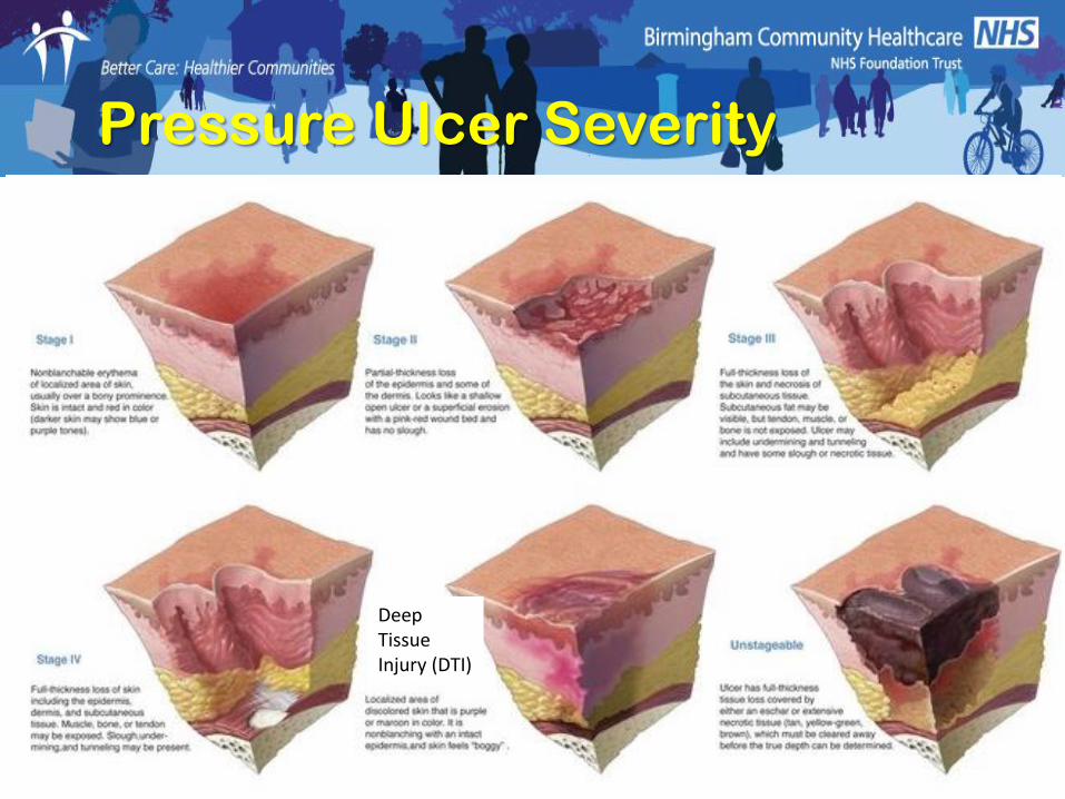
Pressure Ulcers
End of life is defined as a phase in life when an individual experiences physical deterioration, which will eventually cause their death
Skin Changes At Life’s End
• Physiologic changes that occur as a result of the dying process may affect the skin and soft tissues and may manifest as observable (objective) changes in skin colour, turgor, or integrity, or as subjective symptoms such as localized pain. These changes can be unavoidable and may occur with the application of appropriate interventions that meet or exceed the standard of care
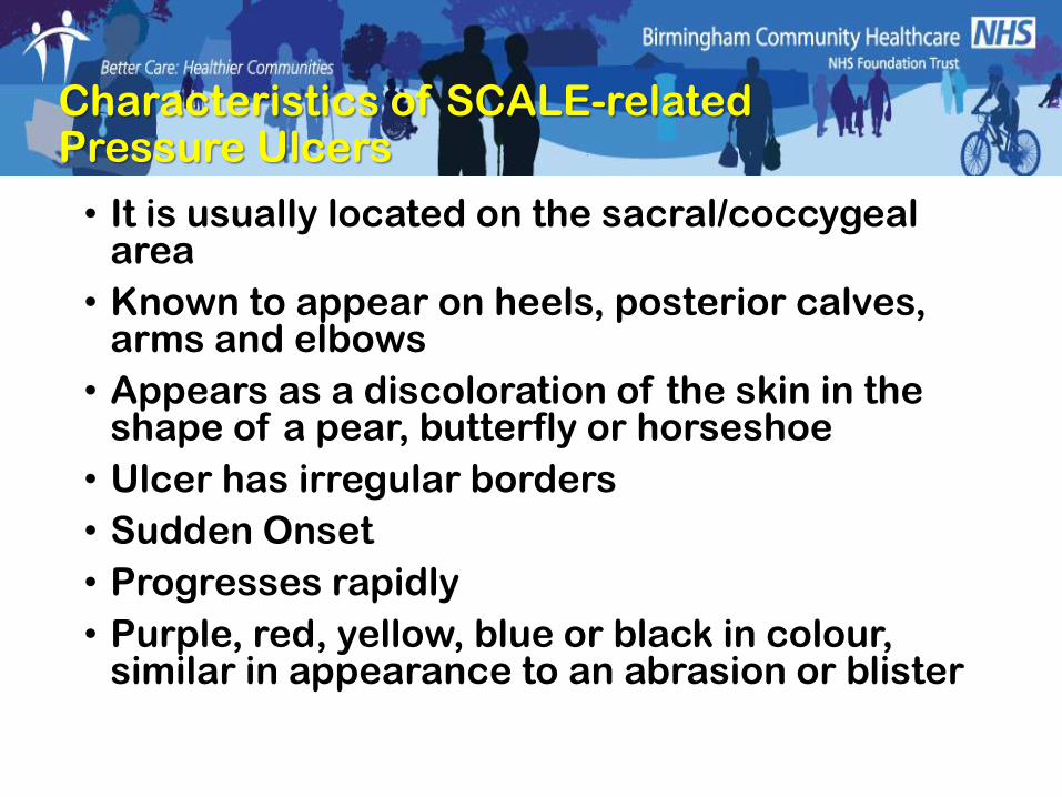
Characteristics of SCALE-related Pressure Ulcers
• It is usually located on the sacral/coccygeal area
• Known to appear on heels, posterior calves, arms and elbows
• Appears as a discoloration of the skin in the shape of a pear, butterfly or horseshoe
• Ulcer has irregular borders
• Sudden Onset
• Progresses rapidly
• Purple, red, yellow, blue or black in colour, similar in appearance to an abrasion or blister


Malignant / Fungating Wounds
• Fungating malignant wounds result from infiltration of the epithelium by cancerous cells
• There are 2 processes:• Ulceration• Proliferation
• Combination of concurrent and progressive processes

Malignant / Fungating Wounds
• Malignant cells distort vascular structures causing fluctuations in blood flow and gives rise to hypoxia
• Malignant cells infiltrate lymphatic vessels, which gives rise to poor lymphatic drainage, a rise in interstitial pressure and vascular collapse
• Vascular collapse and variations in tissue perfusion causes infarction and necrosis
• Anaerobic infection subsequently can ensue
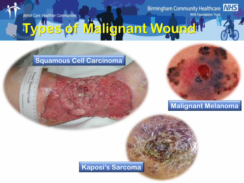
Types of Malignant Wound
Squamous Cell Carcinoma
Malignant Melanoma
Kaposi’s Sarcoma

Types of Malignant Wound
Bowen’s Disease
Basal Cell Carcinoma

Cutaneous Radiation Injury (CRI)
• Injury to the skin and underlying tissues from acute exposure to a large external dose of radiation is referred to as cutaneous radiation injury (CRI)
• Exposure to radiation can damage the basal cell layer of the skin and result in inflammation, erythema, and dry or moist desquamation
• Radiation damage to hair follicles can cause epilation.
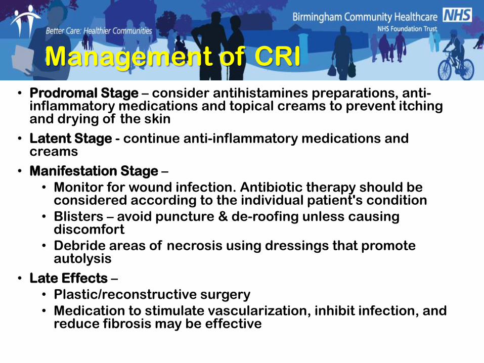
Management of CRI
• Prodromal Stage – consider antihistamines preparations, anti-inflammatory medications and topical creams to prevent itching and drying of the skin
• Latent Stage - continue anti-inflammatory medications and creams
• Manifestation Stage –
• Monitor for wound infection. Antibiotic therapy should be considered according to the individual patient's condition
• Blisters – avoid puncture & de-roofing unless causing discomfort
• Debride areas of necrosis using dressings that promote autolysis
• Late Effects –
• Plastic/reconstructive surgery
• Medication to stimulate vascularization, inhibit infection, and reduce fibrosis may be effective

Skin Tears
• Advanced age – skin thinner, drier, fragile, more vulnerable to trauma
• Protein-calorie malnutrition & catabolism – unintentional weight loss, lean body mass decreased – associated with fragile skin & poor healing
• Skin failure – poor blood supply
• Lymphoedema may increase fragile skin

Common Problems
• Excess exudate
• Malodour
• Bleeding
• Pain
• Fear
• Bulky dressing materials
• Inconvenience

Exudate
• Exudate acts as a reservoir for other aerobic organisms that further complicate malodour
• Need to consider:• Preventing strikethrough
to reduce the frequency of dressing change
• Maintaining a moist environment to minimise pain and bleeding
• Bulk of dressing affecting movement and body image

Malodour
• Debridement of necrosis
• Oral antibiotics to treat the source of the problem, i.e. infection
• Metronidazole gel for dry malodorous wounds –reduces the anaerobic bacteria
• Activated charcoal dressings, especially for exuding wounds
• Maintaining an in-tact dressing
• External control

Necrosis
• Necrotic tissue provides an environment in which anaerobic infection thrives resulting in malodour and exudate
• Autolytic debridement often preferred option to reduce risk of bleeding:• Hydrogels for eschar
• Hydrofibre for soft necrosis - caution
• LarvaE have been used for soft necrosis but caution needed
• Treatment may have to be delayed if patient is receiving radiotherapy

Haemostasis
• Spontaneous bleeding occurs when the tumour erodes the blood vessels and may be compounded by decreased platelet function
• Avoid trauma – use dressings with non-adherent qualities & that require infrequent replacement. Also that are unlikely to cause over proliferation
Haemostasis
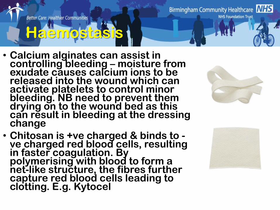
• Calcium alginates can assist in controlling bleeding – moisture from exudate causes calcium ions to be released into the wound which can activate platelets to control minor bleeding. NB need to prevent them drying on to the wound bed as this can result in bleeding at the dressing change
• Chitosan is +ve charged & binds to -ve charged red blood cells, resulting in faster coagulation. By polymerising with blood to form a net-like structure, the fibres further capture red blood cells leading to clotting. E.g. Kytocel

Pain• Assessment paramount:
• To understand the type of pain the patient is experiencing
• Determine the most appropriate treatment
• Monitor the effectiveness of treatment
• Analgesia at regular intervals & prior to dressing replacement
• Prevent pain during dressing changes – true non-adherent dressings that maintain a moist environment to reduce dressing adherence & protect exposed nerve endings. Suitable to manage other symptoms but require infrequent changing

Pain
• Topical anaesthetics such as 2% lidocaine gel applied to the wound bed, but limited evidence to support use
• Topical opioids e.g. Morphine and Diamorphine have been used, but evidence is limited. They are mixed with a hydrogel (about 1 mg of morphine to 1 g of hydrogel for 0.08 to 0.1% mixture). This is usually applied to the wound once daily

Conclusion
• Palliative wound care can be highly complex
• Holistic assessment is paramount to enable high quality of care, manage general symptoms, address psychosocial concerns, & implement strategies aimed at minimizing the unpleasant impact of living with a chronic wound for the patient & his/her circle of care
• Prevention or healing isn’t always possible

Thank you for listening
Don’t forget it’s International Stop Pressure Ulcers Day